111 Case Report
Iranian Journal of Otorhinolaryngology, Vol.26(2),Serial No.75,Apr 2014
A Large Ameloblastic Fibro-odontoma of the Maxillary Sinus
Seyed Ali Banihashem Rad1, Hamed Mortazavi2,*Majid Eshghpour3,
Jahanshah Salehinejad4, Reza Shahakbari3
A b s t r a c t
Introduction:
Ameloblastic fibro-odontoma is a rare, benign, asymptomatic tumor. The term ameloblastic fibro-odontoma was first used by Hooker in 1967 as a separate lesion from ameloblastic odontoma.
Case Report:
This case report describes an eleven years old female with large ameloblastic fibro-odontoma in the right maxillary sinus.
Conclusion:
There is a low potential for recurrence after complete Enucleation of ameloblastic fibro-odontoma, but due to the risk of ameloblastic sarcoma after recurrence, the surgery should be perfect along with a careful follow up.
Keywords:
Ameloblastic fibro-odontoma, Maxillary sinus, Odontogenic tumor.
Received date: 10 Oct 2012 Accepted date: 12 Mar 2013
1
Dental Research Center, Department of Periodontology, Mashhad University of Medical Sciences Mashhad, Iran.
2Department of Oral Medicine, Shahid Beheshti Dental School, Shahid Beheshti University of Medical Sciences
Tehran, Iran.
3
Dental Research Center, Department of Oral and Maxillofacial surgery, Mashhad University of Medical Sciences, Mashhad, Iran.
4
Dental Research Center, Department of Oral and Maxillofacial Pathology, Mashhad University of Medical Sciences, Mashhad, Iran.
*Corresponding Author:
Dental Research Center, Department of Oral and Maxillofacial surgery, Mashhad University of Medical Sciences,
Banihashem Rad SA, et al
112 Iranian Journal of Otorhinolaryngology, Vol. 26(2),Serial No.75,Apr 2014 Introduction
Ameloblastic fibro-odontoma (AFO) is a very rare, benign, asymptomatic, slow growing, expansile mixed odontogenic tumor (1). Odontogenic tumors constitute 0.84% to 1.78% of the histopathological results of oral pathology departments (2). Furthermore, odontogenic tumors account 7% of all oral lesions found in children and adolescents (3). AFO represents 1%-3% of all odontogenic tumors, reaching 4.6% when only the cases in children are mentioned, and 7.9% of odonto-genic tumors expect odontoma (1). The term ameloblastic fibro-odontoma was first used by Hooker in 1967 as a separate lesion from ameloblastic odontoma (4). Reichart and Ries classified this tumor as an ameloblastic ectomesenchymal tumor in 1983 (5). AFO has been also defined by who as "a
neoplasm composed of proliferating
odontogenic epithelium embedded in a cellular ectomesen- chymal tissue that resembles dental papilla with varying degree at inductive change and dental hard tissue formation" (6). It usually occurs in persons less than 20 years old with mean age11.5 years. Most cases are diagnosed between 9 and 11 years old. There is a higher incidence of AFO in men than women. The male to female ratio was reported 1.4:1,1.6:1 and 1.7:1 by Sassi, Boxberger and Minderjahn, respectively (7). However, Hutt did not show sex predilection (8). In a majority of cases, AFO arises in the posterior mandible and is usually associated with an unerupted tooth (1). Involvement of maxillary bone was also reported by Zouhary, Nouri and Miller (9). According to the review of the literature, AFO of the maxillary sinus is an extremely rare which was described by few authors such as Dolanmaz, Nouri and Ozer (10,11).
Radiographically, AFO presents as a
well-defined radiolucency containing
radiopaque areas (11). The aim of this article is to report a case of massive AFO in the maxillary sinus. Due to the risk of recurrence, as well as becoming a
fibrosarcoma, in the absence of proper
treatment or inadequate follow up,
introducing the lesion (due to its rarity) seems to be necessary (12).
Case Report
An 11-year-old girl was referred to the Department of Oral and Maxillofacial Surgery, Mashhad Dental School, Iran for evaluation of a facial swelling of 6 months duration. There was no history of systemic disease and trauma. The extra oral examin- ation revealed an asymptomatic swelling on the right side of the maxilla without signs and symptoms of inflammation.
Intra-orally, a bony hard bulge was palpable in the maxillary vestibule. A full complement of the teeth with the exception of missing second molar was marked able. Oral mucosa was normal and tooth mobility was not seen. Radiographically,
computerized tomography scan (CT)
showed a well-defined, radiolucent lesion in the maxillary sinus which contained several radiopaque materials of varying sizes and shapes (Fig. 1).
Fig 1: The coronal CT scan demonstrates a
very large mixed tumor in the right maxillary sinus. Notice to the size of the lesion and extending to eye floor and wall of the nose.
The panoramic view showed a very large lesion which the maxillary second molar had been involved (Fig.2).
As the clinical and radiological
A Large Ameloblastic Fibro-odontoma of the Maxillary Sinus
Iranian Journal of Otorhinolaryngology, Vol.26 (2),Serial No.75,Apr 2014 113 department of oral and maxillofacial
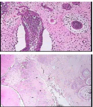
pathology. Pathologist reported: this lesion is lobulated in general configuration and is surrounded by a fibrous capsule.
Fig 2: The panoramic view of the patient
before surgery shows a very large mixed tumor in the right maxillary sinus. The maxillary second molar is involved in the lesion.
The tumor mass is composed
predominantly of a primitive appearing myxoid connective tissue similar to dental pulp. The epithelial component has been compared microsco- pically to the dental lamina that proliferates from oral epithelium in the early stages of the tooth developments. Some foci contain enamel and dentin similar
to compound and complex odontoma (Fig. 3).
Fig 3: Microscopic view of the lesion shows
enamel and dentin similar to compound and complex odontoma and epithelial component.
The tumor mass is composed predominantly of a primitive appearing myxoid connective tissue similar to dental pulp.
These findings were consistent with diagnosis of ameloblastic fibro-odontoma.
After a final histological diagnosis, the patient was submitted for surgical excision and enucleation of the lesion. The patient was taken to the operation room, under general anesthesia an incision was made intraorally. A full thickness mucoperiosteal flap from second incisor to the tuberosity was reflected. After bone removal of the sinus wall, access to the lesion was completed. The lesion and the impacted second molar were enucleated Finally, histopathological examination of the last specimen confirmed the diagnosis of AFO. Postoperatively, after twelve months, no evidence of residual or recurrent disease was found (Fig.4,5).
Fig 4: The panoramic view of the patient 4
weeks after surgery.
Fig 5: The panoramic view of the patient 12
weeks after surgery. The right second premolar is in eruption.
Discussion
Banihashem Rad SA, et al
114 Iranian Journal of Otorhinolaryngology, Vol. 26(2),Serial No.75,Apr 2014
ameloblastic fibroma (12). Slootweg
described that the AFO is an immature complex odontoma (13). Neoplastic behavior and malignant features of AFO has been also reported by Howell and Bregni (14). The most common clinical presentations of AFO are asympto- matic swelling and failure of tooth eruption (12).
The pathologically and radiologically differential diagnosis of AFO are included:
ameloblastoma, odontogenic myxoma,
dentinogerous cyst, odontogenic keratocyst, central giant cell granuloma, histocytosis-X group of lesions, calcifying odontogenic cyst, calcifying epithelial odontogenic tumor,
adenomatoid odontogenic tumor and
immature odontoma (12).
There is controversy over the management of AFO. According to the literature,
conservative surgical excision is an
accepted treatment for this lesion. In most cases, the impacted tooth associated with the tumor is removed at the same time (14). There is a low potential for recurrence. According to Boxberger, in almost all cases recurrences were related to incomplete removal of the lesion at the time of the initial surgery.
Conclusion
Ameloblastic fibro-odontoma (AFO) is a very rare, benign, asymptomatic, slow growing, expansile mixed odontogenic tumor. Note that this tumor is encapsulated, treatment plan is surgical excision. Despite the little potential of this lesion to locally recur, due to a low risk for ameloblastic
sarcoma, a careful follow up is
recommended.
References
1. Santos Tde S, de Carvalho RW, Avelar RL, Dias de Oliveira e Silva E, Frota R, Anjos ED.
Ameloblastic fibro-odontoma in children: report of 2 cases. J Dent Child (Chic) 2011; 78(3):173-7. 2. Cavalcante AS, Anbinder AL, Costa NC, Lima JR, Carvalho YR. Ameloblastic fibro-odontoma: a case report. Med Oral Patol Oral Cir Bucal 2009; 14 (12): e650-3.
3. Guerrisi M, Piloni MJ, Keszler A. Odontogenic tumors in children and adolescents. A 15-year retrospective study in Argentina. Med Oral Patol Oral Cir Bucal 2007; 12(3): E180-5.
4. Hooker SP. Ameloblastic odontoma: A nalaysis of twenty six cases. Oral Surg 1967; 24: 375.
5. Reichart PA, Ries P. Consideration on the classification of odontogenic tumors. Int J Oral Surg 1983; 12(5):323-33.
6. Barnes L, Eveson J, Reichart P, Sidransky D. World Health Organization classification of tumours. Pathology and genetics head and neck tumours. Lyon: IARC Press; 2005. p. 284-5. 7. Sassi LM, Stramandinoli RT, Dissenha JL, Zanferrari FL, Schussel JL. Large ameloblastic fibro-odontoma in a 3 year-old child: case report. RSBO 2011; 8(1):114-6.
8. Hutt PH. Ameloblastic fibro-odontoma: report of a case with documented four-year follow-up. J Oral Maxillofac Surg 1982; 40(1):45-8.
9.Zouhary KJ, Said-Al-Naief N, Waite PD. Ameloblastic fibro-odontoma: expansile mixed radiolucent lesion in the posterior maxilla: a case report. Oral Surg Oral Med Oral Pathol Oral Radiol Endod 2008; 106(4):e15-21.
10. Dolanmaz D, Pampu AA, Kalayci A, Etöz OA, Atici S. An unusual size of ameloblastic fibro-odontoma. Dentomaxillofac Radiol 2008; 37(3): 179-82.
11. Nouri H, Raji A, Ait M'barek B. Ameloblastic fibro-odontoma of the maxillary sinus. Rev Stomatol Chir Maxillofac 2007; 108(5):455-7.
12. Regezi JA, Sciubba JJ, Jordan RCK. Oral pathology: clinical pathologic correlation. St. Louis, Saunders 2008; 227-28.
13. Slootweg PJ. An analysis of the interrelationship of the mixed odontogenic tumors- ameloblastic fibroma, ameloblastic fibro-odontoma, and the odontomas. Oral Surg Oral Med Oral Pathol 1981; 51(3):266-76.