Expressed MicroRNAs in Porcine Sexually Immature and
Mature Testes
Lifan Luo., Lianzhi Ye., Gang Liu, Guochao Shao, Rong Zheng, Zhuqing Ren, Bo Zuo, Dequan Xu,
Minggang Lei, Siwen Jiang, Changyan Deng, Yuanzhu Xiong, Fenge Li*
Key Laboratory of Pig Genetics and Breeding of Ministry of Agriculture, and Key Laboratory of Agricultural Animal Genetics, Breeding and Reproduction of Ministry of Education, Huazhong Agricultural University, Wuhan, People’s Republic of China
Abstract
Background:MicroRNAs (miRNAs) are short non-coding RNA molecules which are proved to be involved in mammalian spermatogenesis. Their expression and function in the porcine germ cells are not fully understood.
Methodology:We employed a miRNA microarray containing 1260 unique miRNA probes to evaluate the miRNA expression patterns between sexually immature (60-day) and mature (180-day) pig testes. One hundred and twenty nine miRNAs representing 164 reporter miRNAs were expressed differently (p,0.1). Fifty one miRNAs were significantly up-regulated and 78 miRNAs were down-regulated in mature testes. Nine of these differentially expressed miRNAs were validated using quantitative RT-PCR assay. Totally 15919 putative miRNA-target sites were detected by using RNA22 method to align 445 NCBI pig cDNA sequences with these 129 differentially expressed miRNAs, and seven putative target genes involved in spermatogenesis includingDAZL,RNF4gene were simply confirmed by quantitative RT-PCR.
Conclusions:Overall, the results of this study indicated specific miRNAs expression in porcine testes and suggested that miRNAs had a role in regulating spermatogenesis.
Citation:Luo L, Ye L, Liu G, Shao G, Zheng R, et al. (2010) Microarray-Based Approach Identifies Differentially Expressed MicroRNAs in Porcine Sexually Immature and Mature Testes. PLoS ONE 5(8): e11744. doi:10.1371/journal.pone.0011744
Editor:Alan Christoffels, University of Western Cape, South Africa
ReceivedNovember 24, 2009;AcceptedJune 29, 2010;PublishedAugust 18, 2010
Copyright:ß2010 Luo et al. This is an open-access article distributed under the terms of the Creative Commons Attribution License, which permits unrestricted use, distribution, and reproduction in any medium, provided the original author and source are credited.
Funding:This work was supported financially by the National Natural Science Foundation of China (30700571), Trans-gene Key Project of China (2009ZX08009-148B), National High Technology Development Project (2007AA10Z162). The funders had no role in study design, data collection and analysis, decision to publish, or preparation of the manuscript.
Competing Interests:The authors have declared that no competing interests exist.
* E-mail: [email protected]
.These authors contributed equally to this work.
Introduction
microRNAs (miRNAs) are small non-coding RNAs (typically 19–23 nucleotides) that play important roles in regulating posttranscriptional translation. The first discovered miRNA, lin-4, is involved in developmental timing in thenematode C. elegans[1]. To date, 10883 miRNA sequences have been published on the Sanger miRNA Registry (http://www.sanger.ac.uk/software/ Rfam/mirna, miRbase Release 14.0). They are increasingly being shown to play vital roles in spermatogenesis, muscle development, feed intake and other important physiological process [2–4]. Such as the myostatin allele in muscle mass QTL interval is characterized by a G to A transition in the 39 un-translated region (UTR) that creates a target site for miR-1 and miR-206 which are highly expressed in skeletal muscle. This causes translational inhibition of the myostatin gene and hence contributes to the muscular hypertrophy of Texel sheep [5].
Spermatogenesis is a complex process through which diploid germ cells proliferate and differentiate into haploid spermatozoa [6]. It is estimated that about 1000 genes involved in spermato-genesis, and 351 of these genes appear to be expressed only in the
male germs [7]. A large number of genes are expressed at grossly higher levels in meiotic and/or early haploid spermatogenic cells than in somatic cells, yet they too are translated inefficiently [8]. Such repression could be reached by ribosomal protein binding with target genes or by some translational control elements which could be bound at 39 UTRs of target genes [9]. miRNAs are a large family of small regulatory elements that direct messenger RNA degradation or disrupt mRNA translation by binding the UTRs and coding sequences (CDS) of target mRNAs [10,11]. For example, miR-122a was suggested targeting a reporter mRNA containing sequences from the 39-UTR of the transition protein 2 (TNP2), a post-transcriptionally regulated testis-specific gene involved in chromatin remodeling during mouse spermatogenesis [2]. The over-expression of miR-34c in HeLa cells led to a shift of the expression profile toward the germinal lineage, and miR-34c could play a role in the late steps of spermatogenesis [12].
spermatocytes) germ cells, suggesting that late meiotic and haploid germ cells are the main source of miRNA production during spermatogenesis [13]. About 54 porcine miRNAs have been identified, however their expression and function in the porcine germ cells are still poorly understood [14–16]. In addition, it is necessary to study porcine miRNAs due to the increasing interest in pig genetics and the benefits of using the domestic pigs as a model for the study of human male infertility. Therefore, we investigated differentially expressed miRNAs between immature and mature testis tissues of Large White boars by miRNAs array analysis, and predicted target genes of these miRNA and analyzed the relationship between those putative genes and spermatogenesis.
Results and Discussion
miRNA microarray analysis
A custom-made mammalian miRNA microarray was used to evaluate the expression of porcine miRNAs. At the design time of the microarray, there were 2522 mammal mature miRNAs including 711 human, 658 mice, 348 rat and 54 porcine mature miRNAs. After removing the redundant sequences, there remained 1260 unique mature miRNA sequences (See probe list of the microarray in Table S1). The microarray contained 1260 probes complementary to these sequences, and all probes were repeated triplicate in one microarray. Microarray hybridization with RNA samples prepared from three 60-day (sexually immature) porcine testes and three 180-day (mature) porcine testes detected expression of 704 miRNAs, with 449.676102.66 miRNAs per sample (Table S2). Of the 704 detectable miRNAs, miRNAs count present in all six samples was 261 (20.71%), which was a little lower than previously reported (28.3%) [17]. Thirty of 54 porcine miRNAs in miRbase release 10.0, were detected (25 in 60-day testes and 27 in 180-day testes).
The microarray data showed that several microRNAs including let-7, miR-923, miR-202, miR-21 and miR-145 were highly expressed in the porcine testes (Table S2). Pairwise significance analysis indicated 129 expressed miRNAs representing 164 miRNAs probes had changed expression profiles between 60-day and 180-day testis samples (p#0.1), and in 180-day (mature) porcine testes down-regulated expression appeared in 78 miRNAs while up-regulated expression appeared in other 51 miRNAs (Table S3). Among 129 differentially expressed miRNAs in pig testes, 8 miRNAs including mmu-let-7e and mmu-miR-181b shared the same expression profile as the previous study in sexually immature/mature mouse testes [18], and 10 miRNAs including hsa-miR-1 and mmu-miR-709 had the same expression profile in the immature/mature rhesus monkey testes [19]. Five miRNAs (mmu-miR-449, rno-miR-34b, mmu-miR-34c hsa-miR-181d, mmu-miR-214) appeared to be differentially expressed in two independent reports [18,19]. Another four miRNAs (mmu-miR-34b, mmu-miR-122a, mmu-miR-16 and mmu-miR-101) were confirmed to be differentially expressed in mouse testes by using conventional Northern blot analysis [2].
Mature miRNA sequences are highly conserved in different animal species and only 1–3 nucleotide differences. For example, there are only 1–2 nucleotide differences in the miR-181b respectively fromMonodelphis domestica,Gorilla,Bos taurusand three members from Homo sapiens. Thus, cross-hybridization among members from the same family and among orthologs across species could explain these microarray results. All above four orthologs of miR-181b (m, g, b, and h) were up-regulated in the 60-day testis (Table S2).
microRNA expression validation
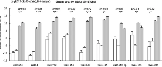
In order to validate the DNA microarray results, real-time RT-PCR with commercially available primers (Table S4) was carried out separately to investigate the differential expression pattern of 9 miRNAs - 663, 1, 762, 143, 638, miR-145, miR-542-3p, miR-155 and miR-705. Our results showed a high level of concordance between these two methods in 8 of the 9 comparisons (Pearson correlation coefficient $0.52, Figure 1). miR-705 has a high Pearson correlation coefficient of 0.52, but a significant difference between immature and mature testes has not been observed using qRT-PCR. And a contradiction expression profile was detected in miR-145. The expression levels varied dramatically, and the variance probably came from biological differences between the samples.
Differentially expressed miRNA character analysis Using a homology search based on genomic survey sequence analysis and microRNA (miRNA) secondary structure, a total of 72 porcine differentially expressed miRNAs were identified and then were located in pig genome covering all chromosomes except SSC14 and SSCY, and 14 (19.44%) miRNAs were mapped to SSCX (Table S5). One reason is that SSCY sequences have not been released yet, and the other is that miRNAs are scarcely located to SSCY. No miRNA was found on the Y chromosome in any species and the densities were greater than twofold those on autosomes in seven of eight mammalian species (p,0.01) [20]. These X-linked miRNAs were expressed in a testis-preferential or testis-specific pattern, suggesting that they have functional roles during spermatogenesis, including the possibility that they contribute to the process of meiotic sex chromosome inactivation, or that they may be essential for post-transcriptional regulation of autosomal mRNAs during the late meiotic and early postmeiotic stages of spermatogenesis [21].
Putative miRNA target gene prediction and expression array of target genes
reproduction, and play important roles in 17 pathways (Table S7). Amony 129 differentially expressed miRNAs, miR-762, miR-149* and miR-663 were the top 3 highest number of miRNA interaction sites.
Real-time RT-PCR was used to validate the target genes which are associated with mammalian testis development and sper-matogenesis. The results showed that in mature testes: AQN-1, HAS3,RNF4gene were down regulated whileSMCPandSPAM1 gene were up regulated (p#0.05); DAZL and SPAG1 had a tendency to be down-regulated, and up-regulated, respectively (Figure 2,DDCtvalue has a negative relationship with the gene
expression level). By comparing the expression profiling of miRNAs and target genes, only about half of these putative target genes remained due to the negative relationship of the expression patterns between miRNA and its target mRNA (Table 1). One of these putative target genes is deleted in
azoospermia like gene (DAZL), targeted by miR-34b, miR-34cetc. In mice, disruption of theDAZLgene leads to loss of germ cells and complete absence of gamete production [25]. An A386G (T54A) mutation occurring in the RNA-binding domain of the DAZL protein has been associated with susceptibility to spermatogenic failure in the Taiwanese [26], and it was associated with the female reproductive traits in pigs [27]. Predictions associated small nuclear RING finger protein RNF4 (SNURF) gene with miR-638, miR-705 and miR-762etc. RNF4 possesses a C-terminal RING finger and acts as a transcription regulator. RNF4 expressed more abundantly in murine adult testis [28]. In adult rat testis, RNF4 mRNA and protein accumulate in postmeiotic round and elongating spermatids, suggesting that this protein is involved in the regulation of processes required for late steps of spermatid maturation, during which vast amount of protein degradation and chromatin Figure 1. Validation of the microarray results using qRT-PCR method.The X-axis represents the miRNAs and the Y-axis shows the relative expression levels of miRNAs (-DCtvalues for qRT-PCR; Log(sample signal, 2) for microarray). The number of biological replicates is three for both
assays. R represents the Pearson correlation coefficient. The significance of differences for the expression between 60-d (immature, 60-day) and 180-d (mature, 180-day) testes was calculated using two-tailed T-test. *, p#0.05; **, p#0.01 (left for qRT-PCR, and right for microarray).
doi:10.1371/journal.pone.0011744.g001
Figure 2. miRNA putative target genes expression in porcine testis was identified by qRT-PCR.The X-axis represents the specific gene and the Y-axis shows theDDCtvalues of genes.DDCtvalue has a negative relationship with the gene expression level, so the smallerDDCtvalue has a
higher expression level. The number of biological replicates is three. The significance of differences for the expression between d (immature, 60-day) and 180-d (mature, 180-60-day) testes was calculated using two-tailed T-test. *, p#0.05; **, p#0.01.
compaction take place [29], and it was associated with the female reproductive traits in pigs [30].
Recently, a novel class of 26–30 nt RNAs (piRNAs) has been described in the testis, where they bind a spermatogenesis-specific protein belonging to the Argonaute protein family called PIWI. Like termed germline small RNAs (gsRNAs), miRNA and short interference RNA and other small RNAs expressed in testes, piRNAs are thought to be involved in gene silencing [31,32]. However, piRNAs are apparently present at low levels in mature spermatozoa, and piRNA sequences are not conserved between species [31,32] The majority of piRNAs are antisense to transposon sequences, suggesting that transposons are the piRNA target [33], but its expression profiles, and function of piRNAs are still poorly understood.
Materials and Methods
Ethics statement
All research involving animals were conducted according to the regulation (No. 5 proclaim of the Standing Committee of Hubei People’s Congress) approved by the Standing Committee of Hubei People’s Congress, P. R. China. Sample collection was approved by the ethics committee of Huazhong Agricultural University (No. 30700571 for this study).
Animals and RNA extraction
Three young Large White boars of 60 days (sexually immature) and 3 of 180 days (sexually mature defined according to the reference [34]) Large White boars were obtained from the pig farm of Huazhong Agricultural University (Wuhan, China). Whole testes were removed from animals and were immediately snap-frozen in liquid nitrogen and stored at280uC. Total RNAs were extracted by Trizol reagent (Invitrogen). All the procedures were carried out according to manufacturer’s protocols.
mParafloTMmicroRNA microarray assay
The custom-made microarray contained 1260 unique miRNA probes generated from 2522 mammalian miRNAs including 711 human, 658 mice, 348 rat and 54 porcine miRNAs (Table S1), and all of the oligonucleotide probes were repeated triplicate in one microarray. MicroRNA microarray analysis was performed by LC Sciences (Houston, TX). Briefly, the assay started from 2 to 5mg total RNA sample, which was size fractionated using a YM-100 Microcon centrifugal filter (from Millipore) and the small RNAs (,300 nt) isolated were 39-extended with a poly(A) tail using poly(A) polymerase. An oligonucleotide tag was then ligated to the poly (A) tail for later fluorescent dye staining; two different tags were used for the two RNA samples in dual-sample experiments. Hybridization was performed overnight on amParafloTM micro-fluidic chip using a micro-circulation pump (Atactic Technologies). On the microfluidic chip, each detection probe consisted of a chemically modified nucleotide coding segment complementary
to target microRNA (from miRBase release 10.0, 1260 miRNA probes in total) or control RNA and a spacer segment of polyethylene glycol to extend the coding segment away from the substrate. Multiple control probes including BKG, PUC2PM-20B, PUC2MM-20B and 5S-rRNA, were used for quality controls of chip production in each chip.
The detection probes were made byin situsynthesis using PGR (photogenerated reagent) chemistry. The hybridization melting temperatures were balanced by chemical modifications of the detection probes. Hybridization used 100mL 66 SSPE buffer (0.90 M NaCl, 60 mM Na2HPO4, 6 mM EDTA, pH 6.8)
containing 25% formamide at 34uC. After hybridization detection used fluorescence labeling using tag-specific Cy3 and Cy5 dyes. Microarray experiments were performed three times using biological samples. Hybridization images were collected using a laser scanner (GenePix 4000B; Molecular Devices, Sunnyvale, CA, USA) and digitized using the Array-Pro image analysis software (Media Cybernetics, Bethesda, MD, USA). Data were analyzed by first subtracting the background and then normalizing the signals using a LOWESS filter (Locally-weighted Regression). A transcript to be listed as detectable must meet at least two conditions: signal intensity higher than 36(background standard deviation) and spot CV,0.5, and CV was calculated by (standard deviation)/(signal intensity) [17]. When repeating probes were present on an array, a transcript was listed as detectable only if the signals from at least 50% of the repeating probes are above detection level. Furthermore, ‘‘bad spots’’ that had signal values deviated more than 50% of average values of repeating spots and/ or spot CV larger than 0.5 were discarded. For two color experiments, the ratio of the two sets of detected signals (log2
transformed, balanced) and p-values of the t-test were calculated. Differentially detected signals were those with a slightly relaxed p-value cutoff of 0.1 [35,36], in case that the true positives were excluded.
Quantitative real-time RT-PCR to validate miRNA expression
The microarray findings were validated using quantitative real-time RT-PCR. Ten nanogram RNAs were 39-extended with a poly (A) tail using poly (A) polymerase, and then reverse transcribed to cDNA using the RT-PCR System (Promega). The expression levels of miRNAs were detected in 60-day porcine testes and 180-day porcine testes by SYBR Green I assay (TOYOBO). Each real-time PCR (in 25mL) included 12.5mL SYBR Green Real-time PCR Master Mix, 350–500 ng cDNA, 0.3mM primers (Table S4). The cycling conditions consisted of 1 cycle at 95uC for 3 min, followed by 40 cycles at 94uC for 20 sec, 58uC for 20 sec, and 72uC for 10 sec, with fluorescence acquisition at 74uC in single mode. The specific PCR products were confirmed by the results of melting curve analysis and agarose gel electrophoresis. cDNAs from three testis samples at each stage were used as template to detect the expression changes
Table 1.Number of binding miRNAs of seven potential target genes involved in spermatogenesis.
NO. Binding miRNAs AQN-1 DAZL HAS3 RNF4 SMCP SPAG1 SPAM-1 Total
Bioinformatics 25 14 55 20 17 19 29 179
Bioinformatics+Microarray Array+qPCR
14 10 25 12 7 9 13 90
Positive percentage 0.56 0.714 0.455 0.6 0.412 0.474 0.448 0.503
of the miRNAs, and all PCRs were performed in triplicate. The miRNA was considered to be undetectable when its Ct value
exceeded 35 in the sample tissue. miRNA expression levels were quantified relatively to the expression of 18S RNA using Gene Expression Macro software (Bio-Rad, Richmond, CA, USA) by employing DCt value. Student’st-test was conducted to identify
differentially expressed miRNAs. Due to the negative relationship between Ct and expression level, an improved method of the
previous report [37] was used to compare the results of qRT-PCR and microarray by plotting the -DCtvalues of qRT-PCR versus the
log2 of the microarray signal for each miRNA.
Bioinformatics analysis
A homology search based on genomic survey sequence analysis and human miRNA secondary structure was used to map the miRNA genes. Human miRNA and its corresponding pri-miRNA sequences were obtained from the Sanger miRNA Registry (http://www.sanger.ac.uk/software/Rfam/mirna). To retrieve homologous pig miRNA genes, the human pri-miRNA sequence was used as a query sequence to search homology using BLASTN (Basic Local Alignment Search Tool-nucleotide) on the NCBI pig sequence database with HTGS or Trace-WGS options. The porcine miRNAs were predicted to be located in the correspond-ing target contig or shot-gun sequences, if the similarity was larger than 80%.
To fully inverstgate the function of the differentially expressed miRNAs, we collected pig cDNA sequences were randomly selected from GenBank and performed a GO term and KEGG pathway annotation using the DAVID gene annotation tool (http://david.abcc.ncifcrf.gov/). Here the ‘‘Full-Length’’ cDNA sequences were used to match with the differentially expressed miRNAs sequence by the RNA22 [38] to predict the putative target genes and corresponding target sites, since target mRNAs could be repressed as efficiently by miRNA-binding sites in the 59 UTR and CDS as in the 39UTR [10,11].
Putative target gene validation
Seven candidate target genes involved in spermatogenesis, includingAQN-1,DAZL,HAS3,RNF4,SMCP,SPAG1andSPAM1 genes, were confirmed by real time RT-PCR. From each sample, 10 mg of total RNA was incubated with two units of RNase-free DNase I (New England BioLabs, Inc.) to remove DNA contamination from RNA. RNAs were 39-extended with a poly (A) tail, then the first cDNA strand was synthesized and used as template for quantitative real-time RT-PCR with gene specific primers (Table S4) and b-actin was selected as the endogenous reference. All PCRs were performed in triplicate and gene expression levels were quantified relatively to the expression of
b-actin using Gene Expression Macro software (Bio-Rad, Rich-mond, CA, USA) by employing an optimized comparative Ct
(DDCt) value method. Student’s t-test was conducted to identify
differentially expressed miRNAs by comparingDDCtvalue of two
groups [39].
Supporting Information
Table S1 All the miRNAs probes used in this research. 2522
mammal mature miRNAs were included. After removing the redundant sequences, 1260 unique mature miRNA sequences remained. The signs (*, 3p, 5p and additional letters) of miRNA names are explained at http://www.mirbase.org/help/nomenclature. shtml.
Found at: doi:10.1371/journal.pone.0011744.s001 (0.21 MB XLS)
Table S2 Average signal of detectable transcripts. Detectable
transcripts must meets at least two conditions: signal intensity higher than 36(background standard deviation) and spot CV ,0.5. CV is calculated by (standard deviation)/(signal intensity). Found at: doi:10.1371/journal.pone.0011744.s002 (0.16 MB XLS)
Table S3 Differentially expressed miRNAs detected in porcine
sexually immature and muture testes tissues (p,0.1). p-value with red, yellow and blue means p,0.01, p,0.05 and p,0.1, respectively.
Found at: doi:10.1371/journal.pone.0011744.s003 (0.04 MB XLS)
Table S4 Primer pairs used to confirm the differentially
expressed miRNAs and target genes.
Found at: doi:10.1371/journal.pone.0011744.s004 (0.02 MB XLS)
Table S5 Characteristics analysis of differentially expressed
miRNAs.
Found at: doi:10.1371/journal.pone.0011744.s005 (0.12 MB XLS)
Table S6 The results of 445 gene cDNA sequence aligned with
129 differentially expressed (DE) miRNAs by RNA22. Part 1: miRNA-mRNA interaction site counts and binding DE miRNAs counts of each potential target gene. Part 2: miRNA-mRNA interaction site counts and target gene counts of each DE miRNA. Found at: doi:10.1371/journal.pone.0011744.s006 (0.50 MB XLS)
Table S7 GO Functional Enrichment and KEGG pathway
annotation of the miRNA potential targets.
Found at: doi:10.1371/journal.pone.0011744.s007 (0.03 MB XLS)
Acknowledgments
We thank the anonymous reviewers for critical reading and discussions of the manuscript. We thank LC Sciences for technical assistance with RNA extraction and microarray studies. The authors also acknowledge the staff of the pig farm of Huazhong Agricultural University for providing pig samples.
Author Contributions
Conceived and designed the experiments: ZR BZ DX ML SJ CD YX FL. Performed the experiments: LL LY. Analyzed the data: LL GL GS FL. Contributed reagents/materials/analysis tools: LL RZ. Wrote the paper: FL.
References
1. Lee RC, Feinbaum RL, Ambros, V (1993) TheC. elegansheterochronic gene lin-4 encodes small RNAs with antisense complementarity to lin-1lin-4. Cell 75: 843–854.
2. Yu Z, Raabe T, Hecht NB (2005) MicroRNA Mirn122a reduces expression of the post-transcriptionally regulated germ cell transition protein 2 (Tnp2) messenger RNA (mRNA) by mRNA cleavage. Biol Reprod 73: 427–433.
3. McCarthy JJ, Esser KA (2007) MicroRNA-1 and microRNA-133a expression are decreased during skeletal muscle hypertrophy. J Appl Physiol 102: 306– 313.
5. Clop A, Marcq F, Takeda H, Pirottin D, Tordoir X, et al. (2006) A mutation creating a potential illegitimate microRNA target site in the myostatin gene affects muscularity in sheep. Nat Genet 38: 813–818.
6. Cooke HJ, Saunders PT (2002) Mouse models of male infertility. Nat Rev Genet 3: 790–801.
7. Schultz N, Hamra FK, Garbers DL (2003) A multitude of genes expressed solely in meiotic or postmeiotic spermatogenic cells offers a myriad of contraceptive targets. Proc Natl Acad Sci U S A 100: 12201–12206.
8. Kleene KC (2001) A possible meiotic function of the peculiar patterns of gene expression in mammalian spermatogenic cells. Mech Dev 106: 3–23. 9. Braun RE (1998) Post-transcriptional control of gene expression during
spermatogenesis. Seminars in Cell and Developmental Biology 9: 483–489. 10. Tay Y, Zhang J, Thomson AM, Lim B, Rigoutsos I (2008) MicroRNAs to
Nanog, Oct4 and Sox2 coding regions modulate embryonic stem cell differentiation. Nature 455: 1124–1128.
11. Lytle, JR, Yario, TA, Steitz, JA (2007) Target mRNAs are repressed as efficiently by microRNA-binding sites in the 59UTR as in the 39UTR. Proc Natl Acad Sci U S A 104: 9667–9672.
12. Bouhallier F, Allioli N, Lavial F, Chalmel F, Perrard MH, et al. (2010) Role of miR-34c microRNA in the late steps of spermatogenesis. RNA 16: 720–731. 13. Ro S, Park C, Sanders KM, McCarrey JR, Yan W (2007) Cloning and
expression profiling of testis-expressed microRNAs. Dev Biol 311: 592–602. 14. Feng Y, Huang TH, Fan B, Zhao SH (2008) Mapping of six miRNAs expressed
in porcine skeletal muscle. Anim Genet 39: 91–92.
15. Sawera M, Gorodkin J, Cirera S, Fredholm M (2005) Mapping and expression studies of the mir17-92 cluster on pig chromosome 11. Mamm Genome 16: 594–598.
16. Huang T, Zhu M, Li X, Zhao S (2008) Discovery of porcine microRNAs and profiling from skeletal muscle tissues during development. PLoS ONE 3: e3225. 17. Amanai M, Brahmajosyula M, Perry ACF (2006) A restricted role for
sperm-borne microRNAs in mammalian fertilization. Biol Reprod 75: 877–884. 18. Yan N, Lu Y, Sun H, Qiu W, Tao D, et al. (2009) Microarray profiling of
microRNAs expressed in testis tissues of developing primates. J Assist Reprod Genet 26: 179–186.
19. Yan N, Lu Y, Sun H, Tao D, Zhang S, et al. (2007) A microarray for microRNA profiling in mouse testis tissues. Reproduction 134: 73–79.
20. Guo X, Su B, Zhou Z, Sha J (2009) Rapid evolution of mammalian X-linked testis microRNAs. BMC Genomics, 10: 97.
21. Song R, Ro S, Michaels JD, Park C, McCarrey JR, et al. (2009) Many X-linked microRNAs escape meiotic sex chromosome inactivation. Nat Genet 41: 488–493.
22. Artavanis-Tsakonas S, Rand MD, Lake RJ (1999) Notch signaling: Cell fate control and signal integration in development. Science 284: 770–776. 23. Fernando MMA, Stevens CR, Sabeti PC, Walsh EC, McWhinnie AJM, et al.
(2007) Identification of two independent risk factors for lupus within the MHC in United Kingdom families. PLoS Genet 3: e192.
24. Voegel JJ, Heine MJ, Zechel C, Chambon P, Gronemeyer H (1996) TIF2, a 160 kDa transcriptional mediator for the ligand-dependent activation function AF-2 of nuclear receptors. EMBO J 15: 3667–3675.
25. Ruggiu M, Speed R, Taggart M, McKay SJ, Kilanowski F, et al. (1997) The mouse Dazla gene encodes a cytoplasmic protein essential for gametogenesis. Nature 389: 73–77.
26. Teng YN, Lin YM, Lin YH, Tsao SY, Hsu CC, et al. (2002) Association of a single-nucleotide polymorphism of the deleted-in- azoospermia -like gene with susceptibility to spermatogenic failure. J Clin Endocrinol Metab 87: 5258–5264. 27. Zhang YH, Mei SQ, Peng XW, Niu BY, Ren ZQ, et al. (2009) Molecular characterization and SNPs analysis of the porcine Deleted in AZoospermia Like (pDAZL) gene. Anim Reprod Sci 112: 415–422.
28. Galili N, Nayak S, Epstein JA, Buck CA (2000) Rnf4, a RING protein expressed in the developing nervous and reproductive systems, interacts with Gscl, a gene within the DiGeorge critical region. Dev Dyn 218: 102–111.
29. Yan W, Hirvonen-Santti SJ, Palvimo JJ, Toppari J, Janne OA (2002) Expression of the nuclear RING finger protein SNURF/RNF4 during rat testis development suggests a role in spermatid maturation. Mech Dev 118: 247–253. 30. Niu BY, Ye LZ, Li FE, Deng CY, Jiang SW, et al. (2009) Identification of polymorphism and association analysis with reproductive traits in the porcine RNF4 gene. Anim Reprod Sci 110: 283–292.
31. Lau NC, Seto AG, Kim J, Kuramochi-Miyagawa S, Nakano T, et al. (2006) Characterization of the piRNA complex from rat testes. Science 313: 363–367. 32. Grivna ST, Pyhtila B, Lin H (2006) MIWI associates with translational machinery and PIWI-interacting RNAs (piRNAs) in regulating spermatogenesis. Proc Natl Acad Sci U S A 103: 13415–13420.
33. Brennecke J, Malone CD, Aravin AA, Sachidanandam R, Stark A, et al. (2008) An epigenetic role for maternally inherited piRNAs in transposon silencing. Science 322: 1387–1392.
34. Egbunike GN (1976) Development of puberty in Large White boars in a humid tropical environment. Acta Anat (Basel) 104: 400–405.
35. Fitzpatrick JM, Johnston DA, Williams GW, Williams DJ, Freeman TC, et al. (2005) An oligonucleotide microarray for transcriptome analysis ofSchistosoma mansoniand its application/use to investigate gender-associated gene expression. Molecular & Biochemical Parasitology 141: 1–13.
36. Wester L, Koczan D, Holmberg J, Olofsson P, Thiesen HJ, et al. (2003) Differential gene expression in pristane-induced arthritis susceptible DA versus resistant E3 rats. Arthritis Res The 5: R361–R372.
37. Chen Y, Gelfond JA, McManus LM, Shireman PK (2009) Reproducibility of quantitative RT-PCR array in mina expression profiling and comparison with microarray analysis. BMC Genomics 10: 407.
38. Miranda K, Huynh T, Tay Y, Ang YS, Tam W, et al. (2006) A pattern-based method for the identification of microRNA binding sites and their corresponding teteroduplexes. Cell 126: 1203–1217.