Plant Identification Based on Leaf Midrib
Cross-Section Images Using Fractal
Descriptors
Núbia Rosa da Silva1,2, João Batista Florindo1, María Cecilia Gómez1,3, Davi
Rodrigo Rossatto4, Rosana Marta Kolb5, Odemir Martinez Bruno1,2 *
1São Carlos Institute of Physics, University of São Paulo, PO Box 369, 13560-970, São Carlos, SP, Brazil,
2Institute of Mathematics and Computer Science, University of São Paulo, USP, São Carlos, São Paulo, Brazil,3Department of Physics, Faculty of Biochemistry and Biological Sciences, National University of Littoral, Santa Fe, Argentina,4Department of Applied Biology, Faculty of Agriculture and Veterinary Sciences, Univ Estadual Paulista, UNESP, Jaboticabal, São Paulo, Brazil,5Department of Biological Sciences, Faculty of Sciences and Letters, Univ Estadual Paulista, UNESP, Assis, São Paulo, Brazil
*bruno@ifsc.usp.br
Abstract
The correct identification of plants is a common necessity not only to researchers but also to the lay public. Recently, computational methods have been employed to facilitate this task, however, there are few studies front of the wide diversity of plants occurring in the world. This study proposes to analyse images obtained from cross-sections of leaf midrib using fractal descriptors. These descriptors are obtained from the fractal dimension of the object computed at a range of scales. In this way, they provide rich information regarding the spa-tial distribution of the analysed structure and, as a consequence, they measure the multi-scale morphology of the object of interest. In Biology, such morphology is of great importance because it is related to evolutionary aspects and is successfully employed to characterize and discriminate among different biological structures. Here, the fractal de-scriptors are used to identify the species of plants based on the image of their leaves. A large number of samples are examined, being 606 leaf samples of 50 species from Brazilian flora. The results are compared to other imaging methods in the literature and demonstrate that fractal descriptors are precise and reliable in the taxonomic process of plant species identification.
Introduction
A series of methodologies and approaches have been performed in the task of understanding and description of the natural world surrounding us [1]. All major areas of scientific knowl-edge, as geology, physics, biology and medical sciences have been searching for patterns that may help in the understanding of natural phenomena [1–3]. In Biology, such aspects started in the ancient Greece, where philosophers tried to describe, identify and classify natural entities (species) based on identifiable traits [4]. The Greek philosopher Theoprasthus performed the a11111
OPEN ACCESS
Citation:da Silva NR, Florindo JB, Gómez MC,
Rossatto DR, Kolb RM, Bruno OM (2015) Plant Identification Based on Leaf Midrib Cross-Section Images Using Fractal Descriptors. PLoS ONE 10(6): e0130014. doi:10.1371/journal.pone.0130014
Academic Editor:Helmut Ahammer, Medical
University of Graz, AUSTRIA
Received:January 19, 2015
Accepted:May 15, 2015
Published:June 19, 2015
Copyright:© 2015 da Silva et al. This is an open
access article distributed under the terms of the
Creative Commons Attribution License, which permits unrestricted use, distribution, and reproduction in any medium, provided the original author and source are credited.
Data Availability Statement:Data are available at
the Harvard Dataverse Database:https://dataverse. harvard.edu/dataset.xhtml?persistentId = doi:10. 7910/DVN/KDZVUM.
Funding:NRS was supported by grant number 2011/
fapesp.br/pt/bolsas/140250/geometria-fractal-e-most famous case, where he proposed a classification system of plant species according to their external morphology, adopting as a classifier their distinct growth forms [5]. Since these an-cient times, san-cientists have proposed a series of manners to perform classification [6–8] and to identify species. Yet, in plants, the older and most adopted methodology used to infer and pro-duce classification system is the observation and description of internal and external plant traits [9,10], associated, in recent times, with the information stored at molecular level [11].
The most common aspects used by specialists to categorize and identify species concern the use of external traits of plants, in where such specialists access information stored in the form, ontogeny and number of elements forming reproductive organs (flowers) and dispersion enti-ties (fruits) [12,13]. The use of such elements produced both good tools to identify species and important classification systems to understand the evolution of groups of species [8,14]. De-spite the importance and significance of such aspects, the analysis of such structures cannot be always employed, as these elements appear only in specific times of year, when plants are re-producing or dispersing their descendants [15]. In such cases, specialists also recur and extract information stored in vegetative parts of plants, especially the leaves, which are available for sampling throughout the year [16].
When assessing vegetative organs as the leaves, there is a chance to confound certain infor-mation provided by their morphological and anatomical analysis [17,18], as leaves are one of the most diverse plant organs in terms of morphology and anatomy [19,20] and such morpho-anatomical traits can vary drastically according to environmental conditions [21]. However, some studies have provided good evidence that the analysis of certain external and internal leaf structures could be of substantial information to aid species classification [22–24]. Until re-cently, information stored on vegetative traits of plants were only extracted by the human eye, which is capable of extracting low amounts of information such as shape, types, divisions, among others. Nowadays, a series of computational methodologies are available to search and extract information to discriminate plant species [21,25], assessing properties such as texture and color, which were not possible to be inferred by conventional analysis. The use of such ap-proaches has been explored with great success, using both external [26–29] and some internal [25] aspects of leaves.
Among the computational analysis of leaf internal structures, only color and texture infor-mation of photosynthetic and protection tissues have been explored with success to discrimi-nate plant species [25]. Nevertheless, leaves have a great diversity of other internal structures that can potentially store information for discrimination patterns [13,30]. One of them is the midrib, which drastically differs between species in its shape and composition of vascular and fundamental tissues [20]. Anatomically, leaf midrib is composed by a set of highly specialized tissues (pholem and xylem) and other cells, which are normally very similar between individu-als of the same species [31], as this region is less plastic than other regions of the leaf blade, as the mesophyll for example [32]. Additionally, the midrib is considered as a stable region re-garding the conservation of its structures when submitted to the image acquisition process. The use of midrib anatomy to discriminate plant species has been recently explored as a new tool to assist plant classification [33,34]. Such studies indicate the great potential of the compu-tational methodologies to explore the patterns of composition and arrangement of tissues and structures in the midrib, which may provide a great additional source of information to the dis-crimination of plant species. In fact, a preliminary approach using only 10 species provided evi-dence for the robustness of such kind of methodology [35].
Considering the several methodologies used to discriminate plant species, many of them successfully made use of latest and advanced methods of image analysis. Most of such methods analyze only the external shape of the leaf; although this can be sufficient in some situations, the addition of internal traits, such as that from midrib, may provide the creation of robust
analise-de-imagens-aplicadas-a-biologia-vegetal/). RMK was supported by grant number 2011/23112-3, Fundação de Amparo à Pesquisa do Estado de São Paulo (http://www.bv.fapesp.br/pt/auxilios/46845/ anatomia-foliar-de-plantas-de-diferentes-formacoes-vegetacionais/). OMB was supported by grant number 2011/01523-1, Fundação de Amparo à Pesquisa do Estado de São Paulo (http://www.bv. fapesp.br/pt/auxilios/30465/metodos-de-visao- computacional-aplicados-a-identificacao-e-analise-de-plantas/); grant number 307797/2014-7, Conselho Nacional de Desenvolvimento Científico e Tecnológico; and grant number 484312/2013-8, Conselho Nacional de Desenvolvimento Científico e Tecnológico.
Competing Interests:The authors have declared
descriptors, able to synthesize all this informational richness in a feature vector, making the discrimination of plants a more feasible task. The efficiency of this kind of analysis turns state-of-the-art texture-based methods, like LBP and Gabor-wavelets, into potentially good methods for the automatic identification of species studied here. For instance, Casanova et al. [36] ob-tained good results by extracting texture features from the leaf surface using Gabor wavelet fil-ters. Still among the texture-based imaging methods in plant leaves, fractal descriptors have demonstrated to be a promising approach mainly to identify species based on the digital repre-sentation of the leaf [26,37,38]. This is a consequence of the complex nature intrinsic to frac-tals, which makes them quite similar to much structures found in the nature and, particularly, in the plant leaves.
Based on the context exposed above, in our study we have applied a combination between two advanced computational methods (fractal-based descriptors, that is, Bouligand-Minkowski [38] and Fourier [39]) to extract and provide species discrimination based on information stored in leaf midribs. The results obtained using 606 leaf samples of 50 species from Brazilian flora demonstrated the robustness of applying this methodology.
Materials and Methods
Image Acquisition
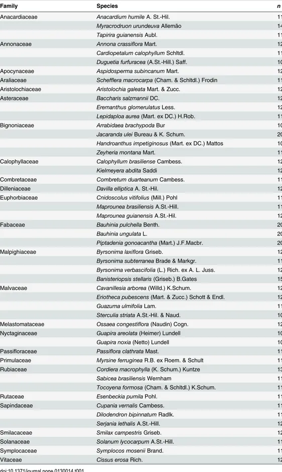
Samples of leaves were collected from 50 species in the Cerrado biome in central Brazil, at IBGE Ecological Reserve (Table 1). IBGE (Brazilian Institute of Geography and Statistics) al-lows the use of samples for scientific research purposes. At least four leaves (one per individual) were sampled for each species. All samples were obtained from fully expanded leaves collected from the third and fourth nodes from the branch tip. Middle regions of the leaf, including the midrib, were fixed in FAA 70 (Formalin, Acetic acid, 70% Alcohol) for 48 hours [40]. These were dehydrated in an ethanol series and embedded in paraffin. The thickness of the cross sec-tions was 8μm. The sections were stained with astra blue 1% and basic fuchsin 1%, both from Sigma, and mounted with Entellan1. The images of midribs were captured in 10x objective lens, using a trinocular microscope Axio Lab A1 coupled to a digital camera Axiocam ICc 1.
The image was pre-processed to remove the background by manually segmenting the region of interest, so that only the region of the midrib was analyzed by the fractal descriptors, as shown inFig 1. In the following, the combination of Bouligand-Minkowski/Fourier fractal de-scriptors proposed in this study was used to obtain the meaningful features of each sample. Fi-nally, these features are employed in the input of a supervised classifier, which predicts the species of each sample. The classification scheme divides the samples into a training and a test-ing set, ustest-ing a 10-fold cross-validation procedure, as described in [41]. The classifier was the Linear Discriminant Analysis (LDA) [41], which has demonstrated to be a suitable method for plant image analysis [38]. The results were compared to other state-of-the-art and classical de-scriptors, that is, Local Binary Patterns [42] and Gabor-wavelets Descriptors [43].
Fractal Geometry
A fractal is a geometric structure characterized by two main properties: infinite self-similarity, that is, at any scale, the object is composed by copies of itself, and infinite complexity, that is, there are different details to be observed at any scale.
Table 1. Family, species and number of samples (n) per species used in the experiments.
Family Species n
Anacardiaceae Anacardium humileA. St.-Hil. 11
Myracrodruon urundeuvaAllemão 14
Tapirira guianensisAubl. 11
Annonaceae Annona crassifloraMart. 12
Cardiopetalum calophyllumSchltdl. 11
Duguetia furfuracea(A.St.-Hill.) Saff. 10
Apocynaceae Aspidosperma subincanumMart. 12
Araliaceae Schefflera macrocarpa(Cham. & Schltdl.) Frodin 11
Aristolochiaceae Aristolochia galeataMart. & Zucc. 12
Asteraceae Baccharis salzmanniiDC. 12
Eremanthus glomerulatusLess. 12
Lepidaploa aurea(Mart. ex DC.) H.Rob. 11
Bignoniaceae Arrabidaea brachypodaBur 10
Jacaranda uleiBureau & K. Schum. 20
Handroanthus impetiginosus(Mart. ex DC.) Mattos 10
Zeyheria montanaMart. 11
Calophyllaceae Calophyllum brasilienseCambess. 12
Kielmeyera abditaSaddi 12
Combretaceae Combretum duarteanumCambess. 11
Dilleniaceae Davilla ellipticaA. St.-Hil. 12
Euphorbiaceae Cnidoscolus vitifolius(Mill.) Pohl 11
Maprounea brasiliensisA.St.-Hill. 11
Maprounea guianensisA.St.-Hil. 12
Fabaceae Bauhinia pulchellaBenth. 20
Bauhinia ungulataL. 20
Piptadenia gonoacantha(Mart.) J.F.Macbr. 20
Malpighiaceae Byrsonima laxifloraGriseb. 12
Byrsonima subterraneaBrade & Markgr. 11
Byrsonima verbascifolia(L.) Rich. ex A. L. Juss. 12
Banisteriopsis stellaris(Griseb.) B.Gates 15
Malvaceae Cavanillesia arborea(Willd.) K.Schum. 12
Eriotheca pubescens(Mart. & Zucc.) Schott & Endl. 12
Guazuma ulmifoliaLam. 11
Sterculia striataA.St.-Hil. & Naud. 10
Melastomataceae Ossaea congestiflora(Naudin) Cogn. 12
Nyctaginaceae Guapira areolata(Heimer) Lundell 10
Guapira noxia(Netto) Lundell 10
Passifloraceae Passiflora clathrataMast. 11
Primulaceae Myrsine ferrugineaR.B. ex Roem. & Schult 11
Rubiaceae Cordiera macrophylla(K. Schum.) Kuntze 13
Sabicea brasiliensisWernham 11
Tocoyena formosa(Cham. & Schltdl.) K.Schum. 11
Rutaceae Esenbeckia pumilaPohl. 11
Sapindaceae Cupania vernalisCambess. 11
Dilodendron bipinnatumRadlk. 11
Serjania lethalisA.St.-Hill. 12
Smilacaceae Smilax campestrisGriseb. 12
Solanaceae Solanum lycocarpumA.St.-Hill. 11
Symplocaceae Symplocos moseniiBrand. 11
Vitaceae Cissus erosaRich. 12
linearly withu, in fractals this relation is exponential and the fractal dimensionDXofXis given by:
DX/limu
!0 logN
logu:
In the real-world there is no fractal structure, in the strict sense of the word, even because the range of scales is always finite. However, it is quite common to find objects with high com-plexity and self-similarity at particular ranges of scales. Based on such observation, several methods have been proposed to obtain meaningful information about an object based on a
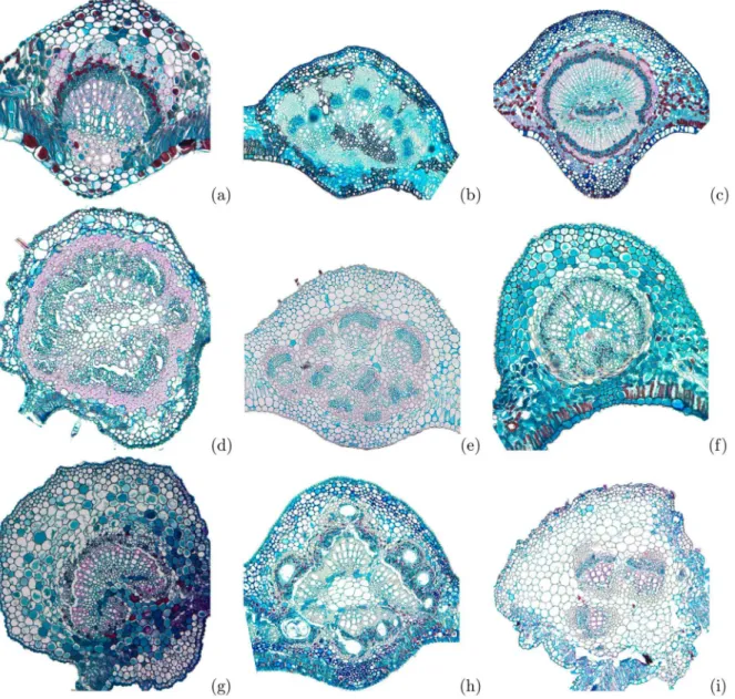
Fig 1. Histological samples of some leaves midrib cross-section used in the experiments.(a)Banisteriopsis stellaris, (b)Cardiopetalum calophyllum, (c)Cordiera macrophylla, (d)Dilodendron bipinnatum, (e)Guapira noxia, (f)Myrsine ferruginea, (g) Sabicea brasiliensis, (h)Tapirira guianensisand (i)
Lepidaploa aurea.
fractal geometry modeling [44–46]. Most of these studies employ the fractal dimension, alone or associated to other traditional measures. There are a number of methods to estimate the fractal dimensionDRof real-world objects. Each one may result in a different value and is more useful for a particular application, but all of them are based in the following bilogarithmic ex-pression:
DR/lim
!0
MðÞ
log;
whereMis the fractality measure and is specific for each method andis the scale parameter. Even though the fractal dimension is a powerful descriptor and enough to model some com-plex systems, it has some outstanding drawbacks. First, it is a unique real value and cannot ex-press all the richness of a structure at all scales. Besides, unlike the case of mathematical fractals, the fractal dimension of real-world objects changes depending on the scale range con-sidered. To make possible a more robust analysis based on fractal geometry, some methods that extend the fractal dimension concept have been proposed, such as the multifractals [47,
48], the multiscale fractal dimension [26,49] and the fractal descriptors [50,51]. This study focus on fractal descriptors, given the remarkable results achieved by this approach in previous studies on plant image analysis [26,29,37,38].
Fractal Descriptors. Fractal descriptors [37,50,51] extend the fractal dimension concept by using all the values in the fractality function. In this way, the set of features (descriptors)d
are given by:
D: log! logMðÞ:
The values of this function can be used directly [38] or after a transform to highlight some particular characteristic of the features [51]. They also can be extracted from the entire image [50] or using a recursive decomposition [51]. In any case, they quantify the morphology of the object of interest and its spatial distribution.
Proposed Methodology
The structural morphology quantified by fractal descriptors is of great importance in the analy-sis of any natural structure and particularly to describe the shape and visual textures of plant leaves, since the leaf morphology is directly affected by its biological structure and evolutionary history. These are key elements to determine the species to which each sample belongs. A num-ber of studies proposed in the literature confirms the efficiency of fractal descriptors in the analysis of leaves. For example, in [37] and [26], fractal descriptors were employed to identify plant species based on the leaf shape with a good accuracy, whereas in [38] the visual texture of the leaf was quantified by means of fractal descriptors and the results confirmed the precision of fractal descriptors as well.
Bouligand-Minkowski Fractal Descriptors
Proposed in [37], the Bouligand-Minkowski fractal descriptors of a gray-level image are ob-tained from the values of dilation volumes used to compute the Bouligand-Minkowski fractal dimension [38]. These descriptors have demonstrated to be a powerful method to analyze plant structures [38].
LetI:[1:M] × [1:N]! <be a function representing the gray-level image. The first step is to map such image onto a three-dimensional surfaceS, where each pixel in the coordinate (x,y) is mapped onto a point with coordinates (x,y,I(x,y)):
S¼ fðx;y;zÞjðx;yÞ 2 ½1 :M ½1 :N;z¼Iðx;yÞg:
In the following, the surface is dilated by a sphere with radiusr, that is, each point with coordi-nates (x,y,z) is replaced by a sphere with center at (x,y,z) and radiusrand the dilated struc-ture corresponds to the points pertaining to the union of such spheres. The radius is increased up to a pre-defined maximumrmaxand the volume of the dilated surfaceV(r) is given by:
VðrÞ ¼P
wDðrÞ½ðx;y;zÞ;
whereχis the indicator function andS(r) is the set of points in the dilated structure:
S>ðrÞ ¼ fðx;y;zÞj½ðx PxÞ 2
þ ðy PyÞ 2
þ ðz PzÞ 2
1=2rg;
where (Px,Py,Pz)2S.
The Bouligand-Minkowski descriptorsDBMare obtained by
DBM ¼ logVðrÞjrmax
r¼0:
Fourier Fractal Descriptors
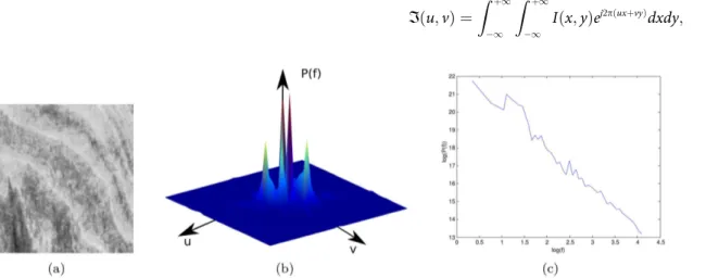
Fourier fractal descriptors [39] are named after the Fourier fractal dimension. This is computed from the logarithmic relation between the Fourier power spectrum and the frequency (Fig 2). At first, the Fourier transformIof the image is obtained by:
Iðu;vÞ ¼
Z þ1
1
Z þ1
1
Iðx;yÞej2pðuxþvyÞdxdy; ð1Þ
Fig 2. Fourier method to estimate the fractal dimension.(a) A texture image. (b) Fourier spectrumP(f). (c) Plot of log(P(f)) × log(f). This curve provides the descriptors of the texture.
wherejis the imaginary number anduandvare the orthogonal components of the frequency
f ¼ ffiffiffiffiffiffiffiffiffiffiffiffiffiffi
u2
þv2
p
. The resulting data is composed by complex numbers without any physical meaning, suggesting to use other measures obtained from the transform, like the power spec-trumP, given by:
P¼R2
þJ2
; ð2Þ
whereRandJare, respectively, the real and imaginary parts of the transform. As stated in [39], the following empirical law is observed for any fractal-like structure:
logðPÞ / logðfÞa;
whereαis a non-negative real-valued exponent used to estimate the fractal dimension. The Fourier fractal descriptors, within an empirically determined range of frequencies [fmin,
fmax], are given by
DF¼ logPðfÞjfmax
fmin:
Karhunen-Lo
è
ve Transform
Let the Bouligand-Minkowski descriptors be represented by a vector withn1components, ~
DBM ¼ fx1;x2; :::;xn1g, and the Fourier descriptors by a vector withn2components, ~
DF ¼ fy1;y2; :::;yn2g. The feature matrix of a database ofmtexture images contains in each
row the descriptors of each image. For the above descriptors, we haveMð1Þ
mn1for the
Bouligand-Minkowski descriptors andMð2Þ
mn2for the Fourier descriptors.
For each feature matrix, covariance matrixSis provided by:
Sði;jÞ ¼
Pn
i¼1ðMð:;iÞ Mð:;iÞÞðMð:;jÞ Mð:;jÞÞ
n 1 ; ð3Þ
wherenis the number of columns in the feature matrix,M(.,i) represents the columniofM
andMð:;iÞis the average column-vector.
The next step is to compute the eigenvalues and eigenvectors ofS. A non-null vectoreis an eigenvector ofSif:
Se¼le; ð4Þ
for any real valueλ.λis an eigenvalue of the matrix.
The eigenvalues ofSare sorted decreasinglyλ1λ2. . .λnand the respective
eigen-vec-torse1,e2,. . .,enare the columns of a linear transform matrixU.
The descriptor matricesM(1)andM(2)are horizontally concatenated intoM(C), such that each row ofM(C)is given byx1,x2,xn1,y1,y2,. . .,yn2. In the following, the combined matrix is multiplied by the transpose ofUgiving rise to the transformed matrix:
DðCÞ¼UTMðCÞ: ð5Þ
Results and Discussion
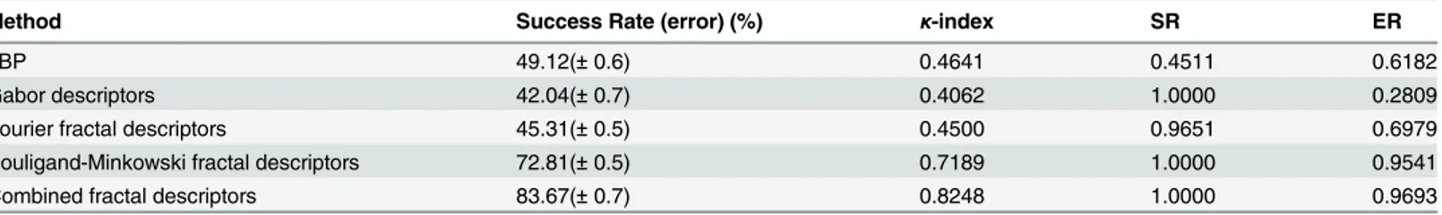
Table 2shows the performance of different texture descriptors in the identification of the ana-lyzed plant species. Besides the ratio of samples correctly classified (Success Rate) and the re-spective cross-validation error, the table also shows three other statistical metrics regarding the robustness of the result, i.e,κ-index, success reliability (SR) and error reliability (ER). Theκ -index quantifies (in statistical terms) how better the classifier is than a random classification. Reliability refers to consistency, it measures the degree of reality and stability of a measure-ment, evaluating if the measure will be the same in every execution. Success and error reliability are metrics derived from thea posterioriprobabilities of the classifier, being the averagea poste-rioriprobability for samples correctly and incorrectly classified, respectively. For each sample, classifiers like LDA output one probability score for each possible class and the class assigned to the sample is that having the highest probability. A reliable method is expected to have this highest probability significantly larger than the sum of all the other probabilities and this is what is assessed by the reliability metric. Generally speaking, the proposed method achieved the greatest rate of plants classified correctly, with a substantial advantage over other classical and state-of-the-art approaches, like LBP for instance. It also presented the highestκindex and a more robust reliability (Gabor presented the same SR, but much smaller ER, while LBP pre-sented smaller values for both SR and ER).
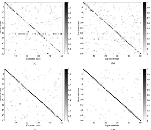
Fig 3shows the confusion matrices for the main compared approaches (LBP, Gabor, Bouli-gand-Minkowski and the proposed method). Confirming its higher success rate value, the combined fractal descriptors provided the most accurate identification of the analyzed species. When compared to LBP and Gabor, the best performance of the proposed method is evidenced by the much smaller number of gray points outside the diagonal. When compared to Bouli-gand-Minkowski, the greater precision of the proposal is not so obvious, but it is observed for some classes, like 3 and 10. These are species where the cross-sections show more periodic pat-terns and where the frequency analysis gives relevant information.
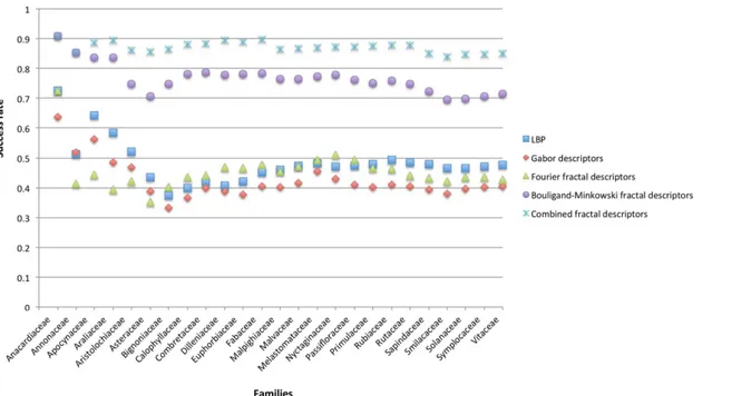
To verify how successful is the use of midrib in identifying species from the same family, the average success rate of the species belonging to the same family was calculated and presented at
Fig 4. When the identification is performed considering the species, the proposed method achieved 83.67% of success rate, however, when the success rate of each family is calculated, the proposed method achieves 87.29% of correct identification. This means that at least 4% of the error is inside the family level, what is expected since the species belonging to the same fam-ily have substantial similarities.
The results above confirm what was expected from the theory background of each method concerning the perspective that each one shows from the image. Unlike Gabor, LBP and other approaches, fractal descriptors are conceived to model the natural composition law of
Table 2. Success rates and other statistical measures of the proposed method compared to other literature approaches to classify the same set of plant species.
Method Success Rate (error) (%) κ-index SR ER
LBP 49.12(±0.6) 0.4641 0.4511 0.6182
Gabor descriptors 42.04(±0.7) 0.4062 1.0000 0.2809
Fourier fractal descriptors 45.31(±0.5) 0.4500 0.9651 0.6979
Bouligand-Minkowski fractal descriptors 72.81(±0.5) 0.7189 1.0000 0.9541
Combined fractal descriptors 83.67(±0.7) 0.8248 1.0000 0.9693
biological structures. Such law is based on the self-replication of elements at different scales whereas this replication is also inherent to the self-similar nature of fractals. Particularly, the method proposed here combines two complementary ways of extracting fractal features. While the dilation volumes in Bouligand-Minkowski express the spatial morphology of the midrib, the Fourier method analyses the complexity of the frequency distribution. The combination by the KL transform results in a solution capable of identifying species using a simple and inex-pensive setup and using a material that can be collected in most cases effortlessly at any time.
The identification of plant species using leaves is naturally a very challenging problem due to the high intra-species dissimilarity and inter-species similarity. Leaf variation occurs at every hierarchical level: within and among plants, populations, and species. In some species subject to different environmental conditions, marked phenotypic differences in leaves can occur during the development. Leaf variation within individuals may also occur regardless of environmental conditions, as part of the normal developmental pattern and seasonal changes, even among sequential leaf position on a stem. Nevertheless, the midrib proved to be a promis-ing structure in the task of identifypromis-ing plants.
Fig 3. Confusion matrices of the main compared approaches.(a) Gabor descriptors. (b) Local Binary Pattern. (c) Bouligand-Minkowski fractal descriptors. (d) Combined fractal descriptors.
In this context, the midrib of a leaf contains vascular bundles, associated fundamental tis-sues (parenchyma and/or collenchyma and/or sclerenchyma) and epidermis. Vascular tistis-sues (xylem and phloem), which compose the midrib bundles vary in quantity and in their spatial disposal. In addition, the vascular system may be formed by a single bundle or be formed by a continuous or an interrupted arch, depending on species [19]. The characteristics of the funda-mental tissues such as cell wall thickness, the presence of secretory cells or structures, and their distribution within the midrib also vary with the species. Similarly, depending on the species, the epidermis can vary depending on the presence or absence of trichomes and their type, the shape and size of its cells, cuticular thickness, etc [52]. Thus, anatomical studies that address the taxonomic aspect traditionally describe these tissues seeking some feature that can distin-guish the species. The qualitative description of these features is a laborious task, however, quantitative data from midrib would be complicated to be obtained by methods which are commonly used in Botany. In this sense, the computational method proposed here obtained very informative measures of texture from the median ribs, being able to differentiate between species. For these reasons, this method is very promising for the present and forthcoming sci-ence, which has sought the automatic identification of species, facilitating studies across the wide diversity of plants occurring in the world.
Conclusions
This study proposed to identify plant species of a tropical savanna of Brazil by extracting fractal descriptors of leaf midrib histological cross-sections. The proposed solution combines Bouli-gand-Minkowski and Fourier fractal descriptors to provide features for the leaf images. These features are categorized by a state-of-the-art classifier method, making possible the correct identification of the species.
Fig 4. Averaged success rate of the species belonging to the same family considering the main compared approaches.
The results confirmed what was expected from the fractal descriptors theory, thus the pro-posed method achieved a great precision in the species identification, outperforming other im-aging techniques and making possible to obtain an automatic and precise categorization using basic biological procedures. We can also conclude that the midrib is a region of the leaf that can provide relevant information in the process of identification of plant species. Therefore, fu-ture studies should take into account both the characteristics of the median vein and of the me-sophyll, which would increase the rate of discrimination among species.
Acknowledgments
Núbia R. Silva, João B. Florindo, Rosana M. Kolb and Odemir M. Bruno are grateful for the support from The State of São Paulo Research Foundation (FAPESP) of the respective grants Nos. 2011/21467-9, 2012/19143-3, 2011/23112-3 and 2011/01523-1. Bruno also acknowledges the financial support of the National Council for Scientific and Technological Development (CNPq), grant Nos. 307797/2014-7 and 484312/2013-8 and Rosana M. Kolb acknowledges PROPe UNESP (edital14/2012/Programa Renove).
Author Contributions
Conceived and designed the experiments: DRR JBF NRS OMB RMK. Performed the experi-ments: JBF NRS. Analyzed the data: DRR JBF NRS OMB RMK. Contributed reagents/materi-als/analysis tools: RMK DRR MCG OMB. Wrote the paper: DRR JBF NRS OMB RMK.
References
1. Mayr E (1985) The Growth of Biological Thought: Diversity, Evolution, and Inheritance. Belknap Press of Harvard University Press.
2. Streckeisen A (1979) Classification and nomenclature of volcanic rocks, lamprophyres, carbonatites, and melilitic rocks: Recommendations and suggestions of the iugs subcommission on the systematics of igneous rocks. Geology 7: 331–335. doi:10.1130/0091-7613(1979)7%3C331:CANOVR%3E2.0. CO;2
3. Streckeisen A (1980) Classification and nomenclature of volcanic rocks, lamprophyres, carbonatites and melilitic rocks iugs subcommission on the systematics of igneous rocks. Geologische Rundschau 69: 194–207. doi:10.1007/BF01869032
4. Stevens PF (1994) The Development of Biological Systematics. Columbia University Press.
5. Greene EL (1983) Landmarks of Botanical History. Number pt. 2 in Landmarks of Botanical History. Stanford University Press.
6. Hennig W, Davis D, Zangerl R (1999) Phylogenetic Systematics. University of Illinois Press.
7. Sneath P, Sokal R (1973) Numerical Taxonomy: The Principles and Practice of Numerical Classifica-tion. A Series of books in biology. W. H. Freeman.
8. Cronquist A (1981) An Integrated System of Classification of Flowering Plants. Columbia University Press.
9. Davis PH, Heywood VH (1963) Principles of Angiosperm Taxonomy. Edinburgh and London: Oliver and Boyd, 1 edition.
10. Stuessy T, Mayer V, Hörandl E (2003) Deep morphology: toward a renaissance of morphology in plant systematics. Regnum vegetabile. A.R.G. Gantner.
11. Endress P (2002) Morphology and angiosperm systematics in the molecular era. The Botanical Review 68: 545–570. doi:10.1663/0006-8101(2002)068%5B0545:MAASIT%5D2.0.CO;2
12. Eyde RH (1975) The bases of angiosperm phylogeny: Floral anatomy. Annals of the Missouri Botanical Garden 62: 521–537. doi:10.2307/2395266
13. Judd WS, Campbell CS, Kellog EA, Stevens PF, Donoghue MJ (2008) Plant Systematics: A Phyloge-netic Approach. Sinauer Associates, third edition, 620 pp.
14. Dahlgren R (1983) General aspects of angiosperm evolution and macrosystematics. Nordic Journal of Botany 3: 119–149. doi:10.1111/j.1756-1051.1983.tb01448.x
16. Rathcke B, Lacey EP (1985) Phenological patterns of terrestrial plants. Annual Review of Ecology and Systematics 16: 179–214. doi:10.1146/annurev.es.16.110185.001143
17. Hickey LJ, Wolfe JA (1975) The bases of angiosperm phylogeny: Vegetative morphology. Annals of the Missouri Botanical Garden 62: 538–589. doi:10.2307/2395267
18. Donoghue MJ, Sanderson MJ (1992) The suitability of molecular and morphological evidence in recon-structing plant phylogeny. In: Soltis P, Soltis D, Doyle J, editors, Molecular Systematics of Plants, Springer US. pp. 340–368.
19. Dickison WC (2000) Integrative Plant Anatomy. San Diego: Academic Press, 546 pp.
20. Evert RF (2006) Esau’s Plant Anatomy, Meristems, Cells, and Tissues of the Plant Body: their Struc-ture, Function, and Development. Wiley, Hoboken, 624 pp.
21. de Mesquita Sá Junior JJ, Rossatto DR, Kolb RM, Bruno OM (2013) A computer vision approach to quantify leaf anatomical plasticity: a case study onGochnatia polymorpha(Less.) Cabrera. Ecological Informatics 15: 34–43. doi:10.1016/j.ecoinf.2013.02.007
22. Foroughbakhch R, Ferry RJ Sr, Hernández-Piñero JL, Alvarado-Vázquez MA, Rocha-Estrada A (2008) Quantitative measures of leaf epidermal cells as a taxonomic and phylogenetic tool for the identification ofStanhopeaspecies (Orchidaceae). Phyton 77: 113–127.
23. Araújo JS, Azevedo AA, Silva LC, Meira R (2010) Leaf anatomy as an additional taxonomy tool for 16 species of Malpighiaceae found in the cerrado area (Brazil). Plant Systematics and Evolution 286: 117–131. doi:10.1007/s00606-010-0268-3
24. Ghimire B, Lee C, Yang J, Heo K (2015) Comparative leaf anatomy of native and cultivatedPinus (Pina-ceae) in Korea: implication for the subgeneric classification. Plant Systematics and Evolution 301: 531–540. doi:10.1007/s00606-014-1090-0
25. Sá Junior JJM, Backes AR, Rossatto DR, Kolb RM, Bruno OM (2011) Measuring and analyzing color and texture information in anatomical leaf cross sections: an approach using computer vision to aid plant species identification. Botany 89: 467–479. doi:10.1139/b11-038
26. Plotze RO, Falvo M, Pádua JG, Bernacci LC, Vieira MLC, Oliveira GCX, et al. (2005) Leaf shape analy-sis using the multiscale minkowski fractal dimension, a new morphometric method: a study with Passi-flora(Passifloraceae). Canadian Journal of Botany 83: 287–301. doi:10.1139/b05-002
27. Wang XF, Du JX, Zhang GJ (2005) Recognition of leaf images based on shape features using a hyper-sphere classifier. In: Huang DS, Zhang XP, Huang GB, editors, Advances in Intelligent Computing, Springer Berlin Heidelberg, volume 3644 ofLecture Notes in Computer Science. pp. 87–96.
28. Mugnai S, Pandolfi C, Azzarello E, Masi E, Mancuso S (2008)Camellia japonica L.genotypes identified by an artificial neural network based on phyllometric and fractal parameters. Plant Systematics and Evolution 270: 95–108. doi:10.1007/s00606-007-0601-7
29. Rossatto DR, Casanova D, Kolb RM, Bruno OM (2011) Fractal analysis of leaf-texture properties as a tool for taxonomic and identification purposes: a case study with species from neotropical Melastomata-ceae (Miconieae tribe). Plant Systematics and Evolution 291: 103–116. doi: 10.1007/s00606-010-0366-2
30. Metcalfe C, Chalk L (1950) Anatomy of the dicotyledons: leaves, stem, and wood in relation to taxono-my, with notes on economic uses. Number v. 2 in Anatomy of the Dicotyledons: Leaves, Stem, and Wood in Relation to Taxonomy, with Notes on Economic Uses. Clarendon Press.
31. Keating RC (1984) Leaf histology and its contribution to relationships in the Myrtales. Annals of the Mis-souri Botanical Garden 71: 801–823. doi:10.2307/2399163
32. Niinemets U, Lukjanova A, Turnbull MH, Sparrow AD (2007) Plasticity in mesophyll volume fraction modulates light-acclimation in needle photosynthesis in two pines. Tree Physiology 8: 1137–1151. doi: 10.1093/treephys/27.8.1137
33. Mantovani A, Pereira TE, Coelho MAN (2009) Leaf midrib outline as a diagnostic character for taxono-my inAnthuriumsectionUrospadixsubsectionFlavescentiviridia(Araceae). Hoehnea 36: 269–277. doi:10.1590/S2236-89062009000200005
34. Dalvi VC, Meira RMSA, Francino DMT, Silva LC, Azevedo AA (2014) Anatomical characteristics as tax-onomic tools for the species ofCurtiaandHockinia(Saccifolieae-Gentianaceae Juss.). Plant Systemat-ics and Evolution 300: 99–112. doi:10.1007/s00606-013-0863-1
35. Silva NR, Florindo JB, Gomez MC, Kolb RM, Bruno OM (2014) Fractal descriptors for discrimination of microscopy images of plant leaves. Journal of Physics: Conference Series 490: 012085.
36. Casanova D, de Mesquita Sa Junior JJ, Bruno OM (2009) Plant leaf identification using gabor wavelets. International Journal of Imaging Systems and Technology 19: 236–243. doi:10.1002/ima.20201
38. Backes AR, Casanova D, Bruno OM (2009) Plant leaf identification based on volumetric fractal dimen-sion. International Journal of Pattern Recognition and Artificial Intelligence (IJPRAI) 23: 1145–1160. doi:10.1142/S0218001409007508
39. Florindo JB, Bruno OM (2012) Fractal descriptors based on fourier spectrum applied to texture analysis. Physica A 391: 4909–4922.
40. Johansen DA (1940) Plant microtechnique. New York, London, McGraw-Hill Book Company, inc.
41. Duda RO, Hart PE (1973) Pattern Classification and Scene Analysis. New York: Wiley.
42. Pietikäinen M, Hadid A, Zhao G, Ahonen T (2011). Computer vision using local binary patterns.
43. Manjunath B, Ma W (1996) Texture features for browsing and retrieval of image data. IEEE Transac-tions on Pattern Analysis and Machine Intelligence 18: 837–842. doi:10.1109/34.531803
44. Tian-Gang L, Wang S, Zhao N (2007) Fractal research of pathological tissue images. Computerized Medical Imaging and Graphics 31: 665–671.
45. Wang H, Siopongco J, Wade LJ, Yamauchi A (2009) Fractal analysis on root systems of rice plants in response to drought stress. Environmental and Experimental Botany 65: 338–344. doi:10.1016/j. envexpbot.2008.10.002
46. Xiang Du J, Zhai CM, Wang QP (2013) Recognition of plant leaf image based on fractal dimension fea-tures. Neurocomputing 116: 150–156. doi:10.1016/j.neucom.2012.03.028
47. Harte D (2001) Multifractals: theory and applications. Boca Raton: Chapman and Hall/CRC.
48. Lopes R, Betrouni N (2009) Fractal and multifractal analysis: A review. Medical Image Analysis 13: 634–649. doi:10.1016/j.media.2009.05.003PMID:19535282
49. Manoel ETM, da F Costa L, Streicher J, Muller GB (2002) Multiscale fractal characterization of three-di-mensional gene expression data. In: Gonçalves LMG, Musse SR, Comba JLD, Giraldi G, Dreux M, edi-tors, SIBGRAPI. Washington, DC: IEEE Computer Society, pp. 269–274.
50. Florindo JB, Backes AR, de Castro M, Bruno OM (2012) A comparative study on multiscale fractal di-mension descriptors. Pattern Recognition Letters 33: 798–806. doi:10.1016/j.patrec.2011.12.016
51. Forindo JB, Bruno OM (2013) Texture analysis by multi-resolution fractal descriptors. Expert Systems with Applications 40: 4022–4028. doi:10.1016/j.eswa.2013.01.007