www.bjorl.org.br
Brazilian Journal of
OtOrhinOlaryngOlOgy
1808-8694/$ - see front matter © 2014 associação Brasileira de Otorrinolaringologia e Cirurgia Cérvico-Facial. Published by Elsevier Editora ltda. all rights reserved.
DOi: 10.5935/1808-8694.20140008
ORIGINAL ARTICLE
Localization of ectopic and supernumerary parathyroid glands
in patients with secondary and tertiary hyperparathyroidism:
surgical description and correlation with preoperative
ultrasonography and Tc99m-Sestamibi scintigraphy
José Santos Cruz de Andrade
a, João Paulo Mangussi-Gomes
a,*, Lillian Andrade da Rocha
b,
Monique Nakayama Ohe
c, Marcello Rosano
a, Murilo Catafesta das Neves
a,
Rodrigo de Oliveira Santos
a,ba Department of Otolaryngology, Head and Neck Surgery, Universidade Federal de São Paulo / Escola Paulista de Medicina
(UNI-FESP/EPM), São Paulo, SP, Brazil
b Department of Nephrology, Universidade Federal de São Paulo / Escola Paulista de Medicina (UNIFESP/EPM), São Paulo, SP, Brazil c Department of Clinical Endocrinology, Universidade Federal de São Paulo / Escola Paulista de Medicina (UNIFESP/EPM), São
Paulo, SP, Brazil
Received 22 June, 2013; accepted 12 October, 2013
KEYWORDS
Hyperparathyroidism; Parathyroid glands; Parathyroidectomy; Chronic renal insufi -ciency;
Ultrasonography; Scintigraphy
Abstract
Introduction: Hyperparathyroidism is an expected metabolic consequence of chronic kidney disease (CKD). Ectopic and/or supernumerary parathyroid glands (PT) may be the cause of sur-gical failure in patients undergoing total parathyroidectomy (PTX).
Aim: To deine the locations of ectopic and supernumerary PT in patients with renal hyperpara
-thyroidism and to correlate intraoperative indings with preoperative tests.
Materials and methods: A retrospective study was conducted with 166 patients submitted to PTX. The location of PT during surgery was recorded and classiied as eutopic or ectopic. The
preoperative localizations of PT found by ultrasonography (USG) and Tc99m-Sestamibi
scintig-raphy (MIBI) were subsequently compared with intraoperative indings.
Results: In the 166 patients studied, 664 PT were found. Five-hundred-seventy-seven (86.4%) glands were classiied as eutopic and 91(13.6%) as ectopic. Eight supernumerary PT were found. The most common sites of ectopic PT were in the retroesophageal and thymic regions. Taken together, USG and MIBI did not identify 56 (61.5%) ectopic glands. MIBI was positive for 69,7% of
all ectopic glands located in the mediastinal and thymic regions.
Conclusion: The presence of ectopic and supernumerary PT in patients with renal hyperpara
-thyroidism is signiicant. Although preoperative imaging tests did not locate most of ectopic
glands, MIBI may be important for identifying ectopic PT in the mediastinal and thymic regions.
© 2014 Associação Brasileira de Otorrinolaringologia e Cirurgia Cérvico-Facial. Published by Elsevier
Editora Ltda. All rights reserved.
Please cite this article as: Andrade JS, Mangussi-Gomes J, Rocha LA, Ohe MN, Rosano M, Neves MC, et al. Localization of ectopic and supernumerary parathyroid glands in patients with secondary and tertiary hyperparathyroidism: Surgical description and correlation with preoperative ultrasonography and Tc99m-Sestamibi scintigraphy. Braz J Otorhinolaryngol. 2014;80:29-34.
* Corresponding author.
Introduction
Hyperparathyroidism is a physiological response to meta-bolic changes that occurs in patients with chronic kidney disease (CKD). In spite of advances in the clinical treatment of these patients, a signiicant number develops parathyroid (PT) hyperfunction and autonomy, with consequent eleva -tion of parathyroid hormone (PTH). When the harmful ef-fects of hyperparathyroidism are identiied, such as severe bone disease, surgical treatment becomes necessary.1
Thus, total parathyroidectomy (PTx) in CKD has been indi-cated in symptomatic patients with marked and non-suppress -ible elevations of PTH.1 The goal of surgery is to correctly
iden-tify and resect all PTs. Although the hyperplastic glands are much larger than normal due to kidney disease, their identii -cation is not always simple.2,3 Moreover, although the inding of four glands is expected in most patients, there is signiicant variability in the number of PTs. Supernumerary glands are present in 2.5% to 30% of patients,4,5 and may be the reason
for surgical treatment failure, if not properly identiied.2,6 There is also considerable variation regarding the lo-cation of the PTs, which are often not found in their usu -al location. The identiication of -all glands may require a thorough exploration of the superior mediastinum regions, thyroid gland, carotid sheath, and retroesophageal area.5,7
As a result, imaging studies have been performed to quantify and locate the PTs before surgery. However, al -though widely used in patients with CKD-associated hyper -parathyroidism, it is not yet known whether ultrasonog -raphy (USG) and 99mTc-sestamibi (MIBI) scintig-raphy can affect surgical outcomes, complication rates, and long-term therapeutic success.8–10
This study aimed to evaluate the main locations of the PTs in patients with secondary (SHPT) and tertiary (THPT) hyperparathyroidism, based on intraoperative indings, and to correlate these indings with the preoperative ex -aminations.
Materials and methods
A cross-sectional study including patients with CKD treated at a tertiary referral hospital was performed. Between Feb -ruary of 2011 and October of 2012, 166 patients (44 diag -nosed with SHPT and 122 diag-nosed with THPT) underwent PTx with presternal parathyroid autotransplantation. All pa -tients were examined by a nephrologist specialized in renal osteodystrophy; surgical indication followed the criteria of clinical treatment failure: hypercalcemia and/or persistent hyperphosphatemia; pruritus; bone pain; fractures or high risk of fractures; skeletal deformities and/or calciications; calciphylaxis; and radiographic evidence of renal osteodys-trophy. Patients who had previously undergone PT surgery were excluded from the analysis.
Patients were referred for surgical evaluation after pre -operative imaging examinations had been performed at the service of origin or at the hospital in which the surgery was performed. USG and MIBI images reports from each patient were analyzed, the number of PTs observed, and their loca -tions were quantiied for each test. In cases where the MIBI image was not available for analysis by the surgeon, the examination report was considered. Preoperative imaging assessments were not performed by the same radiologist, nor in the same radiology department.
PALAVRAS-CHAVE Hiperparatireoidismo; Glândulas paratireoides; Paratireoidectomia; Insuiciência renal crônica;
Ultrassonograia; Cintilograia
Localização de glândulas paratireoides ectópicas e supranumerárias em pacientes com
hiper-paratireoidismo secundário e terciário: descrição cirúrgica e correlação com ultrassonograia e cintilograia Tc99m-Sestamibi pré-operatórios
Resumo
Introdução: O hiperparatireoidismo é uma consequência metabólica esperada na doença renal crônica (DRC). Paratireoides (PT) ectópicas e/ou supranumerárias podem ser causa de falha
cirúrgica nos pacientes submetidos à paratireoidectomia total (PTX).
Objetivo: Deinir cirurgicamente a localização das PT, em pacientes com hiperparatireoidismo
associado à DRC, correlacionar esses achados com os exames pré-operatórios.
Materiais e métodos: Foi conduzido um estudo retrospectivo com 166 pacientes submetidos à PTX. A localização das PT no intraoperatório foi registrada, sendo classiicada como tópica ou ectópica. A localização pré-operatória, deinida pela ultrassonograia (USG) e pela cintilograia
Tc99m-Sestamibi (MIBI), foi comparada com aos achados cirúrgicos.
Resultados: Nos 166 pacientes, foram identiicadas 664 PT. Foram classiicadas como tópicas e
ectópicas 577 (86,4%) e 91(13,6%) glândulas, respectivamente. Oito PT supranumerárias foram en
-contradas (7 tópicas e 1 ectópica). As localizações mais comuns de PT ectópicas foram as regiões re
-troesofágica e tímica. Associadas, a USG e a MIBI não identiicaram 56 glândulas (61,5%) ectópicas. Entretanto, a MIBI foi positiva para 69,7% daquelas localizadas nas regiões tímicas e mediastinal.
Conclusão: A presença de glândulas ectópicas e supranumerárias em pacientes com hiperpara
-tireoidismo associado à DRC é signiicativa. Os exames de imagem pré-operatórios não localiza
-ram a maioria das glândulas ectópicas. A MIBI pode ter importância na identiicação de PT nas regiões tímica e mediastinal.
© 2014 Associação Brasileira de Otorrinolaringologia e Cirurgia Cérvico-Facial. Publicado por Elsevier
All patients were submitted to surgery by the same sur -gical team. The sur-gical technique consisted of convention-al parathyroidectomy with bilaterconvention-al neck exploration and heterotopic presternal intramuscular autotransplantation, as previously described.11
During the procedure, the location of the PT was record -ed; it was considered normal when the lower glands were related to the lower pole of the thyroid gland, and when the upper glands were found near the upper pole of the thyroid, by the entry point of the inferior laryngeal nerve in the cricothyroid muscle.
When the PT was not identiied in its usual position, a systematic exploration of the following areas was per -formed: retroesophageal, carotid sheath, thymic tongue, and upper mediastinum. If even then the PT was not identi -ied, an ipsilateral total lobectomy of the thyroid gland was performed.
For those patients in whom the PT was not identiied intraoperatively, but with persistence or recurrence of dis -ease in the postoperative follow-up, the gland was classiied as ectopic and the case was considered a “surgical failure”. PT fragments were sent for cryopreservation in most pa -tients. Intraoperative frozen pathology was not performed in any case. Although the collection of intraoperative PTH (IOPTH) was performed in all cases (two blood samples, one at anesthetic induction, and another 20 minutes after re-moval of all glands), the result did not affect the surgical decision in any situation, due to the delay in obtaining these results.11
Only patients with a minimum postoperative follow-up period of 6 months were included in the study.
Finally, the data provided by radiological preoperative im -aging were compared with the intraoperative indings for each patient. The present study was approved by the research eth -ics committee of the institution under protocol No. 886/00.
Results
Of the 166 patients studied, 44 had a diagnosis of SHPT and 122 of THPT. Regarding gender, 82 were males and 84 were females. The mean age among patients with SHPT and THPT was 46.3 years (range: 23-67 years) and 48.1 years (range:
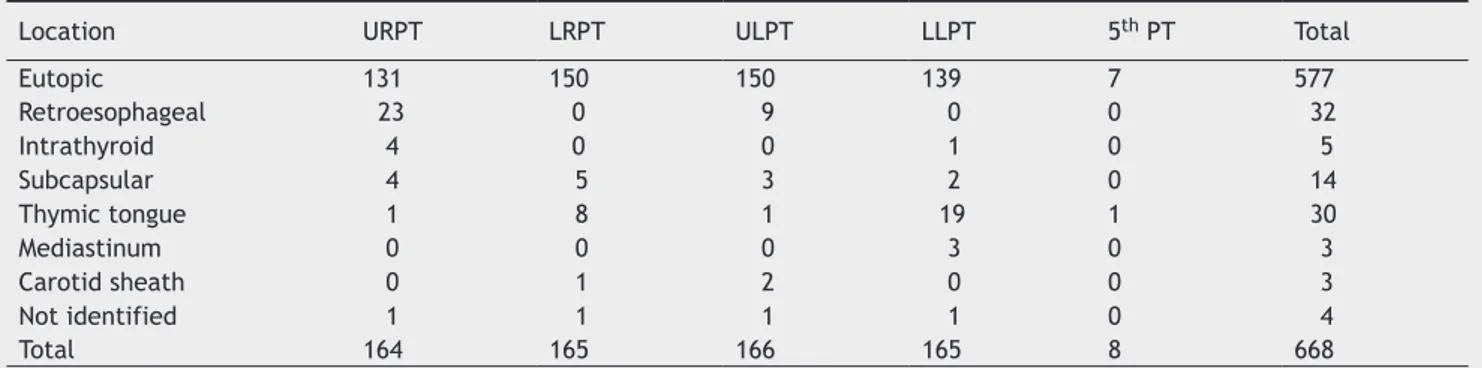
Table 1 Location of parathyroids, according to intraoperative indings.
Location URPT LRPT ULPT LLPT 5th PT Total
Eutopic 131 150 150 139 7 577
Retroesophageal 23 0 9 0 0 32
Intrathyroid 4 0 0 1 0 5
Subcapsular 4 5 3 2 0 14
Thymic tongue 1 8 1 19 1 30
Mediastinum 0 0 0 3 0 3
Carotid sheath 0 1 2 0 0 3
Not identified 1 1 1 1 0 4
Total 164 165 166 165 8 668
URPT, upper right parathyroid; LRPT, lower right parathyroid; ULPT, upper left parathyroid; LLPT, lower left parathyroid; 5th PT,
5th parathyroid (supernumerary).
21-72 years), respectively. The mean duration of dialysis in patients with SHPT was 10.2 years (range: 2-25 years) and in patients with THPT, 6.1 years (range: 0.5 to 17 years).
A total of 664 PTs were found in 166 patients undergoing surgery. Four PTs were identiied in 150 patients; ive PTs were identiied in eight patients (4.8%), and only three PTs in eight other patients (4.8%).
Of the eight patients with only three glands identiied intraoperatively, four (2.4%) did not present clinical and laboratory evidence of disease recurrence. The other four patients (2.4%) had evidence of persistent or recurrent hy -perparathyroidism in the postoperative follow-up, and their missing glands were considered ectopic. Surgical failure was, therefore, 2.4%.
Regarding the location of PTs, 577 (86.4%) glands were classiied as eutopic, and 91 (13.6%) as ectopic (87 located in non-usual position, and four that were not found intra -operatively).
Considering a total number of 668 PTs (ectopic and eu -topic), 664 glands (99.4%) were located. The surgical loca -tion of PTs is shown in Table 1.
Regarding the 87 ectopic glands, the upper-right PT (URPT) and lower-left PT (LLPT) were the most commonly found in non-usual positions, representing, 36.3% and 28.6% of total ectopic indings, respectively.
Regarding the URPT, it was considered ectopic in 24.6% of cases, and the retroesophageal position was the most fre -quent (71.8%). The LLPT, in turn, was classiied as ectopic in 17% of cases, and the thymic region was the most common location (76%).
Of the eight supernumerary PTs that were identiied, seven were located in eutopic positions. Only one was ecto -pic. Fig. 1 shows the location of ectopic PTs.
During the preoperative assessment, 153 patients (92.1%) underwent USG. The number of glands located is shown in Table 2. As for MIBI, 159 patients (95.7%) underwent the ex -amination. The number of glands located is shown in Table 3. The mean number of glands identiied by USG was 1.58 glands/exam. MIBI identiied a mean of 2.41 glands/exam.
The association between ectopic PTs and imaging tests was as follows:
Figure 1 Intraoperative identiication of ectopic parathyroids. The parathyroid glands that were not identiied intraoperatively were also included. URPT, upper right parathyroid; LRPT, lower right parathyroid; ULPT, upper left parathyroid; LLPT, lower left parathyroid;
5th PT, 5th parathyroid (supernumerary).
Table 2 Number of glands located by preoperative ultraso-nography.
Number of glands identified
Number of
examinations Percentage
0 31 20.3%
1 47 30.7%
2 45 29.4%
3 18 11.8%
4 12 7.8%
Total 153 100%
Table 3 Number of glands located by preoperative MIBI.
Number of glands identified
Number of
examinations Percentage
0 14 8.8%
1 32 20.1%
2 30 18.9%
3 38 23.9%
4 45 28.3%
Total 159 100%
• Of the ive intrathyroid PTs, only two were suggested by USG, and three were not identiied either by USG or by MIBI.
• Of the three mediastinal PTs, all were visualized by MIBI.
Regarding the 30 ectopic PTs located in the thymic tongue, ive were not identiied in any of the examinations, ten were identiied only by MIBI, ive were identiied only by USG, and ten were identiied by USG and MIBI.
No ectopic PT located in the subcapsular or retroesoph-ageal region was identiied by USG or MIBI before surgery. When considering only the regions of the upper mediasti-num and thymus, the USG was able to identify 45.5% of ec -topic PTs, all located in the thymic tongue. MIBI identiied 69.7% of the glands in these regions.
Preoperatively, the USG and MIBI correctly identiied 18 (19.8%) and 24 (26.4%) of ectopic PTs, respectively. Regard -ing ectopic PTs, 31 (34.1%) were identiied by USG and/or MIBI; 60 ectopic glands (65.9%) were not identiied by any of the preoperative examinations used in this study.
Discussion
Although IOPTH and frozen pathology were not used to con -irm the removal of all glands intraoperatively, 99.4% of PTs were correctly located in the sample presented. Further -more, only 2.4% of patients had surgical failure in the post -operative follow-up.
The main causes of failure in the surgical treatment of patients with hyperparathyroidism associated with CKD are failure to identify the PTs or presence of supernumerary
Not identiied
Carotid sheath
Mediastinum
Thymic tongue
Subcapsular
Intrathyroid
Retroesophageal
URPT
LRPT
ULPT
LLPT
5th PT
The low accuracy of USG and MIBI in identifying ectopic glands is explained by the limitations inherent to the meth-ods. In the case of retroesophageal PTs, for instance, USG suffers interference from the trachea, and MIBI, from the thyroid.8,9,18 In this series, no retroesophageal gland was suggested by imaging studies.
Fortunately, the capacity of these tests to locate PTs in the upper mediastinum and thymic regions is higher.8,16 During the intraoperative period, the dissection of the ret-roesophageal area is much simpler to perform, when com -pared to the upper mediastinum approach. Mediastinal dis-sections can hardly be performed without any suggestion of the presence of a gland in this region, due to the higher morbidity of this surgical approach.
MIBI was able to identify 69.7% of PTs in the upper me -diastinum and thymic tongue, versus only 45.5% identiied by the USG. All three cases of mediastinal PTs were shown by MIBI.
The superiority of MIBI when compared to USG to iden -tify PTs in the upper mediastinum and thymic regions has been described in the literature. In the study by Vulpio et al., USG did not identify any ectopic PT in the upper mediastinal or thymic regions versus 78% by MIBI.8 Simi-larly, Gasparri et al. demonstrated that preoperative MIBI was able to correctly identify 73.7% of ectopic glands in these regions.18
The real value of preoperative examinations in the con-text of hyperparathyroidism associated with CKD is yet to be determined. Some authors state that they do not change the primary surgery outcomes, as long as it is performed by an experienced surgeon.17,18 However, other authors have obtained good results with preoperative imaging analysis, and recommend the combined use of MIBI and USG in the preoperative evaluation of patients with hyperparathyroid -ism associated with CKD.9
This study was conducted in a public hospital with lim -ited resources. Any technology that helps in the treatment of these patients is always well accepted. However, their unavailability should not prevent or contraindicate the sur-gery. In this study, IOPTH and pathological anatomy were not considered in the intraoperative decisions, which also did not compromise the surgical outcome.
The USG assessment is recommended as the most im-portant preoperative examination, as it is more accessible and provides information regarding PTs and possible thyroid diseases, which are very prevalent in our country.Whenever possible, the additional utilization of MIBI to identify possi-ble ectopic glands in the thymic and mediastinal regions is recommended, although this situation represents a minority of patients.
Conclusion
The presence of supernumerary and ectopic parathyroid glands in patients undergoing PTx for CKD-associated hyper-parathyroidism is signiicant and justiies a careful intraop -erative search. An exploration routine for the most common sites of ectopic gland location is necessary, which in this study were the retroesophageal and thymic regions for the upper and lower parathyroids, respectively. Although preop -erative imaging exams did not identify the majority of ec-glands. Both situations can be related to the existence of
ectopic PTs.5
The frequency of ectopic PTs in the present study was 13.6%, similar to that found by other authors. Gomes et al.3 reported 18.9% ectopic glands, while the rate observed by Vulpio et al. was 12%.8
The main difference compared to the literature, howev -er, is the location of the ectopic glands. While in the pres-ent study the regions where ectopic PTs were most commonly found were, in descending order, retroesophageal areas (35.2% of all ectopic PTs, with upper PTs in 100% of cases) and thymus (32.9% of ectopic PTs, with lower PTs in 90% of cases), the lit -erature describes only the thymus region as the most common for the inding of ectopic PTs (lower, in most cases).3,12-14
All authors agree that the position of the gland is relat-ed to embryological development. The lower PTs originate from the third branchial arch together with the thymus, while the upper PTs originate from the fourth branchi -al arch. The migration of lower PTs, longer and closer to the thymic tissue, would determine the greater variability in their inal position.12-14
Another major cause of failure at the initial surgery is the presence of supernumerary PTs, identiied in 4.8% of patients in the present series. In the literature, the pres-ence of supernumerary glands is quite variable, ranging from 2.5% to 30%.4,5,14 The explanation for such a signii -cant variation may be related to differences in the surgical techniques used in each study.
Therefore, some authors recommend a routine cervical thymectomy during the surgical treatment of patients with hy -perparathyroidism associated with CKD.3,5,13,14 Pattou et al., for instance, in a study of 290 patients submitted to PTx and routine cervical thymectomy due to hyperparathyroidism asso-ciated with CKD, found supernumerary PTs located in the thymic region in 70 individuals (24.1% of total number of patients).5
The fact that routine cervical thymectomy was not per -formed in this study may explain the small number of su-pernumerary PTs found mainly in the thymus and/or upper mediastinum regions. Nevertheless, the surgical failure rate was only 2.4%, similar to that demonstrated by other au -thors. Tominaga et al., for instance, reported a 4.2% failure rate in 1,156 patients who underwent surgery.6
This apparent nonsensical inding is explained, as most supernumerary PTs found in the thymus actually represent small grafts of microscopic embryonic remnants. These grafts were unlikely to have the capacity to promote dis -ease recurrence and/or persistence.
The capacity of imaging studies to locate the PTs preop-eratively, especially ectopic glands, can greatly inluence the surgical outcome.9,15
In this series of patients, the USG located 1.58 PTs/exam and MIBI located 2.41 PTs/exam. Many studies have shown conlicting data on the sensitivity and speciicity of imag -ing studies in this group of patients.8,9,12,16 However, few have correlated the imaging indings with the intraoperative identiication of ectopic glands.
topic glands, MIBI may have an important role as a surgical planning method, in the identiication of ectopic PT located in the upper mediastinal and thymic regions.
Conlicts of interest
The authors declare no conlicts of interest.
References
1. The National Kidney Foundation Kidney Disease Outcomes Quality Initiative (NKF KDOQI). Clinical Practice Guidelines and
Clinical Practice Recommendations for Diabetes and Chronic
Kidney Disease. Am J Kidney Dis. 2007;49:S12-154.
2. Gough I. Reoperative parathyroid surgery: the importan-ce of ectopic location and multigland disease. ANZ J Surg.
2006;76:1048-50.
3. Gomes EMS, Nunes RC, Lacativa PGS, Almeida MH de, Franco FM, Leal CTS, et al. Ectopic and extranumerary parathyroid glands location in patients with hyperparathyroidism secon
-dary to end stage renal disease. Acta Cir Bras. 2007;22:105-9. 4. Wang C. The anatomic basis of parathyroid surgery. Ann Surg.
1976;183:271-5.
5. Pattou FN, Pellissier LC, Noël C, Wambergue F, Huglo DG, Proye
CA. Supernumerary parathyroid glands: frequency and surgical
signiicance in treatment of renal hyperparathyroidism. World J Surg. 2000;24:1330-4.
6. Tominaga Y, Katayama A, Sato T, Matsuoka S, Goto N, Haba T, et al. Re-operation is frequently required when parathyroid
glands remain after initial parathyroidectomy for advanced se-condary hyperparathyroidism in uraemic patients. Nephrol Dial
Transplant. 2003;18:iii65-70.
7. Numano M, Tominaga Y, Uchida K, Orihara A, Tanaka Y, Takagi H. Surgical signiicance of supernumerary parathyroid glands in renal hyperparathyroidism. World J Surg. 1998;22:1098-102. 8. Vulpio C, Bossola M, De Gaetano A, Maresca G, Bruno I, Fad
-da G, et al. Usefulness of the combination of ultrasonography and 99mTc-sestamibi scintigraphy in the preoperative evalua-tion of uremic secondary hyperparathyroidism. Head Neck.
2010;32:1226-35.
9. Périé S, Fessi H, Tassart M, Younsi N, Poli I, St Guily JL, et al.
Usefulness of combination of high-resolution ultrasonography and dual-phase dual-isotope iodine 123/technetium Tc 99m sestamibi scintigraphy for the preoperative localization of hyperplastic parathyroid glands in renal hyperparathyroidism.
Am J Kidney Dis. 2005;45:344-52.
10. Chiu B, Sturgeon C, Angelos P. What is the link between non -localizing sestamibi scans, multigland disease, and persistent
hypercalcemia? A study of 401 consecutive patients undergoing parathyroidectomy. Surgery. 2006;140:418-22.
11. Santos RO, Ohe MN, Carvalho AB, Neves MC, Kunii I,
La-zaretti-Castro M, et al. Total parathyroidectomy with
presternal intramuscular autotransplantation in renal
pa-tients: a prospective study of 66 patients. J Osteoporos. 2012;2012:631243.
12. Mariani G, Gulec SA, Rubello D, Boni G, Puccini M, Pelizzo MR, et al. Preoperative localization and radioguided parathyroid
surgery. J Nucl Med. 2003;44:1443-58.
13. Noussios G, Anagnostis P, Natsis K. Ectopic parathyroid glands and their anatomical, clinical and surgical implications. Exp
Clin Endocrinol Diabetes. 2012;120:604-10.
14. Schneider R, Waldmann J, Ramaswamy A, Fernández ED, Barts
-ch DK, S-chlosser K. Frequency of ectopic and supernumerary intrathymic parathyroid glands in patients with renal hyper
-parathyroidism: analysis of 461 patients undergoing initial parathyroidectomy with bilateral cervical thymectomy. World J Surg. 2011;35:1260-5.
15. Madorin C, Owen RP, Fraser WD, Pellitteri PK, Radbill B, Rinal -do A, et al. The surgical management of renal
hyperparathyroi-dism. Eur Arch Otorhinolaryngol. 2012;269:1565-76.
16. Sukan A, Reyhan M, Aydin M, Yapar AF, Sert Y, Canpolat T, et
al. Preoperative evaluation of hyperparathyroidism: the role of dual-phase parathyroid scintigraphy and ultrasound imaging.
Ann Nucl Med. 2008;22:123-31.
17. Lai ECH, Ching ASC, Leong HT. Secondary and tertiary hyper -parathyroidism: role of preoperative localization. ANZ J Surg.
2007;77:880-2.
18. Gasparri G, Camandona M, Bertoldo U, Sargiotto A, Papotti
M, Raggio E, et al. The usefulness of preoperative dual-pha-se 99mTc MIBI-scintigraphy and IO-PTH assay in the treatment of secondary and tertiary hyperparathyroidism. Ann Surg.