SUMMARY
Objective: To evaluate frequency, anatomic presentation, and quantities of supernumer-ary parathyroids glands in patients with primsupernumer-ary hyperparathyroidism (HPT1) associated with multiple endocrine neoplasia type 1 (MEN1), as well as the importance of thymec-tomy, and the beneits of localizing examinations for those glands. Methods: Forty-one patients with hyperparathyroidism associated with MEN1 who underwent parathyroid-ectomy between 1997 and 2007 were retrospectively studied. he location and number of supernumerary parathyroids were reviewed, as well as whether cervical ultrasound and parathyroid SESTAMIBI scan (MIBI) were useful diagnostic tools. Results: In ive patients (12.2%) a supernumerary gland was identiied. In three of these cases (40%), the glands were near the thyroid gland and were found during the procedure. None of the imaging examinations were able to detect supernumerary parathyroids. In one case, only the pathologic examination could ind a microscopic ith gland in the thymus. In the last case, the supernumerary gland was resected through a sternotomy ater a recur-rence of hyperparathyroidism, ten years ater the initial four-gland parathyroidectomy without thymectomy. MIBI was capable of detecting this gland, but only in the recur-rent setting. Cervical ultrasound did not detect any supernumerary glands. Conclusion:
he frequency of supernumerary parathyroid gland in the HPT1/MEN1 patients studied (12.2%) was signiicant. Surgeons should be aware of the need to search for supernumer-ary glands during neck exploration, besides the thymus. Imaging examinations were not useful in the pre-surgical location of these glands, and one case presented a recurrence of hyperparathyroidism.
Keywords: Multiple endocrine neoplasia type 1; primary hyperparathyroidism; parathy-roid glands; ultrasonography; parathyparathy-roidectomy.
©2012 Elsevier Editora Ltda. All rights reserved.
Study conducted at the Discipline of Head and Neck, Department of Surgery, Hospital das Clínicas, Faculdade de Medicina da Universidade de São Paulo (FMUSP), São Paulo, SP, Brazil
Submitted on: 04/27/2011 Approved on: 02/10/2012
Correspondence to:
André Fernandes d’Alessandro
Av. Dr. Enéas de Carvalho Aguiar, 255
Cerqueira César CEP: 05403-000 São Paulo – SP, Brazil [email protected]
Conflict of interest: None.
Supernumerary parathyroid glands in hyperparathyroidism associated
with multiple endocrine neoplasia type 1
ANDRÉ FERNANDESD’ALESSANDRO1, FÁBIO LUIZDE MENEZES MONTENEGRO2, LENINE GARCIA BRANDÃO3, DELMAR MUNIZ LOURENÇO JR4,
SÉRGIODE ALMEIDA TOLEDO5, ANÓI CASTRO CORDEIRO6
1Resident, General Surgery, Hospital das Clínicas, Faculdade de Medicina da Universidade de São Paulo (FMUSP), São Paulo, SP, Brazil 2PhD in Medicine; Assistant Physician, Discipline of Neck Surgery, Hospital das Clínicas, FMUSP, São Paulo, SP, Brazil
3Full Professor, Department of Surgery, Discipline of Head and Neck, FMUSP, São Paulo, SP, Brazil 4PhD in Endocrinology, FMUSP; Post-doctorate in Endocrinology, FMUSP, São Paulo, SP, Brazil 5Full Professor, FMUSP; Chief, Genetic Endocrinology Unit, FMUSP, São Paulo, SP, Brazil
INTRODUCTION
Multiple endocrine neoplasia type 1 (MEN1) is an autoso-mal dominant disorder characterized by a germline muta-tion in the MEN1 gene that causes neoplastic changes in several endocrine glands, such as pituitary, pancreatic islet cells, and parathyroids1,2. Primary hyperparathyroidism (HPT1), an endocrine disturbance where an overproduc-tion of parathyroid hormone (PTH) leads to elevated se-rum calcium, is usually the irst manifestation of MEN11,3. Most cases of HPT1 are sporadic (90%), but there are also some familial hereditary syndromes (10%), such as MEN1. Single parathyroid adenoma (85%) is the main cause of sporadic HPT1, while HPT1 with MEN1 gener-ally presents an asymmetric parathyroid gland hyperpla-sia3,4. Parathyroidectomy is the treatment of the disease. Many authors recommend partial parathyroidectomy, in which part of or one small gland is let in the neck5,6, and even selective parathyroidectomy of only abnormal mac-roscopic glands has been proposed7. Other authors prefer total parathyroidectomywith forearm autograting as the procedure of choice8. Irrespective to the type of parathy-roidectomy, transcervical thymectomy seems to be advis-able, due to the possibility of carcinoid thymic tumors, an extremely rare but aggressive neoplasm, which afects up to 8% of individuals with MEN1, being a cause of death in these patients with delayed diagnosis3,4. he second reason for this procedure is the embryological and anatomic rela-tion between the inferior parathyroids and the thymus9,10, which is a frequent site of ectopic parathyroid glands (up to 25% of cases) and possible supernumerary glands, when there are more than four parathyroids glands11. hese su-pernumerary glands are an important cause of recurrent and persistent HPT1 ater parathyroidectomy in MEN1 patients11,12. hus, this study aimed to assess the frequency of supernumerary parathyroids in 41 Brazilian MEN1 pa-tients with HPT1, their clinical importance, and the role of the diagnostic methods.
METHODS
his retrospective study evaluates the frequency, anatomic presentation, and quantities of supernumerary parathy-roids glands in 41 patients with HTP1 associated with MEN1 who underwent parathyroidectomy consecutively between 1997 and 2007 at the institution. hese cases have been studied during the screening program for MEN1 that is being currently performed in this hospital13,14. hey were diagnosed with MEN1 as they presented the proband with at least two of three tumors or with genetic tests in relatives of those patients, in accordance with international guide-lines15. Whether the imaging examinations, namely cervi-cal ultrasonography (USG) and parathyroid SESTAMIBI scan (MIBI), were efective to detect supernumary glands was reviewed. Whether prophylactic thymectomy could detect these glands was also studied.
Data collection on the patients’ records searched the following parameters: age, gender, surgical procedure, and USG and MIBI reports. If available, these examinations images were compared with the macroscopic intra-oper-ative indings, such as number, size, and location of the excised glands. he results of imaging were also analyzed when the glands were detected only in the routine histo-logical examination of the thymus.
RESULTS
Forty-one HTP1/MEN1 patients were operated. Nineteen were male and 22 were female. heir ages ranged from 19 to 73 years, with an average of 40.7 years.
In ive patients (12.2%), a ith parathyroid gland was found, and no patient presented more than ive glands. Of these, only one patient was male and four patients were female. heir average age was 44 years (range: 32-59).
In the irst case, a 32-year-old female, USG identiied only one parathyroid gland, and MIBI found two hyper-active glands. Both indings were not correlated with the supernumerary parathyroid. he supernumerary gland was located between the upper right and lower right para-thyroid glands, measuring 0.5 x 0.3 x 0.3 cm. In the second case, a female, 57 years old, USG identiied only one para-thyroid, and MIBI showed three hyperactive glands, but again none of the results was indicative of the supernumer-ary gland. he ith gland was found medially between the let parathyroids, measuring 0.9 x 0.9 x 0.6 cm. In the third case, a female patient, 40 years old, had the same imaging parameters of case 2: only one parathyroid showed by USG and three hyperactive parathyroids in MIBI scan. Both ex-aminations were not correspondent to the intraoperative inding of the supernumerary gland, which was just above the upper let parathyroid, measuring 0.6 x 0.4 x 0.3 cm. In these three cases the supernumerary parathyroids were found during the neck exploration, near the thyroid gland. In all these cases a clear separation of connective tissue was evident with the apparent topic parathyroids, which avoided the risk of the misdiagnosis of supernumerary gland by surgical splitting.
In the fourth case, a male patient of 59 years, none of the imaging studies or the neck exploration found a super-numerary gland, but the histological examination of the thymus revealed a microscopic supernumerary parathyroid.
DISCUSSION
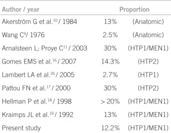
Some anatomic studies showed a frequency of supernu-merary parathyroid glands in the population ranging from 2.5% to 13%, frequently associated with the thymus or the embryological path of the lower parathyroids9,10.
Two studies evaluated cases of supernumerary glands in secondary hyperparathyroidism (HPT2). In one study, 14.3% of patients had a ith parathyroid16. he other re-ported supernumerary parathyroids in up to 30% of para-thyroid operations, and when these glands were not ex-cised, they were responsible for up to 32% of recurrent and persistent HPT217.
hree studies about HTP1 in patients with MEN1 re-lated diferent proportions of supernumerary cases: 13%, more than 20%, and 30%, respectively12,18,19.
he primary hyperplasia of this study presented an in-termediary proportion of supernumerary glands in com-parison to what was published in most anatomic studies and in clinical studies of patients with HTP2, and was low-er than othlow-er studies of MEN1, as shown in Table 2. he diference between clinical and anatomic studies could be explained by the larger size of hyperplastic glands, or by the eventuality of its detection in recurrent cases that is not possible in cadaveric studies.
hompson et al. showed approximately 5% of super-numerary cases in an anatomical study based on clinical cases of HTP1 in general, including single adenomas and hyperplasias not related with MEN120. his may be related to the fact that the vast majority of cases of solitary HTP1 are a single adenoma, where the excision of the afected gland is suicient to resolve the disease, avoiding an exten-sive cervical exploration, reducing the chances of inding an extra gland20.
A plausible explanation for a smaller proportion of su-pernumerary parathyroids in the present study of HPT1 versus that of HPT2 is the possibility of greater parathy-roid growth stimulus in HPT2, which normally presents itself with more cases of supernumerary glands than gen-eral HPT120. he clinical or genetic evaluation allows ear-lier diagnosis of HPT1/MEN121,22, which can determine the surgical treatment in early phases of HTP1 with less developed glands, where there was less time for the growth of mass and volume of these glands.
Among the 41 patients with HPT1/MEN1 studied, only one presented recurrent disease caused by a supernu-merary parathyroid (2.22%). However, it should be noted that many of the present cases have less than ten years of follow-up. hat is not enough time to rule out the possibil-ity of recurrence, which occurs very late, in the authors’ experience. he recurrence of HPT1 in MEN1 can occur later, when compared to HPT2.
he supernumerary parathyroid was found in the thy-mus in two of ive cases. One was identiied only through histology, and the other through the recurrent HPT1 ater many years, conirming the possibility of asymmetrical and asynchronous hyperplasia of parathyroids in MEN1. In this view, studies with less than partial parathyroid-ectomy in which follow-up is inferior to ive to ten years should be analyzed with caution.
he other glands were found during the neck explora-tion, not far from the other parathyroids. hey were in an accessible and visible location for the surgeon. hus, it is suggested that any node in the surgical ield of the para-thyroidectomy should be excised and sent for pathology testing in patients with MEN1-related HPT1. Irrespective to their size or macroscopic aspect, the resection of these
Patient Gender Age SPT found by USG? SPT found by MIBI? Localization of SPT
1 Female 32 No No Between RSPT and RIPT
2 Female 57 No No Right LSPT and LIPT
3 Male 59 No No Intratimic
4 Female 32 No Ten years after first surgery Mediastinal region
5 Female 40 No No Above LSPT
SPT, supernumerary parathyroid; USG, ultrasonography; TPT, topic parathyroid; RSPT, right superior parathyroid; RIPT, right inferior parathyroid; LSPT, left superior parathyroid; LIPT, left inferior parathyroid.
Table 1 – Cases of supernumerary parathyroids
Author / year Proportion
Akerström G et al.10 / 1984 13% (Anatomic)
Wang C9/ 1976 2.5% (Anatomic)
Arnalsteen L; Proye C11 / 2003 30% (HTP1/MEN1)
Gomes EMS et al.16 / 2007 14.3% (HTP2)
Lambert LA et al.26 / 2005 2.7% (HTP1)
Pattou FN et al.17 / 2000 30% (HTP2)
Hellman P et al.18 / 1998 > 20% (HTP1/MEN1)
Kraimps JL et al.19 / 1992 13% (HTP1/MEN1)
Present study 12.2% (HTP1/MEN1)
nodes may eventually show a supernumerary parathyroid, although in most cases a lymph node is reported by the pathologist.
All cases have maintained the location pattern of these glands, always reinforcing the need of thymectomy due to high incidence, and the need for a careful neck exploration during surgery. Arnalsteen et al., in two studies about re-current HPT1, found that supernumerary parathyroid was responsible for 51% of cases recurrent HTP1 in asymmet-ric hyperplasia, including cases of MEN type 110,11.
As in other studies, in the present study imaging exam-inations, such as USG and MIBI, were not efective for lo-calizing supernumerary glands at the initial operation11,16. In fact, many supernumerary glands were small and very diicult to identify by current methods. he success of MIBI scan of hyperactive parathyroids has a close relation-ship with the size and histological features of these glands, but it is useful to suggest an ectopic gland, especially in the mediastinal region23-25. One of the present cases, as well as a report found in the literature25, may serve as example, because ten years ater the irst surgery, during investiga-tion of the recurrent disease, only the MIBI scan detected the supernumerary parathyroid in the mediastinal region. Another method now widely used to detect a possible su-pernumerary gland is intra-operative serum PTH. his method seems to fail in tracking supernumerary glands in MEN1, because the PTH secretion ability of a gland de-pends on its size. hus, the small size of most supernumer-ary glands, still in initial hyperplasic process, may prevent the production of PTH in suicient quantity in order to afect the pattern of PTH decay8. Notwithstanding, it is thought that intraoperative PTH should be employed in these cases if available. Intraoperative PTH can suggest a hyperfunctioning parathyroid, but it would not be a reli-able indicator of microscopic glands. It is hard to predict if or when a small thymic parathyroid gland will cause recurrent HPT. High rates of recurrence (64% at median follow-up of four years) ater parathyroidectomy in MEN1 have been reported26. he persistence of an elevated PTH level ater successful four-gland resection is strongly sug-gestive of a hyperactive supernumerary parathyroid gland, which should be searched for before closing the wound. On the other hand, it would help to stop the extensive ex-ploration when less than four glands were identiied, if a signiicant fall of PTH is demonstrated.
As for the beneit of thymectomy, which is routinely performed at this institution for HPT in MEN1 cases since 1997, despite not being part of this study, it is worth not-ing that another patient followed at the institution, pre-viously submitted to a parathyroidectomy without thy-mectomy, developed a thymic carcinoid tumor, ive years ater the parathyroidectomy. hymectomy is not routinely performed for secondary or tertiary HPT at the institu-tion. No risk of carcinoid tumor is present in renal cases.
Anatomic bases for supernumerary glands are apparently indistinct regarding MEN1, HPT, and renal HPT. Howev-er, clinical behavior seems to be not comparable: in the au-thors’ experience, the recurrence rate is low in renal HPT, and in most cases related to autotransplantation. hese examples illustrate and reinforce the potential beneit of thymectomy in all cases of HPT1 associated with MEN1. In other conditions, such as renal related hyperplasia, the risk of extra surgical time and the potential risk to the in-nominate vein related to this strategy must be considered.
CONCLUSIONS
Supernumerary parathyroid glands in HPT1 with MEN1 were found in 12.2% of the present cases. Surgeons must be aware of the possibility of also inding these glands in the neck during the procedure, although many of them are in the thymus. Imaging studies were not helpful in locat-ing supernumerary glands before the irst surgery in HPT1 with MEN1, but this aspect should not discourage their use, as ectopic glands are also a considerable clinical prob-lem in these patients.
REFERENCES
1. Blackburn M, Diamond T. Primary hyperparathyroidism and familial hyper-parathyroid syndromes. Aust Fam Physician. 2007;36(12):1029-33. 2. Lakhani VT, You YN, Wells SA. he multiple endocrine neoplasia syndromes.
Annu Rev Med. 2007;58:253-65.
3. Hof AO, Hanache OM. Neoplasia endócrina múltipla tipo 1: diagnóstico clínico, laboratorial e molecular e tratamento das doenças associadas. Arq Bras Endocrinol Metab. 2005;49(5):735-46.
4. Ferolla P, Falchetti A, Filosso P, Tomassetti P, Tamburrano G, Avenia N, et al. hymic neuroendocrine carcinoma (carcinoid) in multiple endocrine neoplasia type 1 syndrome: the Italian series. J Clin Endocrinol Metab. 2005;90(5):2603-9.
5. Lambert LA, Shapiro SE, Lee JE, Perrier ND, Truong M, Wallace MJ, et al. Surgical treatment of hyperparathyroidism in patients with multiple endocrine neoplasia type 1. Arch Surg. 2005;140(4):374-82.
6. Carling T, Udelsman R. Parathyroid surgery in familial hyperparathyroid dis-orders. J Intern Med. 2005;257(1):27-37.
7. Lee CH, Tseng LM, Chen JY, Hsiao HY, Yang AH. Primary hyperparathyroid-ism in multiple endocrine neoplasia type 1: individualized management with low recurrence rates. Ann Surg Oncol. 2006;13(1):103-9.
8. Tonelli F, Marcucci T, Fratini G, Tommasi MS, Falchetti A, Brandi ML. Is total parathyroidectomy the treatment of choice for hyperparathyroidism in mul-tiple endocrine neoplasia type 1? Ann Surg. 2007;246(6):1075-82.
9. Wang C. he anatomic basis of parathyroid surgery. Ann Surg. 1976;183(3):271-5.
10. Akerström G, Malmaeus J, Bergström R. Surgical anatomy of human parathy-roid glands. Surgery. 1984;95(1):14-21.
11. Arnalsteen L, Proye C. Surgery of hyperparathyroidism and of its potential recurrence in the MEN I setting. Ann Chir. 2003;128(10):706-9.
12. Arnalsteen L, Quievreux JL, Huglo D, Pattou F, Carnaille B, Proye C. Reopera-tion for persistent or recurrent primary hyperparathyroidism. Seventy-seven cases among 1888 operated patients. Ann Chir. 2004;129(4):224-31. 13. Lourenço-Jr DM, Toledo RA, Coutinho FL, Margarido LC, Siqueira SA, Santos
MA, et al. he impact of clinical and genetic screenings on the management of the multiple endocrine neoplasia type 1. Clinics. 2007;62(4):465-76. 14. Toledo RA, Lourenco DM, Coutinho FL, Quedas E, Mackowiack I, Machado
MC, et al. Novel MEN1 germline mutations in Brazilian families with multiple endocrine neoplasia type 1. Clin Endocrinol (Oxf). 2007;67(3):377-84. 15. Brandi ML, Gagel RF, Angeli A, Bilezikian JP, Beck-Peccoz P, Bordi C, et al.
Guidelines for diagnosis and therapy of MEN type 1 and type 2. J Clin Endo-crinol Metab. 2001;86(12):5658-71.
16. Gomes EMS, Nunes RC, Lacativa PGS, Almeida MH, Franco FM, Leal CTS, et al. Ectopic and extranumerary parathyroid glands location in patients with hyperparathyroidism secondary to end stage renal disease. Acta Cir Bras. 2007;22(2):105-9.
18. Hellman P, Skogseid B, Oberg K, Juhlin C, Akerström G, Rastad J. Primary and reoperative parathyroid operations in hyperparathyroidism of multiple endo-crine neoplasia type 1. Surgery. 1998;124(6):993-9.
19. Kraimps JL, Duh QY, Demeure M, Clark OH. Hyperparathyroidism in mul-tiple endocrine neoplasia syndrome. Surgery. 1992;112(6):1080-6.
20. hompson NW, Eckhauser FE, Harness JK. he anatomy of primary hyper-parathyroidism. Surgery. 1982;92(5):814-21.
21. Montenegro FL, Tavares MR, Cordeiro AC, Ferraz AR, Ianhez LE, Buchpigel CA. Intrathyroidal supernumerary parathyroid gland in hyperparathyroidism ater renal transplantation. Nephrol Dial Transplant. 2007;22(1):293-5. 22. Nilubol N, Beyer T, Prinz RA, Solorzano CC. Mediastinal
hyperfunction-ing parathyroids: incidence, evolvhyperfunction-ing treatment, and outcome. Am J Surg. 2007;194(1):53-6.
23. Calva-Cerqueira D, Smith BJ, Hostetler ML, Lal G, Menda Y, ODorisio TM, et al. Minimally invasive parathyroidectomy and preoperative MIBI scans: corre-lation of gland weight and preoperative PTH. J Am Coll Surg. 2007;205:S38-44. 24. Erbil Y, Kapran Y, Işsever H, Barbaros U, Adalet I, Dizdaroglu F, et al. he
positive efect of adenoma weight and oxyphil cell content on preoperative lo-calization with 99mTc-sestamibi scanning for primary hyperparathyroidism. Am J Surg. 2008;195(1):34-9.