R E S E A R C H
Open Access
Bordonein-L, a new L-amino acid oxidase
from
Crotalus durissus terrificus
snake
venom: isolation, preliminary
characterization and enzyme stability
Karla C. F. Bordon
1, Gisele A. Wiezel
1, Hamilton Cabral
2and Eliane C. Arantes
1*Abstract
Background:Crotalus durissus terrificusvenom (CdtV) is one of the most studied snake venoms in Brazil. Despite presenting several well known proteins, its L-amino acid oxidase (LAAO) has not been studied previously. This study aimed to isolate, characterize and evaluate the enzyme stability of bordonein-L, an LAAO from CdtV.
Methods:The enzyme was isolated through cation exchange, gel filtration and affinity chromatography, followed by a reversed-phase fast protein liquid chromatography to confirm its purity. Subsequently, its N-terminal amino acid sequence was determined by Edman degradation. The enzyme activity and stability were evaluated by a microplate colorimetric assay and the molecular mass was estimated by SDS-PAGE using periodic acid-Schiff staining and determined by mass spectrometry.
Results:The first 39 N-terminal amino acid residues exhibited high identity with other snake venom L-amino acid oxidases. Bordonein-L is a homodimer glycoprotein of approximately 101 kDa evaluated by gel filtration. Its monomer presents around 53 kDa estimated by SDS-PAGE and 58,702 Da determined by MALDI-TOF mass spectrometry. The enzyme exhibited maximum activity at pH 7.0 and lost about 50 % of its activity after five days of storage at 4 °C. Bordonein-L’s activity was higher than the control when stored in 2.8 % mannitol or 8.5 % sucrose.
Conclusions:This research is pioneering in its isolation, characterization and enzyme stability evaluation of an LAAO from CdtV, denominated bordonein-L. These results are important because they increase the knowledge about stabilization of LAAOs, aiming to increase their shelf life. Since the maintenance of enzymatic activity after long periods of storage is essential to enable their biotechnological use as well as their functional studies.
Keywords:Crotalus durissus terrificus, L-amino acid oxidase, Rattlesnake, Enzyme activity, Enzyme stability, Chromatography, Snake venom, Yellow venom, Stabilization
Background
L-amino acid oxidases (LAAOs) are enantioselective flavoenzymes that catalyze the stereospecific oxidative deamination of L-amino acids. An amino acid inter-mediate is hydrolyzed, releasing α-keto acids and am-monia. Concomitantly, the reduced non-covalently bond cofactor–flavin mononucleotide (FMN) or flavin adenine
dinucleotide (FAD) – reoxidizes on molecular oxygen, producing hydrogen peroxide [1].
LAAOs are found in such diverse life forms as bac-teria, marine organisms, fish, cyanobacbac-teria, fungi, green algae, and snake venoms (SV) from the families Crotali-dae, Elapidae and Viperidae [1–12].
SV-LAAOs are, in general, non-covalently bonded to FAD and their FAD-binding site shares sequential simi-larity with human monoamine oxidase, mouse interleu-kin 4-induced, bacterial and fungal LAAOs [1, 13]. SV-LAAOs usually constitute from 0.15 to 5 % of the snake venom protein, with some exceptions, such as the * Correspondence:[email protected]
1Department of Physics and Chemistry, School of Pharmaceutical Sciences of
Ribeirão Preto, University of São Paulo (USP), Avenida do Café, s/n, Ribeirão Preto 14040-903 SP, Brazil
Full list of author information is available at the end of the article
LAAO of Bungarus caeruleus, which represents 25 % of the total protein [14]. Several biological activities have been attributed to SV-LAAOs, including cytotoxicity, mild myo-necrosis, apoptosis induction, induction and/or inhibition of platelet aggregation, as well as hemorrhagic, hemolytic, edematogenic, antibacterial, antiproliferative, antiparasitic and anti-HIV activities [14–25]. These activities are consid-ered the result of the release of hydrogen peroxide, which produces oxidative stress [26]. However, the role of LAAOs in the venom has not been elucidated yet [26].
SV-LAAOs exhibit a wide range of isoelectric points (pI) from about 4.4 to 8.1, although it is unknown whether the different charges result in distinct pharma-cological properties [13]. These enzymes prefer hydro-phobic L-amino acids, because of substrate specificity related to side-chain binding sites [27].
LAAO activity is inhibited in the presence of ethylenedi-aminetetraacetic acid (EDTA), N-ethylmaleimide, phenyl-methanesulfonyl fluoride (PMSF), glutathione and 1, 10-phenanthroline, since its cofactor is reduced under these conditions [14]. Furthermore, bivalent cations show differ-ent effects on LAAO activity. Manganese and calcium ions do not affect its specific activity. The LAAO fromC. adamanteus requires Mg2+, while those from Lachesis mutaandBothrops braziliare inhibited by Zn2+[14].
The cytotoxic effect of Bl-LAAO fromB. leucurusvenom was inhibited by about 25 % in the presence of catalase, an enzyme that cleaves hydrogen peroxide [17]. Additionally, the LAAOs of Naja naja kaouthia and Calloselasma rhodostoma venoms were inhibited by polyphenols from
Areca catechuandQuercus infectoriaextracts evaluated by
in vitrotests [28]. Although the ethylacetate extract from
Azima tetracanthaleaves exerts anin vitroinhibitory activ-ity on toxic enzymes fromB. caeruleusandVipera russelli
venoms, LAAOs from neither venom was inhibited [29]. The LAAOs have shown maximum absorbance at 465 and 380 nm because of their bond with FAD [13]. Small changes in the absorption spectra of SV-LAAOs were observed after inactivation by freezing and thawing or modification of the ionic composition and pH condi-tions, indicating alterations in the microenvironment of the FAD cofactor [30]. Most of the studies in this area were published in the 1950s and 1960s [31–35]. One example is the inactivation of an LAAO isolated fromC. adamanteus venom by high temperature and freezing. The higher the temperature or the pH of storage buffer, the higher the enzymatic inactivation, an inactivation that may be lower in the presence of chloride ions. On the other hand, at lower temperatures (freezing), the in-activation and storage buffer pH are inversely related. However, chloride ions were not able to prevent enzym-atic inactivation in this case [31, 32]. Further studies showed that the inactivation of LAAOs causes changes in optical rotatory dispersion whereas the redox properties
of free flavin are similar to those of the inactive enzyme [33, 35]. The change in redox properties suggests the loss of most interactions between flavin and apoprotein. Raibe-kas and Massey [36] extracted the cofactor of the LAAO from C. adamanteus venom at pH 3.5, rebound it at pH 8.5 and restored the enzymatic activity in the presence of 50 % glycerol followed by dialysis at 4 °C against 0.1 M Tris–HCl buffer, pH 7.5, containing 0.1 M KCl [36].
Due to their participation in metabolic pathways involving nitrogen and their antimicrobial, antiviral and antitumor effects, SV-LAAOs are considered a promising biotechnological agent and a tool for in-vestigating cellular processes [13, 14]. However, di-verse conditional factors that can reduce the stability of biocatalysts – including temperature, pH, oxida-tive stress, the solvent, binding of metal ions or co-factors, and the presence of surfactants – limit the industrial use of enzymes [37, 38]. Working under oper-ational conditions of enzyme stability, the process costs are reduced [37], since the enzyme is active when in use and keeps active over time [39].
Two reports have shown that the presence of univalent ions or substrates for LAAOs and analogues of the pros-thetic group (competitive inhibitors) prevents the inacti-vation of some SV-LAAOs [32, 40]. However, no additional studies have addressed the use of additives to maintain LAAO activity, which is highly desirable for in-dustrial applications.
The use of additives to maintain proteins in their ac-tive forms is widespread throughout the pharmaceutical industry. For example, cyclodextrins are employed as ex-cipients in pharmaceutical formulations in order to avoid protein aggregations to keep the protein in its ac-tive form [41]. There is a huge diversity of addiac-tives that act as cryoprotectants. Sugars and polyols, such as su-crose and mannitol, respectively, are used as protein sta-bilizers since they are able to interact with protein through hydrogen bonds to replace the protein-water molecular interactions [42, 43]. Amino acids are also used as cryoprotectants [43]. Usually, adjuvants are employed at a percentage that ranges from 0.5 to 2 %, although higher concentrations have already been tested [44–46].
Therefore, this study isolated an LAAO fromC. durissus terrificus venom (CdtV), denominated bordonein-L, and evaluated the effect of different additives (mannitol, sucrose, L-Lys and L-Gly) as cryoprotectants for the enzyme.
Methods
Isolation of bordonein-L
Cdt yellow venom from the Ribeirão Preto region (21° 10′
of the Brazilian Institute of Environment and Renewable Natural Resources (IBAMA).
Desiccated CdtV (1 g) was purified through cation ex-change chromatography, as described by Bordon et al.
[47]. The CM5 fraction obtained in the first chromato-graphic step was fractionated on a HiPrep 16/60 Sepha-cryl S-100 HR column (1.6 × 60 cm, GE Healthcare, Sweden) equilibrated and eluted with 0.05 M sodium acetate buffer containing 0.15 M NaCl, pH 5.5, at a flow rate of 0.5 mL/min. The subfraction CM5S2 was applied on two 1-mL HiTrap Heparin HP col-umns (GE Healthcare) connected in a series equili-brated with 0.05 M sodium acetate buffer, pH 5.5. Adsorbed proteins were eluted using a step concen-tration gradient from 0 to 100 % of buffer B (1 M NaCl in the same buffer) at a 1.0 mL/min flow rate. To assess its purity degree, the peak H7 (LAAO bordonein-L) was submitted to RP-FPLC, as de-scribed by Bordon et al. [47].
Determination of proteins
Total proteins were determined by the 280/205 nm ab-sorption method [48].
Determination of molecular mass
SDS-PAGE (10 %) was run according to the descrip-tion of Laemmli [49]. The gel was stained with PlusOne Coomassie PhastGel Blue R-350 (GE Healthcare, Sweden) whereas periodic acid-Schiff (PAS) staining was employed to detect glycoproteins [50]. The hyaluronidase CdtHya1, a glycoprotein recently isolated from CdtV, was used as the control [47].
The molecular mass of bordonein-L was estimated by gel filtration chromatography on a Superdex 200 10/300GL column (GE Healthcare) calibrated with the following protein molecular mass standards: 12.4, 29, 66, 150 and 200 kDa (Sigma-Aldrich Co., United States). Blue dextran (2000 kDa, Sigma-Aldrich Co.) was used to determine the void volume. The column was equilibrated whereas the standards and the en-zyme were eluted with the same buffer used on HiPrep 16/60 Sephacryl S-100 HR column. Each standard was filtered individually through the Super-dex column and a calibration curve was constructed.
The molecular mass of bordonein-L was also analyzed by a MALDI-TOF mass spectrometer (Ultraflex II, Bruker Daltonics, Germany). MS spectrum was acquired in posi-tive linear mode in the mass range 10,000-70,000 Da. TFA 0.1 % (10μL) was added to the lyophilized enzyme. This solution was mixed (1:1) with sinapinic acid (20 mg/mL in 50/50 0.2 % ACN/TFA, v/v); and 2μL of this mixture was spotted on a MALDI plate (384 positions) using the dried droplet method.
Bordonein-L sequencing andin silicoanalysis
The N-terminal of bordonein-L was determined by Edman degradation in an automated protein sequencer model PPSQ-33A (Shimadzu Co., Japan) and compared with sequences deposited in the Basic Local Alignment Search Tool (BLAST) [51]. The alignment was created by MultAlin Interface Page [52] and the figure was gen-erated by ESPript [53] server.
LAAO activity
The LAAO activity of bordonein-L was performed through a microplate colorimetric assay according to modifications on the Kishimoto and Takahashi method [54]. Bordonein-L was incubated at 37 °C for 60 min with 0.002 M o-phenylenediamine (OPD) (Sigma-Aldrich Co.), 1 U/mL horseradish peroxidase (Sigma-Aldrich), 0.005 M L-Leucine (Sigma-Aldrich) and 0.05 M Tris–HCl buffer, pH 7.0. The reaction was stopped with 2 M H2SO4and the absorbance was measured at 492/630 nm. LAAO activity was also eval-uated at different pH levels (5.0-9.0).
LAAO stability
The evaluation of LAAO stability was performed for 40 days at different concentration levels (1.4 %, 2.8 % and 8.5 %) of mannitol, sucrose, L-lysine and L-glycine, stored at 4 °C. Bordonein-L activity was also evaluated after being frozen (−20 °C) for a period of five days. The evaluation of enzymatic activity after lyophilization was performed as soon as this process was finished. The as-says were carried out according to the LAAO activity assay previously described. Control consisted of bordonein-L in the absence of additives and storage at 4 °C. The enzyme was protected from light under all the tested conditions.
Statistical analysis
LAAO activity data were expressed as mean ± standard error of mean (SEM). The analysis of variance (ANOVA) test was employed to evaluate data on LAAO activity in the presence of additives and to compare lyophilized, frozen and LAAO at 4 °C (five days), whereas the ttest was utilized to compare LAAO stability after freezing versus already lyophilized. They were statistically signifi-cant whenp< 0.05.
Results
Isolation of bordonein-L
Bordonein-L was purified in three chromatographic steps: cation exchange, molecular exclusion and affinity chromatography.
S-100 HR column and LAAO activity was detected in the CM5S2 fraction (Fig. 1b), which was submitted to affinity chromatography on a HiTrap Heparin HP column. Thus, pure LAAO (peak H7), denominated bordonein-L, was ob-tained (Fig. 1c). The pure enzyme represents 48.3 % of the total activity and 0.5 % of the total protein of the venom (Table 1). Bordonein-L was then applied on a C4 column
(Fig. 1d) and the main peak was submitted to Edman degradation.
Determination of molecular mass
SDS-PAGE under non-reducing conditions indicated that the peak H7 (bordonein-L) showed a high degree of purity while its monomer presented around 53 kDa
a
b
c
d
Fig. 1Isolation of Bordonein-L. Absorbance was monitored at 280 nm, at 25 °C, using a FPLC Äkta Purifier UPC-10 system. The dotted lines represent the concentration gradient. The vertical bars indicate the LAAO activity.aCdtV (1 g) was dispersed in 50 mL of 0.05 M sodium acetate buffer, pH 5.5 (buffer A) and the supernatant was fractionated on a CM-cellulose-52 column (1.0 × 40 cm) using a concentration gradient from 0 to 100 % of buffer B (1 M NaCl in buffer A).bThe fraction CM5 was filtered on a HiPrep 16/60 Sephacryl S-100 HR column (1.6 × 60 cm) using 0.05 M sodium acetate buffer containing 0.15 M NaCl, pH 5.5.cAffinity chromatography of the CM5S2 fraction on HiTrap Heparin HP column (two 1-mL columns connected in series) using a concentration gradient from 0 to 100 % of buffer B.dReversed-phase FPLC of H7 (bordonein-L) on a C4 column (0.46 × 25 cm, 5μm particles) using a concentration gradient from 0 to 100 % of solution B (60 % acetonitrile in 0.1 % TFA)
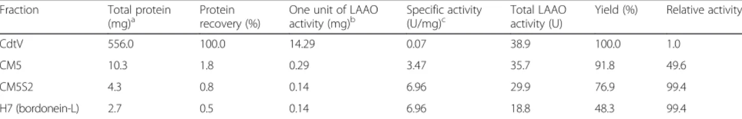
Table 1Specific activity and recovery of active fractions eluted during the purification procedure of bordonein-L
Fraction Total protein
(mg)a Proteinrecovery (%) One unit of LAAOactivity (mg)b Specific activity(U/mg)c Total LAAOactivity (U) Yield (%) Relative activity
CdtV 556.0 100.0 14.29 0.07 38.9 100.0 1.0
CM5 10.3 1.8 0.29 3.47 35.7 91.8 49.6
CM5S2 4.3 0.8 0.14 6.96 29.9 76.9 99.4
H7 (bordonein-L) 2.7 0.5 0.14 6.96 18.8 48.3 99.4
a
Total protein quantified by absorbance method 280/205 nm (SCOPES, 1974)
bLAAO activity unit (U): amount of protein (mg) able to release 1.0
μmol of H2O2per minute cSpecific activity: amount of H
(Fig. 2a), versus 56 kDa under reducing conditions (data not shown). Periodic acid-Schiff (PAS) staining evi-denced that bordonein-L is a glycoprotein (Fig. 2b). The molecular mass of 58,702 Da was determined by MALDI-TOF (linear positive mode) mass spectrometry (Fig. 2c). Gel filtration under non-reducing conditions revealed a protein of approximately 101 kDa (Fig. 2d), indicating that bordonein-L is a dimer protein.
In silicoassays
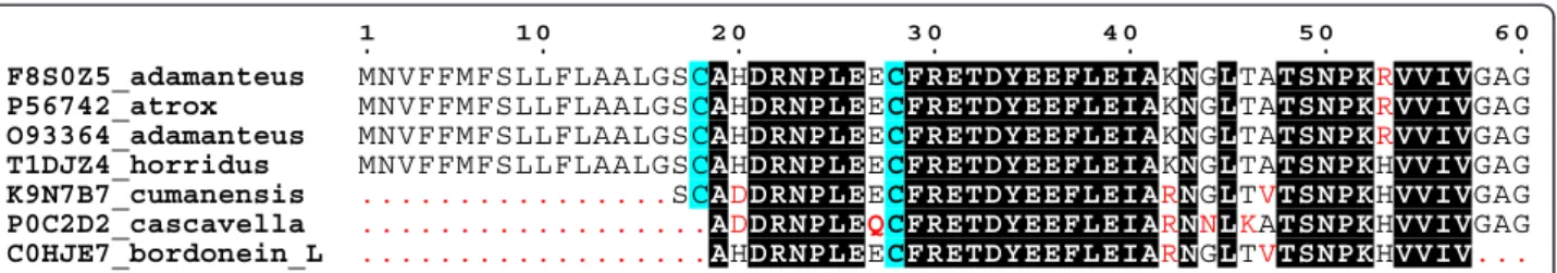
The sequence of the first 39 N-terminal amino acid resi-dues from bordonein-L was determined by Edman deg-radation and appears in the UniProt Knowledgebase
under the accession number C0HJE7. This primary se-quence exhibited high identity with other SV-LAAOs of the genusCrotalus(Fig. 3).
LAAO activity and stability
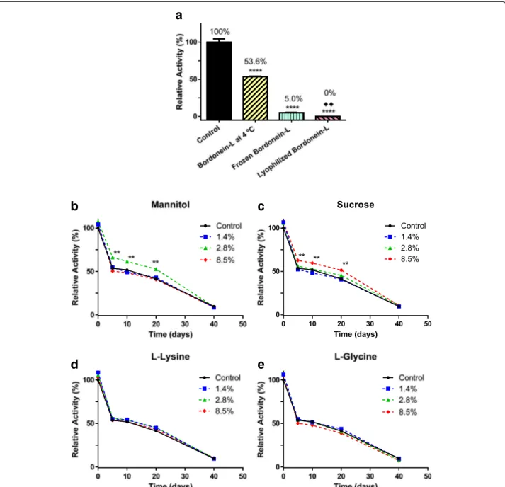
Bordonein-L showed an optimum pH of 7.0 (Fig. 4) and lost around 50 % of its activity in the first five days of storage at 4 °C (Fig. 5a-e). The frozen bordonein-L did not show enzymatic activity after lyophilization (Fig. 5a). Low activity (5 %) was also seen after thawing (Fig. 5a). Furthermore, LAAO activity was statistically significant when freezing and lyophilization were compared (Fig. 5a). L-lysine and L-glycine were not able to avoid
a
c
d
b
the loss of activity at the tested concentrations (Fig. 5d and e). Bordonein-L activity was decreased when stored in 2.8 % mannitol, but during the course of the deter-mined period of time (20 days), it was higher than the control. The enzymatic activity was the same as the con-trol in the presence of other mannitol concentrations (1.4 % and 8.5 %) (Fig. 5b). On the other hand, 8.5 % su-crose kept bordonein-L more active than control during the first 20 days. Other tested sucrose concentrations were unable to keep bordonein-L more active than the control in the same time period (Fig. 5c).
Discussion
There are 78 and 51 known primary sequences of SV-LAAOs deposited in the NCBI and UniProt databanks, respectively. However, the LAAO fromCrotalus durissus terrificus venom (CdtV), one of the most studied snake venoms in Brazil, had not been assessed previously.
This is the first report of an LAAO from CdtV, denominated bordonein-L. The enzyme was isolated in three chromatographic steps and represented 0.5 % of the soluble venom protein. The specific activity for the
soluble venom was 0.07 against 6.96 for bordonein-L, representing a 99.4-fold purification. The fractionation of 1 g of CdtV yielded only 2.7 mg of bordonein-L, a yield approximately four fold lower than the one ob-tained from the purification of 1 g of C. adamanteus
venom [36]. However, its recovery is within the range from 0.15 to 5 % of the total protein observed in other snake venoms [14]. Significant differences in activity and protein concentration are observed even in snake venoms from the same species and region, as recently reported for the Cdt venom from the Botucatu region (SP, Brazil) [55].
Bordonein-L is a homodimeric glycoprotein. Molecu-lar sieve chromatography under non-reducing conditions revealed a protein of approximately 101 kDa, while its mass was estimated at about 53 kDa by SDS-PAGE and 58,702 Da by mass spectrometry. SV-LAAOs are usually homodimeric FAD-binding glycoproteins with a molecu-lar mass of around 110–150 kDa when measured by gel filtration under non-denaturing conditions and around 50–70 kDa when assayed by SDS-PAGE under reducing and non-reducing conditions [13]. Our results indicate that bordonein-L is a non-covalently associated homodi-mer, as reported for most SV-LAAOs.
The sequence of the first 39 N-terminal amino acid residues of bordonein-L exhibited identity with other SV-LAAOs, since the amino-terminal region is highly conserved. A high degree of similarity (>84 %) has been described among the primary sequences of SV-LAAOs even when comparing distinct genera [14].
Bordonein-L exhibited more than 80 % of relative ac-tivity in the pH range from 5.5 to 8.0, showing max-imum activity at pH 7.0. Other SV-LAAOs show an active conformation at a pH ranging from 5.5 to 7.5, be-ing inactivated at extremely basic pHs [34]. We observed an approximately 50 % loss of LAAO activity in the first five days of storage at 4 °C, almost complete inactivation after freezing and thawing, and total inactivation after lyophilization. The activity of the LAAO isolated from
C. adamanteus, which shares high sequence identity with bordonein-L, is also greatly decreased by freezing Fig. 3Multiple sequential alignment of snake venom L-amino acid oxidases from the genusCrotalus. Initial N-terminal of bordonein-L [Swiss-Prot: C0HJE7, bottom] and LAAOs from crotalic venoms:C. adamanteus[Swiss-Prot: F8S0Z5, O93364],C. atrox[Swiss-Prot: P56742],C. horridus[Swiss-Prot: T1DJZ4],C. d. cumanensis[Swiss-Prot: K9N7B7–fragment] andC. d. cascavella[Swiss-Prot: P0C2D2–fragment]. The highly conserved residues in bordonein-L are highlighted in black. The amino acid residues in red indicate low consensus. Cys residues are shaded in blue. The alignment and figure were generated by the servers MultAlin [52] and ESPript [53], respectively
[31, 32]. Other SV-LAAOs presented similar results [13]. Therefore, we suggest that bordonein-L be kept at 4 °C and near neutral pH to avoid its inactivation.
In relation to the stability of bordonein-L, L-glycine and L-lysine did not prevent the loss of enzymatic activ-ity during the 40 days of storage at 4 °C, probably be-cause they are not able to effectively interact with the active site in contrast to the hydrophobic L-amino acids
and competitive inhibitors. L-glycine is the smallest amino acid and this small size may hinder its interaction with the catalytic site of bordonein-L. On the other hand, the amino acid L-lysine presents high polarity and the presence of polar groups might disrupt hydrophobic interactions. Hydrophobic L-amino acids, e.g. L-leucine, were not tested in this study as cryoprotectants because they are usually the preferred substrates of LAAOs
a
b
c
d
e
Fig. 5Bordonein-L stability.aEvaluation of stability after five days at−20 °C and 4 °C and as soon as the lyophilization was finished. The stability was
also evaluated for 40 days in the presence of (b) mannitol, (c) sucrose, (d) L-lysine and (e) L-glycine. All samples were kept protected from light. Each point represents the mean ± S.E.M. (n= 3) at each additive concentration (**p< 0.0001 compared to the respective control using one-way ANOVA test). Each bar represents the mean ± S.E.M. (n= 3) at 4 °C, freezing and lyophilization conditions (****p< 0.0001 when 4 °C, freezing and lyophilization was compared to control and when freezing and lyophilization compared to 4 °C using one-way ANOVA test;♦♦p< 0.05 when freezing and lyophilization
whereas changes in the amino acid concentration would occur due to their concomitant oxidation during the ac-tivity assay, which would prevent the correct quantifica-tion of the LAAO activity [32].
Bordonein-L’s activity was higher than the control dur-ing the first 20 days when stored in 2.8 % mannitol or 8.5 % sucrose. At those concentrations, mannitol and su-crose interacted with bordonein-L through hydrogen bonds, which probably stabilized the enzyme by re-placing the water molecular interactions, as reported for other proteins [42, 43]. However, after 40 days of stor-age, bordonein-L lost almost all of its activity even in the presence of additives. The rapid loss of activity (around 50 %) in the first five days and activity loss even in the presence of additives lead us to speculate that an alteration in the cofactor, such as oxidation or reduction, and/or changes in the catalytic site are responsible for the loss of LAAO activity since they may hinder the interaction among flavin, protein and substrate. The re-duction of enzymatic activity as a result of FAD loss or conformational alterations was reported in other LAAOs [30, 33, 35]. Some conformational changes at the cata-lytic site were also suggested for gyroxin, another en-zyme isolated from CdtV, whose catalytic efficiency was decreased in the presence of Mn2+and Cu2+[56].
The incorporation of additives to improve the stabilization of enzymes is the oldest and one of the most reliable en-zyme stabilization methods, being employed in the most marketed enzyme formulations [57]. Since LAAOs are con-sidered a promising biotechnological agent and a tool to in-vestigate cellular processes, the retention of its enzymatic activity over time is essential [13, 14].
Conclusions
An LAAO, denominated bordonein-L, was isolated from CdtV and presented higher enzymatic activity than the control when stored in 2.8 % mannitol or 8.5 % sucrose. These results may help the search for new additives to be used in stabilizing the LAAO, with the objective of increasing the shelf life of the enzyme.
Abbreviations
ANOVA:Analysis of variance; BLAST: Basic local alignment search tool; CdtV:Crotalus durissus terrificusvenom; EDTA: Ethylenediaminetetraacetic acid; FAD: Flavin adenine dinucleotide; FMN: Flavin mononucleotide; LAAO: L-amino acid oxidase; MALDI-TOF: Matrix assisted laser desorption ionization time of flight; OPD: O-phenylenediamine; PAS: Periodic acid-Schiff; pI: Isoelectric point; PMSF: Phenylmethanesulfonyl fluoride; RP-FPLC: Reversed-phase fast protein liquid chromatography; SDS-PAGE: Sodium dodecyl sulphate polyacrylamide gel electrophoresis; SEM: Standard error of mean; SV: Snake venom..
Competing interests
The authors declare that there are no competing interests.
Authors’contributions
KCFB worked on the isolation and characterization of the enzyme and drafted the manuscript. GAW carried out the enzyme stability assays and
helped in drafting the manuscript. HC participated in the design of the study and analysis of the results. ECA is the corresponding author and designer of the research. All authors read and approved the final manuscript.
Acknowledgements
This study received financial support from the State of São Paulo Research Foundation (FAPESP–grant n. 2011/23236-4; scholarship to GAW, n. 2014/06170-8), Coordination for the Improvement of Higher Education Personnel (CAPES–scholarship to GAW), National Council for Scientific and Technological Development (CNPq process 303689/2013-7) and the Support Nucleus for Research on Animal Toxins (NAP-TOXAN-USP, grant n. 12–125432.1.3). The authors would like to thank Prof. Dr. Norberto Peporine Lopes for providing the MALDI-TOF mass spectrometer used in this study. The authors also acknowledge the biologist Luiz Henrique Anzaloni Pedrosa for extracting the snake venom and Iara Aimê Cardoso for technical assistance. Thanks are also due to the Center for the Study of Venoms and Venomous Animals (CEVAP) of UNESP for enabling the publication of this special collection (CNPq process 469660/2014-7).
Author details
1Department of Physics and Chemistry, School of Pharmaceutical Sciences of
Ribeirão Preto, University of São Paulo (USP), Avenida do Café, s/n, Ribeirão Preto 14040-903 SP, Brazil.2Department of Pharmaceutical Sciences, School
of Pharmaceutical Sciences of Ribeirão Preto, University of São Paulo (USP), Ribeirão Preto, SP, Brazil.
Received: 28 November 2014 Accepted: 21 July 2015
References
1. Costa TR, Burin SM, Menaldo DL, de Castro FA, Sampaio SV. Snake venom L-amino acid oxidases: an overview on their antitumor effects. J Venom Anim Toxins incl Trop Dis. 2014;20:23.
2. Stumpf PK, Green DE. L-amino acid oxidase ofProteus vulgaris. J Biol Chem. 1944;153:387–99.
3. Geueke B, Hummel W. A new bacterial L-amino acid oxidase with a broad substrate specificity: purification and characterization. Enzyme Microb Technol. 2002;31(1–2):77–87.
4. Coudert M, Vandecasteele JP. Characterization and physiological function of a soluble L-amino-acid oxidase inCorynebacterium. Arch Microbiol. 1975;102(2):151–3.
5. Tong H, Chen W, Shi W, Qi F, Dong X. SO-LAAO, a novel L-amino acid oxidase that enablesStreptococcus oligofermentansto outcompete Streptococcus mutansby generating H2O2from peptone. J Bacteriol. 2008;190(13):4716–21.
6. Yang HC, Johnson PM, Ko KC, Kamio M, Germann MW, Derby CD, et al. Cloning, characterization and expression of escapin, a broadly antimicrobial FAD-containing L-amino acid oxidase from ink of the sea hareAplysia californica. J Exp Biol. 2005;208(Pt 18):3609–22.
7. Palenik B, Morel FMM. Comparison of cell-surface L-amino-acid oxidases from several marine-phytoplankton. Mar Ecol Prog Ser. 1990;59:195–201. 8. Kitani Y, Kikuchi N, Zhang G, Ishizaki S, Shimakura K, Shiomi K, et al. Antibacterial action of L-amino acid oxidase from the skin mucus of rockfishSebastes schlegelii. Comp Biochem Physiol B Biochem Mol Biol. 2008;149(2):394–400.
9. Kasai K, Ishikawa T, Komata T, Fukuchi K, Chiba M, Nozaka H, et al. Novel L-amino acid oxidase with antibacterial activity against methicillin-resistant Staphylococcus aureusisolated from epidermal mucus of the flounder Platichthys stellatus. FEBS J. 2010;277:453–65.
10. Bockholt R, Masepohl B, Kruft V, Wittmann-Liebold B, Pistorius EK. Partial amino acid sequence of an L-amino acid oxidase from the cyanobacterium SynechococcusPCC6301, cloning and DNA sequence analysis of the aoxA gene. Biochim Biophys Acta. 1995;1264(3):289–93.
11. Nuutinen JT, Marttinen E, Soliymani R, Hilden K, Timonen S. L-Amino acid oxidase of the fungusHebeloma cylindrosporumdisplays substrate preference towards glutamate. Microbiology. 2012;158(Pt 1):272–83. 12. Piedras P, Pineda M, Muñoz J, Cárdenas J. Purification and characterization
of an L-amino-acid oxidase fromChlamydomonas reinhardtii. Planta. 1992;188(1):13–8.
14. Izidoro LFM, Sobrinho JC, Mendes MM, Costa TR, Grabner AN, Rodrigues VM, et al. Snake venom L-amino acid oxidases: trends in pharmacology and biochemistry. Biomed Res Int. 2014; doi.org/10.1155/2014/196754. 15. Bregge-Silva C, Nonato MC, de Albuquerque S, Ho PL, de Azevedo IL J,
Vasconcelos Diniz MR, et al. Isolation and biochemical, functional and structural characterization of a novel L-amino acid oxidase fromLachesis mutasnake venom. Toxicon. 2012;60(7):1263–76.
16. Souza DH, Eugenio LM, Fletcher JE, Jiang MS, Garratt RC, Oliva G, et al. Isolation and structural characterization of a cytotoxic L-amino acid oxidase fromAgkistrodon contortrix laticinctussnake venom: preliminary
crystallographic data. Arch Biochem Biophys. 1999;368(2):285–90. 17. Naumann GB, Silva LF, Silva L, Faria G, Richardson M, Evangelista K, et al.
Cytotoxicity and inhibition of platelet aggregation caused by an L-amino acid oxidase fromBothrops leucurusvenom. Biochim Biophys Acta. 2011;1810(7):683–94.
18. Alves RM, Antonucci GA, Paiva HH, Cintra AC, Franco JJ, Mendonça-Franqueiro EP, et al. Evidence of caspase-mediated apoptosis induced by L-amino acid oxidase isolated fromBothrops atroxsnake venom. Comp Biochem Physiol A Mol Integr Physiol. 2008;151(4):542–50.
19. Li R, Zhu S, Wu J, Wang W, Lu Q, Clemetson KJ. L-amino acid oxidase from Naja atravenom activates and binds to human platelets. Acta Biochim Biophys Sin (Shangai). 2008;40(1):19–26.
20. Ali SA, Stoeva S, Abbasi A, Alam JM, Kayed R, Faigle M, et al. Isolation, structural, and functional characterization of an apoptosis-inducing L-amino acid oxidase from leaf-nosed viper (Eristocophis macmahoni) snake venom. Arch Biochem Biophys. 2000;384(2):216–26.
21. Wei JF, Yang HW, Wei XL, Qiao LY, Wang WY, He SH. Purification, characterization and biological activities of the L-amino acid oxidase from Bungarus fasciatussnake venom. Toxicon. 2009;54(3):262–71.
22. Vargas Muñoz LJ, Estrada-Gomez S, Nuñez V, Sanz L, Calvete JJ. Characterization and cDNA sequence ofBothriechis schlegeliiL-amino acid oxidase with antibacterial activity. Int J Biol Macromol. 2014;69:200–7. 23. Lee ML, Chung I, Fung SY, Kanthimathi MS, Hong TN. Antiproliferative
activity of king cobra (Ophiophagus hannah) venom L-amino acid oxidase. Basic Clin Pharmacol Toxicol. 2014;114(4):336–43.
24. Yun Z, Yongtang Z, Yujie Z. Application of snake venom L-amino acid oxidase in preparing AIDS treating medicine. PAT - CN1526445.
25. Zhang YJ, Wang JH, Lee WH, Wang Q, Liu H, Zheng YT, et al. Molecular characterization ofTrimeresurus stejnegerivenom L-amino acid oxidase with potential anti-HIV activity. Biochem Biophys Res Commun. 2003;309(3):598–604.
26. Fox JW. A brief review of the scientific history of several lesser-known snake venom proteins: L-amino acid oxidases, hyaluronidases and
phosphodiesterases. Toxicon. 2013;62:75–82.
27. Ponnudurai G, Chung MC, Tan NH. Purification and properties of the L-amino-acid oxidase from Malayan pit viper (Calloselasma rhodostoma) venom. Arch Biochem Biophys. 1994;313(2):373–8.
28. Leanpolchareanchai J, Pithayanukul P, Bavovada R. Anti-necrosis potential of polyphenols against snake venoms. Immunopharmacol Immunotoxicol. 2009;31(4):556–62.
29. Janardhan B, Shrikanth VM, Mirajkar KK, More SS.In vitroscreening and evaluation of antivenom phytochemicals fromAzima tetracanthaLam. leaves againstBungarus caeruleusandVipera russelli. J Venom Anim Toxins incl Trop Dis. 2014;20:12.
30. Massey V, Curti B. On the reaction mechanism ofCrotalus adamanteus L-amino acid oxidase. J Biol Chem. 1967;242(6):1259–64.
31. Curti B, Massey V, Zmudka M. Inactivation of snake venom L-amino acid oxidase by freezing. J Biol Chem. 1968;243:2306–14.
32. Kearney EB, Singer TP. The L-amino acid oxidases of snake venom. III. Reversible inactivation of L-amino acid oxidases. Arch Biochem Biophys. 1951;33(3):377–96.
33. Wellner D. Evidence for conformational changes in L-amino acid oxidase associated with reversible inactivation. Biochemistry. 1966;5(5):1585–91. 34. Coles CJ, Edmondson DE, Singer TP. Reversible inactivation of L-amino acid
oxidase. Properties of the three conformational forms. J Biol Chem. 1977;252(22):8035–9.
35. Soltysik S, Byron CM, Einarsdottir GH, Stankovich MT. The effects of reversible freezing inactivation and inhibitor binding on redox properties of L-amino-acid oxidase. Biochim Biophys Acta. 1987;911(2):201–8.
36. Raibekas AA, Massey V. Glycerol-induced development of catalytically active conformation ofCrotalus adamanteusL-amino acid oxidasein vitro. Proc Natl Acad Sci U S A. 1996;93(15):7546–51.
37. van den Burg B, Eijsink VG. Selection of mutations for increased protein stability. Curr Opin Biotechnol. 2002;13(4):333–7.
38. Eijsink VGH, Gaseidnes S, Borchert TV, van den Burg B. Directed evolution of enzyme stability. Biomol Eng. 2005;22(1–3):21–30.
39. Drago GA, Gibson TD. Enzyme stability and stabilisation: applications and case studies. In: Hofman M, Thonart P, editors. Engineering and Manufacturing for Biotechnology. Netherlands: Springer; 2002. p. 361–76. Series: Focus on Biotechnology.
40. Kearney EB, Singer TP. The inactivation of L-amino acid oxidase by inorganic phosphate and arsenate. Arch Biochem. 1949;21(1):242–5.
41. Serno T, Geidobler R, Winter G. Protein stabilization by cyclodextrins in the liquid and dried state. Adv Drug Deliv Rev. 2011;63(13):1086–106. 42. Allison SD, Chang B, Randolph TW, Carpenter JF. Hydrogen bonding between
sugar and protein is responsible for inhibition of dehydration-induced protein unfolding. Arch Biochem Biophys. 1999;365(2):289–98.
43. Arakawa T, Prestrelski SJ, Kenney WC, Carpenter JF. Factors affecting short-term and long-term stabilities of proteins. Adv Drug Deliv Rev. 2001;46(1–3):307–26. 44. Namaldi A, Calik P, Uludag Y. Effects of spray drying temperature and
additives on the stability of serine alkaline protease powders. Dry Technol. 2006;24(11):1495–500.
45. Xie GF, Timasheff SN. Mechanism of the stabilization of ribonuclease A by sorbitol: preferential hydration is greater for the denatured then for the native protein. Protein Sci. 1997;6(1):211–21.
46. Liu W, Wang DQ, Nail SL. Freeze-drying of proteins from a sucrose-glycine excipient system: effect of formulation composition on the initial recovery of protein activity. AAPS Pharm Sci Tech. 2005;6(2):E150–7.
47. Bordon KC, Perino MG, Giglio JR, Arantes EC. Isolation, enzymatic characterization and antiedematogenic activity of the first reported rattlesnake hyaluronidase fromCrotalus durissus terrificusvenom. Biochimie. 2012;94(12):2740–8.
48. Scopes RK. Measurement of protein by spectrophotometry at 205 nm. Anal Biochem. 1974;59(1):277–82.
49. Laemmli UK. Cleavage of structural proteins during the assembly of the head of bacteriophage-T4. Nature. 1970;227:680–5.
50. Doerner KC, White BA. Detection of glycoproteins separated by nondenaturing polyacrylamide gel electrophoresis using the periodic acid-schiff stain. Anal Biochem. 1990;187(1):147–50.
51. Basic Local Alignment Search Tool. http://blast.ncbi.nlm.nih.gov/Blast.cgi. 52. Corpet F. Multiple sequence alignment with hierarchical-clustering. Nucleic
Acids Res. 1988;16(22):10881–90.
53. Gouet P, Courcelle E, Stuart DI, Metoz F. ESPript: analysis of multiple sequence alignments in PostScript. Bioinformatics. 1999;15(4):305–8. 54. Kishimoto M, Takahashi T. A spectrophotometric microplate assay for L-amino
acid oxidase. Anal Biochem. 2001;298(1):136–9.
55. Lourenço Jr A, Zorzella Creste CF, de Barros LC, dos Santos LD, Pimenta DC, Barraviera B, et al. Individual venom profiling ofCrotalus durissus terrificus specimens from a geographically limited region: crotamine assessment and captivity evaluation on the biological activities. Toxicon. 2013;69:75–81. 56. Barros LC, Soares AM, Costa FL, Rodrigues VM, Fuly AL, Giglio JR, et al.
`Biochemical and biological evaluation of gyroxin isolated fromCrotalus durissus terrificusvenom. J Venom Anim Toxins incl Trop Dis. 2011;17(1):23–33 [http://www.scielo.br/scielo.php?pid=S1678-91992011000100004&script= sci_arttext]