Furanocoumarins Is Widespread throughout the
Actinomycetales
Gauri V. Lapalikar, Matthew C. Taylor*, Andrew C. Warden, Colin Scott, Robyn J. Russell, John G. Oakeshott
Ecosystem Sciences, Commonwealth Science and Industrial Research Organisation, Canberra, Australian Capital Territory, Australia
Abstract
Two classes of F420-dependent reductases (FDR-A and FDR-B) that can reduce aflatoxins and thereby degrade them have previously been isolated fromMycobacterium smegmatis. One class, the FDR-A enzymes, has up to 100 times more activity than the other. F420is a cofactor with a low reduction potential that is largely confined to theActinomycetalesand some ArchaeaandProteobacteria. We have heterologously expressed ten FDR-A enzymes from diverseActinomycetales, finding that nine can also use F420H2to reduce aflatoxin. Thus FDR-As may be responsible for the previously observed degradation of aflatoxin in otherActinomycetales. The one FDR-A enzyme that we found not to reduce aflatoxin belonged to a distinct
clade (herein denoted FDR-AA), and our subsequent expression and analysis of seven other FDR-AAs fromM. smegmatis
found that none could reduce aflatoxin. Certain FDR-A and FDR-B enzymes that could reduce aflatoxin also showed activity with coumarin and three furanocoumarins (angelicin, 8-methoxysporalen and imperatorin), but none of the FDR-AAs tested showed any of these activities. The shared feature of the compounds that were substrates was ana,b-unsaturated lactone moiety. This moiety occurs in a wide variety of otherwise recalcitrant xenobiotics and antibiotics, so the FDR-As and FDR-Bs
may have evolved to harness the reducing power of F420 to metabolise such compounds. Mass spectrometry on the
products of the FDR-catalyzed reduction of coumarin and the other furanocoumarins shows their spontaneous hydrolysis to multiple products.
Citation:Lapalikar GV, Taylor MC, Warden AC, Scott C, Russell RJ, et al. (2012) F420H2-Dependent Degradation of Aflatoxin and other Furanocoumarins Is
Widespread throughout theActinomycetales. PLoS ONE 7(2): e30114. doi:10.1371/journal.pone.0030114 Editor:John R. Battista, Louisiana State University and A & M College, United States of America ReceivedSeptember 29, 2011;AcceptedDecember 9, 2011;PublishedFebruary 27, 2012
Copyright:ß2012 Lapalikar et al. This is an open-access article distributed under the terms of the Creative Commons Attribution License, which permits unrestricted use, distribution, and reproduction in any medium, provided the original author and source are credited.
Funding:This project was funded under CSIRO Synthetic Enzymes Emerging Science Initiative. The funders had no role in study design, data collection and analysis, decision to publish, or preparation of the manuscript.
Competing Interests:The authors have declared that no competing interests exist. * E-mail: m.taylor@csiro.au
Introduction
Aflatoxins are complex aromatic compounds containing a,b -unsaturated lactone moieties that are produced most notably by Aspergillus flavus and Aspergillus parasiticus and have high chronic toxicity and carcinogenicity in animals, including humans [1]. Although relatively recalcitrant to biodegradation, aflatoxins are known to be degraded by some species from the order Actinomycetales, specifically Nocardia corynebacteriodes [2], Rhodococcus erythropolis, Mycobacterium fluorantheniorans sp. Nov DSM44556T [3,4,5] and Mycobacterium smegmatis [6]. The degradation of aflatoxin inM. smegmatis has been shown to involve two families of F420H2-dependent reductases [6], but the role of these enzymes
in the other species is unknown. The five enzymes characterized from one of these families (the FDR-A family) have 10 to 100 fold more activity for the aflatoxins than do the seven characterized from the other (FDR-B) family [6].
F420 is a naturally occurring deazaflavin derivative found in
methanogenicArchaea, theActinomycetalesand someProteobacteria[7]. It is a flavin analog (synthesized from a riboflavin intermediate) that differs from other flavin-derived cofactors, such as flavin mononucleotide (FMN), in having a negatively charged phospho-lactoc-glutamyl chain as opposed to a phosphate attached to its
ribitol group. The phospholactoc-glutamyl chain may prevent the cofactor from diffusing across the cell membrane [8,9]. Despite its structural similarity to FMN, F420 is chemically more akin to
NAD(P)H as a cofactor in that it only takes part in hydride transfers, and not other oxidative reactions as FMN does. F420also
has a redox potential of2350 mV [10,11], which is slightly more negative than NAD(P)H (2320 mV) [12], and much more so than FMN (2230 mV) [13].
F420 is reduced by F420-dependent glucose 6-phosphate
dehydrogenase (FGD) as the first step in the pentose phosphate pathway in a number of species ofMycobacteriaandNocardia[7]. In other species, such asRhodococcusandNocardioides, F420is reduced
by F420:NADPH oxidoreductase [14,15]. Reduced F420has been
implicated in functions as diverse as NO reduction [16], denitration of picric acid [14,17], malachite green decolorisation [18], antibiotic biosynthesis [19,20] and PA-824 antituberculosis drug activation [21,22,23], as well as aflatoxin degradation [6]. The unique chemistry and low reduction potential of F420 could
enable it to reduce many of these otherwise relatively recalcitrant molecules.
Recent annotations of sequencedActinomycetales genomes have identified three families of potential F420-dependent enzymes [24],
reduce aflatoxins. These two families are structurally and phylogenetically related to the FMN-dependent pyridoxamine 59-phosphate oxidase (PNPOx) family. The FDR-A family also includes the deazaflavin-dependent nitroreductase (Ddn) Rv3547, fromMycobacterium tuberculosis, that has been shown to reduce and activate the anti-tuberculosis drug PA-824 [23]. There is no experimental evidence as to whether any other enzyme in this family can act as a nitroreductases. The FDR-B enzymes are more closely related to the PNPOx enzymes, but none of the FDR-Bs tested was able to catalyze the oxidation of pyridoxamine 59 -phosphate to pyridoxal -phosphate (vitamin B6) in the presence of FMN [6]. The third family containing F420-dependent enzymes
are TIM barrel proteins that are phylogenetically related to the lumazine synthase enzymes [24]. The functionally described F420
-dependent members of this class include FGD [24,25] and enzymes involved in antibiotic biosynthesis [19,20].
To determine how widespread aflatoxin degrading activity is within theActinomycetaleswe have cloned and expressed ten FDR-A enzymes from various species across four suborders within the Actinomycetales. We have further characterized these enzymes with other furanocoumarins, specifically angelicin, imperatorin and 8-methoxypsoralen (8-MOP) plus khellin, all of which are defence compounds exuded by plant roots that are structurally related to aflatoxins as well as some other structurally related compounds. Some of these molecules also proved to be substrates for some of the FDR-As. Mass spectrometry was carried out to identify reaction products.
Materials and Methods
Chemicals
Aflatoxins B1 and G1 were obtained from Sigma-Aldrich (Australia) and Fermentek (Israel), at greater than 98% purity. Angelicin, imperatorin, coumarin, 8-methoxypsoralen, hydro-prene, khellin and glucose-6-phosphate were obtained from Sigma-Aldrich, at greater than 98% purity. F420 was prepared
from soluble fractions ofM. smegmatisextracts, as per the methods of Isabelleet al.[8].
Cloning, expression and purification of recombinant proteins
Ten previously uncharacterized M. smegmatis fdr-Agenes were amplified from genomic DNA of strain mc2155 using Platinum high fidelity Taq polymerase (Invitrogen, USA) and the primer sequences shown in Table S1. One previously characterizedfdr-A, MSMEG_5998, was also re-isolated as a truncated gene (because it had not expressed well originally and sequence analysis suggested that the 27 N-terminal residues of the encoded protein may be superfluous). The amplicons corresponding to all these genes were recombined into the destination vector pDEST17 via the pDONR201 plasmid of the Gateway System (Invitrogen). Genes for ten other FDR-A enzymes chosen from nine different strains within theActinomycetales(Frankia alniACN14a,Janibacter sp HTCC2649, Marine actinobacterium PHSC20C1, Mycobacterium tuberculosis H37Rv, Mycobacterium vanbaalenii PYR-1, Nocardia farcinica IFM 10152, Rhodococcus erythropolis PR4 (two genes), Rhodococcus jostiiRHA1,Streptomyces coelicolorA3(2)) were synthesized commercially (GeneArt, Germany) in E. coli-codon-optimized form and confirmed by sequencing (Micromon, Australia). The ten genes were then recombined into the Gateway destination vector pDEST17. The protein accession numbers for the FDR-A and related enzymes studied here, and their sources, are shown in Table S2.
Expression and purification of the enzymes encoded by these newly amplified or synthesized genes, and re-expression/ purification of other FDR-As and FDR-Bs of Tayloret al. [6], were carried out following the methods of those authors. Briefly, plasmid DNA was obtained via the QiaQuick mini-prep kit (Qiagen) and transformed intoE. coliBL21 AI cells (Invitrogen). The cells were grown in Luria-Bertani medium containing 100mg/ml ampicillin for 2 hours at 37uC (when the OD reached about 0.6) at which point they were induced with 0.2% L-arabinose (Sigma-Aldrich) at 28uC for 2 hours. The cells were centrifuged and the cell pellet resuspended in lysis buffer (50 mM NaH2PO4, 300 mM NaCl, 20 mM imidazole, pH 8.0). The cells
were then lysed with an EmulsiFlex-C3 homogeniser (ATA Scientific, Australia) and the recombinant proteins were purified from the soluble cell extract by nickel affinity chromatography on a 0.5 ml bed-volume Ni-NTA resin (Invitrogen). Bound proteins were eluted with increasing concentrations of 250–500 mM imidazole. The purity of the proteins was observed on NuPAGEH
NovexH 10% Bis-Tris gels (Invitrogen, Australia) run at 120 V and stained with Coomassie Brilliant Blue. The purified proteins were dialyzed and stored at 4uC in 50 mM NaH2PO4, pH 8.0.
The concentrations of the purified proteins were determined by measuring absorbance at 280 nm using a NanoDrop Spectro-photometer ND1000 (Thermo Fisher Scientific, Australia), with the extinction coefficient of each protein (Table S2) estimated using Vector NTI (Invitrogen).
Enzyme activity assays
Expressed enzymes were tested for activity against aflatoxin G1 (AFG1) and aflatoxin B1 (AFB1) in the presence of F420H2as per
the methods of Tayloret al.[6]. The assays were carried out in 20 or 200ml volumes at 22uC with a reaction mix consisting of 0.1–
1mM enzyme, 10mM F420, 2.5 mM glucose-6-phosphate,
0.45mM FGD, 50 mM Tris HCl, pH 7.5, and 100mM aflatoxin.
Assays with other potential substrates were set up in the same way except that substrate concentrations were adjusted to suit their aqueous solubility; imperatorin was added at 148mM, 8-MOP at
58mM, angelicin at 135mM, coumarin at 100mM, khellin at
190mM and hydroprene at 90mM. Time course high
perfor-mance liquid chromatography (HPLC) assays were conducted by sampling 5ml of the reaction mix at defined intervals (2 min–
30 min depending on the reaction), using an Agilent 1200 HPLC autosampler, and injecting onto an Agilent Zorbax Eclipse XDB-C18 column (3.5mm, 2.1630 mm). The aflatoxins were separated as per the methods of Taylor et al.[6]. Coumarin, angelicin, 8-MOP and khellin were separated isocratically with 30% acetonitrile and 0.5% acetic acid in water (v/v). Imperatorin and hydroprene were separated isocratically with 50% acetonitrile and 0.5% acetic acid. Substrates were monitored at 365 nm for aflatoxin, 325 nm for coumarin, 254 nm for imperatorin, 8-MOP, angelicin and khellin, and 265 nm for hydroprene. Specific activities for the substrates were determined by quantifying the loss of substrate using Chemstation software (Agilent). Correlations between activities on different substrates were assessed using Sigma Plot 11.0 (Systat Software, USA).
Phylogenetic methods
FDR-A enzymes (TIGR00026/PF04075/DUF385) within the Actinomycetaleswere identified by searching the NCBI databases on 17/08/2010. 164 FDR-A amino acid sequences (details in Fig. S1) were recovered from twenty different Actinomycetales strains (M. smegmatis and the nine strains from which new FDR-As were cloned and expressed above, plusFrankia sp.EAN1pec,Kineococcus radiotolerans, Mycobacterium avium 104,Mycobacterium bovisAF2122/ 97, Mycobacterium gilvum PYR-GCK, Mycobacterium sp. JLS, Mycobacterium leprae, Mycobacterium ulcerans Agy99 and Saccharopoly-spora erythraeaNRRL 2338 andStreptomyces galilaeus). 75 FDR-B and PNPOx enzymes (TIGR00026/PF01243) (fromE. coli,Frankia alni ACN14a, Homo sapiens, Mycobacterium sp. JLS, M. smegmatis, M. tuberculosisH37Ra andM. vanbaaleniiPYR-1), recovered by similar methods, were used as outgroups. Amino acid sequences were aligned by ClustalW and a phylogenetic tree constructed from full-length sequences using the Minimum Evolution method as implemented in MEGA 4.0 [26], with pairwise deletion, Poisson correction and 1000 bootstrap replicates.
Results
Orthologues of FDR-A enzymes from numerous Actinomycetalesreduce aflatoxin
Genes encoding ten FDR-A enzymes from nine different Actinomycetaleswere synthesized, cloned and expressed (Fig. S2) to determine how widespread the FDR-catalyzed reduction of
aflatoxins might be within this order. Four of the strains (N. farcinica, R. jostii, S. coelicolor A3(2) and the nitrogen fixing plant symbiont F. alni) were isolated from soil and therefore might encounter aflatoxins, but the remainder were from environments far less likely to contain aflatoxins (M. vanbaaleniiis from estuarine sediments;Janibactersp. HTCC2649, marineActinobacteriumandR. erythropolisPR4 are from marine environments; andM. tuberculosis H37Rv is a laboratory strain of a human pathogen). The ten enzymes (Table S2) were selected because of their close sequence similarity to one or other of the three previously characterized FDR-A enzymes, MSMEG_3356, _2850 and _5998 from M. smegmatis, with the greatest activities against AFG1 (>10mmol -min21mmol21 enzyme) [6]. Qualitative analysis of substrates AFG1 and AFB1 after overnight incubation with the enzymes and F420H2 revealed that nine of the enzymes could reduce both
aflatoxins, the exception being the SCO7200 enzyme from the soil bacterium S. coelicolor A3(2), which was unable to reduce either (data not shown).
FDR-A enzymes fall into two functionally distinct clades
To understand the distribution of the active and inactive FDR-A enzymes from the various Actinomycetales tested above, we constructed a phylogenetic tree of 164 FDR-As from twenty sequencedActinomycetalesgenomes (includingM. smegmatisand the nine species selected above; Fig. S1). The number of FDR-As identified in these genomes ranged from one inM. lepraeup to 15 inMycobacteriumspecies JLS and M. smegmatis. This tree (Fig. 1),
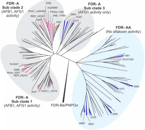
Figure 1. Phylogeny of 164 FDR-A enzymes from 20Actinomycetalesgenomes.Phylogenetic methods and the genomes used are given in the Materials and Methods. For clarity, only the FDR-As that have been characterized biochemically are labeled (M. smegmatis previously characterized (red),M. smegmatischaracterized herein (blue) and the otherActinomycetalesspecies characterized herein (pink)). TheM. smegmatis
enzymes are labeled with their locus tag numbers (the MSMEG_ prefix being deleted for clarity) while the enzymes from the other species are labeled with their complete locus tag. All the characterized enzymes are also given simple number designations (in brackets) to aid comparison with later figures. A fully labeled version of this phylogeny is given as Figure S1.
which we rooted with the sequences of PNPOx and FDR-B enzymes, revealed two major clades of FDR-As, one of which contained all 19 of the enzymes shown above and by Tayloret al. [6] to have activity against either or both of AFG1 and AFB1 (and for which we retain the name FDR-A), and the other of which contained the SCO7200 enzyme above from S. coelicolor that lacked activity against either (and which we rename FDR-AA). All 20 genomes had at least one representative in each clade (apart from M. leprae which has only one FDR-AA enzyme), although there was considerable unevenness in the distribution of species’ representatives across subclades (see also Fig. S1).
The phylogeny also showed three distinct subclades within the FDR-A clade, with all the previously characterized M. smegmatis enzymes within subclades 1 and 2. The third subclade contained three previously uncharacterized M. smegmatis enzymes (MSMEG_3909, _5215 and _6325) (Fig. 1). We therefore cloned and expressed these three enzymes, plus sevenM. smegmatis FDR-AA enzymes and assayed their activities against AFB1 and AFG1. Qualitative analysis after overnight incubations showed that the seven additional FDR-AA enzymes could not reduce AFG1 or AFB1 while the three FDR-A3 enzymes were all active against AFG1, albeit not AFB1. These results suggest that some aflatoxin reduction activity is generally present in the FDR-A clade and generally absent in the FDR-AA clade, and that the activity in the third subclade of the FDR-As does not extend to AFB1.
FDR enzymes reduce furanocoumarins
All 17 FDR-A and eight FDR-AA enzymes expressed and purified in this study, along with twoM. smegmatisFDR-B enzymes, MSMEG_3380 and MSMEG_6848, from Tayloret al. [6], were assayed quantitatively for activity against AFG1 and AFB1 and six other potential substrates; the a,b-unsaturated esters angelicin, imperatorin, 8-MOP, coumarin and hydroprene, plus the a,b -unsaturated ketone, khellin.
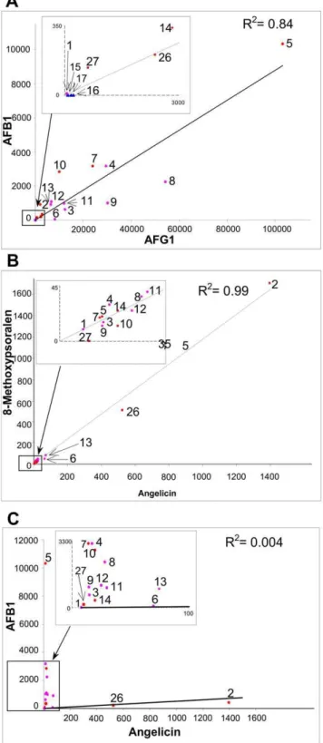
Values for specific activities with AFB1 and AFG1 for the FDR-A enzymes from the otherActinomycetalesare broadly comparable to those of the previously characterized M. smegmatis FDR-As (Fig. 2). There is again a strong trend for higher activities with AFG1 than AFB1, although overall there is a strong positive correlation between the two activities (Fig. 3). The most active enzyme for both substrates remains MSMEG_5998 (10364 and 10.360.2mmol min21mmol21 enzyme for AFG1 and AFB1,
respectively). The next highest activity for AFB1 was for the M. vanbaalenii enzyme, which sits in the same subclade (FDR-A1) as MSMEG_5998, Mvan_5261 (3.160.6mmol min21mmol21
en-zyme) and the M. smegmatis enzymes MSMEG_2850 and MSMEG_3356 (3.1 and 2.8mmol/min/mmol enzyme, respective-ly). The next three highest values for AFG1 activity were from other species:R. erythropolis, RER_34350;R. jostii, RHA1_ro00484 and M. vanbaalenii, Mvan_5261 (54.360.5, 30.260.2 and 29.560.6mmol min21mmol21 enzyme, respectively), although only one of the latter is from a soil organism (R. jostii). The subclades FDR-A1 and -A2 containing MSMEG_5998 and MSMEG_3356 contain all of the higher activity enzymes but there is also considerable variation within these subclades with regards to their aflatoxin reduction capability.
All of the FDR-A1 and FDR-A2 enzymes across the different species which were able to reduce both AFG1 and AFB1 were also able to reduce angelicin and 8-MOP, whereas no activity for these two furanocoumarins was observed for the three FDR-A3s that could only reduce AFG1 or for any of the FDR-AA enzymes. Interestingly, the two FDR-Bs could reduce angelicin and one could also reduce 8-MOP. Specific activities for the two furanocoumarins were strongly correlated with each other
(r2= 0.99) but bore little relationship to aflatoxin activities (r2,0.01) (Fig. 3).
With the exception of one FDR-A enzyme, MSMEG_2027, and the FDR-B, MSMEG_3380, activities against angelicin and 8-MOP were quite low. These two enzymes were also the only ones from M. smegmatis that had any activity against coumarin and imperatorin (Fig. 2). In fact, their activity for the furanocoumarins was approximately 2 fold greater than their AFB1 activity (in contrast to the other FDR-A enzymes which had at least 10 fold more activity for AFB1 than the furanocoumarins). This showed that some FDR enzyme activities have diverged so as to utilise differenta-bunsaturated esters. Thus, most of the FDR-A1, -A2 and a couple of FDR-B enzymes are capable of significant substrate promiscuity.
No activity with hydroprene and khellin was seen for any of the FDR enzymes. In the case of khellin this was probably because it is an a,b-unsaturated ketone rather than an ester and its a,b -unsaturated bond is stabilized by a methyl group. Hydroprene is a linear molecule that does have ana,b-unsaturated ester moiety but its double bond is also stabilized by a methyl group at the b -carbon.
Unlike aflatoxin [6], the reduced furanocoumarins yielded detectable breakdown products, as measured by the absorbance at 254 nm. The presence of these products was quantified over a 12 hour time course taking measurements every 20 minutes; the HPLC trace and time course for 8-MOP are shown in Fig. 4 A and B, respectively. In all cases the substrate (1) is reduced to an unstable dihydro reaction product (Fig. 4C, 2) as confirmed by the increase in molecular weight of 2.02 m/z, as measured by LCMS ToF. The dihydro-furanocoumarin was then spontaneously hydrolyzed into three secondary products (3A–3C), all appearing after the detection of the initial reduction product. The first of the secondary reaction products (3A) corresponds to a ring opening hydrolysis as previously characterized by a similar reduction by XenA [27]. However, the other two products (3B and 3C) were observed with an m/z ratio of 121.08 greater than the initial reaction product. This corresponds to the condensation of Tris buffer with the unstable dihydro-furanocoumarin reaction prod-uct. The dihydro-furanocoumarin product may react with Tris by two methods, which may account for the two observed product peaks. Firstly, it may be ring opened by any one of the three hydroxyl groups of Tris by alcoholytic ring opening [28]. Secondly, the carbon associated with the ester functional group may be attacked by the amine via nucleophilic substitution. Further experiments are required to confirm these reaction products by NMR.
Discussion
In previous work we discovered five FDR-A enzymes fromM. smegmatiswhich could all reduce aflatoxins [6]. We have now tested another ten FDR-A enzymes from various other Actinomycetales from a wide range of ecological niches and found that nine of them can also reduce both the aflatoxins (AFB1 and AFG1) that we have assayed. This suggests that aflatoxin reducing activity is wide-spread in this family of enzymes and, as the family of enzymes is itself widespread in theActinomycetales, it suggests that a capacity to degrade aflatoxins could be widespread in this order. This is consistent with previous reports of aflatoxin degradation in other species of Actinomycetales, such as N. corynebacteriodes [2], R. erythropolis, andM. fluoranthenivoranssp. Nov DSM44556T[3,4,5].
enzymes. Functional analysis of seven comparatively close relatives of SCO7200 from M. smegmatis then found that they were also incapable of reducing the aflatoxins. It appears that the clade containing these enzymes, which we denote here as FDR-AA, represents a distinct functional as well as phylogenetic entity. This functional distinction was borne out by our subsequent analysis of the enzymes’ activities for various furanocoumarins, which likewise showed that some furanocoumarin activity was common among the FDR-As but lacking in the FDR-AAs tested.
There are two other lines of evidence in our data showing functional evolution within the FDR-A/FDR-AA phylogeny. Firstly, we found a small subclade of FDR-As, the FDR-A3s, that could reduce AFG1 but not AFB1. Secondly, the capability of reducing at least some of the furanocoumarins proved to be widespread among the other FDR-As and absent from the three A3s and eight AAs tested. The functions of the FDR-AAs thus remain unknown. In fact, to the best of our knowledge there is no empirical functional data for any of the FDR-AA enzymes that we could find in our database searches.
Indeed we have no direct evidence that the FDR-AA clade even uses F420. Given their relationship to the highly conserved
FMN-utilizing PNPOx enzymes [6], it is possible that the FDR-AA enzymes may have evolved to use another flavin cofactor, perhaps FMN or maybe even another Actinomycetales-specific flavin analogue such as roseoflavin [29,30,31]. It is notable that the structural and aminoacid sequence identity of the FDRA and -AA enzymes (Fig. S3) shows that the putative phosphate binding residue W76 is highly conserved throughout both classes, suggesting a conserved need for the cofactor to contain a ribitol phosphate chain. Furthermore, all of the other residues identified in the active site are conserved in both FDR-A and -AA, with the notable exception of Y124, which is only highly conserved in the active FDR-A enzymes but not in the -AA enzymes. Further mutagenesis and functional assays are required to identify the role of these residues in the FDR enzymes and provide insight into the functional evolution of this family.
While the FDR-As are generally able to reduce the aflatoxins and some of them can also reduce coumarin and certain furanocoumarins, there are large, quantitative differences in the activities of the different FDR-As for these compounds. This activity variation is highly correlated for the two aflatoxins and also for the two furanocoumarins angelicin and 8-MOP, but there is very little correlation between activities for the aflatoxin and the two furanocoumarin substrates. The aflatoxins and (furano)cou-marins in this study may represent only a small proportion of the possible substrates for these enzymes because thea,b-unsaturated lactone moiety that appears to be required for activity occurs in a wide variety of xenobiotics and antibiotics. Such compounds may include UV oxidised polyaromatic hydrocarbons [32], plant derived lactones such as digitalis [33] and other antibiotics including leptomycin [34]. It remains to be determined as to whether the aflatoxins or (furano)coumarins are in fact physiolog-ical substrates for any of the enzymes here; some of the host organisms have ecologies which would not likely encompass these specific compounds.
Once the (furano)coumarins are reduced by the FDR-As they are likely to be further catabolized in theActinomycetalesthrough the catechol catabolic pathway, providing substrates for the citric acid Figure 3. Pairwise correlations among the AFB1, AFG1,
angelicin and 8-methoxypsoralen activities of FDR-A1/2/3 and FDR-B enzymes.The R2values for each pair of activities are given on each graph. Enzymes are numbered as per Fig. 2.
doi:10.1371/journal.pone.0030114.g003
Figure 2. Specific activities of heterologously expressed and purified FDR-A and -B enzymes against different substrates. M. smegmatisenzymes characterized previously (red) or herein (blue) and enzymes from the otherActinomycetalesspecies characterized herein (pink) are color-coded and labeled as per Fig. 1 and their phylogenetic relationships are shown on the left (reformatted from Fig. 1). Subclades that have been condensed are labelede. Specific activities are given as means and standard errors of at least three biological replicates. Substrates were tested with 1mM enzyme except for MSMEG_5998 (0.1mM) for AFG1 and AFB1 and MSMEG_2027 (0.5mM) for imperatorin. ND – Not detected.
cycle [35]. In the case of coumarin, dihydrocoumarin resulting from the FDR-catalyzed reduction reaction would be spontane-ously hydrolyzed to 3-hydroxyphenyl propionic acid, which could be further hydrolyzed to 2,3-dihydroxyphenyl propionic acid by melilotate hydroxylase [36], enabling ring meta-cleavage by extradiol dioxygenases [37,38]. After cleavage, the resulting
succinate would enter the citric acid cycle. The other furanocou-marin compounds are similarly reduced by the FDRs and after further hydrolysis should be ring cleaved by extradiol dioxygenases and lysed to produce succinate, thus also providing a carbon source. Other metabolic fates might also be possible for the possible hydrolyzed intermediates, such as through condensation Figure 4. Reduction and spontaneous hydrolysis of 8-MOP.8-MOP was reduced with MSMEG_2027 and analysed by LCMS. Spectra from both diode array (A) and MS ToF were recorded, diode array peaks are labeled with [M+H]+. The reaction was monitored every 20 minutes and the area of
by 4-coumarate-CoA ligase [39]. Further experiments are required to confirm the hydrolyzed ring products of dihydrocou-marin, and the metabolic fate of coumarin and its furano derivatives in the F420containingActinomycetales.
To gain further insight into the potential substrates for the FDR enzymes we interrogated Biocyc (http://biocyc.org/comp-geno-mics) for potential operons with at least partly conserved gene complements in which the genes encoding theM. smegmatis FDR-A/AA/B enzymes might sit (Table S3). Only one gene, encoding the FDR-A1 enzyme MSMEG_5998, was predicted to be in such an operon. One of the other two genes in this operon, encoding the enzyme MSMEG_5997, was found to be conserved in some related species but is not annotated with any putative function or structural domain. The other, encoding MSMEG_5996, is predicted to be an acetyl-CoA acetyl transferase gene. Further analysis using NCBI gene view and BLAST revealed that regions in the immediate vicinity of genes encoding M. vanballenii, R. erthropolis and M. tuberculosis homologues of MSMEG_5998 also contain acetyl-CoA acetyl transferase genes (Table S3). Acetyl-CoA acetyl transferases are known to perform a broad range of functions in central metabolism and biosynthesis [40]. They are also involved inActinomycete- specific pathways such as mycothiol formation [41]. Genes encoding FDR-As in Streptomyces species have been found in antibiotic biosynthetic operons [42,43,44], although their exact role in the biosynthesis has not been determined. No operons with at least partially conserved gene complements were found for the genes encoding enzymes in the FDR subclades -A2, -A3 or in the FDR-AA and -B clades.
F420 has been shown to be a non-essential cofactor in M. smegmatis[6,18] but this and other studies have shown that F420is
required for the metabolism of several otherwise recalcitrant molecules. As noted above, the aflatoxins and plant furanocou-marins studied here may only be model substrates for most of the FDR-A enzymes, many of which are in organisms found in environments not likely to encounter such compounds. The FDR-As are a divergent family with few conserved amino acids, thus suggesting the ability to adapt to new substrates and perhaps new cofactors. We have proposed a role of these enzymes in the secondary metabolism of toxic compounds, including antibiotic biosynthesis. We further suggest that these enzymes, although diverse, may be important to the F420-containingActinomycetalesto
adapt to the harsh environments in which they can be found, aiding in the catabolism of many otherwise recalcitrant molecules.
Supporting Information
Figure S1 Phylogenetic tree of FDR-A enzymes used in
this study.
(TIF)
Figure S2 SDS-PAGE of his-tagged FDR-A enzymes from
ten differentActinomycetalesspecies.Whole cell (WC) and soluble protein (S) fractions of each protein were recombinantly expressed inE. coliBL21-AI cells and separated on a 10% SDS-PAGE with a Biorad (Australia) Precision Plus Protein Standards molecular weight marker (M).E. coliBL 21- AI cells were used as control (control), The expressed proteins were: FRAAL1295 (1), SCO7200 (2), A20C1_10575 (3), RER_09240 (4), nfa28980 (5), JNB_17993 (6), RER_34350 (7), RHA1_ro00484 (8), Mvan_5261 (9) and Rv3547 (10).
(TIF)
Figure S3 Comparison of putative deazaflavin pocket
and glutamate chain binding sites in the FDR-As.Amino acid substitutions within the putative deazaflavin binding pocket as well as the c-glutamate chain are shown in comparison to the crystal structure of MSMEG_3356. Conserved amino acids compared with MSMEG_3356 are shown with an * (in red). FDR-A enzymes showing specific activities above 10,000 nmol/ min/mmol enzyme are highlighted with a grey background.
FDR-A enzymes are arranged in the same sequence as in Figure 2. (TIF)
Table S1 Primers used for protein expression work.
(DOCX)
Table S2 Genes expressed in this study.
(DOCX)
Table S3 Genes observed in the same reading frame as
FDR genes.
(DOCX)
Author Contributions
Conceived and designed the experiments: MCT GVL JGO RJR CS ACW. Performed the experiments: MCT GVL. Analyzed the data: MCT GVL JGO RJR CS ACW. Contributed reagents/materials/analysis tools: MCT GVL CS ACW. Wrote the paper: GVL MCT JGO.
References
1. Wagacha JM, Muthomi JW (2008) Mycotoxin problem in Africa: Current status, implications to food safety and health and possible management strategies. International Journal of Food Microbiology 124: 1–12.
2. Ciegler A, Lillehoj EB, Peterson RE, Hall HH (1966) Microbial Detoxification of Aflatoxin. Applied Microbiology 14: 934–&.
3. Teniola OD, Addo PA, Brost IM, Farber P, Jany KD, et al. (2005) Degradation of aflatoxin B-1 by cell-free extracts ofRhodococcus erythropolisandMycobacterium fluoranthenivoranssp nov DSM44556(T). International Journal of Food Microbi-ology 105: 111–117.
4. Hormisch D, Brost I, Kohring GW, Giffhorn E, Kroppenstedt RM, et al. (2004)
Mycobacterium fluoranthenivorans sp nov., a fluoranthene and aflatoxin B-1 degrading bacterium from contaminated soil of a former coal gas plant. Systematic and Applied Microbiology 27: 653–660.
5. Alberts JF, Engelbrecht Y, Steyn PS, Holzapfel W, van Zyl W (2006) Biological degradation of aflatoxin B-1 byRhodococcus erythropoliscultures. International Journal of Food Microbiology 109: 121–126.
6. Taylor MC, Jackson CJ, Tattersall DB, French N, Peat TS, et al. (2010) Identification and characterization of two families of F420 H2-dependent
reductases fromMycobacteriathat catalyse aflatoxin degradation. Mol Microbiol 78: 561–575.
7. Purwantini E, Gillis TP, Daniels L (1997) Presence of F420-dependent
glucose-6-phosphate dehydrogenase inMycobacteriumandNocardiaspecies, but absence fromStreptomycesandCorynebacteriumspecies and methanogenic Archaea. FEMS Microbiology Letters 146: 129–134.
8. Isabelle D, Simpson DR, Daniels L (2002) Large-scale production of coenzyme F420-5,6 by usingMycobacterium smegmatis. Applied and Environmental
Microbi-ology 68: 5750–5755.
9. Bashiri G, Rehan AM, Greenwood DR, Dickson JMJ, Baker EN (2010) Metabolic Engineering of Cofactor F420Production inMycobacterium smegmatis.
Plos One 5: e15803.
10. de Poorter LMI, Geerts WJ, Keltjens JT (2005) Hydrogen concentrations in methane-forming cells probed by the ratios of reduced and oxidized coenzyme F-420. Microbiology-Sgm 151: 1697–1705.
11. Jacobson F, Walsh C (1984) Properties of 7,8-Didemethyl-8-Hydroxy-5-Deazaflavins Relevant to Redox Coenzyme Function in Methanogen Metab-olism. Biochemistry 23: 979–988.
12. Kakinuma K, Kaneda M, Chiba T, Ohnishi T (1986) Electron-Spin-Resonance Studies on a Flavoprotein in Neutrophil Plasma-Membranes - Redox Potentials of the Flavin and Its Participation in Nadph Oxidase. Journal of Biological Chemistry 261: 9426–9432.
13. Draper RD, Ingraham LL (1968) A Potentiometric Study of Flavin Semiquinone Equilibrium. Archives of Biochemistry and Biophysics 125: 802–&.
14. Ebert S, Rieger PG, Knackmuss HJ (1999) Function of coenzyme F420in aerobic
catabolism of 2,4,6-trinitrophenol and 2,4-dinitrophenol byNocardioides simplex
FJ2-1A. Journal of Bacteriology 181: 2669–2674.
16. Purwantini E, Mukhopadhyay B (2009) Conversion of NO2 to NO by reduced coenzyme F420protectsmycobacteriafrom nitrosative damage. Proceedings of the
National Academy of Sciences of the United States of America 106: 6333–6338. 17. Ebert S, Fischer P, Knackmuss HJ (2001) Converging catabolism of 2,4,6-trinitrophenol (picric acid) and 2,4-dinitrophenol byNocardioides simplexFJ2-1A. Biodegradation 12: 367–376.
18. Guerra-Lopez D, Daniels L, Rawat M (2007)Mycobacterium smegmatismc(2) 155 fbiC and MSMEG_2392 are involved in triphenylmethane dye decolorization and coenzyme F420biosynthesis. Microbiology-Sgm 153: 2724–2732.
19. Li W, Khullar A, Chou S, Sacramo A, Gerratana B (2009) Biosynthesis of Sibiromycin, a Potent Antitumor Antibiotic. Applied and Environmental Microbiology 75: 2869–2878.
20. Gerratana B (2010) Biosynthesis, synthesis, and biological activities of pyrrolobenzodiazepines. Med Res Rev.
21. Singh R, Manjunatha U, Boshoff HIM, Ha YH, Niyomrattanakit P, et al. (2008) PA-824 Kills Nonreplicating Mycobacterium tuberculosis by Intracellular NO Release. Science 322: 1392–1395.
22. Choi KP, Bair TB, Bae YM, Daniels L (2001) Use of transposon Tn5367 mutagenesis and a nitroimidazopyran-based selection system to demonstrate a requirement for fbiA and fbiB in coenzyme F420biosynthesis byMycobacterium
bovisBCG. Journal of Bacteriology 183: 7058–7066.
23. Manjunatha UH, Boshoff H, Dowd CS, Zhang L, Albert TJ, et al. (2006) Identification of a nitroimidazo-oxazine-specific protein involved in PA-824 resistance inMycobacterium tuberculosis. Proceedings of the National Academy of Sciences of the United States of America 103: 431–436.
24. Selengut JD, Haft DH (2010) Unexpected Abundance of Coenzyme F420
-Dependent Enzymes in Mycobacterium tuberculosis and Other Actinobacteria. Journal of Bacteriology 192: 5788–5798.
25. Bashiri G, Squire CJ, Moreland NJ, Baker EN (2008) Crystal structures of F420
-dependent glucose-6-phosphate dehydrogenase FGD1 involved in the activation of the anti-tuberculosis drug candidate PA-824 reveal the basis of coenzyme and substrate binding. Journal of Biological Chemistry 283: 17531–17541. 26. Tamura K, Dudley J, Nei M, Kumar S (2007) MEGA4: Molecular evolutionary
genetics analysis (MEGA) software version 4.0. Molecular Biology and Evolution 24: 1596–1599.
27. Griese JJ, R PJ, Schwarzinger S, Dobbek H (2006) Xenobiotic reductase A in the degradation of quinoline by Pseudomonas putida 86: physiological function, structure and mechanism of 8-hydroxycoumarin reduction. J Mol Biol 361: 140–152.
28. Berkessel A, Cleemann F, Mukherjee S, Muller TN, Lex J (2005) Highly efficient dynamic kinetic resolution of azlactones by urea-based bifunctional organoca-talysts. Angewandte Chemie-International Edition 44: 807–811.
29. Otani S, Takatsu M, Nakano M, Kasai S, Miura R, et al. (1974) Roseoflavin, a New Antimicrobial Pigment from Streptomyces. Journal of Antibiotics 27: 88–89.
30. Mack M, Grill S (2006) Riboflavin analogs and inhibitors of riboflavin biosynthesis. Applied Microbiology and Biotechnology 71: 265–275. 31. Grill S, Busenbender S, Pfeiffer M, Kohler U, Mack M (2008) The bifunctional
flavokinase/flavin adenine dinucleotide synthetase from Streptomyces davawensis
produces inactive flavin cofactors and is not involved in resistance to the antibiotic roseoflavin. Journal of Bacteriology 190: 1546–1553.
32. Pitts JN (1983) Formation and Fate of Gaseous and Particulate Mutagens and Carcinogens in Real and Simulated Atmospheres. Environmental Health Perspectives 47: 115–140.
33. Brodersen R, Kjaer A (1946) The Antibacterial Action and Toxicity of Some Unsaturated Lactones. Acta Pharmacologica et Toxicologica 2: 109–120. 34. Hamamoto T, Uozumi T, Beppu T (1985) Biosynthetic-Studies of Leptomycins.
Journal of Antibiotics 38: 533–535.
35. Barnes MR, Duetz WA, Williams PA (1997) A 3-(3-hydroxyphenyl)propionic acid catabolic pathway inRhodococcus globerulusPWD1: cloning and character-ization of the hpp operon. J Bacteriol 179: 6145–6153.
36. Strickland S, Massey V (1973) The purification and properties of the flavoprotein melilotate hydroxylase. J Biol Chem 248: 2944–2952.
37. Bugg TD, Sanvoisin J, Spence EL (1997) Exploring the catalytic mechanism of the extradiol catechol dioxygenases. Biochem Soc Trans 25: 81–85. 38. Spence EL, Kawamukai M, Sanvoisin J, Braven H, Bugg TD (1996) Catechol
dioxygenases from Escherichia coli (MhpB) and Alcaligenes eutrophus (MpcI): sequence analysis and biochemical properties of a third family of extradiol dioxygenases. Journal of Bacteriology 178: 5249–5256.
39. Kaneko M, Ohnishi Y, Horinouchi S (2003) Cinnamate:coenzyme A ligase from the filamentous bacteriumStreptomyces coelicolorA3(2). J Bacteriol 185: 20–27. 40. Lynen F, Ochoa S (1953) Enzymes of Fatty Acid Metabolism. Biochimica et
Biophysica Acta 12: 299–314.
41. Bornemann C, Jardine MA, Spies HSC, Steenkamp DJ (1997) Biosynthesis of mycothiol: elucidation of the sequence of steps in Mycobacterium smegmatis. Biochemical Journal 325: 623–629.
42. Alexeev I, Sultana A, Mantsala P, Niemi J, Schneider G (2007) Aclacinomycin oxidoreductase (AknOx) from the biosynthetic pathway of the antibiotic aclacinomycin is an unusual flavoenzyme with a dual active site. Proc Natl Acad Sci U S A 104: 6170–6175.
43. Chung JY, Fujii I, Harada S, Sankawa U, Ebizuka Y (2002) Expression, purification, and characterization of AknX anthrone oxygenase, which is involved in aklavinone biosynthesis in Streptomyces galilaeus. J Bacteriol 184: 6115–6122.