Distinct Upstream Role of Type I IFN
Signaling in Hematopoietic Stem Cell-Derived
and Epithelial Resident Cells for Concerted
Recruitment of Ly-6C
hi
Monocytes and NK
Cells via CCL2-CCL3 Cascade
Erdenebileg Uyangaa1☯, Jin Hyoung Kim1☯, Ajit Mahadev Patil1☯, Jin Young Choi1, Seong Bum Kim1, Seong Kug Eo1,2*
1College of Veterinary Medicine and Bio-Safety Research Institute, Chonbuk National University, Iksan, Republic of Korea,2Department of Bioactive Material Sciences, Graduate School, Chonbuk National University, Jeonju, Republic of Korea
☯These authors contributed equally to this work. *vetvirus@chonbuk.ac.kr
Abstract
Type I interferon (IFN-I)-dependent orchestrated mobilization of innate cells in inflamed tis-sues is believed to play a critical role in controlling replication and CNS-invasion of herpes simplex virus (HSV). However, the crucial regulators and cell populations that are affected by IFN-I to establish the early environment of innate cells in HSV-infected mucosal tissues are largely unknown. Here, we found that IFN-I signaling promoted the differentiation of CCL2-producing Ly-6Chimonocytes and IFN-γ/granzyme B-producing NK cells, whereas deficiency of IFN-I signaling induced Ly-6Clomonocytes producing CXCL1 and CXCL2. More interestingly, recruitment of Ly-6Chimonocytes preceded that of NK cells with the lev-els peaked at 24 h post-infection in IFN-I–dependent manner, which was kinetically associ-ated with the CCL2-CCL3 cascade response. Early Ly-6Chimonocyte recruitment was governed by CCL2 produced from hematopoietic stem cell (HSC)-derived leukocytes, whereas NK cell recruitment predominantly depended on CC chemokines produced by resi-dent epithelial cells. Also, IFN-I signaling in HSC-derived leukocytes appeared to suppress Ly-6Ghineutrophil recruitment to ameliorate immunopathology. Finally, tissue resident
CD11bhiF4/80himacrophages and CD11chiEpCAM+dendritic cells appeared to produce
ini-tial CCL2 for migration-based self-amplification of early infiltrated Ly-6Chimonocytes upon
stimulation by IFN-I produced from infected epithelial cells. Ultimately, these results decipher a detailed IFN-I–dependent pathway that establishes orchestrated mobilization of Ly-6Chi
monocytes and NK cells through CCL2-CCL3 cascade response of HSC-derived leukocytes and epithelium-resident cells. Therefore, this cascade response of resident– to-hematopoi-etic–to-resident cells that drives cytokine–to-chemokine–to-cytokine production to recruit orchestrated innate cells is critical for attenuation of HSV replication in inflamed tissues.
a11111
OPEN ACCESS
Citation:Uyangaa E, Kim JH, Patil AM, Choi JY, Kim SB, Eo SK (2015) Distinct Upstream Role of Type I IFN Signaling in Hematopoietic Stem Cell-Derived and Epithelial Resident Cells for Concerted Recruitment of Ly-6ChiMonocytes and NK Cells via CCL2-CCL3 Cascade. PLoS Pathog 11(11): e1005256. doi:10.1371/journal.ppat.1005256
Editor:Pinghui Feng, University of Southern California, UNITED STATES
Received:June 22, 2015
Accepted:October 9, 2015
Published:November 30, 2015
Copyright:© 2015 Uyangaa et al. This is an open access article distributed under the terms of the
Creative Commons Attribution License, which permits unrestricted use, distribution, and reproduction in any medium, provided the original author and source are credited.
Data Availability Statement:All relevant data are within the paper and its Supporting Information files.
Author Summary
Herpes simplex virus type 1 and 2 (HSV-1 and HSV-2) are the most common cause of genital ulceration in humans worldwide with lifelong latent infection after peripheral rep-lication in mucosal tissues. Furthermore, acquisition of human immunodeficiency virus (HIV) is increased in HSV-infected individuals, underscoring the contribution of this virus in facilitating increased susceptibility to other microbial pathogens. Therefore, it is imperative to characterize the host defense to HSV infection and identify key components that regulate virus resistance, in order to devise therapeutic strategy. Although type I inter-feron (IFN-I)-dependent orchestrated mobilization of innate cells in inflamed tissues is considered a key player to control replication and CNS-invasion of HSV, the regulators and cell population that are affected by IFN-I to establish the orchestrated environment of innate cells in HSV-infected tissues are largely unknown. In the present study, we demon-strate that IFN-I signal governs the sequential recruitment of Ly-6Chimonocytes and then NK cells into mucosal tissues, depending on CCL2-CCL3 cascade mediated by HSC-derived leukocytes and epithelial resident cells, respectively. Also, tissue resident
CD11bhiF4/80himacrophages and CD11chiEpCAM+dendritic cells were involved in pro-ducing the initial CCL2 for migration-based self-amplification of rapidly infiltrated Ly-6Chimonocytes through stimulation by IFN-I produced from infected epithelial cells. This study deciphers detailed IFN-I-dependent pathway that establishes orchestrated mobiliza-tion of Ly-6Chimonocytes and NK cells through CCL2-CCL3 cascade.
Introduction
Herpes simplex virus types 1 and 2 (HSV-1 and HSV-2) are significant human pathogens and the most common cause of genital ulceration in humans worldwide [1,2]. Typically, infection with HSV-1 or -2 via genital routes results in a lifelong latent infection of the host after periph-eral replication in mucosal tissues [3], thereby providing potential transmission to neighbor hosts in response to reactivation. In addition, acquisition of human immunodeficiency virus (HIV) is increased 2- to 3-fold in HSV-infected individuals, underscoring the contribution of this virus in facilitating increased susceptibility to other microbial pathogens [4–6]. Conse-quently, it is imperative to characterize the host defense to HSV infection and identify key com-ponents that regulate virus resistance in order to devise a strategy that will reduce viral
prevalence.
The development of a mouse model of vaginal HSV infection has provided a significant contribution to our understanding of innate and adaptive immune responses to HSV infection in mucosal tissues [7,8]. In the rodent model, viral replication is initially limited to the vaginal mucosa [1–3] followed by spread into the central nervous system (CNS) upon retrograde trans-port of virions into the sacral ganglia, resulting in fatal paralysis [1–3]. Investigators have dem-onstrated the importance of the innate immune responses, including natural killer (NK), NKT cells, monocytes, and neutrophils as well as the production of type I interferons (IFN-α/β), interleukin (IL)-12, and IL-18, in suppressing viral replication and reducing virus-mediated mortality [9–13]. Also, robust IFN-γ-producing T-helper 1 (Th1) CD4+T-cell immunity is required for protection against primary and secondary HSV-1 or -2 infection via the mucosal route [14–16]. Therefore, orchestrated mobilization of effector innate and adaptive immune cells (monocytes, NK, and T cells) in mucosal tissues appears to play a crucial role in providing effective protection against HSV infection without detrimental pathology.
NK cells are known to play a critical role in HSV control by recognizing and killing infected cells upon engagement of NK cell-activating receptors by altered self or viral ligand pairs expressed by infected cells [17–19]. These antiviral NK cells are thought to be regulated by type I IFN (IFN-I) signals through promotion of cell entry into the cell cycle, proliferation, survival, and cytotoxicity [20,21]. Some of these effects may be indirectly mediated by IL-15, although this is debatable [20,21]. Also, the coordinated roles of CD11b+Ly-6Chimonocytes in various immunopathogeneses caused by pathogenic infection were recently described [22,23]. After infection, CD11b+Ly-6Chimonocytes emigrate from the bone marrow (BM) into the blood-stream through CCR2-receptor–mediated signaling and differentiate into dendritic cells (DCs) that produce tumor necrosis factor (TNF)-αand nitric oxide (NO) [24,25]. These cells, known as Tip-DCs, are required for lysis and clearance of pathogens such asListeria monocytogenes
[25] andToxoplasma gondii[26]. Furthermore, monocyte-derived DCs appear to migrate from the inflamed tissues to the draining lymph node (DLN) and prime naïve CD4+T cells in Leish-mania majorinfection [27]. Likewise, the chemokine CCL2 recruits Ly-6Chimonocytes to mucosal tissues after HSV infection, where they appear to exert tailored protective immunity by secreting antimicrobial factors (TNF-α, NO) and stimulating antiviral Th1 immunity through differentiation into inflammatory DCs [28]. In accordance with these findings, IFN-I have been shown to regulate CD11b+Ly-6Chimonocyte recruitment during viral infection [29,30], as well as during chronic inflammation [31], by inducing CCL2. Therefore, defining the role of IFN-I signaling in the differentiation and recruitment of monocytes is required to better understand inflammatory responses for the development of therapeutic strategies. How-ever, the crucial regulatory factors and the cell populations that are affected by IFN-I to estab-lish the orchestrated mobilization of innate immune cells such as NK cells and CD11b+Ly-6Chi monocytes in mucosal tissues following mucosal HSV infection are largely unknown.
Therefore, the experiments presented here were undertaken (a) to define the role of IFN-I in the regulation of innate immune cell populations such as monocytes and NK cells, and (b) to identify specific cell populations and molecular regulators affected by IFN-I that are involved in establishing early orchestrated mobilization of innate cells in mucosal tissues following mucosal HSV infection. Somewhat intriguingly, our data revealed that IFN-I signaling regu-lated the sequential recruitment of CD11b+Ly-6Chimonocytes followed by NK cells, which was closely associated with the cascade response of CCL2 and CCL3 production in mucosal tis-sues; i.e., CD11b+Ly-6Chimonocyte infiltration peaked at around 24 h after infection, whereas NK cell infiltration peaked at 48 h post-infection (pi). Furthermore, early recruitment of CD11b+Ly-6Chimonocytes was dominantly regulated by CCL2 produced from hematopoietic stem cell (HSC)-derived leukocytes, whereas NK cell recruitment depended on CCL3 and other CC chemokines produced from resident epithelium cells. Also, early CCL2 proteins secreted from tissue resident CD11bhiF4/80himacrophages and CD11chiEpCAM+DCs upon stimulation of IFN-I produced from infected epithelium were required for migration-based self-amplification of CD11b+Ly-6Chimonocytes in mucosal tissues. Taken together, these results provide new insights into the role of IFN-I signaling by defining a unique cascade path-way in the regulation of chemokine responses required for the early orchestrated mobilization of innate immune cells.
Results
IFN-I signaling is involved in the rapid and concerted recruitment of
Ly-6C
himonocytes and CD11c
+DCs
required to control shedding of infectious virus (S1C Fig) and to prevent CNS invasion of HSV-1 from mucosal tissues (S1D–S1H Fig). Furthermore, it seemed likely that IFN-I signal-ing played a crucial role in establishsignal-ing an early orchestrated environment of cytokine and che-mokine expression for viral clearance (S2 Fig). Notably, BL/6 mice showed markedly enhanced expression of type I IFNs (IFN-α/β), CCL2, CCL3, and CCR5 (involved in recruitment of monocytes and NK cells), and CXCL10 (involved in recruitment of Th1 CD4+T cells) in the vaginal tract, compared toIFNARKO mice.
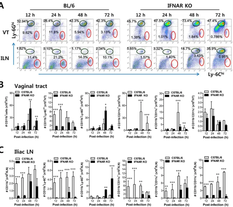
However, the crucial regulatory factors that establish the early orchestrated environment of infiltrated leukocytes in mucosal tissues and their cellular source have been not defined in depth. CD11b+Ly-6Chimonocytes play a pivotal role in exerting direct antimicrobial activity or further differentiate into inflammatory DCs, which participate in innate and adaptive pro-tective immunity [24,25]. In addition, since our preliminary data showed that expression of the chemokine CCL2, a major factor in monocyte recruitment, was early and markedly enhanced in the vaginal tract of WT BL/6 mice upon mucosal HSV-1 infection (S2 Fig), we performed kinetic examinations of recruited myeloid-derived leukocyte subpopulations in the vaginal tract and its draining LN (iliac LN) 12, 24, 48, and 72 h after infection. IFN-I signal was found to be critical for recruitment of CD11b+Ly-6Chimonocytes in vaginal tract and iliac LN during the early phase of HSV-1 mucosal infection (Fig 1A). Notably, WT BL/6 mice showed early infiltration of CD11b+Ly-6Chimonocytes with a 10- to 20-fold increase in the vaginal tract and iliac LN at 24 h after infection, compared toIFNARKO mice. In contrast,IFNARKO mice contained a greater number of accumulated CD11b+Ly-6Ghigranulocytes in the vaginal tract with levels peaking at 48 h pi, and the number of iliac CD11b+Ly-6Ghigranulocytes increased up to 72 h pi. These results indicate that maximal infiltration of CD11b+Ly-6Chimonocytes in the vaginal tract of BL/6 mice is established at around 24 h after infection, whereas the infiltra-tion of CD11b+Ly-6Ghigranulocytes inIFNARKO mice peaked at 48 h pi. When total accu-mulated subpopulations of myeloid-derived leukocytes and DCs were examined during the early phase after HSV-1 infection (12, 24, 48, and 72 h pi), maximum accumulation of CD11b +-Ly-6Chimonocytes was detected in the vaginal tract of BL/6 mice with 4- to 5-fold increased levels at 24 h pi, despite a moderately increased number of total CD11b+myeloid-derived cells inIFNAR KOmice (Fig 1B). In contrast, the total number of CD11+DCs and its myeloid DC subpopulation (CD11b+CD11c+) was increased in the vaginal tract of BL/6 mice, with a delayed peak at 48 h pi compared toIFNARKO mice. Likewise, iliac LN of BL/6 mice also showed early increased levels of total CD11b+Ly-6Chimonocytes with saturated levels at 24 h pi, and levels of CD11c+DCs, the myeloid DC subset (CD11c+CD11b+) and other DC subsets (CD11b−CD11c+) peaked at around 48 h pi, whereas inIFNARKO mice CD11b+Ly-6Ghi gran-ulocytes showed gradually increasing levels up to 72 h pi (Fig 1C). To summarize, interestingly, these results indicate that infiltration of CD11b+Ly-6Chimonocytes (levels of which peaked at 24 h pi) precede that of CD11c+DCs (levels of which peaked at 48 h) in mucosal tissues upon HSV-1 infection. Therefore, these results suggest that IFN-I signaling plays a crucial role in establishing early infiltration of CD11b+Ly-6Chimonocytes and CD11c+DCs in the submu-cosa area of the vaginal tract with a concerted pattern of recruitment, which could subsequently provide a well-controlled inflammatory reaction and adaptive protective immunity.
IFN-I signal differentially induces functional activation of Ly-6C
hiand
Ly-6C
lomonocytes
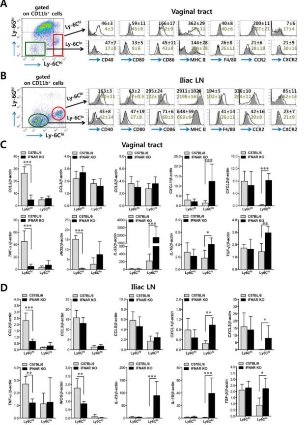
also have the ability to present Ag as professional antigen-presenting cells similar to DCs dur-ing mucosal inflammation caused by HSV-1 infection. To this end, we further characterized CD11b+Ly-6Chiand CD11b+Ly-6Clomonocytes for antigen-presenting molecules and activa-tion markers. CD11b+Ly-6Chimonocytes from the vaginal tract of both WT BL/6 andIFNAR
KO mice expressed higher levels of Ag presenting-related molecules (CD40, CD80, CD86, and Fig 1. IFN-I signaling is essential to induce the rapid and concerted recruitment of Ly-6Chimonocytes and CD11c+DCs in mucosal and draining LN tissues. (A)Leukocyte infiltration of vaginal tract and iliac LN in infected BL/6 orIFNARKO mice.(B,C)Accumulated number of infiltrated leukocyte subsets. Cells were prepared from vaginal tract (B; VT) and iliac LN (C; ILN) by collagenase digestion at 12, 24, 48, and 72 h pi and subcellular proportions of CD11b+Ly-6Chiand CD11b+Ly-6Ghileukocytes were determined using flow cytometric analysis. Values in the representative dot-plots denote the average percentages of each population derived from at least four independent samples after gating on CD11b+cells. Data in the bar chart represent the
average±SD of values derived from three individual experiments (n= 4–5).*,p<0.05;**,p<0.01;***,p<0.001 compared with the levels of BL/6 orIFNAR KO mice at the indicated time point.
MHC II) than CD11b+Ly-6Clomonocytes, and these molecules showed significantly higher expression levels in CD11b+Ly-6Chimonocytes derived from WT BL/6 mice compared to those fromIFNARKO mice (Fig 2A). Also, CD11b+Ly-6Chimonocytes derived from vaginal tract of BL/6 mice displayed upregulated expression of CCR2, the receptor of CCL2. Similarly, CD11b+Ly-6Chimonocytes in iliac LN of BL/6 mice also showed upregulated expression levels of Ag-presenting–related molecules and activation markers compared to those ofIFNARKO mice (Fig 2B). Since CD11b+Ly-6Chimonocytes also exert antimicrobial activity by secreting antimicrobial factors (TNF-α, NO), we next examined the expression of cytokines and chemo-kines in CD11b+Ly-6Chiand CD11b+Ly-6Clomonocytes derived from vaginal and iliac LN tis-sues. Intriguingly, CD11b+Ly-6Chiand CD11b+Ly-6Clomonocytes showed contrasting expression patterns of cytokines and chemokines in WT BL/6 andIFNARKO mice (Fig 2C). CD11b+Ly-6Chimonocytes derived from vaginal tract of WT BL/6 mice showed markedly higher expression of TNF-αand iNOS cytokines (the main factors of TipDC) and the CCL2 chemokine (the ligand of CCR2 as a major monocyte chemotactic factor) than those ofIFNAR
KO mice. In contrast, CD11b+Ly-6Clomonocytes from vaginal tract ofIFNARKO mice expressed high mRNA levels of cytokines IL-23, IL-10, and TGF-βand CXC chemokines (CXCL1, CXCL2). Similarly, iliac CD11b+Ly-6Chimonocytes derived from WT BL/6 mice showed upregulated expression of CCL2, TNF-α, and iNOS, whereas the expression of CXC chemokines (CXCL1 and CXCL2) and cytokines (IL-23, IL-10, and TGF-β) was increased in iliac CD11b+Ly-6Clomonocytes ofIFNARKO mice (Fig 2D). Collectively, these results indi-cate that IFN-I signal is crucial for determining the characteristics of CD11b+Ly-6Chiand CD11b+Ly-6Clomonocytes. Also, somewhat surprisingly, IFN-I signaling differentially affected the functional maturation of CD11b+Ly-6Chiand CD11b+Ly-6Clomonocytes during mucosal HSV-1-induced inflammation.
Essential role of IFN-I signal in concerted recruitment of NK cells to
mucosal tissues and their activation
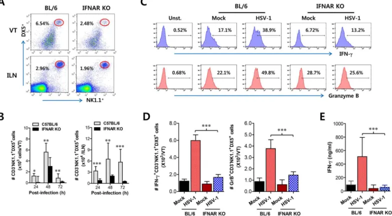
Together with CD11b+Ly-6Chimonocytes, NK cells are a critical innate cell population for pro-viding protection against mucosal HSV-1 infection via production of IFN-γ[17–19]. Since CD11b+Ly-6Chimonocytes and CD11c+DCs were shown to be consecutively recruited to the vaginal tract, we were also interested in the recruitment kinetics and activation of NK cells in IFNAR-ablated mice. Before virus infection, the frequency and number of CD3−NK1.1+DX5+ NK cells were comparable in vaginal tract and iliac LN of both BL/6 andIFNARKO mice, except liver tissue (S3 Fig), which indicates that IFN-I signaling may not contribute to basal recruitment of NK cells in primary target tissues of HSV-1 mucosal infection. However, it was revealed that IFN-I signaling plays a crucial role in recruiting CD3−NK1.1+DX5+NK cells into vaginal tract after HSV-1 infection. Notably, WT BL/6 mice displayed delayed recruitment of CD3−NK1.1+DX5+NK cells in the vaginal tract with the levels peaking at 48 h pi, compared to CD11b+Ly-6Chimonocytes, and an increased frequency of NK cells, compared toIFNARKO mice (Fig 3A). Also, iliac NK cell frequency was moderately increased in WT BL/6 mice com-pared toIFNARKO mice. Supporting these findings, total accumulated number of
CD3-NK1.1+DX5+NK cells in vaginal tract and iliac LN was greater in WT BL/6 mice, with levels peaking at 48 h pi, than inIFNARKO mice (Fig 3B). In addition, when the activation lev-els of NK cells were examined by analysis of NK cells producing IFN-γand granzyme B, the ablation of IFN-I signal resulted in reduced production of IFN-γand granzyme B from
3E). Collectively, these results indicate that NK cell recruitment in vaginal tract follows that of CD11b+Ly-6Chimonocytes like CD11c+DCs, and that IFN-I signal also has a critical contribu-tion to the early concerted recruitment and activacontribu-tion of CD11b+Ly-6Chimonocytes and NK cells in mucosal tissues following HSV-1 infection.
Cascade responses of CCL2-CCL3 are kinetically associated with
concerted recruitment of Ly-6C
himonocytes and NK cells
Sequential responses of chemokines appear to be necessary for the selective and tailored envi-ronment of infiltrated leukocytes that provides effective protection against mucosal HSV-1 infection, even though the chemokine responses can be redundant [32,33]. Moreover, our data demonstrated that infiltration of CD11b+Ly-6Chimonocytes preceded that of NK cells during mucosal inflammation; CD11b+Ly-6Chimonocyte infiltration peaked at around 24 h pi, IFNARKO mice at 24 h pi. Data denote the average±SD of values derived from three individual experiments (n= 5–6).*,p<0.05;**,p<0.01;***,p<0.001
for comparison between the indicated groups.
doi:10.1371/journal.ppat.1005256.g002
Fig 3. IFN-I signaling is involved in connected recruitment and activation of NK cells to CD11b+Ly-6Chimonocytes. (A)NK cell infiltration of infected
BL/6 orIFNARKO mice.(B)Total accumulated NK cell number. Cells were prepared from vaginal tract (VT) and iliac LN (ILN) by collagenase digestion at 24, 48, and 72 h pi and used for analysis of NK cells. Values in dot-plots denote the average percentages of NK1.1+DX5+NK cells derived from four independent samples after gating on CD3-negative cells at 48 h pi.(C)Frequency of IFN-γor granzyme B-producing cells among NK cells. The production of IFN-γand granzyme B by CD3−NK1.1+DX5+NK cells was determined by intracellular staining after stimulation of vaginal NK cells with PMA plus ionomycin at 48 h pi.
Values in histograms represent the average percentages of IFN-γor granzyme B-producing cells among CD3−NK1.1+DX5+NK cells.(D)Absolute number of
IFN-γor granzyme B-producing NK cells. Total number of IFN-γor granzyme B-producing CD3−NK1.1+DX5+NK cells in vaginal tract was enumerated by
flow cytometric analysis using intracellular and surface staining at 48 h pi.(E)Secreted IFN-γlevels in vaginal tract. Secreted IFN-γlevels were determined by ELISA at 48 h pi using vaginal lavages. Data in bar chart represent the average±SD of values derived from three individual experiments (n= 4–5).**,
p<0.01;***,p<0.001 compared with the levels ofIFNARKO mice.
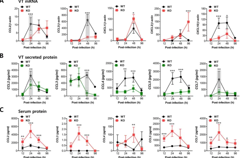
whereas NK cell infiltration peaked at 48 h after mucosal HSV-1 infection. Therefore, we were interested in the regulatory molecules that are involved in establishing this tailored environ-ment of sequential CD11b+Ly-6Chimonocyte and NK cell infiltration in vaginal tissues. To this end, we performed a kinetic examination of the expression and secretion of several CC and CXC chemokines in vaginal tissues and lavages at different time points up to 96 h pi. Somewhat intriguingly, our data revealed that CCL2 and CCL3 expression was evident early, with cascade responses peaking at subsequent time points. Hence, expression of CCL2, a ligand of CCR2, began as early as 12 h pi in vaginal tract of WT BL/6 mice and increased sharply in a time-dependent manner (Fig 4A). Maximal levels of vaginal CCL2 induction in WT mice were observed at around 24 h pi and had declined 80% by 96 h pi, whereasIFNARKO mice showed a delayed and lower expression of vaginal CCL2 with levels gradually increasing after 48 h pi. Vaginal expression of CCL3, a ligand of CCR1 and CCR5, peaked at 48 h pi in WT mice, whereasIFNARKO mice showed a drastically decreased expression level of CCL3. Other CXC chemokines (CXCL1, CXCL2) that are involved in recruitment of neutrophils via CXCR2 showed higher expression levels in the vaginal tract ofIFNARKO mice compared to WT BL/6 mice. Notably, CXCL2 expression gradually increased inIFNARKO mice after 48 h pi, whereas WT BL/6 mice showed decreased levels at 96 h pi. Regarding secreted chemokine proteins in vaginal lavages, ligands of CCR2 (CCL2, CCL7) were secreted at a higher level in WT BL/6 mice with reaching a maximum level at 24 h pi, compared to secretion inIFNARKO mice (Fig 4B). In contrast, ligands of CCR5 (CCL3, CCL4, CCL5) showed delayed peaks at around 48 h pi and such levels were markedly higher in WT mice than inIFNARKO mice. These results indicate that cascade responses of CC chemokines, especially CCL2 and CCL3, appear to be closely associated with consecutive recruitment of CD11b+Ly-6Chimonocytes and NK cells in the vaginal tract of WT BL/6 mice, whereas enhanced induction of CXC chemokine (CXCL1, CXCL2) inIFNARKO mice appears to be involved in neutrophil recruitment in the vaginal tract. However, systemic levels of most chemokine proteins were markedly higher in the sera of
IFNARKO mice compared to WT BL/6 mice (Fig 4C). Although cellular sources for huge amount of chemokine secretion in sera ofIFNARKO mice were not defined, it is likely that various cell types derived fromIFNARKO mice, including epithelial cells, fibroblast, and endo-thelial cells, are highly susceptible to HSV-1 replication, thereby secreting chemokines in sera by severe inflammation in whole body. In addition, this result suggests that enhanced chemo-kine levels in the serum may cause disturbed trafficking of various leukocytes inIFNARKO mice.
IFN-I signal on leukocytes derived from HSC lineage is required for
normal recruitment of Ly-6C
himonocytes, but not NK cells
Since CD11b+Ly-6Chimonocytes and NK cells showed concerted recruitment with levels peak-ing at different time points associated with the cascade response of CCL2-CCL3 in the vaginal tract, we were interested in dissecting the contribution of resident (stromal and epithelial cells) and HSC-derived leukocytes in this process. To achieve this, we used a BM chimeric model of WT BL/6 andIFNARKO mice. As expected,IFNARKO recipients ofIFNARKO BM donor cells (KO-KO chimera) showed a diminished frequency of CD11b+Ly-6Chimonocytes in vagi-nal and iliac LN, compared to WT recipients of WT BM donor cells (WT-WT chimera) (Fig
5A). Also, somewhat intriguingly,IFNARKO recipients of WT BM donor cells (WT-KO
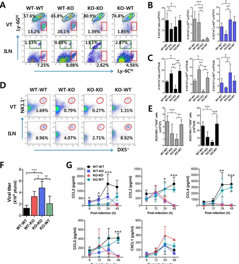
HSC lineage are essential for the recruitment of CD11b+Ly-6Chimonocytes in vaginal and iliac LN tissues. When the absolute number of recruited CD11b+Ly-6Chimonocytes was deter-mined, the essential role of IFN-I receptor on leukocytes derived from the HSC lineage in vagi-nal CD11b+Ly-6Chimonocyte recruitment was revealed again, even though KO-WT chimeras showed slightly higher levels of total recruited CD11b+cells compared to the other chimeras (Fig 5B). In contrast, IFN-I receptors in HSC-derived leukocytes appeared to be dispensable for the recruitment of CD11b+Ly-6Ghineutrophils in the vaginal tract, because WT-KO chime-ras showed a reduced number of neutrophils compared to KO-KO chimechime-ras. Also, consider-able recruitment of iliac CD11b+Ly-6Chimonocytes in both WT-KO and KO-WT chimeras highlighted the importance of IFN-I receptors in both HSC-derived and resident cells for lym-phoid tissues (Fig 5C). Therefore, these results indicate that IFN-I signal in some leukocytes derived from the HSC lineage plays a crucial role in early recruitment of CD11b+Ly-6Chi monocytes in the vaginal tract with relatively less contribution to recruitment of CD11b+ Ly-6Chimonocytes in draining LN, even though specific leukocytes derived from the HSC lineage Fig 4. Sequential responses of chemokines in response to mucosal infection with HSV-1. (A)Expression of chemokine mRNA in vaginal tract of BL/6 orIFNARKO mice following mucosal infection with HSV-1. The levels of chemokine mRNAs were determined by real-time qRT-PCR using total RNA extracted from vaginal tract (VT) tissues at 12, 24, 48, and 96 h pi.(B)Secreted levels of chemokine proteins in vaginal tract.(C)Serum chemokine protein levels. The levels of chemokine proteins were determined by CBA using vaginal lavages and sera at 12, 24, 48, and 96 h pi. Data show the average±SD of values derived from three individual experiments (n= 5–6). Areas highlighted by gray color denote interesting points.*,p<0.05;**,p<0.01;***,p<0.001 compared with the levels inIFNARKO mice at the indicated time point.
to involve in the recruitment of Ly-6Chimonocytes were not defined. Another intriguing result is that, unlike vaginal CD11b+Ly-6Chimonocyte recruitment, the recruitment of NK cells in the vaginal tract and iliac LN obviously depended dominantly on IFN-I receptor in resident cells; hence, KO-WT chimera showed completely recovered recruitment of CD3−NK1.1+DX5+ NK cells in vaginal tract and iliac LN, compared to diminished recruitment of NK cells in KO-KO and WT-KO chimeras (Fig 5D and 5E). In addition, it was likely that recovered recruitment of CD11b+Ly-6Chimonocytes and NK cells in WT-KO and KO-WT chimeras contributed to the control of HSV-1 replication in the vaginal tract (Fig 5F). Collectively, these results suggest that IFN-I signaling in resident cells is dominantly responsible for recruiting NK cells in inflammatory mucosal tissues, in contrast to CD11b+Ly-6Chimonocytes that depend on the IFN-I signal in BM-derived HSCs.
To further characterize the distinct dependence of vaginal recruitment of CD11b+Ly-6Chi monocytes and NK cells on IFN-I signal, we examined cascade responses of CC and CXC che-mokines in vaginal lavages obtained from four BM chimeric models following mucosal HSV-1 infection. CCL2 secretion was gradually increased in WT-KO chimera with levels detectable at 6 h, increasing at 24 h pi, and then sharply decreasing at 48 h pi (Fig 5G). However, KO-WT chimeras displayed delayed secretion of CCL2 starting at 24 h pi and gradually increasing to 48 h pi. These data indicate that HSC-derived leukocytes in vaginal tract may be dominant for production of CCL2 before 24 h pi, which is then replaced by CCL2 produced from resident cells of vaginal tract. In contrast, CCL3 as well as CCL4 and CCL5 showed secretion patterns that gradually increased from 24 h pi with maximum secretion at 48 h pi in KO-WT chimeras, suggesting that secretion of CCL3, together with several CCR5-binding CC chemokines, may be more dependent on resident cells after 24 h pi and subsequently involved in inducing infil-tration of NK and T cells in the vaginal tract. However, CXCL1, a ligand of CXCR2 for neutro-phil infiltration, intriguingly showed highest levels in KO-KO chimeras, which displayed increased infiltration of CD11b+Ly-6Ghineutrophils. Ultimately, these results indicate that early infiltration of CD11b+Ly-6Chimonocytes depends on CCL2 produced from HSC-derived leukocytes before 24 h pi, whereas CCL3, CCL4, and CCL5 produced from resident cells after 48 h pi are dominant regulators that induce infiltration of NK cells and CD11c+DCs, and then T cells at a later phase.
Orchestrated response of HSC-derived leukocytes and resident cells for
consecutive recruitment of Ly-6C
himonocytes and NK cells via CC
chemokines
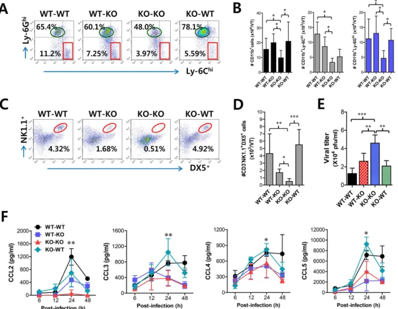
To further confirm the differential dependence of CD11b+Ly-6Chimonocyte and NK cell infil-tration on CC chemokines produced by HSC-derived leukocytes and mucosal resident cells, we used BM chimeric model of WT BL/6 and CCL2 KO mice. Since CCL2 KO recipients of WT BM donor cells (WT-KO) displayed a significantly higher frequency of vaginal CD11b+Ly-6Chi monocytes compared to CCL2 KO recipients of CCL2 KO BM donor cells (KO-KO) and WT recipients of CCL2 KO BM donor cells (KO-WT), CCL2 produced by HSC-derived leukocytes appeared to play a dominant role in the early recruitment of CD11b+Ly-6Chimonocytes in vaginal tract (Fig 6A). However, CC chemokines produced by vaginal resident cells were also Cells prepared from VT and ILN of the recipients were used to analyze the frequency (D) and total number (E) of CD3−NK1.1+DX5+NK cells at 48 h pi.(F)
Viral titers in vaginal lavages. Viral titers in vaginal lavages collected at 48 h pi were determined by plaque assay.(G)Chemokine secretion in vaginal lavages of the recipients. The levels of chemokine proteins were determined by CBA using vaginal lavages at 6, 12, 24, and 48 h pi. Values in dot-plots represent the average percentages of each population derived from four independent samples, and data in graphs represent the average±SD of values derived from three individual experiments (n= 4–5).*,p<0.05;**,p<0.01;***,p<0.001 compared with the level of the indicated group.
considered to have a residual role in the recruitment of CD11b+Ly-6Chimonocytes because WT-KO chimeras still showed a lower frequency of vaginal CD11b+Ly-6Chimonocytes than WT recipients of WT BM donor cells (WT-WT). Supporting these findings, when the absolute number of CD11b+Ly-6Chimonocytes in the vaginal tract was determined, a significant role of CCL2 produced by HSC-derived leukocytes was evident in the early recruitment of CD11b+ Ly-6Chimonocytes in the vaginal tract (Fig 6B). Moreover, CCL2 produced from HSC-derived leukocytes and resident cells was shown to partially contribute to vaginal recruitment of CD11b+Ly-6Ghineutrophils, because KO-KO chimeras showed a decreased number of vaginal CD11b+Ly-6Ghineutrophils compared to WT-KO and KO-WT chimeric models (Fig 6B). In addition, our data revealed that CCL2 played a certain role in recruiting NK cells, because KO-KO chimeras showed diminished NK cell frequency and number compared to WT-WT chimeras (Fig 6C and 6D). However, recruitment of CD3−NK1.1+DX5+NK cells in vaginal tract appeared to dominantly depend on CC chemokines produced by resident cells together with CCL2, because KO-WT chimeras displayed a comparable frequency of NK cells to WT-WT chimeras. Also, WT-KO chimeras showed a higher frequency of NK cells in the vagi-nal tract than KO-KO chimeras, which indicates that CCL2 produced by HSC-derived leuko-cytes contributes in part to NK cell recruitment. Viral burden in vaginal lavages reflected the differential dependence of CD11b+Ly-6Chimonocytes and NK cells on CCL2 produced from HSC-derived and resident cells (Fig 6E). We also examined the levels of secreted CC and CXC chemokines in vaginal lavages of four CCL2 KO BM chimeric models. As expected, WT-WT chimeras showed the highest CCL2 levels, which peaked at 24 h pi and declined by 48 h pi, whereas a considerable amount of secreted CCL2 protein was detected in WT-KO chimeras with a delayed pattern that started at around 24 h pi and gradually increased until 48 h pi (Fig 6F), indicating that early infiltrated HSC-derived leukocytes producing CCL2 may be evident from around 24 h pi. In contrast withIFNARKO BM chimera experiment, secretion of CCL2 protein was evident at higher levels in KO-WT chimeras at 24 h pi compared to WT-KO chi-meras. This implies that vaginal resident cells could compensate for the CCL2 supply at a cer-tain point in the absence of CCL2 production by infiltrated leukocytes. Other CC chemokines (CCL3, CCL4, CCL5) that are involved in NK and Th CD4+T cell recruitment were detected at higher levels in WT-WT and KO-WT chimeras compared to WT-KO and KO-KO chimeras, suggesting that the supply of those CC chemokines is mostly accomplished by vaginal resident cells after 24 h pi. Moreover, maximum secretion of CCR5 ligands (CCL3, CCL4, CCL5) was observed in vaginal tract of KO-WT chimera at around 24 h pi, which implies that partial CCL2 ablation could change sequential production of other chemokines, due to compensation roles. Collectively, these results indicate that CCL2 produced by HSC-derived leukocytes is pre-dominantly responsible for the accumulation of CD11b+Ly-6Chimonocytes within 24 pi, whereas recruitment of NK cells in the vaginal tract appears to be mediated by the CC chemo-kine pool achieved by vaginal resident cells from 24 to 48 h pi, together with CCL2.
Critical role of IFN-I signaling on tissue resident CD11b
hiF4/80
himacrophages and CD11c
hiEpCAM
+DCs in the initial production of
CCL2 proteins
recruitment of CD11b+Ly-6Chimonocytes, which subsequently provide amplified recruitment of more CD11b+Ly-6Chimonocytes via CCL2. To this end, we examined CCL2 and CCL3 expression in tissue resident CD11bhiand CD11chicell subpopulations at 12 h pi, before the recruitment of CD11b+Ly-6Chimonocytes peaked. Interestingly, initial expression of CCL2 was dominantly achieved in CD11bhiF4/80himacrophages and CD11chiEpCAM+DCs sorted from vaginal tract of WT BL/6 mice, compared to those ofIFNARKO mice (Fig 7A). Also, other CD11bhimyeloid cells (CD11bhiF4/80lo) and CD11chiDCs (CD11chiEpCAM−), but not Fig 6. CCL2 produced from infiltrated leukocytes derived from HSC lineage plays a dominant role in mucosal recruitment of CD11b+Ly-6Chi monocytes.BM cells from BL/6 (WT) orCCL2KO (KO) mice were grafted to lethally irradiated BL/6 orCCL2KO recipient mice, which were infected i.vag. with HSV-1.(A,B)CD11b+Ly-6Chiinfiltration of infected recipients in vaginal tract. Cells were prepared from the vaginal tract by collagenase digestion at 24 h pi and subcellular proportions of CD11b+Ly-6Chiand CD11b+Ly-6Ghileukocytes were determined using flow cytometric analysis.(C,D)Frequency and absolute number of infiltrated NK cells in vaginal tract of the recipients. Cells prepared from the vaginal tract of the recipients were used to analyze the frequency (C) and total number (D) of CD3−NK1.1+DX5+NK cells at 48 h pi.(E)Viral titers in vaginal lavages. Viral titers in vaginal lavages collected at 48 h
pi were determined by plaque assay.(F)Chemokine secretion in vaginal lavages of the recipients. The levels of chemokine proteins were determined by CBA using vaginal lavages at 6, 12, 24, and 48 h pi. Values in dot-plots represent the average percentages of each population derived from four independent samples, and data in graphs represent the average±SD of values derived from three individual experiments (n= 4–5).*,p<0.05;**,p<0.01;***,p<0.001 compared with the level of the indicated group.
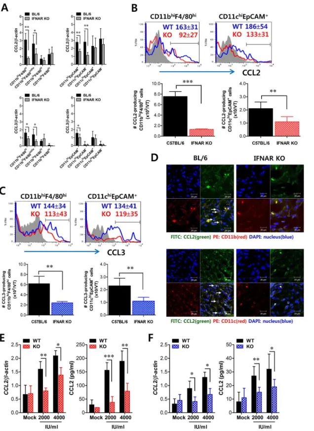
Fig 7. Critical role of IFN-I signaling on resident CD11bhiF4/80himacrophages and CD11chiEpCAM+DCs in the production of initial CCL2 protein for early migration of CD11b+Ly-6Chimonocytes. (A)Early expression of CCL2 and CCL3 by sorted CD11bhi
F4/80himacrophages and CD11chiEpCAM+ DCs. Subsets of macrophages (CD11bhiF4/80hi, CD11bhiF4/80int/lo, CD11bloF4/80hi, and CD11bloF4/80lo) and DCs (CD11chiEpCAM+, CD11chiEpCAM−,
CD11cloEpCAM+, and CD11cloEpCAM−) were sorted 12 h after mucosal HSV-1 infection, and CCL2 and CCL3 expression was analyzed with real-time
CD11bloand CD11clocells, showed significantly higher levels of CCL2 expression in WT BL/6 compared toIFNARKO mice. In addition, CCL3 expression was also apparent in CD11bhiF4/ 80himacrophages and CD11chiEpCAM+DCs derived from WT BL/6 mice, even though levels of CCL3 expression were much lower than those of CCL2. To confirm these findings, we enu-merated CCL2-producing CD11bhiF4/80himacrophages and CD11chiEpCAM+DCs in vaginal tract of both WT BL/6 and IFNAR KO mice at 12 h pi. As expected, increased expression of CCL2 protein in vaginal CD11bhiF4/80himacrophages and CD11chiEpCAM+DCs of WT BL/6 mice was observed, and a higher number of CCL2-producing CD11bhiF4/80himacrophages and CD11chiEpCAM+DCs was detected in vaginal tract of WT BL/6 mice, compared with
IFNARKO mice (Fig 7B). Similarly, vaginal CD11bhiF4/80himacrophages and
CD11chiEpCAM+DCs derived from WT BL/6 mice showed higher expression of CCL3 com-pared to those ofIFNARKO mice (Fig 7C). The production of CCL2 protein by vaginal CD11bhiF4/80himacrophages and CD11chiEpCAM+DCs was further visualized using confocal microscopy (Fig 7D). In consistent, CCL2-producing CD11b+and CD11c+cells were detected at a higher frequency in vaginal tract of WT BL/6 mice thanIFNARKO mice. Also, F4/80+ cells producing CCL2 protein were detected at a higher frequency in the vaginal tract of WT BL/6 thanIFNARKO mice (S4 Fig). Since IFN-I signaling plays an important role in inducing CCL2 protein by stimulation with IFN-I proteins produced from epithelial cells through IFI-16 and STING recognition upon HSV-1 infection [34,35], we examined the expression of CCL2 from vaginal CD11bhiF4/80himacrophages and CD11chiEpCAM+DCs sorted from WT BL/6 andIFNARKO mice upon stimulation with IFN-αprotein. As expected, CD11bhiF4/80hi mac-rophages and CD11chiEpCAM+DCs derived from vaginal tract of WT BL/6 mice showed higher expression of CCL2 protein than those ofIFNARKO mice (Fig 7E and 7F), even though CCL2 expression in CD11chiEpCAM+DCs was detected at lower levels than CD11bhiF4/80hi macrophages. Similarly, other CC chemokines (CCL3, CCL4, CCL5), but not CXC chemo-kines, were expressed with slightly higher levels in CD11bhiF4/80himacrophages and CD11chiEpCAM+DCs derived from vaginal tract of WT BL/6 mice, compared to those of
IFNARKO (S5 Fig). Collectively, these results suggest that tissue resident CD11bhiF4/80hi mac-rophages and CD11chiEpCAM+DCs are candidates for supplying the initial CCL2 protein upon stimulation by IFN-I produced from infected epithelial cells, thereby inducing early infil-tration of CD11b+Ly-6Chimonocytes into mucosal tissues.
Tissue resident CD11b
hiF4/80
himacrophages and CD11c
hiEpCAM
+DCs play a role in establishing early orchestrated infiltration of Ly-6C
himonocytes and NK cells
Since tissue resident CD11bhiF4/80himacrophages and CD11chiEpCAM+DCs were proposed to be the initial key cell populations for recruiting early CD11b+Ly-6Chimonocytes for migra-tion-based self-amplification, we investigated the contribution of these cell populations in establishing early orchestrated infiltration of CD11b+Ly-6Chimonocytes and NK cells in vagi-nal tract. To examine the role of CD11bhiF4/80himacrophages we used the depletion model of macrophages and CD11chiEpCAM+DCs producing CCL2 (B) and CCL3 (C) were determined by flow cytometric analysis at 12 h pi. Blue line, HSV-1-infected BL/6; Red line, HSV-HSV-1-infected IFNAR KO; Gray line, mock-infected.(D)Confocal microscopic analysis of early CCL2 production by resident CD11b+macrophages and CD11c+DCs. CD11b+macrophages and CD11c+DCs producing CCL2 protein were visualized by confocal microscopy at 12 h pi. CD11b+macrophages and CD11c+DCs producing CCL2 protein are denoted by white arrows.(E,F)Essential role of IFN-I signaling in the production of CCL2 protein from CD11bhiF4/80himacrophages and CD11chiEpCAM+DCs. Vaginal CD11bhiF4/80himacrophages (E) and CD11chiEpCAM+DCs (F) isolated from WT andIFNARKO mice were stimulated with recombinant IFN-α(2,000 and 4,000 IU/ml) for 6 h. The expression and production of CCL2 were determined by real-time qRT-PCR and CBA using stimulated cells and collected culture media, respectively. Data represent the average±SD of values derived from three individual experiments (n= 4–5).*,p<0.05;**,p<0.01;***,p<0.001 compared with the level of the indicated group.
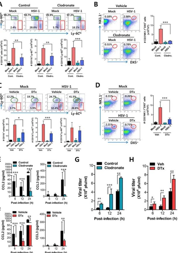
CD11bhiF4/80hicells using clodronate liposome, as WT BL/6 mice showed an approximately 60% reduction in CD11bhiF4/80himacrophages after administration of clodronate liposomes (S6A and S6B Fig). Ablation of tissue resident CD11bhiF4/80himacrophages reduced the accu-mulated number of infiltrated CD11b+Ly-6Chimonocytes, but the frequency of CD11b+ Ly-6Chimonocytes was not obviously changed after gating on CD11b+cells because of the effect of clodronate liposomes on total CD11b+myeloid cells (Fig 8A). Thus, the accumulated num-ber of total CD11b+and CD11b+Ly-6Ghigranulocytes was also reduced. Since clodronate lipo-somes affect total CD11b+myeloid cells, including tissue resident CD11bhiF4/80hi
macrophages, we also used GdCl3, which has been known as macrophage-selective inhibitor
[36]. Our data revealed that GdCl3administration selectively reduced the frequency and
accu-mulated number of CD11b+Ly-6Chimonocytes in vaginal tract (S7A Fig). Also, the recruit-ment of NK cells in vaginal tract was apparently decreased in both frequency and accumulated number by the inhibition of tissue resident CD11bhiF4/80himacrophages using cloronate lipo-somes (Fig 8B) and GdCl3(S7B Fig). In addition, we used CD11c-DTR mice that allow
condi-tional DC depletion upon DT injection (S6C and S6D Fig) to examine the role of
CD11chiEpCAM+DCs in the recruitment of early CD11b+Ly-6Chimonocytes. Consistently, ablation of CD11chiEpCAM+DCs reduced the frequency and accumulated number of CD11b
-+Ly-6Chimonocytes and NK cells in vaginal tract (Fig 8C and 8D). These results suggest that
tissue resident CD11bhiF4/80himacrophages and CD11chiEpCAM+DCs play an important role in recruiting the early infiltration of CD11b+Ly-6Chimonocytes to foster the orchestrated infiltration of subsequent leukocytes.
To further understand the role of CD11bhiF4/80himacrophages and CD11chiEpCAM+DCs in recruiting initial CD11b+Ly-6Chimonocytes for migration-related self-amplification, we examined CCL2 and CCL3 production in vaginal tracts of mice in which CD11bhiF4/80hi mac-rophages and CD11chiEpCAM+DCs were depleted. Ablation of tissue resident CD11bhiF4/ 80himacrophages and CD11chiEpCAM+DCs diminished production of CCL2 protein as early as 6 h pi, but CCL3 production was reduced at 24 h pi (Fig 8E and 8F) with a delayed pattern, which appeared to result from decreased infiltration of CD11b+Ly-6Chimonocytes. In support of these findings, decreased infiltration of early CD11b+Ly-6Chimonocytes in vaginal tract of mice depleted for CD11bhiF4/80himacrophages and CD11chiEpCAM+DCs was closely associ-ated with enhanced replication of HSV-1 (Fig 8G and 8H). Selective inhibition of CD11bhiF4/ 80himacrophages by GdCl3 also resulted in an enhanced viral burden in vaginal lavages (S7C
Fig). Therefore, these results indicate that tissue resident CD11bhiF4/80himacrophages and
CD11chiEpCAM+DCs in vaginal tract are key cellular regulators for the initial recruitment of CD11b+Ly-6Chimonocytes, thereby establishing an orchestrated environment of subsequent leukocyte infiltration through their migration-based self-amplification to provide early viral clearance.
Discussion
depend on the CCL2-CCL3 cascade mediated by HSC-derived leukocytes and resident cells, respectively. Furthermore, tissue resident CD11bhiF4/80himacrophages and CD11chiEpCAM+ DCs were involved in producing the initial CCL2 for migration-based self-amplification of rap-idly infiltrated CD11b+Ly-6Chimonocytes through stimulation by IFN-I produced from infected epithelial cells. To the best of our knowledge, this is the first report that deciphers the cellular and molecular cascade pathway to regulate IFN-I-dependent recruitment of CD11b +-Ly-6Chimonocytes and NK cells into inflammatory mucosal tissues.
Orchestrated mobilization of innate and adaptive immune cells between the vaginal epithe-lium and the draining (iliac) LNs during mucosal HSV-1 infection is critical for the control of viral replication, thereby preventing CNS invasion. However, the redundancy of chemokine recognition by immune cells sometimes makes the interpretation of results difficult. The che-mokine receptor CCR2 is expressed in CD11b+Ly-6Chimonocytes as well as a subset of B, NK, NKT cells, pDCs, and activated T cells [37–39]. However, since the number of B, NKT cells, pDCs, and activated T cells in vagina mucosa ofCCR2KO mice is comparable to that of WT mice following mucosal HSV infection [28], CCR2 is thought to play a role in the recruitment of CD11b+Ly-6Chimonocytes and NK cells. In addition, NK cell recruitment is also regulated by other CC chemokines, including CCL3, CCL4, and CCL5, as a result of expression of CCR5 on NK cells [40–42], and CXCL10, a ligand of CXCR3, was suggested to mediate NK cell mobi-lization in vaginal tissue as a principal chemoattractant molecule that is liberated early after genital HSV-2 infection [43]. Consequently, CCR2 is considered the main chemokine receptor involved in the recruitment of CD11b+Ly-6Chimonocytes, whereas several chemokine recep-tors appear to be involved in the recruitment of NK cells. Likewise, our data demonstrate that the production of CCL2, a ligand of CCR2, is kinetically associated with the vaginal recruit-ment of CD11b+Ly-6Chimonocytes, which peaked at 24 h pi. CD11b+Ly-6Chimonocytes egress massively from bone marrow into the bloodstream in a CCR2-dependent manner and are recruited via the CCL2-CCR2 axis into infected sites, where they exert direct antimicrobial activities or further differentiate into inflammatory DCs [24,25]. However, the cellular source of CCL2 for the recruitment of CD11b+Ly-6Chimonocytes into mucosal tissues was not clearly defined. Our experiments usingIFNARKO BM chimeric models demonstrate that some leuko-cytes derived from the HSC lineage play a dominant role in the recruitment of CD11b+Ly-6Chi monocytes into vaginal tissues as well as iliac LNs because onlyIFNARKO mice reconstituted by WT BM cells (WT-KO) showed comparable recruitment of CD11b+Ly-6Chimonocytes into vaginal and iliac LN tissues to that of WT recipients of WT BM cells (WT-WT). This result contrasts starkly with the fact that IFN-I signals on corneal resident cells play an impor-tant role in recruiting CD11b+Ly-6Chimonocytes in the corneal area [13], but is in line with a report that IFN-I signaling on the hematopoietic cell lineage plays a dominant role in leukocyte recruitment into inflammatory lung tissues after influenza infection [29]. Supportively, CCL2 production in vaginal tissues of WT-KO BM chimeric model gradually increased up to 24 h pi, after which CCL2 level decreased. This implies that recruitment of CD11b+Ly-6Chimonocytes may be governed by other chemokines produced from resident cells after 24 h pi or that CCL2 pool produced up to 24 h is sufficient to recruit major CD11b+Ly-6Chimonocytes. The former monocyte infiltration by CD11chiDCs. CD11c-DTR Tg mice were treated with DT to deplete CD11chiDCs and infected i.vag. with HSV-1. The infiltration of CD11b+Ly-6Chimonocytes was determined by flow cytometric analysis at 24 h pi.(D)Regulation of NK cell infiltration by CD11chiDCs. CD3−NK1.1+DX5+
NK cells in the vaginal tract were analyzed in DT-treated CD11c-DTR mice at 48 h pi.(E,F)Resident CD11bhiF4/80himacrophages and CD11chiDCs provide very early CCL2 production. The levels of CCL2 and CCL3 proteins in vaginal lavages were determined in clodronate- and DT-treated mice at 6, 12, and 24 h pi.(G,H)Viral replication in vaginal tract of mice depleted for CD11bhiF4/80himacrophages and CD11chiDCs. Viral titers in vaginal tract of clodronate- and DT-treated mice were determined by plaque assay at 6, 12, and 24 h pi. Data in the bar chart represent the average±SD of values derived from three
individual experiments (n= 4–5).*,p<0.05;**,p<0.01;***,p<0.001 compared with the level of the indicated group.
speculation is supported by the fact that CCL2 level in vaginal lavages ofCCL2KO-WT BM chimera gradually increased after 24 h pi, suggesting that resident cells such as epithelial cells resistant toγ-irradiation might produce CCL2 and other CC chemokines with delayed patterns compared to HSC-derived leukocytes. However, as the recruitment of CD11b+Ly-6Chi mono-cytes peaked at 24 h pi, CCL2 produced from HSC-derived leukomono-cytes within 24 h pi is consid-ered to be responsible for the major infiltration of CD11b+Ly-6Chimonocytes. Our
experiments usingCCL2KO BM chimeras in part support this, becauseCCL2KO recipients of WT BM cells (WT-KO) elicited a higher frequency of CD11b+Ly-6Chimonocytes than KO-KO and KO-WT BM chimeras, indicating that CCL2 produced from HSC-derived leuko-cytes plays a dominant role in recruiting early CD11b+Ly-6Chimonocytes in vaginal tissues. Consequently, the turning point at which the recruitment of CD11b+Ly-6Chimonocytes is replaced by recruitment of other cell populations, including NK cells and CD11c+DCs, appeared to occur between 24 and 48 h pi.
In addition to CD11b+Ly-6Chimonocytes, neutrophils are a major population of innate immune cells that are involved in immunopathology [44,45], although their role in viral infec-tion remains controversial. According to our data, it is likely that IFN-I signaling is a negative regulator in the recruitment of CD11b+Ly-6G+neutrophils into vaginal tissues. Indeed, whereas IFN-I signaling seems to play a general role in promoting CD11b+Ly-6Chimonocytes [29–31], its effect on CD11b+Ly-6G+neutrophil trafficking and function are quite divergent between dif-ferent infection models [46,47]. We show here thatIFNARKO mice produced higher amounts of CXCL1 and CXCL2, which are the main ligands of CXCR2 expressed on neutrophils. It was recently reported that IFN-I abrogates the recruitment of neutrophils to ganglia by directly sup-pressing CXCL2 expression by monocytes [48]. This is in line with our finding that infiltrated CD11b+Ly-6Clocells sorted fromIFNARKO mice, but not CD11b+Ly-6Chimonocytes, expressed high levels of CXCL1 and CXCL2 compared to those from WT mice. However, the most interesting result is that the recruitment of CD11b+Ly-6G+neutrophils depends mostly on resident cells, because only WT mice reconstituted byIFNARKO BM cells showed identical lev-els of infiltrated neutrophils to the KO-KO chimeras. This indicates that IFN-I signaling on HSC-derived leukocytes suppresses the recruitment of neutrophils in vaginal tissues following HSV-1 infection, thereby ameliorating aberrant immunopathology. Considering thatIFNAR
KO mice lack neutrophil recruitment in inflamed tissues in bacterial infection models [49], it seems likely that IFN-I signaling in HSC-derived leukocytes performs divergent roles to recruit specific innate cells depending on the context and specific details of the infection model.
In contrast to the recruitment of CD11b+Ly-6Chimonocytes, and somewhat intriguingly, NK cell recruitment into vaginal and iliac LN tissues is delayed and dependent on resident cells such as epithelial and stromal cells that are resistant toγ-irradiation. It is possible that CCL3, CCL4, and CCL5, which are ligands of CCR5 expressed on NK cells, are the main regulators of NK cell recruitment because their expression was kinetically associated with the recruitment of NK cells. Also, our data support the residual role of HSC-derived leukocytes in recruiting NK cells in vaginal and iliac LN tissues because of the increased accumulation of NK cells in
IFNARKO recipients of WT BM cells (WT-KO) compared with KO-KO BM chimera. The role of CCL3 and CCL5 in NK cell recruitment has been extensively described [40–42], and our data are further strengthened by striking evidence that decreased CCL3 production in
CD11b+Ly-6Clomonocytes probably do not play a residual role in recruiting vaginal NK cells because there was no difference in CCL3 and CCL5 production between monocytes sorted from WT andIFNARKO mice. Instead, CCL2 produced from CD11b+Ly-6Chimonocytes appears to share responsibility in recruiting NK cells with CCL3 and CCL5 produced from resi-dent cells, becauseCCL2KO recipients of WT BM cells (WT-KO) showed a higher number of vaginal NK cells than KO-KO BM chimeras. Despite debatable issue, CCL2-CCR2 axis is believed to play a certain role in recruiting NK cells to infected sites [51], which is consistent with our result thatCCL2KO recipients ofCCL2KO BM cells showed reduced infiltration of NK cells in vaginal tissues following HSV-1 infection. Consequently, the recruitment of late-comer NK cells in vaginal tissue appears to be partly regulated by the earlier recruitment of first-mover CD11b+Ly-6Chimonocytes, which mostly depends on HSC-derived leukocytes.
CCL2 chemokine is widely studied because of its upregulation in autoimmune disease, tumors, and infections. Although CCL2 production has been shown to be regulated by TNF-α
and STAT2 signals [52,53], CCL2 production can be induced by stimulation with IFN-I alone through an IFN-responsive element in theCCL2upstream promoter [13]. Following HSV-1 mucosal infection, initial viral replication is limited to mucosa epithelium of the vaginal tract, where IFN-I is produced through the action of IFI-16/p204 and the downstream adaptor mole-cule STING [13,34,35,50]. This IFN-I appears to stimulate neighboring epithelial cells and tissue resident immune cells to induce the expression of CCL2 [13,50]. As another interesting finding, our data suggest the importance of vaginal tissue resident CD11bhiF4/80himacrophages and CD11chiEpCAM+DCs in providing the initial CCL2 protein responsible for recruiting early CD11b+Ly-6Chimonocytes within 12 h pi, although CD11c−EpCAM+epithelial cells can also produce CCL2 protein through IFN-I stimulation [13,50]. It is likely that the amount of CCL2 protein produced from vaginal tissue resident CD11bhiF4/80himacrophages and
CD11chiEpCAM+DCs is low, due to basally low number of these cell populations in vaginal tis-sue. However, the initial CCL2 may be able to recruit early CD11b+Ly-6Chimonocytes, which in turn provide more CCL2 protein to establish self-amplification of CD11b+Ly-6Chimonocyte migration. This notion is strengthened by a previous study showing that CD11b+F4/80+ macro-phages in the liver provide initial CCL2 protein to recruit NK cells in a MCMV infection model [51]. The vaginal tract is composed of three principal layers: (i) the mucosa epithelium com-posed of stratified nonkeratinized squamous epithelial tissue, (ii) the submucosa of vascularized connective tissue, and (iii) the muscularis layer composed of smooth muscle [54]. CD11bhiF4/ 80himacrophages and CD11chiEpCAM+DCs, which are responsive to stimulation by IFN-I [54], are strategically localized in both the stratified squamous epithelial layer and the submuco-sal lamina propria. Therefore, the data presented here suggest that IFN-I produced from initially infected epithelial cells stimulate tissue resident CD11bhiF4/80himacrophages and
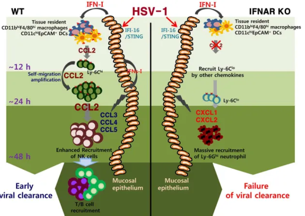
The role of CD11b+Ly-6Chimonocytes in controlling parasitic and bacterial infections is well described [6,24–28], but the contribution of these cells in HSV infection has only recently begun to grab the attention [13,28]. The majority of studies suggest a predominant role of neu-trophils and NK cells in containing HSV replication during the innate immune response [55,56], whereas the mechanism of the antimicrobial activity by CD11b+Ly-6Chimonocytes is ambiguous. Although our data discount direct antiviral action of IFN-I on HSV-1 replication [57,58], we surmise a coordinated role of early infiltrated CD11b+Ly-6Chimonocytes in estab-lishing orchestrated mobilization of innate immune cells to contain HSV-1 replication in vagi-nal tissues. Furthermore, considering that IFN-I sigvagi-naling plays an important role in HSC proliferation and differentiation [59–61],IFNRAKO mice may contain different cell constitu-tion from WT mice during infecconstitu-tion. Specific cell populaconstitu-tions directly affected by IFN-I during hematopoiesis and their contributions to unique cell population in peripheral tissues has been recently described [29]. Supportively, IFN-I–dependent differentiation of CD11b+Ly-6Chi monocytes producing CCL2 appears to play a critical role in establishing the orchestrated mobilization of innate cells, whereas the differentiation of CD11b+Ly-6Clomonocytes produc-ing CXCL1, IL-23, IL-10, and TGF-βis likely to induce aberrant sequential inflammation in vaginal tissue ofIFNARKO mice. This finding is also strengthened by the recent report that Fig 9. IFN-I–dependent cascade pathway for concerted recruitment of CD11b+Ly-6Chimonocytes and NK cells in mucosal tissues following HSV-1 infection.At the very initial phase of infection, CCL2 is initially produced from tissue resident CD11bhiF4/80himacrophages and CD11chiEpCAM+DCs through stimulation with IFN-I proteins produced from mucosal epithelium via the action of IFI-16 and STING in response to vaginal infection with HSV-1, thereby recruiting very early CD11b+Ly-6Chimonocytes within 12 h pi. Subsequently, the early infiltrated CD11b+Ly-6Chimonocytes contribute to migration-based self-amplification through the supply of higher levels of CCL2 at around 24 h pi. Delayed production of other CC chemokines (CCL3, CCL4, CCL5) from infected and uninfected epithelial cells, together with CCL2 provided by monocytes, then contributes to the recruitment of late-comer NK cells at 48 h pi. The lack of an initial CCL2 supply inIFNARKO mice results in reduced recruitment of CD11b+Ly-6Chimonocytes; instead, CXC chemokines (CXCL1, CXCL2) primarily produced from resident cells and CD11b+Ly-6Clomonocytes probably induce massive recruitment of CD11b+Ly-6G+neutrophils.
IFN-I attenuates neutrophil infiltration to prevent severe tissue damage caused by uncontrolled inflammation [48]. In conclusion, our findings elucidate a detailed IFN-I-dependent pathway that establishes the orchestrated mobilization of CD11b+Ly-6Chimonocytes and NK cells through consecutive production of CCL2 and CCL3 chemokines from HSC-derived leukocytes and epithelium-resident cells. Our data further imply that the cascade responses of resident-to-hematopoietic-to-resident cells for cytokine-to-chemokine-to-cytokine production to recruit innate immune cells are critical for the attenuation of HSV replication in inflamed tissues.
Materials and Methods
Ethics statement
All animal experiments described in the present study were conducted at Chonbuk National University according to the guidelines set by the Institutional Animal Care and Use tees (IACUC) of Chonbuk National University and were pre-approved by the Ethical Commit-tee for Animal Experiments of Chonbuk National University (Permission code 2013–0028). Animal research protocol in this study followed the guideline set up by the nationally recog-nized Korea Association for Laboratory Animal Sciences (KALAS).
Animals
Female C57BL/6 (H-2b) mice (6–8 weeks old) were purchased from Samtako (O-San, Korea).
CCL2knockout (KO) andCD11c-DTRtransgenic (Tg) mice (B6.FVB-Tg Itgax-DTR/EGFP 57Lan/J [DTR]) were obtained from The Jackson Laboratories (Bar Harbor, ME, USA).IFNAR
KO mice deficient in the receptor for type I IFNs (IFN-α/β) were described elsewhere [29]. All mice were genotyped and bred in the animal facilities of Chonbuk National University.
Cells, viruses, and in vivo viral infection
The HSV-1 McKrae strain was propagated in Vero cells (CCL81; ATCC, Manassas, VA, USA) using DMEM supplemented with 2.5% FBS, penicillin (100 U/ml), and streptomycin (100 U/ ml). The Vero cells were infected with HSV-1 at a multiplicity of infection (MOI) of 0.01 and incubated in a CO2incubator for 1 h at 37°C with shaking at 15-min intervals. After absorption,
the inoculum was removed, and 5 ml of maintenance medium containing 2.5% FBS was added. Approximately 2–3 dpi, cultures of the host cells showing an 80–90% cytophatic effect were har-vested and titrated by conventional plaque assay using Vero cells. The virus stocks were stored in aliquots at -80°C until use. For in vivo viral challenge, the mice were pretreated with proges-terone to synchronize their estrous cycles as described earlier [62,63]. Briefly, mice were subcu-taneously (s.c.) injected with medroxyprogesteron 17-acetate (Depoprovera [DP]; Sigma-Aldrich, St. Louis, MO, USA) at 2 mg per mouse. Five days after injection of DP, the mice were challenged intravaginally (i.vag.) with the HSV-1 McKrae strain (1×10⁶PFU). Infected mice were examined daily for vaginal inflammation, neurological illness, and death, as described pre-viously [63]. Mice were scored 1 to 5 depending on the clinical severity of disease (0, no change; 1, mild inflammation; 2, moderate swelling; 3, severe inflammation; 4, paralysis; 5, death).
Antibodies and reagents
The monoclonal antibodies used for flow cytometric analysis and other experiments were pur-chased from eBioscience (San Diego, CA, USA), BD Bioscience (San Diego, CA, USA), and R&D Systems (Minneapolis, MN, USA), which included: FITC-conjugated anti-mouse CD3ε
granzyme B (NGZB), CD11b (M1/70), CD326 (EpCAM) (G8.8), CCR2 (475301), and CXCR2 (242216); PE-Cy-7–conjugated anti-mouse NK1.1 (PK136), PerCP-Cy5.5–conjugated anti-mouse Ly-6C (HK1.4), PerCP-conjugated anti-anti-mouse CCL3(MIP-1 alpha) (DNT3CC); allo-phycocyanin (APC)-conjugated anti-mouse Ly-6G (1A8) and F4/80 (BM8); biotin-conjugated anti-mouse pan-NK cells, CD49b-Integrin alpha 2 (DX5), F4/80 (BM8); and streptavidin-con-jugated APC.
Quantitation of viral burden and cytokine/chemokine expression
Quantitative real-time PCR for viral burden and cytokine/chemokine expression. Viral burden and cytokine/chemokine expression were assessed by real-time qPCR using genomic DNA extracted from collected tissues and real-time qRT-PCR using total RNA extracted from collected tissues and sorted cells, respectively. After extraction of genomic DNA and total RNA, real-time qPCR was performed using a CFX96 Real-Time PCR Detection system (Bio-Rad Laboratories, Hercules, CA, USA). To determine the expression of cytokine and chemo-kine mRNAs, total RNA was subjected to reverse transcription with High-Capacity cDNA Reverse Transcription Kits (Applied Biosystems, Foster City, CA, USA) for real-time qPCR. The reaction mixture contained 2μl of template cDNA, 10μl of 2× SYBR Primix Ex Taq, and
200 nM specific primers (S1 Table) at a final volume of 20μl. The reaction mixes were
dena-tured at 95°C for 30 s and then subjected to 45 cycles of 95°C for 5 s and 60°C for 20 s. After the reaction cycle was completed the temperature was increased from 65°C to 95°C at a rate of 0.2°C/15 s and fluorescence was measured every 5 s to construct a melting curve. A control sample that contained no template DNA was run with each assay, and all determinations were performed at least in duplicate to ensure reproducibility. The authenticity of the amplified product was determined by melting curve analysis. Viral burden was expressed as viral DNA copy number per microgram of genomic DNA, and the expression of cytokines and chemo-kines was expressed as relative fold expression after normalization to the housekeeping geneβ -actin. All data were analyzed using Bio-Rad CFX Manager, version 2.1 analysis software (Bio-Rad Laboratories).
Cytokine bead array (CBA). The levels of chemokine proteins in vaginal lavages and sera were measured by the cytometric bead array (CBA) specific for CCL2 (MCP-1), CCL3 (MIP-1α), CCL4 (MIP-1β), CCL5 (RANTES), CCL7 (MCP-3), and CXCL1 (KC), according to the manufacturer’s protocols (BD Bioscience).
Analysis of infiltrated leukocytes in vaginal tract and iliac lymph node
Vaginal tract and iliac LN were collected from WT andIFNARKO mice infected with HSV-1 McKrae strain (1×10⁶PFU/mouse) at 12, 24, 48, and 72 h pi. Leukocytes that had infiltrated into the vaginal tract and iliac LN were harvested, homogenized by gently pressing through a 100-mesh tissue sieve, and digested with 25μg/ml collagenase type IV (Worthington Biochem,Freefold, NJ, USA) in RPMI medium for 1 h at 37°C in a shaking incubator. Cells were counted and stained for CD11b, Ly6G, Ly6C, CD3, CD4, CD8, and NK1.1 with directly conjugated antibodies (eBioscience) for 30 min at 4°C. Finally, the cells were fixed with 10% formaldehyde. Data collection and analysis were performed with a FACSCalibur flow cytometer (Becton Dickson Medical Systems, Sharon, MA, USA) and FlowJo (Tree Star, San Carlos, CA, USA) software.
Analysis of NK cell activity
(Sigma-Aldrich). Vaginal leukocytes were prepared from WT andIFNARKO mice at 48 h pi and stimulated with PMA and ionomycin in the presence of monensin (2μM) to induce the
expression of IFN-γ(PMA 50 ng/ml plus ionomycin 750 ng/ml for 2 h) and GrB (PMA 50 ng/ ml plus ionomycin 750 ng/ml for 4 h). After stimulation, cells were surface-stained using FITC anti-mouse-CD3ε, PE-Cy7 anti-mouse NK1.1, biotin-conjugated anti-mouse pan-NK cells and anti-CD49b (DX5) antibodies, and streptavidin-APC for 30 min at 4°C. The cells were washed twice with FACS buffer containing monensin. After fixation, the cells were permeabi-lized with 1× permeabilization buffer (eBioscience) and stained intracellularly with PE-conju-gated anti-mouse IFN-γ(XMG1.2) and granzyme B (16G6) antibodies in permeabilization buffer for 30 min at 4°C. Finally, the cells were washed twice with PBS and analysis was per-formed with a FACS Calibur flow cytometer (Becton Dickson Medical Systems) using FlowJo (Tree Star) software.
Generation of bone marrow chimeric mice
C57BL/6,IFNARKO, andCCL2KO mice wereγ-irradiated with one dose of 950 rads. Within 12 h, recipient mice were reconstituted with bone marrow (BM) cells (1.5×10⁷cells/mouse) derived from C57BL/6,IFNARKO, andCCL2KO donor mice, respectively. The recipient mice were given sulfamethoxazole and trimethoprim in their drinking water for the next 10 days. Recipient mice were allowed to reconstitute their hematopoietic stem cell (HSC) population for 4 to 6 weeks. Chimerism was confirmed prior to experimental use. BM chimeric mice were infected i.vag. with HSV-1 McKrae strain (1×10⁶or 1×10⁷PFU/mouse) and sacrificed at the indicated days pi. Cells that infiltrated into vaginal tissues and iliac LN were analyzed by flow cytometry.
Confocal microscopy
For fluorescence staining, mice were infected i.vag. with HSV-1 McKrae strain (1×10⁶PFU/ mouse) and sacrificed at 12 h pi. Vaginal tracts were collected and frozen in optimum cutting temperature (OCT) compound. Sections of 6–7μm thickness were cut, air-dried, and fixed in
cold solution (1:1 mixture of acetone and methanol) for 15 min at -20°C. After washing with PBS three times, non-specific binding was blocked with 10% normal goat serum and cells were permeabilized with 0.1% Triton X-100. Staining was performed by incubating sections over-night in moist chambers at 4°C with biotin-conjugated anti-mouse myeloid cell marker CD11b, DC marker CD11c, or macrophage marker F4/80 plus FITC-conjugated anti-mouse CCL2. Primary antibodies were detected with PE-conjugated streptavidin. Nuclei were coun-terstained with DAPI (4'6-diamidino-2-phenylindole; Sigma-Aldrich). Fluorescence was observed using a confocal laser scanning microscope (Carl Zeiss, Zena, Germany).
Depletion of localized CD11b
hiF4/80
himacrophages and
CD11c
hiEpCAM
+DCs
Temporal depletion of localized CD11bhiF4/80himacrophages in the vaginal tract was achieved by both i.v. and i.vag. administration of clodronate liposome or PBS liposome (250μl for i.v.
and 10μl/VT for i.vag.) 24 h before challenge. For depletion of CD11chiEpCAM+DCs,
CD11c-DTR Tg mice were administered diphtheria toxin (DT) via both i.v. and i.vag. routes at 12 and 24 h before challenge (9 ng/g for i.v. and 320 ng/VT for i.vag.). In some experiments, gadolin-ium chloride (III) (GdCl3; 10 mg/kg for i.v. and 10 mg/VT for i.vag. at -24 h pi) was