Submitted10 May 2016 Accepted 5 September 2016 Published29 September 2016
Corresponding author Christina A. Kellogg, [email protected]
Academic editor
Mauricio Rodriguez-Lanetty
Additional Information and Declarations can be found on page 16
DOI10.7717/peerj.2529
Distributed under
Creative Commons Public Domain Dedication
OPEN ACCESS
Bacterial community diversity of the
deep-sea octocoral
Paramuricea placomus
Christina A. Kellogg1, Steve W. Ross2and Sandra D. Brooke3
1St. Petersburg Coastal and Marine Science Center, US Geological Survey, St. Petersburg, FL,
United States of America
2Center for Marine Science, University of North Carolina at Wilmington, Wilmington, NC,
United States of America
3Coastal and Marine Laboratory, Florida State University, St. Teresa, FL, United States of America
ABSTRACT
Compared to tropical corals, much less is known about deep-sea coral biology and ecology. Although the microbial communities of some deep-sea corals have been described, this is the first study to characterize the bacterial community associated with the deep-sea octocoral, Paramuricea placomus. Samples from five colonies of
P. placomuswere collected from Baltimore Canyon (379–382 m depth) in the Atlantic Ocean off the east coast of the United States of America. DNA was extracted from the coral samples and 16S rRNA gene amplicons were pyrosequenced using V4-V5 primers. Three samples sequenced deeply (>4,000 sequences each) and were further analyzed. The dominant microbial phylum was Proteobacteria, but other major phyla included Firmicutes and Planctomycetes. A conserved community of bacterial taxa held in common across the threeP. placomuscolonies was identified, comprising 68– 90% of the total bacterial community depending on the coral individual. The bacterial community ofP. placomusdoes not appear to include the genusEndozoicomonas, which has been found previously to be the dominant bacterial associate in several temperate and tropical gorgonians. Inferred functionality suggests the possibility of nitrogen cycling by the core bacterial community.
SubjectsBiodiversity, Ecology, Marine Biology, Microbiology
Keywords Cold-water coral, Bacteria, Gorgonian, Submarine canyon, Microbiome
INTRODUCTION
Cold-water corals provide critical three-dimensional habitat, creating biodiversity hot spots in the deep ocean (Buhl-Mortensen & Mortensen, 2005;Miller et al., 2012;Roberts et al., 2009). In addition to creating habitat for fishes and invertebrates, these corals are themselves landscapes for microbial associates (Penn et al., 2006;Yakimov et al., 2006). Studies on human gut microbes are revealing connections between internal microbiota and host-organism health and immunity (Turnbaugh et al., 2007); likewise, coral-associated microbiota are connected to the health and resilience of their hosts (Pantos et al., 2003; Ritchie, 2006). Bacterial pathogens or dysbiosis (microbial imbalance) have been linked to coral disease outbreaks on tropical reefs (e.g.,Bythell, Pantos & Richardson, 2004;Mouchka, Hewson & Harvell, 2010) and mass die-offs of temperate corals (mainly gorgonians) (Bally & Garrabou, 2007;Hall-Spencer, Pike & Munn, 2007;Vezzulli et al., 2010). These studies have resulted in considerable attention and research into coral-associated microbes,
particularly since the application of molecular tools and DNA sequencing in 2001 (Rohwer et al., 2001).
While much of the focus has remained on reef-building stony corals, increasing attention is being paid to gorgonians which also host diverse bacterial communities. Regardless of tropical, temperate, or deep-sea, most gorgonian bacterial microbiomes are dominated by Proteobacteria (Bayer et al., 2013;Brück et al., 2007;Correa et al., 2013;Duque-Alarcón, Santiago-Vázquez & Kerr, 2012; La Rivière, Garrabou & Bally, 2015; La Rivière et al., 2013;Penn et al., 2006;Ransome et al., 2014;Robertson et al., 2016;Sunagawa, Woodley & Medina, 2010;Vezzulli et al., 2013). However, Tenericutes (specificallyMycoplasmas) (Gray et al., 2011;Holm & Heidelberg, 2016) and recently Spirochaetes (Holm & Heidelberg, 2016; Lawler et al., 2016; Van de Water et al., 2016) have also been shown to be dominant or co-dominant in a few species. In gammaproteobacterial-dominated gorgonians, often the dominant genus wasEndozoicomonas(Bayer et al., 2013;Correa et al., 2013;La Rivière et al., 2013;Ransome et al., 2014;Robertson et al., 2016;Vezzulli et al., 2013), while a common alphaproteobacterial genus was Stenotrophomonas(Bayer et al., 2013;Brück et al., 2007; Correa et al., 2013;Robertson et al., 2016). Comparable research on deep-sea corals has been slower, due to the high cost and difficulty of obtaining samples. Most of the focus has been on the deep-sea stony coral, Lophelia pertusa(Galkiewicz et al., 2011;Hansson et al., 2009;Kellogg, Lisle & Galkiewicz, 2009;Neulinger et al., 2008;Schöttner et al., 2009; Yakimov et al., 2006). However, microbial studies have been conducted on a few deep-sea octocorals (Gray et al., 2011;Lawler et al., 2016;Penn et al., 2006).
Paramuricea placomus(Linnaeus, 1758) is a plexaurid gorgonian coral endemic to the North Atlantic Ocean, occurring on both sides of the Atlantic basin (Buhl-Mortensen & Buhl-Mortensen, 2014;Simpson, Eckelbarger & Watling, 2005). In the western Atlantic, it has been documented on seamounts and deep reef areas along the east coast of the United States and into the Gulf of Mexico (Lumsden et al., 2007). Although commonly encountered as by-catch in fishing nets, little is known about P. placomus (Simpson, Eckelbarger & Watling, 2005). A temperate congener (P. clavata) in the Mediterranean was the focus of microbiological studies (La Rivière, Garrabou & Bally, 2010;La Rivière et al., 2013;Vezzulli et al., 2013;Vezzulli et al., 2010).
During a 2012 research expedition in and near Baltimore Canyon off the mid-Atlantic coast of the United States, we collected samples of P. placomus in order to describe the bacterial associates of a deep-sea Paramuriceaspecies. Based on reports that have shown tropical (Littman et al., 2009;Rohwer et al., 2002), temperate (Van de Water et al., 2016), and cold-water (Kellogg, Lisle & Galkiewicz, 2009;Lawler et al., 2016) corals to have conserved bacterial communities associated with them, we hypothesized thatP. placomus
would also have a specific bacterial community with an identifiable core component shared by all individuals.
MATERIALS AND METHODS
Sample site and collections
Samples ofP. placomuswere collected from a single area in Baltimore Canyon (38◦ 09.08′
N, 73◦
50.26′
Figure 1 Multibeam sonar map of Baltimore Canyon showingP. placomussample collection location (white arrow).Locations of all ROV dives made during the 2012 cruise are indicated by black lines. Depth contours are in meters.
the NOAA shipNancy Foster. In spite of a number of other dives in this canyon (Fig. 1), this coral species was only observed in this one relatively small area. Collections were made using the remotely-operated vehicle (ROV)Kraken II(Univ. of Connecticut) during dive number ROV-2012-NF-19 between 10:30–11:30 Eastern Daylight Time (14:30–15:30 Coordinated Universal Time (UTC)). The sample site was a flat plateau with a depth range of 379–382 m, water temperature of 5.8–6.0 ◦C and salinity of 35.0. Samples NF12.19Q2, NF12.19Q5, NF12.19Q6 and NF12.19Q7 were all within one meter of each other and were collected without repositioning the ROV. This was done both to save time on a dive with multiple other objectives (repositioning the ROV can be time consuming and stirs up the substrate reducing visibility) and because the collections were being shared with a coral genetics group that was interested in the relationships of the close colonies. The ROV was moved a short distance away (2 to 3 m) to collect NF12.19Q1, at which point all the quivers dedicated to microbiology samples were filled. All coral colonies had adult galatheid squat lobster (Eumunida picta) associates, except NF12.19Q2, which hosted several galatheids that were too small to identify. Specimen NF12.19Q1 was a larger colony, with a dark purple stalk and mainly yellow polyps, in contrast to the other four specimens which had much paler, lavender stalks, and more variegated yellow and lavender polyps (Figs. 2Band
2C).
A
C B
Figure 2 Images ofP. placomus. (A)In situphoto of NF12.19Q1 (white arrow) showing general site rugosity and proximity (>1 m) of otherP. placomuscolonies (black arrows). For scale, red laser dots
Small pieces of each coral colony (ca. 5–15 cm) were removed using the ROV’s manipulator arm and placed into individual polyvinyl chloride (PVC) quivers that had been washed, ethanol sterilized, filled with freshwater, and sealed with a rubber stopper while the ROV was on deck. The freshwater evacuated at depth when the quiver was opened to receive the coral sample, so that only seawater local to the coral samples was entrained during collection. Each coral sample was placed in a separate quiver and sealed at depth to prevent microbial contamination from other corals or different water masses during ascent. All collections occurred during 1 h. Upon recovery of the ROV (7 h after collection), the samples were removed from the quivers using ethanol-sterilized forceps, trimmed if necessary with ethanol-sterilized shears (to select a part of the coral sample that was not in contact with the ROV collection claw), and placed into individual, sterile 50 mL tubes. The tubes were filled with RNAlater solution (Life Technologies, Grand Island, NY, USA) to preserve the samples, placed at 4 ◦
C overnight to allow the fixative to infiltrate the samples, and then transferred to−20 ◦C until ready for processing. If sufficient biomass remained, specimen photos were taken of the samples (Fig. 2B) and tissue was shared with a research group working on octocoral phylogenies.
Nucleic acid extraction
Microbial community DNA was extracted from P. placomus following the protocol described in Sunagawa, Woodley & Medina (2010). Rather than grinding the sample, the protocol was modified by clipping a small piece (one polyp and its attached piece of central skeleton) from each octocoral sample using sterile forceps and shears and then placing it into a bead tube supplied with the PowerPlant DNA extraction kit (MO BIO, Carlsbad, CA, USA). Polyps were always collected from the part of the coral sample furthest from the end grasped during collection to limit any contamination from the ROV claw. The Sunagawa protocol was further modified by increasing the proteinase K (Ambion, Grand Island, NY, USA) incubation to 90 min from the protocol’s stated 60 min. Extracted DNA was quantified using the PicoGreen DNA quantification kit (Invitrogen, Grand Island, NY, USA) and the presence of bacterial DNA was confirmed by PCR amplification of 16S rRNA genes, using primers Eco8F (Edwards et al., 1989) and 1492R (Stackebrandt & Liesack, 1993), AmpliTaq Gold polymerase, and thermal cycled as follows: 1 cycle of 95 ◦C for 15 min; 30 cycles of 95 ◦
C for 1 min, 54 ◦
C for 1 min, and 72 ◦
C for 2 min; and a final extension of 72 ◦
C for 10 min.
16S rRNA gene pyrosequencing
The DNA extractions were sent to Selah Genomics (Greenville, SC) for sequencing. Each of the fiveP. placomussamples was amplified using barcoded primers (Integrated DNA Technologies, Inc., Coralville, IA, USA) targeting the V4-V5 hypervariable region of the 16S rRNA gene (563F/926R); V4-forward: 5′
-AYTGGGYDTAAAGNG and V5-reverse: 5′
-CCGTCAATTYYTTTRAGTTT (Claesson et al., 2010) Amplification was done using Roche’s FSHF (High Fidelity) polymerase and the following cycling parameters: 1 cycle of 95 ◦
C for 2 min; 35 cycles of 95 ◦
C for 30 s, 55 ◦
C for 30 s, and 72 ◦
C for 1 min; and a final extension of 72 ◦
C for 4 min. The amplicons then underwent 454 pyrosequencing
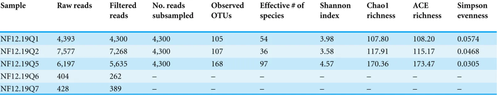
Table 1 Summary statistics for 454 sequencing of 16S rRNA genes fromP. placomus.
Sample Raw reads Filtered reads
No. reads subsampled
Observed OTUs
Effective # of species
Shannon index
Chao1 richness
ACE richness
Simpson evenness
NF12.19Q1 4,393 4,300 4,300 105 54 3.98 107.80 108.20 0.0574 NF12.19Q2 7,577 7,268 4,300 107 36 3.58 117.91 115.17 0.0468 NF12.19Q5 6,197 5,635 4,300 168 97 4.57 170.36 173.47 0.0305
NF12.19Q6 404 262 – – – – – – –
NF12.19Q7 428 389 – – – – – – –
using Titanium FLX chemistry. Sequence data from all five samples were deposited in the NCBI Sequence Read Archive (SRA) under BioProject numberPRJNA297333.
Bioinformatics
A fully commented workflow describing the analysis conducted, as well as all the resulting output files, is available as a USGS data release athttp://dx.doi.org/10.5066/F7HQ3WZZ
Operat
ional taxonom
ic units
Sequences per sample NF12-19Q1
NF12-19Q2
NF12-19Q5
2000
0 1000 3000 4000
150
100
50
0
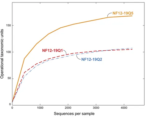
Figure 3 Rarefaction curves for bacterial diversity in three individuals ofP. placomuscollected from Baltimore Canyon.Although there were five samples collected, only three were successfully sequenced. Operational taxonomic units (calculated using a 97% sequence similarity cutoff) identified by 454 pyrose-quencing of 16S rRNA genes.
1993). SIMPER was run using abundance data with no transformation, using the sample names (NF12.19Q1, NF12.19Q2, NF12.19Q5) as factors.
RESULTS AND DISCUSSION
During this cruise 18 ROV dives were conducted from the head to the mouth of Baltimore Canyon, over sections of hardbottom (usually compacted mud) along both walls (Fig. 1). A depth range of 234–1,001 m was covered, with the bulk of the dives at 300–600 m. While other octocoral species, such asParagorgia arboreaandPrimnoa resedaeformiswere encountered at multiple locations in this canyon, P. placomuswas only found at one location in Baltimore Canyon (Fig. 1). During a subsequent cruise in 2013,P. placomus
was also sighted at a single location in Norfolk Canyon (37 02.96 N, 74 37.03 W; 448 m); unfortunately, the coral was identified from video review after the cruise, so no samples could be collected for comparison. In both cases, theP. placomushabitat was a flat rocky terrace on top of a steep wall. In Baltimore Canyon, the patch ofP. placomusconsisted of similarly-sized colonies (<1 m tall), with each colony typically less than one meter from another, possibly suggesting a single recruitment event (Fig. 2A). In Norfolk Canyon, the larger colonies were of similar size to those in Baltimore Canyon, but other smaller colonies were also observed. Like most corals, successful colonization ofParamuriceasp. requires hard substrata (Doughty, Quattrini & Cordes, 2013). Most of the octocoral species in these canyons were observed on underhangs or steep walls and boulders, presumably where sediment deposition was minimized. Colonies ofP. placomus, however, were only
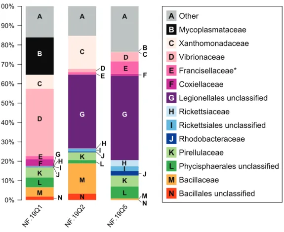
Figure 4 Relative abundance of common taxonomic groups inP. placomussamples.Families (or the nearest identifiable phylogenetic level) that represent≥1% of the total taxa are shown. All remaining taxa are summarized as ‘‘Other’’ [A]. B=Tenericutes. Gammaproteobacterial groups are C–G,
alphapro-teobacterial groups are H–J, Planctomycetes are K–L, and Firmicutes are M–N. *See note in reference to Francisellaceae inTable 2.
found on relatively flat terrace-type habitat that had a veneer of sediment. Many flat terrace areas were observed in both canyons but distribution of this species was very limited, suggesting that other factors influence successful recruitment and colonization. These may include larval delivery (controlled by water currents), a lack of appropriate settlement cues, and sub-optimal water conditions (e.g., particle load) (Doughty, Quattrini & Cordes, 2013; Mienis et al., 2012). Also, sister species likeP. clavataare surface brooders, which often results in short dispersal distances and was suggested as a reason for clumped distributions of the genus in the Gulf of Mexico (Doughty, Quattrini & Cordes, 2013).
Bacterial diversity associated withP. placomusin Baltimore Canyon
We chose to use an open OTU picking method so as to capture maximum diversity, including novel environmental sequences that do not have close matches in reference databases. The P. placomusShannon index values (range =3.58–4.57; Table 1) were similar to those seen for tropical stony corals likeOrbicella annularis(Barott et al., 2011), but were higher than those seen for unimpacted P. clavata(range =0.8–1.3) in the Mediterranean (Vezzulli et al., 2013), suggesting higher bacterial species richness compared to the temperate sister species (with the obvious caveat that the three studies did not use identical methodologies). Converting the Shannon index values to effective number of species (Hill, 1973), also known as true diversities (Table 1), allows a linear comparison of the magnitude of true diversity differences between two communities. The diversity of healthyP. clavatabacterial communities with Shannon index values of 0.8–1.3 (Vezzulli et al., 2013) is equivalent to that of communities with 2–4 equally-common species, compared to a range of 36–97 equally-common species (i.e., effective number of species) inP. placomus(Table 1). Using this metric,P. placomusbacterial communities are roughly 9 to 50 times more diverse than those associated withP. clavata.
As seen in most octocorals (Brück et al., 2007;Duque-Alarcón, Santiago-Vázquez & Kerr, 2012; Gray et al., 2011; Sunagawa, Woodley & Medina, 2010;Webster & Bourne, 2007), the bacterial community was dominated by proteobacterial sequences (>50% relative abundance). Other major phyla included the Firmicutes (10%) and Planctomycetes (10%). Tenericutes were 20% of sample NF12.19Q1, and less than 1% in the other two samples. Present at or below 1% relative abundance were Acidobacteria, Actinobacteria, Bacteroidetes, Chlamydiae, Chloroflexi, Cyanobacteria, Fusobacteria, Gemmatimonadetes, Lentisphaerae, and Verrucomicrobia.
At the microbial family level, pronounced differences occurred between sample NF12.19Q1 and samples NF12.19Q2 and NF12.19Q5; most notably, reductions in unclassified Legionellales and alphaproteobacterial groups, and increases in Vibrionaceae, and Mycoplasmataceae (Fig. 4). Similarity percentages (SIMPER) analysis (Clarke, 1993) showed the average dissimilarity between NF12.19Q1 and NF12.19Q2 or NF12.19Q5 was 50.63–51.03, and the groups responsible for more than 5% of that dissimilarity were Legionellales (11.9%), Vibrionaceae (7.1–9.4%), and Mycoplasmataceae (7.7–9.0%). The average dissimilarity between samples NF12.19Q2 and NF12.19Q5 was 44.04 and was driven by Xanthomonadaceae (7.5%) and Bacillaceae (6.8%).
Mycoplasma sp. sequences were previously found associated with other deep-sea corals, including the scleractinianL. pertusa(Kellogg, Lisle & Galkiewicz, 2009;Neulinger et al., 2009;Neulinger et al., 2008), and octocorals including a bamboo coral (Penn et al., 2006),Cryogorgia koolsae(Gray et al., 2011) andPlumarella superba(Gray et al., 2011). A phylogenetic comparison revealed two ‘coral’Mycoplasmaclades: the deep-sea octocoral clones clustered together with sequences from the tropical coralMuricea elongata, and the sequences fromL. pertusaformed a separate cluster (Gray et al., 2011). We aligned the 329 bpMycoplasma16S rRNA sequence found inP. placomusagainst V4-V5 regions derived from nearly full-length 16S rRNA sequences available fromL. pertusa(GenBank Accession number AM911412.1), bamboo coral (DQ395563.1), and M. elongata (DQ917875.1,
DQ917898.1), to determine if it clustered in either of the two deep-sea coral clades. Note
that we were unable to include other octocoral sequences because the clones were too short and did not include enough of the V4-V5 variable region for alignment. TheMycoplasma
fromP. placomusdid not cluster with any of the other coral mycoplasmal sequences. All threeP. placomussamples showed higher relative abundance of gammaproteobacte-rial families (Fig. 4, letters C–G) compared to alphaproteobacterial families (Fig. 4, letters H–J). The bacterial community of temperate sister-speciesP. clavata, is heavily dominated by Gammaproteobacteria (>90%), whereas other corals from the family Plexauridae (Cryogorgia koolsae(Gray et al., 2011).Eunicea fusca(Duque-Alarcón, Santiago-Vázquez & Kerr, 2012)Muricea elongata(Ranzer, Restrepo & Kerr, 2006), andSwiftia exertia(Brück et al., 2007)) tend to have a roughly equal distribution of Alpha- and Gammaproteobacteria, but at much lower relative abundances (ca. 15–37% each).
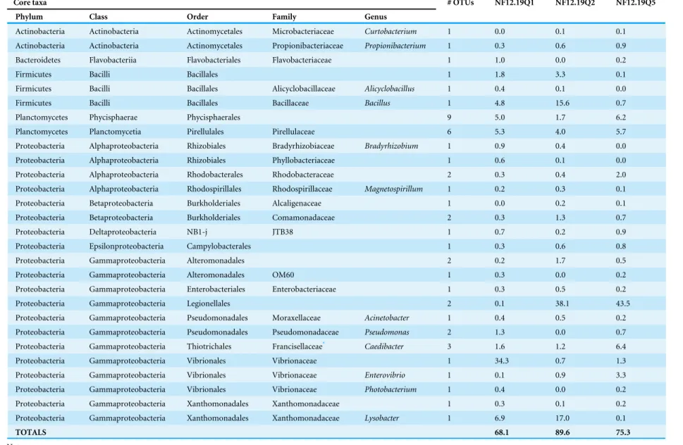
Table 2 Core bacterial taxa shared by threeP. placomussamples and their relative abundance in each sample, as a percentage of the total taxa.
Core taxa # OTUs NF12.19Q1 NF12.19Q2 NF12.19Q5
Phylum Class Order Family Genus
Actinobacteria Actinobacteria Actinomycetales Microbacteriaceae Curtobacterium 1 0.0 0.1 0.1
Actinobacteria Actinobacteria Actinomycetales Propionibacteriaceae Propionibacterium 1 0.3 0.6 0.9
Bacteroidetes Flavobacteriia Flavobacteriales Flavobacteriaceae 1 1.0 0.0 0.2
Firmicutes Bacilli Bacillales 1 1.8 3.3 0.1
Firmicutes Bacilli Bacillales Alicyclobacillaceae Alicyclobacillus 1 0.4 0.1 0.0
Firmicutes Bacilli Bacillales Bacillaceae Bacillus 1 4.8 15.6 0.7
Planctomycetes Phycisphaerae Phycisphaerales 9 5.0 1.7 6.2
Planctomycetes Planctomycetia Pirellulales Pirellulaceae 6 5.3 4.0 5.7
Proteobacteria Alphaproteobacteria Rhizobiales Bradyrhizobiaceae Bradyrhizobium 1 0.9 0.4 0.0
Proteobacteria Alphaproteobacteria Rhizobiales Phyllobacteriaceae 1 0.6 0.1 0.0
Proteobacteria Alphaproteobacteria Rhodobacterales Rhodobacteraceae 2 0.3 0.4 2.0
Proteobacteria Alphaproteobacteria Rhodospirillales Rhodospirillaceae Magnetospirillum 1 0.2 0.3 0.1
Proteobacteria Betaproteobacteria Burkholderiales Alcaligenaceae 1 0.0 0.2 0.1
Proteobacteria Betaproteobacteria Burkholderiales Comamonadaceae 2 0.3 1.3 0.7
Proteobacteria Deltaproteobacteria NB1-j JTB38 1 0.7 0.2 0.9
Proteobacteria Epsilonproteobacteria Campylobacterales 1 0.3 0.6 0.8
Proteobacteria Gammaproteobacteria Alteromonadales 2 0.2 1.7 0.5
Proteobacteria Gammaproteobacteria Alteromonadales OM60 1 0.3 0.0 0.2
Proteobacteria Gammaproteobacteria Enterobacteriales Enterobacteriaceae 1 0.3 0.5 0.2
Proteobacteria Gammaproteobacteria Legionellales 2 0.1 38.1 43.5
Proteobacteria Gammaproteobacteria Pseudomonadales Moraxellaceae Acinetobacter 1 0.4 0.5 0.2
Proteobacteria Gammaproteobacteria Pseudomonadales Pseudomonadaceae Pseudomonas 2 1.3 0.0 0.7
Proteobacteria Gammaproteobacteria Thiotrichales Francisellaceae* Caedibacter 3 1.6 1.2 6.4
Proteobacteria Gammaproteobacteria Vibrionales Vibrionaceae 1 34.3 0.7 1.3
Proteobacteria Gammaproteobacteria Vibrionales Vibrionaceae Enterovibrio 1 0.1 0.9 3.3
Proteobacteria Gammaproteobacteria Vibrionales Vibrionaceae Photobacterium 1 0.4 0.0 0.2
Proteobacteria Gammaproteobacteria Xanthomonadales Xanthomonadaceae 1 0.3 0.1 0.2
Proteobacteria Gammaproteobacteria Xanthomonadales Xanthomonadaceae Lysobacter 1 6.9 17.0 0.1
TOTALS 68.1 89.6 75.3
Notes.
*Caedibacterare actually incertae sedis, being closely related to bothLegionellaandFrancisella, but placed in Francisellaceae by Greengenes database. A value of 0.0 means <0.1%.
K
ellogg
e
t
al.
(2016),
P
eerJ
,
DOI
10.7717/peerj.2529
Composition of conserved core bacterial community
Sequences shared by all three coral samples were examined to identify core bacterial taxa (characterized to the lowest possible taxon, down to genus, Table 2). It is recognized that this population ofP. placomus is both isolated and likely highly clonal, and therefore may not reflect all the bacterial diversity present across the large geographic range of this coral species, or could contain regionally-specific taxa. While other studies have included bacterial groups present in 30–50% of the coral samples as part of the core (Ainsworth et al., 2015), given the small sample size of this study we have opted for the most conservative approach; requiring the OTU be present in 100% of the samples. For each sample, the relative abundance of each taxon is shown in relation to the total taxa (Table 2). For samples NF12.19Q5 and NF12.19Q2 the core taxa make up 75 to nearly 90% of the total community, suggesting a strongly species-specific bacterial community. The core taxa constitute 68% of sample NF12.19Q1, in spite of its visibly different appearance at the family level (Fig. 4). This can be contrasted against the microbiome of the temperate gorgonianEunicella cavolini, where only 7 of 2067 OTUs (less than 1%) were shared across 9 samples (Bayer et al., 2013).
A member of theP. placomuscore (Table 2),Propionibacterium, was recently identified as a conserved member of the core microbiome across a number of tropical and mesophotic corals (Ainsworth et al., 2015). The authors used fluorescently-labeled probes to localize the
Propionibacteriumto the corals’ endosymbiotic algae (zooxanthellae) and speculated these bacteria had a role in facilitating the success of the dinoflagellate-coral symbiosis, and/or meeting the coral host’s energy requirements. Interestingly,Propionibacteriumspp. have been cultivated or detected by molecular techniques from both azooxanthellate (Lawler et al., 2016;Santiago-Vázquez et al., 2007) and zooxathellate corals (Bayer et al., 2013; De Castro et al., 2010;Nithyanand, Manju & Pandian, 2011). This raises an interesting question as to whether these conserved bacteria have multiple roles, or play a different role in azooxanthellate corals.
TheP. placomus core bacterial community includes Alpha-, Beta-, Delta-, Epsilon-and Gammaproteobacteria (Table 2). Legionellales sequences have not been commonly detected in association with corals. Single clones of Legionellales were obtained from the skeletons of Mediterranean coralsCladocora caespitosaandBalanophyllia europaea(Meron et al., 2012) and less than 10 clones similar toLegionella feeleiiwere associated with diseased colonies of temperate gorgonianEunicella verrucosa(Ransome et al., 2014). A recent study of three deep-sea coral species within the family Anthothelidae detected this order as a minor component of their bacterial communities (Lawler et al., 2016). This bacterial order is defined as comprising facultative and obligate intracellular parasites, many of which are associated with free-living protozoa. It therefore remains to be determined if Legionellales are direct associates of the gorgonian, or infect amoebae that are part of the holobiont microbiome (Garrity, Bell & Lilburn, 2005;Rowbotham, 1986).
In addition to the dominant phylotypes of Legionellales, another major component of the core gammaproteobacterial taxa, particularly for sample NF12.19Q1, is the Vibrionaceae, including the genera Enterovibrioand Photobacterium(Table 2). Vibrionaceae are a common component of coral microbiota, having been found in healthy tropical (Bourne & Munn, 2005;Chimetto et al., 2008;Daniels et al., 2011;Lampert et al., 2006) and cold-water corals (Galkiewicz et al., 2011;Gray et al., 2011). While this bacterial group has been linked to a number of diseases in tropical (e.g.,Arotsker et al., 2009;Ben-Haim & Rosenberg, 2002; Cervino et al., 2004) and temperate (Bally & Garrabou, 2007;Hall-Spencer, Pike & Munn, 2007) corals, pathogenicity seems to be driven by water temperatures greater than 20 ◦
C. While other members of the order Xanthomonadales have been found in association with corals (Brück et al., 2007;Cárdenas et al., 2012;Rohwer et al., 2002),Lysobacter was only known from freshwater and soil environments (Christensen, 2005). However, this genus’s ability to degrade chitin (Christensen, 2005) would be useful for a coral host that feeds on zooplankton.Lysobacterare named for their antimicrobial activity, active against not only bacteria, but also yeasts, filamentous fungi, and nematodes (Christensen, 2005), suggesting this genus has a potential role in protecting and maintaining the microbial balance of the coral.
A component of the core microbiome ofP. placomuswas identified as Francisellaceae (Table 2andFig. 4). However, when those sequences were run through RDP Classifier (Wang et al., 2007) they were identified as Thiotrichales incertae sedis, genusCaedibacter. Further evaluation using BLAST (Altschul et al., 1990) showed that the closest matches in GenBank included uncultured Caedibacter clones derived from the coral Orbicella faveolata (FJ425613, FJ425621). The genusCaedibacter consists of endosymbionts of
Parameciumand has been shown to be polyphyletic (Beier et al., 2002), including both Alphaproteobacteria similar to Rickettsiales and Gammaproteobacteria similar to both
LegionellaandFrancisella(as seen here). As with Legionellales, the presence of this bacterial group hints at the presence of eukaryotic members (i.e., protist hosts) in these coral microbiomes.
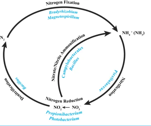
B ac ill us Nitrogen Reduction Magnetospirillum Bradyrhizobium Nitrogen Fixation Photobacterium Propionibacterium De ni tr if ic at io
n itr N
ifi ca tio n P ir el lu la ce ae N itra te/N itri te A
mm
onifi
catio n
NO2– NO3–
N2
NH4
+ (NH 3) Ca mp ylo bac tera les Bac illu s
Figure 5 Core bacterial groups potential roles within the nitrogen cycle.A number of the bacterial groups present within the core microbiome of these threeP. placomussamples were previously recognized for their roles within the nitrogen cycle. This diagram illustrates a simplified overview of the bacterial groups with their possible functions. This figure was adapted from one presented inWegley et al. (2007).
Core: Nitrogen cycling?
A recent characterization of the core microbiome of the deep-sea octocoral Anthothela grandiflorarevealed the possibility of a nearly complete nitrogen cycle, based on previously described abilities of particular bacteria present in the coral (Lawler et al., 2016). With this in mind, we examined the core bacterial community associated with the three
P. placomuscolonies (Table 2) and determined that a similar possibility exists for this coral species (Fig. 5).
The alphaproteobacterial genusBradyrhizobiumis typically an intracellular nitrogen-fixing symbiont in the root nodules of plants; however, some free-living strains of
Mohamed et al., 2010) and the deep-sea octocoralAlcyonium grandiflorum(Lawler et al., 2016), and may be conducting nitrification. Propionibacterium(Allison & Macfarlane, 1989) andPhotobacterium(Thyssen & Ollevier, 2005) have been shown to reduce nitrate to nitrite.Bacillusspp. have been shown to carry out denitrification (Verbaendert et al., 2011) and also nitrate/nitrite ammonification (Hoffmann et al., 1998). Lastly, members of the Campylobacterales are known to reduce nitrate and nitrite to ammonium (De Vries et al., 1980). Nitrogen cycling has recently been confirmed in the deep-sea coralLophelia pertusa
(Middelburg et al., 2015), and given thatP. placomusis likely to have an inconsistent diet of nano-zooplankton and detrital particulates (Ribes, Coma & Gili, 1999), the ability to recycle and retain nitrogen would be beneficial (Fig. 5). Further work with metagenomics and transcriptomics remains to be conducted to confirm this hypothesis.
CONCLUSIONS
Based on these three samples from the Baltimore Canyon population, the deep-sea plexaurid octocoralP. placomushas a species-specific bacterial community that shows very little overlap with previously characterized temperate gorgonians, including sister-species
P. clavata. The bacterial community of this species is dominated by Proteobacteria but has similar diversity to that of tropical stony corals. Conserved core bacterial taxa comprise 68– 90% of the total community, leaving roughly 10–30% individual variability between coral colonies. Additional sampling from other locations is required to confirm the consistency of these findings across the coral’s geographic range. The composition of the core suggests that nitrogen cycling may be carried out by the bacterial associates. This study is the first description of the bacterial microbiota of a deep-sea paramuricid species and provides a baseline for comparison by future studies to address questions regarding biogeography, ecology, and resilience of these corals to anthropogenic impacts and changing climate.
ACKNOWLEDGEMENTS
Thanks are extended to the captain and crew of the NOAA shipNancy Foster and to theKraken II ROV team. The authors also thank Ashley Shade (Michigan State Univ.) and the Explorations in Data Analysis for Metagenomic Advances in Microbial Ecology (EDAMAME) Workshop for critical lessons in workflow organization and bioinformatics tools.
Michael Rhode (UNC-Wilmington) produced the multibeam map of Baltimore Canyon used inFig. 1and provided assistance at sea. Dawn Goldsmith (USGS) formattedFig. 4
using R. Betsy Boynton (USGS) drafted the inset map used inFig. 1and prepared final versions of all the figures. Scott France and Rachel Clostio (University of Louisiana at Lafayette) provided the coral genetic results confirming host species identity.
Any use of trade names is for descriptive purposes only and does not imply endorsement by the US government.
ADDITIONAL INFORMATION AND DECLARATIONS
Funding
Funding for this project was provided by the US Geological Survey’s Ecosystems Mission Area, Environments Program through the Outer Continental Shelf study on Mid-Atlantic Canyons. Additional funding was sponsored by the National Oceanographic Partnership Program and supplied by the Bureau of Ocean Energy Management (BOEM) contract number M10PC00100 (contracted to CSA Ocean Sciences, Inc.). The Nancy Foster and Kraken II were provided by the NOAA Office of Ocean Exploration. The funders had no role in study design, data collection and analysis, decision to publish, or preparation of the manuscript.
Grant Disclosures
The following grant information was disclosed by the authors:
US Geological Survey’s Ecosystems Mission Area, Environments Program. National Oceanographic Partnership Program.
Bureau of Ocean Energy Management (BOEM): M10PC00100. NOAA Office of Ocean Exploration.
Competing Interests
The authors declare there are no competing interests.
Author Contributions
• Christina A. Kellogg conceived and designed the experiments, performed the experiments, analyzed the data, contributed reagents/materials/analysis tools, wrote the paper, prepared figures and/or tables, reviewed drafts of the paper.
• Steve W. Ross contributed reagents/materials/analysis tools, prepared figures and/or tables, reviewed drafts of the paper.
• Sandra D. Brooke contributed reagents/materials/analysis tools, reviewed drafts of the paper.
DNA Deposition
The following information was supplied regarding the deposition of DNA sequences: NCBI Sequence Read Archive (SRA) under BioProject numberPRJNA297333.
Data Availability
The following information was supplied regarding data availability:
REFERENCES
Ainsworth TD, Krause L, Bridge T, Torda G, Raina J-B, Zakrzewski M, Gates RD, Padilla-Gamiño JL, Spalding HL, Smith C, Woolsey ES, Bourne DG, Bongaerts P, Hoegh-Guldberg O, Leggat W. 2015.The coral core microbiome identifies rare bacterial taxa as ubiquitous endosymbionts.The ISME Journal9:2261–2274
DOI 10.1038/ismej.2015.39.
Allison C, Macfarlane GT. 1989.Dissimilatory nitrate reduction byPropionibacterium acnes.Applied and Environmental Microbiology55:2899–2903.
Altschul SF, Gish W, Miller W, Myers EW, Lipman DJ. 1990.Basic local alignment search tool.Journal of Molecular Biology215:403–410
DOI 10.1016/S0022-2836(05)80360-2.
Arotsker L, Siboni N, Ben-Dov E, Kramarsky-Winter E, Loya Y, Kushmaro A. 2009.Vibriosp. as a potentially important member of the Black Band Disease (BBD) consortium inFaviasp. corals.FEMS Microbiology Ecology70:515–524
DOI 10.1111/j.1574-6941.2009.00770.x.
Bally M, Garrabou J. 2007.Thermodependent bacterial pathogens and mass mortalities in temperate benthic communities: a new case of emerging disease linked to climate change.Global Change Biology13:2078–2088DOI 10.1111/j.1365-2486.2007.01423.x. Barott KL, Rodriguez-Brito B, Janouškovec J, Marhaver K, Smith JE, Keeling P,
Rohwer FL. 2011.Microbial diversity associated with four functional groups of benthic reef algae and the reef-building coralMontastraea annularis.Environmental Microbiology13:1192–1204DOI 10.1111/j.1462-2920.2010.02419.x.
Bayer T, Arif C, Ferrier-Pagès C, Zoccola D, Aranda M, Voolstra CR. 2013.Bacteria of the genusEndozoicomonasdominate the microbiome of the Mediterranean gorgonian coralEunicella cavolini.Marine Ecology Progress Series479:75–84
DOI 10.3354/meps10197.
Bazylinski DA, Dean AJ, Schüler D, Phillips EJP, Lovley DR. 2000.N2-dependent growth and nitrogenase activity in the metal-metabolizing bacteria, Geobac-terandMagnetospirillumspecies.Environmental Microbiology2:266–273
DOI 10.1046/j.1462-2920.2000.00096.x.
Beier CL, Horn M, Michel R, Schweikert M, Görtz H-D, Wagner M. 2002.The genus
Caedibacter comprises endosymbionts ofParameciumspp. related to the Rick-ettsiales (Alphaproteobacteria) and toFrancisella tularensis(Gammaproteobacteria).
Applied and Environmental Microbiology 68:6043–6050
DOI 10.1128/AEM.68.12.6043-6050.2002.
Beleneva IA, Dautova TI, Zhukova NV. 2005.Characterization of communities of heterotrophic bacteria associated with healthy and diseased corals in Nha Trang Bay (Vietnam).Microbiology74:579–587 DOI 10.1007/s11021-005-0106-8.
Ben-Haim Y, Rosenberg E. 2002.A novelVibriosp. pathogen of the coralPocillopora damicornis.Marine Biology 141:47–55DOI 10.1007/s00227-002-0797-6.
Van Bleijswijk JDL, Whalen C, Duineveld GCA, Lavaleye MSS, Witte HJ, Mienis F. 2015.Microbial assemblages on a cold-water coral mound at the SE Rockall
Bank (NE Atlantic): interactions with hydrography and topography.Biogeosciences Discussions12:1509–1542DOI 10.5194/bgd-12-1509-2015.
Bourne DG, Munn CB. 2005.Diversity of bacteria associated with the coralPocillopora damicornisfrom the Great Barrier Reef.Environmental Microbiology7:1162–1174
DOI 10.1111/j.1462-2920.2005.00793.x.
Brück TB, Brück WM, Santiago-Vázquez LZ, McCarthy PJ, Kerr RG. 2007.Diversity of the bacterial communities associated with the azooxanthellate deep water octocorals
Leptogorgia minimata,Iciligorgia schrammi, andSwiftia exertia.Marine Biotechnology 9:561–576DOI 10.1007/s10126-007-9009-1.
Buhl-Mortensen P, Buhl-Mortensen L. 2014.Diverse and vulnerable deep-water biotopes in the Hardangerfjord.Marine Biology Research10:253–267
DOI 10.1080/17451000.2013.810759.
Buhl-Mortensen L, Mortensen PB. 2005. Distribution and diversity of species associated with deep-sea gorgonian corals off Atlantic Canada. In: Freiwald A, Roberts JM, eds.
Cold-water corals and ecosystems. Berlin: Springer-Verlag, 849–879.
Bythell JC, Pantos O, Richardson LL. 2004. White plague, white band, and other ‘‘white’’ diseases. In: Rosenberg E, Loya Y, eds.Coral health and disease. Berlin: Springer-Verlag, 351–365.
Caporaso JG, Bittinger K, Bushman FD, Desantis TZ, Andersen GL, Knight R. 2010a. PyNAST: a flexible tool for aligning sequences to a template alignment. Bioinformat-ics26:266–267DOI 10.1093/bioinformatics/btp636.
Caporaso JG, Kuczynski J, Stombaugh J, Bittinger K, Bushman FD, Costello EK, Fierer N, Gonzalez Peña A, Goodrich JK, Gordon JI, Huttley GA, Kelley ST, Knights D, Koenig JE, Ley RE, Lozupone CA, McDonald D, Muegge BD, Pirrung M, Reeder J, Sevinsky JR, Turnbaugh PJ, Walters WA, Widmann J, Yatsunenko T, Zaneveld J, Knight R. 2010b.QIIME allows analysis of high-throughput community sequencing data.Nature Methods7:335–336DOI 10.1038/nmeth.f.303.
Cárdenas A, Rodriguez-R LM, Pizarro V, Cadavid LF, Arévalo-Ferro C. 2012.Shifts in bacterial communities of two caribbean reef-building coral species affected by white plague disease.The ISME Journal6:502–512DOI 10.1038/ismej.2011.123.
Cardini U, Bednarz VN, Naumann MS, Van Hoytema N, Rix L, Foster RA, Al-Rshaidat MMD, Wild C. 2015.Functional significance of dinitrogen fixation in sustaining coral productivity under oligotrophic conditions.Proceedings of the Royal Society of London Series B-Biological Sciences282:20152257DOI 10.1098/rspb.2015.2257. Cervino JM, Hayes RL, Polson SW, Polson SC, Goreau TJ, Martinez RJ, Smith GW.
2004.Relationship ofVibriospecies infection and elevated temperatures to yellow blotch/band disease in Caribbean corals.Applied and Environmental Microbiology 70:6855–6864DOI 10.1128/AEM.70.11.6855-6864.2004.
Chimetto LA, Brocchi M, Thompson CC, Martins RCR, Ramos HR, Thompson FL. 2008.Vibrios dominate as culturable nitrogen-fixing bacteria of the Brazil-ian coralMussismilia hispida.Systematic and Applied Microbiology31:312–319
Christensen P. 2005.Family I. Xanthomonadaceae, genus IV.Lysobacter.Bergey’s Manual of Systematic Bacteriology2:95–101.
Claesson MJ, Wang Q, O’Sullivan O, Greene-Diniz R, Cole JR, Ross RP, O’Toole PW. 2010.Comparison of two next-generation sequencing technologies for resolving highly complex microbiota composition using tandem variable 16S rRNA gene regions.Nucleic Acids Research38:e200DOI 10.1093/nar/gkq873.
Clarke KR. 1993.Non-parametric multivariate analyses of changes in community struc-ture.Australian Journal of Ecology18:117–143
DOI 10.1111/j.1442-9993.1993.tb00438.x.
Correa H, Haltli B, Duque C, Kerr R. 2013.Bacterial communities of the gor-gonian octocoralPseudopterogorgia elisabethae.Microbial Ecology66:972–985
DOI 10.1007/s00248-013-0267-3.
Daniels CA, Zeifman A, Heym K, Ritchie KB, Watson CA, Berzins I, Breitbart M. 2011.Spatial heterogeneity of bacterial communities in the mucus ofMontastraea annularis.Marine Ecology Progress Series426:29–40DOI 10.3354/meps09024. De Castro AP, Dias Araújo Jr S, Reis AMM, Moura RL, Francini-Filho RB, Pappas Jr G,
Rodrigues TB, Thompson FL, Krüger RH. 2010.Bacterial community associated with healthy and diseased reef coralMussismilia hispidafrom Eastern Brazil.
Microbial Ecology59:658–667DOI 10.1007/s00248-010-9646-1.
De Vries W, Niekus HGD, Boellaard M, Stouthamer AH. 1980.Growth yields and energy generation byCampylobacter sputorumsubspecies bubulus during growth in continuous culture with different hydrogen acceptors.Archives of Microbiology 124:221–227DOI 10.1007/BF00427730.
DeSantis TZ, Hugenholtz P, Larsen N, Rojas M, Brodie EL, Keller K, Huber T, Dalevi D, Hu P, Andersen GL. 2006.Greengenes, a chimera-checked 16S rRNA gene database and workbench compatible with ARB.Applied and Environmental Micro-biology72:5069–5072DOI 10.1128/AEM.03006-05.
Doughty CL, Quattrini AM, Cordes EE. 2013.Insights into the population dynamics of the deep-sea coral genusParamuriceain the Gulf of Mexico.Deep-Sea Research Part II-Topical Studies in Oceanography99:71–82DOI 10.1016/j.dsr2.2013.05.023. Duque-Alarcón A, Santiago-Vázquez LZ, Kerr RG. 2012.A microbial community
analysis of the octocoralEunicea fusca.Electronic Journal of Biotechnology15:1–9. Edgar RC. 2010.Search and clustering orders of magnitude faster than BLAST.
Bioinfor-matics26:2460–2461DOI 10.1093/bioinformatics/btq461.
Edwards U, Rogall T, Blocker H, Emde M, Bottger EC. 1989.Isolation and di-rect complete nucleotide determination of entire genes: characterization of a gene coding for 16S ribosomal RNA.Nucleic Acids Research17:7843–7853
DOI 10.1093/nar/17.19.7843.
Filippidou S, Junier T, Wunderlin T, Lo C-C, Li P-E, Chain PS, Junier P. 2015. Under-detection of endospore-formingFirmicutesin metagenomic data.Computational and Structural Biotechnology Journal13:299–306DOI 10.1016/j.csbj.2015.04.002.
Gade D, Schlesner H, Glöckner FO, Amann R, Pfeiffer S, Thomm M. 2004. Identifica-tion of Plantomycetes with order-, genus-, and strain-specific 16S rRNA-targeted probes.Microbial Ecology47:243–251DOI 10.1007/s00248-003-1016-9.
Galkiewicz JP, Pratte ZA, Gray MA, Kellogg CA. 2011.Characterization of culturable bacteria isolated from the cold-water coralLophelia pertusa.FEMS Microbiology Ecology 77:333–346DOI 10.1111/j.1574-6941.2011.01115.x.
Garrity GM, Bell JA, Lilburn T. 2005.Order VI: Legionellales.ord. nov. Bergey’s Manual of Systematic Bacteriology 2:210.
Gray MA, Stone RP, McLaughlin MR, Kellogg CA. 2011.Microbial consortia of gorgonian corals from the Aleutian islands.FEMS Microbiology Ecology 76:109–120
DOI 10.1111/j.1574-6941.2010.01033.x.
Hall-Spencer JM, Pike J, Munn CB. 2007.Diseases affect cold-water corals too:Eunicella verrucosa(Cnidaria: Gorgonacea) necrosis in SW England.Diseases of Aquatic Organisms76:87–97DOI 10.3354/dao076087.
Hansson L, Agis M, Maier C, Weinbauer MG. 2009.Community composition of bacte-ria associated with cold-water coralMadrepora oculata: within and between colony variability.Marine Ecology Progress Series397:89–102DOI 10.3354/meps08429. Hill MO. 1973.Diversity and eveness: a unifying notation and its consequences.Ecology
54:427–432DOI 10.2307/1934352.
Hoffmann T, Frankenberg N, Marino M, Jahn D. 1998.Ammonification inBacillus subtilisutilizing dissimilatory nitrite reductase is dependent onresDE.Journal of Bacteriology180:186–189.
Holm JB, Heidelberg KB. 2016.Microbiomes ofMuricea californicaandM. fruticosa: comparative analyses of two co-occurring eastern Pacific octocorals.Frontiers in Microbiology7:Article 917DOI 10.3389/fmicb.2016.00917.
Kellogg CA. 2015.Cold-water coral microbiomes (Paramuricea placomus) from Baltimore Canyon: raw and processed data: US Geological Survey Data release
DOI 10.5066/F7HQ3WZZ.
Kellogg CA, Lisle JT, Galkiewicz JP. 2009.Culture-independent characterization of bacterial communities associated with the cold-water coralLophelia pertusain the northeastern Gulf of Mexico.Applied and Environmental Microbiology 75:2294–2303
DOI 10.1128/AEM.02357-08.
Kellogg CA, Piceno YM, Tom LM, DeSantis TZ, Gray MA, Andersen GL. 2014. Comparing bacterial community composition of healthy and dark spot-affectedSiderastrea sidereain Florida and the Caribbean.PLoS ONE9:e108767
DOI 10.1371/journal.pone.0108767.
Kellogg CA, Piceno YM, Tom LM, DeSantis TZ, Gray MA, Zawada DG, Andersen GL. 2013.Comparing bacterial community composition between healthy and white plague-like disease states inOrbicella annularisusing PhyloChipTMG3 microarrays.
PLoS ONE8:e79801DOI 10.1371/journal.pone.0079801.
Kunin V, Engelbrektson A, Ochman H, Hugenholtz P. 2010.Wrinkles in the rare biosphere: pyrosequencing errors can lead to artificial inflation of diversity estimates.
Kuykendall LD. 2005.Family VII. Bradyrhizobiaceae, genus I.Bradyrhizobium. Bergey’s Manual of Systematic Bacteriology2:438–443.
La Rivière M, Garrabou J, Bally M. 2010.Spatial and temporal analysis of bacterial diversity associated with the Mediterranean gorgonianParamuricea clavata.Rapport Commission International pour l’exploration scientifique de la Mer Mediterranée, Monaco39:769.
La Rivière M, Garrabou J, Bally M. 2015.Evidence for host specificity among dominant bacterial symbionts in temperate gorgonian corals.Coral Reefs34:1087–1098
DOI 10.1007/s00338-015-1334-7.
La Rivière M, Roumagnac M, Garrabou J, Bally M. 2013.Transient shifts in bacterial communities associated with the temperate gorgonianParamuricea clavatain the northwestern Mediterranean Sea.PLoS ONE 8:e57385
DOI 10.1371/journal.pone.0057385.
Lampert Y, Kelman D, Dubinsky Z, Nitzan Y, Hill RT. 2006.Diversity of culturable bacteria in the mucus of the Red Sea coralFungia scutaria.FEMS Microbiology Ecology 58:99–108DOI 10.1111/j.1574-6941.2006.00136.x.
Lawler SN, Kellogg CA, France SC, Clostio RW, Brooke SD, Ross SW. 2016. Coral-associated bacterial diversity is conserved across two deep-seaAnthothelaspecies.
Frontiers in Microbiology7:Article 458DOI 10.3389/fmicb.2016.00458.
Lema KA, Willis BL, Bourne DG. 2012.Corals form characteristic associations with symbiotic nitrogen-fixing bacteria.Applied and Environmental Microbiology 78:3136–3144DOI 10.1128/AEM.07800-11.
Lesser MP, Falcón LI, Rodríguez-Román A, Enríquez S, Hoegh-Guldberg O, Iglesias-Prieto R. 2007.Nitrogen fixation by symbiotic cyanobacteria provides a source of nitrogen for the scleractinian coralMontastraea cavernosa.Marine Ecology Progress Series346:143–152DOI 10.3354/meps07008.
Littman RA, Willis BL, Pfeffer C, Bourne DG. 2009.Diversities of coral-associated bac-teria differ with location, but not species, for three acroporid corals on the Great Bar-rier Reef.FEMS Microbiology Ecology68:152–163
DOI 10.1111/j.1574-6941.2009.00666.x.
Logan NA, De Vos P. 2009.Genus I. Bacillus.Bergey’s Manual of Systematic Bacteriology 3:21–128.
Lumsden SE, Hourigan TF, Bruckner AW, Dorr G. 2007.The state of deep coral ecosystems of the United States. NOAA Technical Memorandum CRCP-3 ed. Silver Spring, MD.
Meron D, Rodolfo-Metalpa R, Cunning R, Baker AC, Fine M, Banin E. 2012.Changes in coral microbial communities in response to a natural pH gradient.The ISME Journal 6:1775–1785DOI 10.1038/ismej.2012.19.
Middelburg JJ, Mueller CE, Veuger B, Larsson AI, Form A, Van Oevelen D. 2015. Discovery of symbiotic nitrogen fixation and chemoautotrophy in cold-water corals.
Scientific Reports5:Article 17962DOI 10.1038/srep17962.
Mienis F, Duineveld GCA, Davies AJ, Ross SW, Seim H, Bane J, Van Weering T.CE. 2012.The influence of near-bed hydrodynamic conditions on cold-water corals
in the Viosca Knoll area, Gulf of Mexico.Deep-Sea Research Part I-Oceanographic Research Papers60:32–45DOI 10.1016/j.dsr.2011.10.007.
Miller RJ, Hocevar J, Stone RP, Fedorov DV. 2012.Structure-forming corals and sponges and their use as fish habitat in Bering Sea submarine canyons.PLoS ONE 7:e3385DOI 10.1371/journal.pone.0033885.
Mohamed NM, Saito K, Tal Y, Hill RT. 2010.Diversity of aerobic and anaerobic ammonia-oxidizing bacteria in marine sponges.The ISME Journal4:38–48
DOI 10.1038/ismej.2009.84.
Morrow KM, Moss AG, Chadwick NE, Liles MR. 2012.Bacterial associates of two Caribbean coral species reveal species-specific distribution and geo-graphic variability.Applied and Environmental Microbiology78:6438–6449
DOI 10.1128/AEM.01162-12.
Mouchka ME, Hewson I, Harvell CD. 2010.Coral-associated bacterial assemblages: current knowledge and the potential for climate-driven impacts.Integrative and Comparative Biology50:662–674DOI 10.1093/icb/icq061.
Neulinger SC, Gärtner A, Järnegren J, Ludvigsen M, Lochte K, Dullo W-C. 2009. Tissue-associated ‘‘CandidatusMycoplasma corallicola’’ and filamentous bacteria on the cold-water coralLophelia pertusa(Scleractinia).Applied and Environmental Microbiology75:1437–1444DOI 10.1128/AEM.01781-08.
Neulinger SC, Järnegren J, Ludvigsen M, Lochte K, Dullo W-C. 2008. Phenotype-specific bacterial communities in the cold-water coralLophelia pertusa(Scleractinia) and their implications for the coral’s nutrition, health, and distribution.Applied and Environmental Microbiology74:7272–7285DOI 10.1128/AEM.01777-08.
Nithyanand P, Manju S, Pandian SK. 2011.Phylogenetic characterization of culturable actinomycetes associated with the mucus of the coralAcropora digitiferafrom Gulf of Mannar.FEMS Microbiology Letters314:112–118
DOI 10.1111/j.1574-6968.2010.02149.x.
Nithyanand P, Pandian SK. 2009.Phylogenetic characterization of culturable bacterial diversity associated with the mucus and tissue of the coralAcropora digitiferafrom the Gulf of Mannar.FEMS Microbiology Ecology69:384–394
DOI 10.1111/j.1574-6941.2009.00723.x.
Oksanen J, Blanchet FG, Friendly M, Kindt R, Legendre P, Mcglinn D, Minchin PR, O’Hara RB, Simpson GL, Solymos P, Stevens M.HH, Szoecs E, Wagner H. 2016. vegan: Community Ecology Package. R package version 2.4-0.Available athttps: // cran.r-project.org/ package=vegan.
Pantos O, Bongaerts P, Dennis PG, Tyson GW, Hoegh-Guldberg O. 2015. Habitat-specific environmental conditions primarily control the microbiomes of the coral
Seriatopora hystrix.The ISME Journal9:1916–1927DOI 10.1038/ismej.2015.3. Pantos O, Cooney RP, Le Tissier MDA, Barer MR, O’Donnell AG, Bythell JC. 2003.The
bacterial ecology of a plague-like disease affecting the Caribbean coralMontastrea annularis.Environmental Microbiology5:370–382
Penn K, Wu D, Eisen JA, Ward N. 2006.Characterization of bacterial communities asso-ciated with deep-sea corals on Gulf of Alaska seamounts.Applied and Environmental Microbiology72:1680–1683DOI 10.1128/AEM.72.2.1680-1683.2006.
Quince C, Lanzén A, Curtis TP, Davenport RJ, Hall N, Head IM, Read LF, Sloan WT. 2009.Accurate determination of microbial diversity from 454 pyrosequencing data.
Nature Methods6:639–641 DOI 10.1038/NMETH.1361.
R Core Development Team. 2015.R: a language and environment for statistical computing. Vienna: R Foundation for Statistical Computing. Available athttp: // www.R-project.org/.
Ransome E, Rowley SJ, Thomas S, Tait K, Munn CB. 2014.Disturbance to conserved bacterial communities in the cold-water gorgonian coralEunicella verrucosa.FEMS Microbiology Ecology90:404–416DOI 10.1111/1574-694.12398.
Ranzer LK, Restrepo PF, Kerr RG. 2006.Microbial community profiles of bleached and wild-typeMuricea elongata.Available athttp:// www.ncbi.nlm.nih.gov/ popset/ 134140623?report=genbank.
Ribes M, Coma R, Gili J-M. 1999.Heterogeneous feeding in benthic suspension feeders: the natural diet and grazing rate of the temperate gorgonianParamuricea clavata(Cnidaria: Octocorallia) over a year cycle.Marine Ecology Progress Series 183:125–137DOI 10.3354/meps183125.
Rideout JR, He Y, Navas-Molina JA, Walters WA, Ursell LK, Gibbons SM, Chase J, McDonald D, Gonzalez A, Robbins-Pianka A, Clemente JC, Gilbert JA, Huse SM, Zhou H-W, Knight R, Caporaso JG. 2014.Subsampled open-reference clustering creates consistent, comprehensive OTU definitions and scales to billions of sequences.PeerJ 2:e545DOI 10.7717/peerj.545.
Ritchie KB. 2006.Regulation of microbial populations by coral surface mucus and mucus-associated bacteria.Marine Ecology Progress Series322:1–14
DOI 10.3354/meps322001.
Roberts JM, Wheeler A, Freiwald A, Cairns S. 2009.Cold-water corals: the biology and geology of deep-sea coral habitats. Cambridge: Cambridge University Press. Robertson V, Haltli B, McCauley EP, Overy DP, Kerr RG. 2016.Highly variable
bacterial communities associated with the octocoralAntillogorgia elisabethae.
Microorganisms4:Article 23DOI 10.3390/microorganisms4030023.
Roder C, Bayer T, Aranda M, Kruse M, Voolstra CR. 2015.Microbiome structure of the fungid coralCtenactis echinataaligns with environmental differences.Molecular Ecology 24:3501–3511DOI 10.1111/mec.13251.
Rohwer F, Breitbart M, Jara J, Azam F, Knowlton N. 2001.Diversity of bacteria associated with the Caribbean coralMontastraea franksi.Coral Reefs20:85-91
DOI 10.1007/s003380100138.
Rohwer F, Seguritan V, Azam F, Knowlton N. 2002.Diversity and distribu-tion of coral-associated bacteria.Marine Ecology Progress Series243:1–10
DOI 10.3354/meps243001.
Rowbotham TJ. 1986.Current views on the relationships between amoebae, legionellae and man.Israel Journal of Medical Sciences22:678–689.
Santiago-Vázquez LZ, Brück TB, Brück W, Duque-Alarcón AP, McCarthy PJ, Kerr RG. 2007.The diversity of the bacterial communities associated with the azooxanthellate hexacoralCirrhipathes lutkeni.The ISME Journal1:654–659
DOI 10.1038/ismej.2007.77.
Schöttner S, Hoffmann F, Wild C, Rapp HT, Boetius A, Ramette A. 2009.Inter- and intra-habitat bacterial diversity associated with cold-water corals.The ISME Journal 3:756–759DOI 10.1038/ismej.2009.15.
Schüler D, Schleifer K-H. 2005.Family I. Rhodospirillaceae, genus IV.Magnetospirillum. Shashar N, Cohen Y, Loya Y, Sar N. 1994.Nitrogen fixation (acetylene reduction) in
stony corals: evidence for coral-bacteria interactions.Marine Ecology Progress Series 111:259–264DOI 10.3354/meps111259.
Simpson AW, Eckelbarger J, Watling L. 2005.Some aspects of the reproductive biology ofParamuricea placomus(Octocorallia) from the Gulf of Maine [Abstract 1195].
Integrative and Comparative Biology45.
Stackebrandt E, Liesack W. 1993. Nucleic acids and classification. In: Goodfellow M, O’Donnell AG, eds.Handbook of new bacterial systematics. London: Academic Press, 152–189.
Sunagawa S, Woodley CM, Medina M. 2010.Threatened corals provide underexplored microbial habitats.PLoS ONE5:e9554DOI 10.1371/journal.pone.0009554.
Thoma JN. 2013.Molecular and morphological diversity of sea fans with emphasis on deep-sea octocorals of the order Alcyonacea Lamouroux 1812. PhD dissertation, University of Louisiana at Lafayette.
Thyssen A, Ollevier F. 2005.Genus IIPhotobacterium.Bergey’s Manual of Systematic Bacteriology2:546–552.
Turnbaugh PJ, Ley RE, Hamady M, Fraser-Liggett C, Knight R, Gordon JI. 2007.The human microbiome project: exploring the microbial part of ourselves in a changing world.Nature449:804–810DOI 10.1038/nature06244.
Van de Water JAJM, Ainsworth TD, Leggat W, Bourne DG, Willis BL, Van Oppen MJH. 2015.The coral immune response facilitates protection against microbes dur-ing tissue regeneration.Molecular Ecology24:3390–3404DOI 10.1111/mec.13257. Van de Water JAJM, Melkonian R, Junca H, Voolstra CR, Reynaud S, Allemand D,
Ferrier-Pagès C. 2016.Spirochaetes dominate the microbial community associated with the red coral Corallium rubrum on a broad geographic scale.Scientific Reports 6:27277DOI 10.1038/srep27277.
Verbaendert I, Boon N, De Vos P, Heylen K. 2011.Denitrification is a common feature among members of the genusBacillus.Systematic and Applied Microbiology 34:385–391DOI 10.1016/j.syapm.2011.02.003.
Vezzulli L, Pezzati E, Huete-Stauffer C, Pruzzo C, Cerrano C. 2013.16SrDNA pyrose-quencing of the Mediterranean gorgonianParamuricea clavatareveals a link among alterations in bacterial holobiont members, anthropogenic influence and disease outbreaks.PLoS ONE8:e67745DOI 10.1371/journal.pone.0067745.
Sea.Environmental Microbiology12:2007–2019
DOI 10.1111/j.1462-2920.2010.02209.x.
Wang Q, Garrity GM, Tiedje JM, Cole JR. 2007.Naive Bayesian classifier for rapid assignment of rRNA sequences into the new bacterial taxonomy.Applied and Environmental Microbiology73:5261–5267DOI 10.1128/AEM.00062-07. Webster NS, Bourne DG. 2007.Bacterial community structure associated with the
Antarctic soft coral,Alcyonium antarcticum.FEMS Microbiology Ecology 59:81–94
DOI 10.1111/j.1574-6941.2006.00195.x.
Wegley L, Edwards R, Rodriguez-Brito B, Liu H, Rohwer F. 2007.Metagenomic analysis of the microbial community associated with the coralPorites astreoides.
Environmental Microbiology9:2707–2719DOI 10.1111/j.1462-2920.2007.01383.x. Wickham H. 2009.ggplot2: elegant graphics for data analysis. New York: Springer-Verlag. Williams WM, Viner AB, Broughton WJ. 1987.Nitrogen-fixation (acetylene-reduction)
associated with the living coralAcropora variabilis.Marine Biology94:531–535
DOI 10.1007/BF00431399.
Yakimov MM, Cappello S, Crisafi E, Trusi A, Savini A, Corselli C, Scarfi S, Giuliano L. 2006.Phylogenetic survey of metabolically active microbial communities associated with the deep-sea coralLophelia pertusafrom the Apulian plateau, Central Mediter-ranean Sea.Deep Sea Research53:62–75DOI 10.1016/j.dsr.2005.07.005.