Low-dose CT pulmonary angiography on a
15-year-old CT scanner: a feasibility study
Moritz Kaup, Tatjana Gruber-Rouh, Jan E Scholtz,
Moritz H Albrecht, Andreas Bucher, Claudia Frellesen,
Thomas J Vogl and Martin Beeres
Abstract
Background: Computed tomography (CT) low-dose (LD) imaging is used to lower radiation exposure, especially in vascular imaging; in current literature, this is mostly on latest generation high-end CT systems.
Purpose: To evaluate the effects of reduced tube current on objective and subjective image quality of a 15-year-old 16-slice CT system for pulmonary angiography (CTPA).
Material and Methods:CTPA scans from 60 prospectively randomized patients (28 men, 32 women) were examined in this study on a 15-year-old 16-slice CT scanner system. Standard CT (SD) settings were 100 kV and 150 mAs, LD settings were 100 kV and 50 mAs. Attenuation of the pulmonary trunk, various anatomic landmarks, and image noise were quantitatively measured; contrast-to-noise ratios (CNR) and signal-to-noise ratios (SNR) were calculated. Three independent blinded radiologists subjectively rated each image series using a 5-point grading scale.
Results:CT dose index (CTDI) in the LD series was 66.46% lower compared to the SD settings (2.490.55 mGy versus 7.421.17 mGy). Attenuation of the pulmonary trunk showed similar results for both series (SD 409.5591.04 HU; LD 380.43 HU93.11 HU;P¼0.768). Subjective image analysis showed no significant differences between SD and LD settings regarding the suitability for detection of central and peripheral PE (central SD/LD, 4.88; intra-class correl-ation coefficients [ICC], 0.894/4.83; ICC, 0.745; peripheral SD/LD, 4.70; ICC, 0.943/4.57; ICC, 0.919; allP>0.4).
Conclusion: The LD protocol, on a 15-year-old CT scanner system without current high-end hardware or post-processing tools, led to a dose reduction of approximately 67% with similar subjective image quality and delineation of central and peripheral pulmonary arteries.
Keyword
Computed tomography pulmonary angiography (CTPA); low-dose imaging; CT pulmonary angiography; CT radiation exposure
Date received: 5 November 2016; accepted: 22 November 2016
Introduction
Computed tomographic pulmonary angiography (CTPA) is widely used to evaluate patients who are clinically suspected of having pulmonary embolism (PE) to either confirm or rule out this diagnosis (1,2).
Even though multiple techniques are designed to reduce radiation exposure, thoracic CT imaging still exposes patients to a higher amount of radiation than other imaging modalities (e.g. X-ray). However, radi-ation exposure in chest CT imaging has decreased remarkably in recent years using low kV imaging, itera-tive reconstruction algorithms, and campaigns by
radiological societies around the world, such as the ‘‘Image Wisely’’ campaign from the Radiological Society of North America (RSNA) and the American College of Radiology (ACR) (3–5).
Department of Diagnostic and Interventional Radiology, Clinic of the Goethe University, Frankfurt, Germany
Corresponding author:
Martin Beeres, Department of Diagnostic and Interventional Radiology, Clinic of the Goethe University, Haus 23C UG, Theodor-Stern-Kai 7, 60590 Frankfurt, Germany.
Email: [email protected]
Acta Radiologica Open 5(12) 1–6
!The Foundation Acta Radiologica 2016
Reprints and permissions:
sagepub.co.uk/journalsPermissions.nav DOI: 10.1177/2058460116684371 journals.sagepub.com/home/arr
State-of-the-art CT scanners implement rapid ima-ging strategies to reduce radiation exposure, including wide detectors, fast gantry rotation times, two detec-tors, or a combination of these strategies (5–10). However, these high-definition CT systems are not available in every radiology department. Even in elderly CT machines, fast tube detector rotation times of
0.5 s/rotation (rot) are possible and can be used in different clinical situations (11). Concerning the colli-mation, sub-millimeter resolution is possible as well. Iterative reconstruction and automated tube potential selection are tools which are designed for up-to-date CT scanner generations. Although these technologies could be used in older CT systems, the software is often not designed to work on older computer systems. Therefore, the aim of this study was to investigate the influence of low-dose (LD) imaging in a 15-year-old 16-slice CT system on image quality, noise, artifacts, and radiation exposure during CTPA examinations.
Material and Methods
Patients and CT protocols
This single-center study was approved by the local Ethics Committee and written informed consent was obtained by all patients. Data of consecutive patients that underwent clinically indicated CT of the chest for ruling out PE between January 2013 and April 2014 were analyzed. All CT examinations included in this study were diagnostic and did not require repeating because of unsatisfactory image quality or artifacts.
Patients were divided into two groups of 30 individ-uals, each based on the chosen dose setting (Table 1). Both groups underwent chest CT examination on a single-source 16-slice CT (Somatom Sensation 16, Siemens Healthcare, Forchheim, Germany) with a 1.2 pitch, 161.5 mm collimation, and 100 kV tube poten-tial. The reference current was set 150 mAs (group A) in standard (SD) settings and 50 mAs (group B) in LD settings (Table 2). Gantry rotation time was 0.5 s. Automatic exposure control was used for all scans
(CARE Dose 4D, Siemens Healthcare, Forchheim, Germany). Data were acquired in the caudocranial dir-ection during inspiratory breath-hold.
Contrast enhancement was achieved by injecting a fixed amount of 60 mL of iodinated contrast material (iodine concentration of 400 mg/mL, Imeron 400, Bracco Imaging, Konstanz, Germany), followed by a 50-mL saline chaser bolus injected through an 18–20 G intravenous access on the patient’s forearm at a flow of 4 mL/s using a double-syringe power injector (Injektron CT2, Medtron, Saarbruecken, Germany).
For a quick overview of CTA images, these were reconstructed at a slice thickness of 5-mm slice thick-ness and 5-mm increments using an angiographic window (center, 100 HU; width, 700 HU) with a medium-soft convolution kernel (B30f).
For further analysis, transverse 2.0-mm slices in 1.5-mm increments were reconstructed. For three-dimen-sional evaluation, coronal and sagittal reformations with a 2-mm slice thickness in 2-mm increments were reconstructed.
Image analysis
One observer (with 7 years of experience reading chest CT scans), who was blinded to the scan protocol, per-formed attenuation measurements in regions of interest (ROI) by using transverse sections on a medical work-station (Syngo Multimodality Workplace; Siemens, Forchheim, Germany). In each patient and for each target structure, three ROIs were prescribed on three consecutive transverse sections depicting the respective target structure. For the pulmonary arteries, these measurements were performed in the bifurcation of the pulmonary trunk and in the artery of segments 1 and 10. Additional ROI evaluations were performed in the subcutaneous fat at the level of the pulmonary trunk, the major pectoral muscle and the liver. To min-imize bias from single measurements, we calculated the
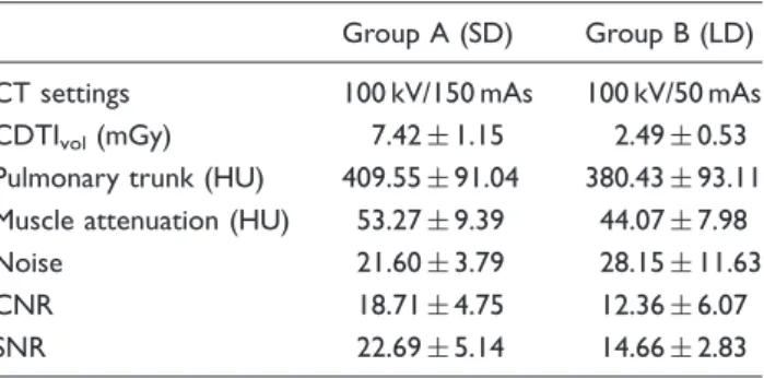
Table 2. Results of objective image analysis.
Group A (SD) Group B (LD)
CT settings 100 kV/150 mAs 100 kV/50 mAs
CDTIvol(mGy) 7.421.15 2.490.53
Pulmonary trunk (HU) 409.5591.04 380.4393.11 Muscle attenuation (HU) 53.279.39 44.077.98
Noise 21.603.79 28.1511.63
CNR 18.714.75 12.366.07
SNR 22.695.14 14.662.83
Data are meanstandard deviation.
CNR, contrast-to-noise ratio; LD, low dose; SD, standard dose; SNR, signal-to-noise ratio.
Table 1. Patient demographics.
Group A (SD) Group B (LD)
Patients (n) 30 30
Men (n) 12 16
Women (n) 18 14
Age (years) 66.55 (37–85) 68.68 (36–78) BMI (kg/m2) 24.3 (19.8–29.7) 23.8 (18.9–28.8)
average of three measurements. Based on these meas-urements, signal-to-noise ratio (SNR) and contrast-to-noise-ratio (CNR) were determined according to the following equation: SNR¼Attenuation/Background noise (BN); CNR¼(Attenuation of pulmonary trunk – Attenuation of muscle)/BN.
Subjective image evaluation was carried out by three independent radiologists, with 5, 6, and 9 years, respectively, of CT imaging experience regarding the suitability and detection of central and peripheral PE and image noise in each patient. Observers were blinded to the shown image series, but were aware of the clinical suspicion of PE. Image quality rating was performed using a five-point Likert scale (5, excellent; 4, good; 3, moderate; 2, fair; 1, unacceptable). Inter-observer agreement was calculated using intra-class correlation coefficient (ICC).
Radiation exposure
To estimate patient radiation dose, we recorded the volume CT dose index (CTDIvol, mGy) and dose
length product (DLP, mGycm) from the patient CT protocol.
Statistical analysis
Analyses were performed computer-based with dedi-cated software (STATA 13 IC, StataCorp LP, College Station, TX, USA). Continuous variables were expressed as median and range. Continuous variables were tested for normal distribution using the Kolmogorov Smirnov–Lilliefors test, corrected accord-ing to Dallal–Wilkinson as appropriate. Statistical sig-nificance was investigated with the Student’s t-test for unpaired samples, if values followed a normal distribu-tion. Otherwise, the U-test according to Wilcoxon– Mann–Whitney was applied. APvalue<0.05 was con-sidered statistically significant.
Furthermore, the inter-rater reliability was calcu-lated using the ICC. The ICC value was interpreted in the following way: ICC <0.20, slight agreement; 0.21–0.40, fair agreement; 0.41–0.60, moderate agree-ment; 0.61–0.80, substantial agreeagree-ment; and 0.81–1.0, almost perfect agreement.
Results
Sixty consecutive individuals with suspected PE under-went CTPA without complications. No significant dif-ferences were observed in age (SD 66.5512.79; LD 68.6811.66), and gender (SD 12 men, 18 women; LD 16 men, 14 women) between individuals included in groups A (SD) and B (LD). Patient demographic data are shown in Table 1. All examinations were
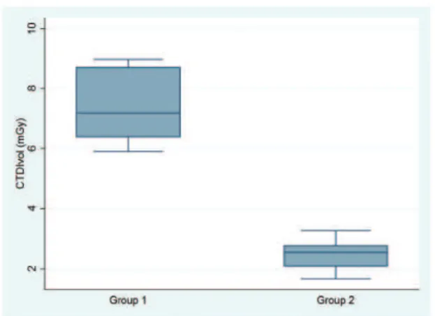
considered to reach an image quality level high enough to rule out PE (Fig. 1). A total of eight cases of PE were found in our study cohort, respectively (3 in group A, 5 in group B). All PEs were detected by all readers without significant difference in size or distribu-tion between both groups.
The radiation dose (CDTIvol) was significant
lower in LD conditions compared to SD parameters (2.490.55 mGy versus 7.421.17 mGy) (Fig. 1).
Objective image evaluation
Evaluation of image noise as an objective image quality parameter showed that the SD protocol resulted in significantly less image noise compared to LD param-eters (P<0.01). The attenuation of the pulmonary trunk showed similar results in both groups (SD 409.5591.04 HU; LD 380.43 HU93.11 HU;
P¼0.768). Hence, there were no significant differences of the attenuation of the major pectoral muscles (SD 53.279.39 HU; LD 44.077.98 HU;P¼0.584).
The highest calculated CNR of the pulmonary was found in the SD series (18.714.75); the CNR in the LD settings was significantly lower (12.366.07;
P<0.01). All details from objective image analysis are summarized in Table 3.
The SNR was highest in the SD settings, followed by the LD series (SD 22.695.14; LD 14.662.83;
P<0.01).
Subjective image evaluation
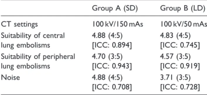
Summarization of image quality scores from all three observers are shown in Table 3.
The evaluation of the subjective ratings revealed no significant differences between the SD and LD settings in regard of the suitability of the detection of central and peripheral lung embolisms. Both ratings showed excellent suitability (central SD/LD, 4.88;
ICC, 0.894/4.83; ICC, 0.745; peripheral SD/LD, 4.70; ICC, 0.943/4.57; ICC, 0.919; all P>0.4).
Similar to the objective measured noise levels, the subjective noise rating consecutively resulted in a signifi-cant difference between SD and LD settings (4.88; ICC, 0.708 versus 3.71; ICC, 0.728;P<0.01) (Figs 2 and 3).
Discussion
All CT examinations were conducted without any problems or side effects. Patient demographics were
not significantly different in both groups in terms of gender, body habitus, and age (Table 1). Our results show that sufficient LD examinations are possible on an old CT scanner system without affecting image qual-ity too much. A lot of promising new techniques have been introduced to reduce radiation exposure while pre-serving image quality (12,13). However, some of these techniques are not available for old scanner generations such as the model we used in our analysis. To buy a new CT scanner is a huge investment for a radiology department and buying a new CT scanner takes often years of budget planning as well as discussions with hospital managers and manufacturers. To date, tech-niques frequently in use are: automated tube potential selection, iterative reconstruction algorithms, high-pitch CT imaging, and low-kV imaging (14–17). In par-ticular, low-kV imaging is often used in combination with some of the mentioned other techniques, especially iterative reconstruction algorithms to reduce image noise (10,18). Iterative reconstruction algorithms help to reduce these effects and to gain sufficient image qual-ity even in images that might appear too noisy in con-ventional filtered back projection (FBP).
In one recent paper Laqumani, et al. analyzed an 80 kV protocol on a 256-slice CT scanner using iterative
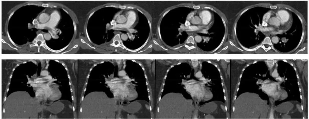
Fig. 2. PE in the left main pulmonary artery. LD CT protocol (axial, coronal, and sagittal view). Table 3. Results of subjective image analysis.
Group A (SD) Group B (LD)
CT settings 100 kV/150 mAs 100 kV/50 mAs Suitability of central
lung embolisms
4.88 (4:5) [ICC: 0.894]
4.83 (4:5) [ICC: 0.745] Suitability of peripheral
lung embolisms
4.70 (3:5) [ICC: 0.943]
4.57 (3:5) [ICC: 0.919]
Noise 4.88 (4:5)
[ICC: 0.708]
3.71 (3:5) [ICC: 0.728]
Data based on ratings from all three observers. Global ICC: 0.925.
reconstruction algorithms in a selected patient cohort (body weight<80 kg) (19). A CTDIvolof 2.30.8 mGy
was reported, which is a good value in CT imaging of the pulmonary vascular system. However, comparing this study to our data is challenging because no iterative reconstruction algorithms were used and tube-detector systems made substantial improvements over the past decade.
A study by Montet et al. compared a FBP recon-structed protocol with a new iterative reconstruction protocol. The examinations were carried out on a high-end CT scanner system (Discovery 750 HD, GE Healthcare, Milwaukee, WI, USA) (17). The FBP proto-col was adjusted to 100 kV, 250 Ref. mAs resulting in a mean CTDIvolof 8.0 (1.8) mGy. Compared to these
values, our protocols remained below the presented CTDIvol(Table 1). However, using iterative
reconstruc-tion algorithms in this study decreased the radiareconstruc-tion exposure to a mean CTDIvol of 0.59 mGy. We could
show a median CTDIvolof 2.490.53 mGy in our LD
cohort. Compared to the 8 mGy in the FBP group on this high-end CT system, we think our values are a good choice for LD CT imaging on an elderly CT system.
Another recent study by Pontana et al. analyzed a reduced dose setting for ruling out PE on a dual-source CT (20). From the full-dose setting they calculated a LD dataset of 120 kV and 66 mAs in the LD group. A mean CTDIvol of 2.76 (0.79) mGy was reported
in the LD group, which is comparable to the values of our analysis. Acute PE was sufficiently ruled out even in the LD protocol using FBP. However, since the focus of this analysis was the evaluation of iterative reconstruction on image quality, the segmental and sub-segmental analysis, in particular, revealed a better delineation of the thrombotic material within the ves-sels using iterative reconstruction algorithms.
Comparing the dose values of recent literature to those presented in our current study, our radiation
dose values seem to be in line even with latest generation CT systems using FBP image recon-struction. As a main result of our study, dose reduction in CTPA is possible and can be achieved easily. To adjust the CT scanner system for the first time to LD values can be challenging because of the fear of gaining insufficient image quality. We wanted to relieve the readers of this manuscript from this fear and want to encourage the use of LD CT protocols even in older CT systems.
Some limitations of our study need to be addressed. First, the overall number of patients in our study was small; further studies with a larger cohort are required. Second, we did not investigate iterative reconstruction algorithms as well as effects of newer tube potential selection in our study. However, since we wanted to show how to deal with an older CT system, these soft-ware and hardsoft-ware tools were simply not available when the CT system was built. Third, additional ana-lysis of reduced contrast material would have been desirable, this will be part of upcoming future investigations.
In conclusion, our results demonstrate that using a LD protocol on a 15-year-old CT scanner system in CTPA allows for a dose reduction of approximately 67% with similar subjective image quality and delinea-tion of central and peripheral pulmonary arteries in clinical routine.
Declaration of conflicting interests
The author(s) declared no potential conflicts of interest with respect to the research, authorship, and/or publication of this article.
Funding
The author(s) received no financial support for the research, authorship, and/or publication of this article.
References
1. Bach AG, Schmoll HJ, Beckel C, et al. Pulmonary embol-ism in oncologic patients: frequency and embolus burden of symptomatic and unsuspected events. Acta Radiol 2014;55:45–53.
2. Crawford F, Andras A, Welch K, et al. D-dimer test for excluding the diagnosis of pulmonary embolism. Cochrane Database Syst Rev 2016;8:CD010864. 3. Brink JA, Amis ES Jr. Image Wisely: a campaign to
increase awareness about adult radiation protection. Radiology 2010;257:601–602.
4. Dougeni E, Faulkner K, Panayiotakis G. A review of patient dose and optimisation methods in adult and paediatric CT scanning. Eur J Radiol 2012;81:e665–683. 5. Fleischmann D, Boas FE. Computed tomography–old ideas and new technology. Eur Radiol 2011;21:510–517. 6. Kroft LJ, Roelofs JJ, Geleijns J. Scan time and patient
dose for thoracic imaging in neonates and small children using axial volumetric 320-detector row CT compared to helical 64-, 32-, and 16- detector row CT acquisitions. Pediatr Radiol 2010;40:294–300.
7. Lell MM, May M, Deak P, et al. High-pitch spiral com-puted tomography: effect on image quality and radiation dose in pediatric chest computed tomography. Invest Radiol 2011;46:116–123.
8. Bauer RW, Schell B, Beeres M, et al. High-pitch dual-source computed tomography pulmonary angiography in freely breathing patients. J Thorac Imaging 2012;27: 376–381.
9. Kerl JM, Lehnert T, Schell B, et al. Intravenous contrast material administration at high-pitch dual-source CT pul-monary angiography: test bolus versus bolus-tracking technique. Eur J Radiol 2012;81:2887–2891.
10. Bodelle B, Klement D, Kerl JM, et al. 70 kV computed tomography of the thorax: valence for computer-assisted nodule evaluation and radiation dose - first clinical results. Acta Radiol 2014;55:1056–1062.
11. Beeres M, Wichmann JL, Paul J, et al. CT chest and gantry rotation time: does the rotation time influence image quality? Acta Radiol 2015;56:950–954.
12. Fleischmann D, Boas FE. Computed tomography– old ideas and new technology. Eur Radiol 2011;21: 510–517.
13. McCollough CH, Bruesewitz MR, Kofler JM Jr. CT dose reduction and dose management tools: overview of avail-able options. Radiographics 2006;26:503–512.
14. Lee JW, Lee G, Lee NK, et al. Effectiveness of adaptive statistical iterative reconstruction for 64-slice dual-energy computed tomography pulmonary angiography in patients with a reduced iodine load: comparison with standard computed tomography pulmonary angiog-raphy. J Comput Assist Tomogr 2016;40:777–783. 15. Scholtz JE, Wichmann JL, Husers K, et al.
Third-generation dual-source CT of the neck using automated tube voltage adaptation in combination with advanced modeled iterative reconstruction: evaluation of image quality and radiation dose. Eur Radiol 2016;26: 2623–2631.
16. Bucher AM, Kerl MJ, Albrecht MH, et al. Systematic comparison of reduced tube current protocols for high-pitch and standard-high-pitch pulmonary CT angiography in a large single-center population. Acad Radiol 2016;23: 619–627.
17. Matsubara K, Sakuda K, Nunome H, et al. 128-slice dual-source CT coronary angiography with prospectively electrocardiography-triggered high-pitch spiral mode: radiation dose, image quality, and diagnostic acceptabil-ity. Acta Radiol 2016;57:25–32.
18. Sauter A, Koehler T, Fingerle AA, et al. Ultra low dose CT pulmonary angiography with iterative reconstruction. PLoS One 2016;11:e0162716.
19. Laqmani A, Regier M, Veldhoen S, et al. Improved image quality and low radiation dose with hybrid itera-tive reconstruction with 80 kV CT pulmonary angiog-raphy. Eur J Radiol 2014;83:1962–1969.