DOI: 10.14260/jemds/2014/1961
CASE REPORT
Journal of Evolution of Medical and Dental Sciences/ Volume 3/ Issue 05/February 03, 2014 Page 1160
STRANGULATED FEMORAL HERNIA IN A MALE PATIENT
–
DIAGNOSTIC
DILEMMA
Sapna Purushotham1, Srinivas B. Kulkarni2, Kshirsagar3, Mudasser Rehan4, Manjunath V5
HOW TO CITE THIS ARTICLE:
Sapna Purushotham, Srinivas B. Kulkarni, Kshirsagar, Mudasser Rehan, Manjunath V. Strangulated Femoral Hernia in a Male Patient – Diagnostic Dilemma. Journal of Evolution of Medical and Dental Sciences 2014; Vol. 3, Issue 05, February 03; Page: 1160-1163, DOI: 10.14260/jemds/2014/1961
ABSTRACT: Strangulated femoral hernia in a male patient is a very rare clinical presentation. Here we report a case of right inguinal swelling mimicking that of lymphadenopathy, later developing perforation due to strangulation of femoral hernia. Laparotomy with ileostomy was done.
KEYWORDS:Femoral hernia, Ritcher s hernia, Strangulated femoral hernia
INTRODUCTION: Femoral hernia is an uncommon condition which has been reported in less than 5% of all abdominal wall hernias, with a female to male preponderance of 1.8:1. This is an elusive condition that despite having life threatening complications are often undiagnosed in asymptomatic patients. The diagnosis of a strangulated femoral hernia still presents a challenge to all clinicians. Till
date only to cases of Richter s type of strangulated femoral hernia have been reported, thus
prompting us to report this rare case.
CASE REPORT: A 58 year old male patient presented to our outpatient department with history of swelling in the right inguinal region since 6 days and vague pain abdomen since 2days. Patient had no other significant past history.
On examination of the patient, there appeared to be a 3x4 cm swelling in the right inguinal region below and lateral to the pubic tubercle, firm to soft in consistency and non-tender. On abdominal examination, the abdomen was found to be soft with no distension and mild tenderness in the umbilical region. Bowel sounds were well appreciated. Routine blood analysis was normal.
A diagnosis of right inguinal lymphadenopathy was made and an ultrasound examination of the swelling and abdomen was advised. Initial ultrasound showed right inguinal mass - ?lymph node mass with minimal Ascites and grade I prostatomegaly.
Fine needle aspiration of the swelling – smears studied show polymorphous population of lymphocytes, neutrophils, plasma cells and foamy histiocytes – suggestive of suppurative lesion.
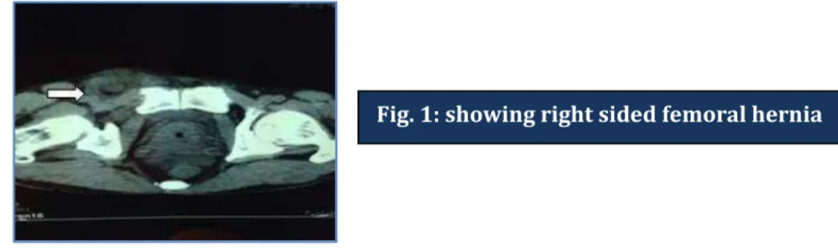
As patient developed abdominal distension on day 4, a contrast enhanced CT scan was advised which showed – right femoral hernia with bowel loops as contents with dilated small bowel loops and extensive pneumo-peritoneum with mild ascites suggestive of hollow viscous perforation.
DOI: 10.14260/jemds/2014/1961
CASE REPORT
Journal of Evolution of Medical and Dental Sciences/ Volume 3/ Issue 05/February 03, 2014 Page 1161 Subsequently, the patient was taken up for exploratory laparotomy, where the presence of a femoral hernia was confirmed, containing part of the ileal wall at the neck of the hernial sac 20cm from the ileo-caecal junction (Richter s hernia). The part of the ileal wall within the sac was perforated but viable. Pus was found in the femoral hernial sac.
Following reduction of the ileum, the perforated part of the ileum brought out as a loop ileostomy. The femoral ring defect, repaired internally by approximating the ilio-pubic tract to the
Cooper s ligament. Counter-incision made over the inguinal region to drain the pus. Peritoneal lavage
done and abdominal drain placed in-situ.
Fig. 2: showing air in the hernia sac Fig. 3: Pneumoperitoneum
Fig. 4: Intra-operative finding
DOI: 10.14260/jemds/2014/1961
CASE REPORT
Journal of Evolution of Medical and Dental Sciences/ Volume 3/ Issue 05/February 03, 2014 Page 1162
The patient s recovery and post-operative period were uneventful. Patient was advised
closure of ileostomy after 6-8 weeks.
DISCUSSION: Femoral hernia is not as common as inguinal hernia. It is often associated with incarceration or strangulation, resulting in peritonitis and mortality. The ratio of femoral hernia relative to all groin hernias is reported to be less than 5% in adults 1, 2, 3. The peak distribution is in
the 50s, with a slight decrease in the 60s and 70s. As for sex distribution, femoral hernia is 4 to 5 times more common in female than in male 4, 5.
Despite the fact that femoral hernias account for only 2-4% of all groin hernias, their timely and correct diagnosis is vital due to the increased mortality associated with emergency surgery for their complications 6. This, however, is not always easy. Femoral hernias are commonly missed or
misdiagnosed as less serious conditions, leaving surgeons to deal with their complications in the acute setting, where mortality has been found to be 10fold 6, 7, 8. Differential diagnosis of femoral
hernia includes lymphadenopathy, saphenous vein varicosity, pseudohernia, femoral artery pseudoaneurysm, soft tissue masses.
The hernia sac commonly consists of small bowel or omentum, but uncommon cases have been reported, where the herniating structures were caecum, appendix, colon, Meckel s diverticulum, ovaries, testes, stomach and kidneys 8, 9.
According to Dahlstrand et al, who published the largest series of femoral hernia repairs to date 6, out of 3, 980 femoral hernia repairs 1430 (35.9%) were emergencies, compared to just 5.4%
for inguinal cases. Furthermore, 22.7% of the emergency procedures for femoral hernias required bowel resection compared to 5.4% for inguinal hernias, whereas that percentage of bowel resection in elective femoral hernia repairs was only 0.6%. Dahlstrand et al also demonstrated that women were more likely than men to require surgery for femoral hernias (5:3 ratio). The risk for emergency surgery for women was also significantly higher (40.6 vs. 28.1%).
Two other similar cases have been reported in literature. First was reported by Duari in 1966
10. He presented the case of a 79-year old man with a right groin abscess that was incised by his GP
and found to contain evil-smelling pus . Duari obviously did not have access to modern abdominal
imaging at the time and performed exploratory laparotomy on the patient, during which he discovered a femoral hernia containing part of the caecal wall and the base of the appendix. Arkoulis
et al in presented the second reported case of a femoral hernia of the Richter s variety
containing caecum and appendix that, following strangulation and perforation, manifested as a groin abscess 11.
REFERENCES:
1. Glassow F. Femoral hernia. Review of 2105 repairs in a 17 year period. Am J Surg 1985; 150:353–6.
2. Waddington RT. Femoral hernia: a recent appraisal. Brit J Surg 1971;58:920–2.
3. Maingot R. Choice of operation for femoral hernia, with special reference to McVay s technique. Brit J Clin Practice 1968;22:323–9.
DOI: 10.14260/jemds/2014/1961
CASE REPORT
Journal of Evolution of Medical and Dental Sciences/ Volume 3/ Issue 05/February 03, 2014 Page 1163 7. Naude GP, Ocon S, Bongard F. Femoral Hernia: The Dire Consequences of a Missed
Diagnosis. Am J Emerg Med. 1997 Nov;15(7):680–2
8. Dahlstrand U, Wollert S, Nordin P, Sandblom G, Gunnarsson U. Emergency femoral hernia repair. A study based on a national register. Ann Surg2009;249:672–6.
9. Patel RB, Vasava N, Hukkeri S. Non-obstructed femoral hernia containing ascending colon, caecum, appendix and small bowel with concurrent bilateral recurrent inguinal hernia. Hernia 2012 Apr;16(2):211–3.
10.Duari M. Strangulated femoral hernia—a Richter s type containing caecum and base of appendix. Postgrad Med J 1966 Nov;42(493):726–8.
11.N Arkoulis, G Savanis and G Simatos Richter s type strangulated femoral hernia containing caecum and appendix masquerading as a groin abscess. Ox J Med, journal of surgical case reports, vol 2012:6, 6.
AUTHORS:
1. Sapna Purushotham 2. Srinivas B. Kulkarni 3. Kshirsagar
4. Mudasser Rehan 5. Manjunath V.
PARTICULARS OF CONTRIBUTORS:
1. Assistant Professor, Department of General Surgery, Sapthagiri Institute of Medical Sciences, Bangalore.
2. Senior Resident, Department of General Surgery, Sapthagiri Institute of Medical Sciences, Bangalore.
3. Professor, Department of General Surgery, Sapthagiri Institute of Medical Sciences, Bangalore.
4. Assistant Professor, Department of General Surgery, Sapthagiri Institute of Medical Sciences, Bangalore.
5. Assistant Professor, Department of General Surgery, Sapthagiri Institute of Medical Sciences, Bangalore.
NAME ADDRESS EMAIL ID OF THE CORRESPONDING AUTHOR: Dr. Sapna Purushotham,
#66, Tapas , Taponagara Main Road,
Chikkagubbi,
Off Hennur Bagalur Road, Bangalore – 560077.
E-mail: sap.sapu@gmail.com