Dragana Kova~evi}
,
1, 2D`enana A. Detanac,
3Vujica Markovi},
1, 2Aleksandra Radosavljevi},
1Krstina Doklesti},
4D`email S. Detanac,
3Svetislav Milenkovi}
1, 2TREATMENT WITH CYCLOSPORINE A
IN SERPIGINOUS CHOROIDITIS:
A CASE REPORT
Primljen/Received 23. 09. 2012. god. Prihva}en/Accepted 01. 11. 2012. god.
Summary:Serpiginous choroiditis is a rare clinical entity. The clinical course of serpiginous choroiditis is very variable, there is no universal marker of treatment success, and even among experts there is debate about what is the most appropriate treatment. The aim of this paper is to describe a case of serpiginous choroiditis treated with Cyclosporine A at a tertiary uveitis referral centre.
Key words:serpiginous choroiditis, Cyclosporine A, serpiginous choroidopathy, fluorescein angiography.
Introduction
Serpiginous choroiditis (SC) is a clinically defi-ned disorder characterized by destruction of the inner choroid and the retinal pigment epithelium (RPE), as well as, secondary involvement of the retina. It is a ra-re, usually bilateral, chronic, progressive, recurrent in-flammation of the RPE, choriocapillaris and choroid of unknown etiology (1). It generally constitutes less than 5% of posterior uveitis in most epidemiological reports (2). The disease primarily affects healthy young to middle-aged adults, with a higher prevalence reported in males than females (3). There is no clear racial pre-dilection and no familial association (4). Most cases of serpiginous choroiditis are not associated with syste-mic disease although there are isolated reports of serpi-ginous choroiditis occurring in the presence of syste-mic diseases such as Crohn’s disease (5), celiac disease (6), extrapyramidal dystonia (7), polyarteritis nodosa
(8), and sarcoidosis (9) that are most likely coinciden-tal. The pathogenesis of serpiginous choroiditis rema-ins unknown. An infectious etiology, immunological derangements, and vascular disorders have been stud-ied in order to find the connection with this condition. Disease presentation and course are variable, and an incomplete understanding of its etiology hinders at-tempts at formulating an effective treatment strategy. The goals of any successful therapy should be the rapid control of active lesions during recurrences, and the prevention of further recurrences and progression of the disease. As it is a rare condition with an insidious clinical progression, the conduct of any clinical trial with sufficient power would be difficult.
Case report
A 55-year-old woman presented to the Eye Clinic (Clinical Center of Serbia, Belgrade), in November 2010, with a one month history of blurred vision on her right eye. The patient had gradual deterioration of vi-sion on her left eye for 20 years. She had sought multi-ple consultations before she came to the Clinic, but had no defined diagnosis. She denied any family history of ocular disease.
Ophthalmological examination revealed a visual acuity of 6/60 in the right eye and counting fingers on 0.5 meters in the left eye. Intraocular pressure was 14 mmHg in both eyes. Anterior segment findings were within normal. Fundus examination of the right eye re-vealed an area of chorioretinal geographic atrophy at the posterior pole which involved the lower half of the fo-vea. There was an active lesion in infero-temporal ma-cula. Fundus examination of the left eye revealed a
lar-1 Clinic for Eye Diseases, Clinical Center of Serbia, Belgrade, Serbia 2 Belgrade University, School of Medicine, Serbia
3 General hospital Novi Pazar, Serbia
4 Clinic of Emergency Surgery, Clinical Center of Serbia, Belgrade, Serbia
2012; 7(2): 103–106 UDK: 617.723-002-085.33
ge, geographic chorioretinal scar at the posterior pole which involved macula, with no signs of active disease.
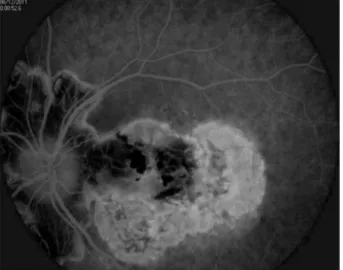
The old lesion in the left eye demonstrated block-age of fluorescence corresponding to the areas of RPE hypertrophy with staining on the edges (Figure 1). Flu-orescein angiography (FA) of the right eye revealed a large hypofluorescent lesion which, in the late phases of the angiogram, had a hyperfluorescent margins — findings typical of an inactive serpiginous lesion and presence of a small area of active disease in infero-tem-poral macula (Figure 2).
A clinical examination revealed an erythrocyte se-dimentation rate of 30 mm in the first hour (normal range 1–12 mm), white blood cells count of 14.5 109/L (3.4–9.7 109/L) with neutrophilia 11.7 109/L (2.1–6.5 109/L) and raised levels of urea 10.2 mmol/L (2.5–7.5 mmol/L). Tests for connective tissue disorders were negative, serum angiotensin converting enzyme was within the normal range and chest X-ray was normal. Infectious serologies (toxoplasmosis, Borrelia burg-dorferi, HIV, herpesvirus, citomegalovirus, varicel-la-zoster virus) were within the normal limits.
Based on the clinical presentation, fluorescein angi-ography, and negative work up for systemic or infectious disease, a diagnosis of serpiginous choroiditis was made.
The patient’s medical history was reviewed for li-ver disease or other contraindications to immunosup-pressive therapy. Baseline complete blood count and liver function tests were performed before recommen-ding the treatment options. Risks, benefits, and alterna-tives were discussed thoroughly, and patient was given an opportunity to consider her therapeutic options.
Treatment with oral Cyclosporine A 300 mg (3 x 100 mg) per day and 8 mg of intravenous (IV) dexameth-asone per day was initiated. After five days, dose of
dexa-methasone was reduced to 4 mg IV for the next three days, followed by oral prednisone 40 mg as a single dose in the morning. The patient was discharged with visual acuity of 6/10 in the right eye, and unchanged visual acu-ity in the left. She continued the treatment with Cyclospo-rine A 300 mg/day and oral prednisone 40 mg/day.
In the follow-up period, patient underwent Snel-len visual acuity (VA) testing, slit-lamp examination, and fundus examination with indirect ophthalmoscopy and a 78-diopter lens.
The first follow-up visit was 3 weeks after initiation of therapy and every 6 to 8 weeks thereafter. The patient was monitored with a complete blood count, Cyclospori-ne A blood level and liver function test (aspartate amino-transferase and alanine aminoamino-transferase levels) every 2 months. Cyclosporine blood levels were obtained to mo-nitor patient compliance and potential toxicity. The pati-ent was also specifically queried at each visit about the presence of potential adverse reactions associated with Cyclosporine A. Drug dosage was adjusted according to the therapeutic response and side effects. Once the choro-iditis appeared not active, oral prednisone was tapered to a lower dosage and finally discontinued. The goal of cyclosporine treatment was inactivity of the lesions for approximately 12 to 24 months, after which the drug was tapered and discontinued.
Patient had decreased ocular inflammation in the right eye within 2 weeks of the initiation of the treat-ment. She was able to taper and discontinue oral pred-nisone within 3 months when the visual acuity was 6/9 in the right eye. The patient continued to be under Cyclosporine A 300 mg (3 x 100 mg) per day, and after 4 months, dose of Cyclosporine A was reduced to 200 104 Dragana Kova~evi}, D`. A. Detanac, V. Markovi}, A. Radosavljevi}, K. Doklesti}, D`. S. Detanac, S. Milenkovi}
Fig. 1. Inactive lesion showing early hypofluorescence secondary to atrophy
of choriocapillaris and progressive hyperfluorescence at the margins of the lesion
Fig. 2. Fundus fluorescein angiography of an active lesion (early and late phase): active lesion in macula
is represented by the blockage of the fluorescence in the early phase and the indistinct margins and more diffuse staining and leakage progressively
mg (2 x 100 mg) per day. The visual acuity was 6/9, and the best-corrected vision was 6/6 (–0.75 Dsph) n the right eye, in that period. Cyclosporine A blood le-vel after nine months of treatment, was 536.6 ng/ml (recommended blood level after 6–12 months of treat-ment is 100–150 ng/ml), which was the reason for dis-continuing of that immunosuppressive agent. The oral prednisone dosage was then increased to the initial le-vel of 60 mg/d. Four weeks later, patient had deteriora-tion of the best-corrected vision to 6/8 (–0.75 Dsph) in the right eye. For that period she tapered the oral pred-nisone to 40 mg/day. After 1 month the best-corrected vision dropped to 6/12 (–0.75 Dsph). Oral Cyclospori-ne A (200 mg/d) was added with triamcinoloCyclospori-ne aceto-nide injections subconjunctivally. The patient is cur-rently on 10 mg/day oral prednisone and 200 mg/day Cyclosporine A, and the clinical status is stable. The visual acuity in the right eye is 6/30 and the best-cor-rected vision is 6/12 (–0.75 Dsph). To the date, the pati-ent has not prespati-ented with choroidal neovasculariza-tion (CNV) as a complicaneovasculariza-tion of SC and no serious ad-verse reactions related to Cyclopsorine A, such as he-patotoxicity and nephrotoxicity were noted.
Discussion
Our patient was a healthy 55-year-old female, with no comorbidities, whose blurring of central vision in the right eye started one month before she came to the Clinic. Her past, twenty years long ocular history was significant for a gradual blurring of vision in the left eye before blurring of central vision started in the right eye. Although disease involvement is usually bi-lateral, the typical presentation is asymmetric, includ-ing our patient, startinclud-ing first in the one eye, with a dec-rease in central vision, metamorphopsia or the devel-opment of scotomata that correspond exquisitely with visible fundus lesions. As with our patient, there are typically no inflammatory cells or flare seen in the an-terior segment or anan-terior vitreous. Classic (peripapil-lary geographic) variant, including our patient, acco-unts for about 80% of the cases of serpiginous choroi-ditis reported in the literature (10). The active disease begins with ill-defined patches of grayish or creamy yellow subretinal infiltrates originating in the peripa-pillary region and progressing centrifugally in an irreg-ular serpentine fashion.
The disease is characterized by multiple recurren-ces at variable intervals, ranging from months to years. About two-thirds of patients with serpiginous choroi-ditis have scars in one or both eyes at initial presenta-tion, and most patients are asymptomatic until the ma-cula is involved (11). Visual loss is directly correlated with the proximity of the lesion to the fovea.
Histopathological studies have demonstrated dif-fuse and focal infiltrates of lymphocytes in the choroid, particularly at the margin of the serpiginous lesions, which implies an inflammatory component to the dise-ase (12). This is the rationale for the use of anti-inflam-matory and immunosuppressive therapies for SC. So-me authors believe that systemic and periocular corti-costeroids may be helpful in the active phase of the dis-ease (13). On the other hand, recent long-term fol-low-up studies have suggested that therapy with immu-nosuppressive agents is the best option to treat active SC, as steroids alone did not prevent recurrences (14). The spectrum of alternative immunosuppressive thera-pies for serpiginous chorioretinitis ranges from monot-herapy with corticosteroids or other agents alone to tri-ple therapy with multitri-ple agents. Prognosis regarding the visual function is generally thought to be poor in this disease. Macular involvement, with consequent decrea-sed visual acuity, occurs in up to 88% of patients and ap-proximately 50% could be expected to have recurrence in 5 years (15). Based on the studies reported so far, the rapid control of any active lesions with aggressive im-munosuppression and thereafter the maintence on ap-propriate immunosuppression for at least 6 months to prevent any immediate recurrence can be considered for the initial management of patients with serpiginous cho-roiditis. Subsequent treatment will depend not only on the severity of the disease, e.g., foveal threatening lesi-ons in an only seeing eye, but also on the general health of the patient and other concerns such as fertility and the response to initial immunosuppressive therapy (16). One of the treatment algorithm based on current knowl-edge is using systemic corticosteroids and periocular steroidal injections as the first line to control active lesi-ons, with immunosuppressive therapy such as cyclospo-rine A, azathiopcyclospo-rine or mycophenolate mofetil used concurrently as monotherapy for maintenance of remis-sion (17). Cases that don not respond to this approach may then be candidates for a combination therapy simi-lar to triple-therapy or alkylating agents (18).
patient is currently on 10 mg/day oral prednisone and 200 mg/day Cyclosporine A, visual acuity and inflammation are stable and she had no constitutional symptoms severe enough to necessitate ceasing cyclosporine A.
Conclusion
Our results suggest that Cyclosporine A used in combination with corticosteroids is a safe and
accepta-ble option for treating patients with active SC. To dem-onstrate the success of any therapeutic approach for serpiginous choroiditis, a long-term follow-up with se-rial fundus photographs and fluorescein angiograms to show disease non-progression is required. Further mul-ticentric studies are required to evaluate the etiology, pathogenesis, natural history and the efficacy of differ-ent treatmdiffer-ent strategies for this rare disease.
106 Dragana Kova~evi}, D`. A. Detanac, V. Markovi}, A. Radosavljevi}, K. Doklesti}, D`. S. Detanac, S. Milenkovi}
Sa`etak
CIKLOSPORIN A U TERAPIJI SERPIGINOZNOG HOROIDITISA:
PRIKAZ SLU^AJA
Dragana Kova~evi},1, 2D`enana A. Detanac,3Vujica Markovi},1, 2Aleksandra Radosavljevi},1 Krstina Doklesti},4D`email S. Detanac,3Svetislav Milenkovi}1, 2
1 — Klinika za o~ne bolesti, Klini~ki centar Srbije, Beograd; 2 — Medicinski fakultet Univerziteta u Beogradu; 3 — Op{ta bolnica Novi Pazar; 4 — Klinika za urgentnu hirurgiju, Klini~ki centar Srbije, Beograd
Serpiginozni horoiditis je redak klini~ki entitet. Prirodni tok bolesti je veoma promenljiv, ne postoji univerzalni pokazatelj terapijskog uspeha, ~ak i me|u ekspertima jo{ uvek postoji debata oko najprikladnijeg terapijskog pristupa. Cilj ovog rada je da opi{e slu~aj
serpiginoznog horoiditisa primarno le~enog Ciklospo-rinom A u tercijarnoj referentnoj ustanovi.
Klju~ne re~i:serpiginozni horoiditis, Ciklosporin A, serpiginozna horoidopatija, fluoresceinska angio-grafija.
REFERENCE
1. Abu el-Asrar AM: Serpiginous (geographical) choroidi-tis. Int Ophthalmol Clin.1995; 35: 87–91.
2. Chang JH, Wakefield D. Uveitis: a global perspecti-ve. Ocul Immunol Inflamm. 2002; 10: 263–79.
3. Akpek EK, Jabs DA, Tessler HH, et al. Successful treat-ment of serpiginous choroiditis with alkylating agents. Ophthal-mology. 2002; 109: 1506–13.
4. Gupta V, Agarwal A, Gupta A, et al. Clinical characteri-stics of serpiginous choroidopathy in North India. Am J Ophthal-mol. 2002; 134: 47–56.
5. Ugarte M, Wearne IM. Serpiginous choroidopathy: an unusual association with Crohn’s disease. Clin Exp Ophthal-mol. 2002; 30: 437–39.
6. Mulder CJ, Pena AS, Jansen J, Oosterhuis JA. Celiac dise-ase and geographic (serpiginous) choroidopathy with occurrence of thrombocytopenic purpura. Arch Intern Med. 1983; 143: 842.
7. Richardson RR, Cooper IS, Smith JL. Serpiginous cho-roiditis and unilateral extrapyramidal dystonia. Ann Ophthal-mol. 1981; 13: 15–19.
8. Pinto Ferreira F, Faria A, Ganhao F. Periarteritis nodosa with initial ocular involvement. J Fr Ophtalmol. 1995; 18: 788–93.
9. Edelsten C, Stanford MR, Graham EM. Serpiginous choroiditis: an unusual presentation of ocular sarcoidosis. Br J Ophthalmol. 1994; 78: 70–71.
10. Nussenblatt RB Whitcup SM. Uveitis: fundamentals and clinical practice. 4th ed. St. Louis: Mosby; 2010.
11. Lim WK, Buggage RR, Nussenblatt RB. Serpiginous choroidopathy: major review. Surv Ophthalmol. 2005; 50: 231–44. 12. Wu JS, Lewis H, Fine SL, et al. Clinicopathologic find-ings in a patient with serpiginous choroiditis and treated choroi-dal neovascularization. Retina. 1989; 9: 292–301.
13. Akpek EK, Jabs DA, Tessler HH, Joondeph BC, Foster CS. Successful treatment of serpiginous choroiditis with alkyla-ting agents. Ophthalmology. 2002; 109: 1506–13.
14. Munteanu G, Munteanu M, Zolog I. Serpiginous cho-roiditis — clinical study. Oftalmologia. 2001; 52: 72–80.
15. Christmas NJ, Oh KT, Oh DM, Folk JC. Long-term follow-up of patients with serpiginous choroiditis. Retina. 2002; 22: 550–56.
16. Svetislav Milenkovic, Vesna Jaksic, Natalija Jakovic, Ivan Stefanovic, Dijana Risimic, Jelena Paovic, James C Folk. Di-agnostic and therapeutic challenges. Retina. 2010; 30(9): 1546–48. 17. Araujo AAQ, Wells AP, Dick AD, Forrester JV. Early treatment with cyclosporin in serpiginous choroidopathy main-tains remission and good visual outcome. Br J Ophthalmol. 2000; 84: 979–82.
18. Hooper PL, Kaplan HJ. Triple agent immunosuppression in serpiginous choroiditis. Ophthalmology. 1991; 98: 944–51.
19. Lee DK, Suhler EB, Augustin W, Buggage RR. Serpi-ginious choroidopathy presenting as choroidal neovasculariza-tion. Br J Ophthalmol. 2003; 87: 1184–97.
Correspondence to/Autor za korespondenciju
dr D`enana Detanac Sutjeska bb, Novi Pazar
