Submitted7 June 2016 Accepted 29 November 2016 Published26 January 2017
Corresponding author Chee Sian Kuan,
cs_sam85@yahoo.com.my
Academic editor Abhishek Kumar
Additional Information and Declarations can be found on page 20
DOI10.7717/peerj.2841
Copyright 2017 Looi et al.
Distributed under
Creative Commons CC-BY 4.0
OPEN ACCESS
Genomic insight into pathogenicity
of dematiaceous fungus
Corynespora
cassiicola
Hong Keat Looi1, Yue Fen Toh1, Su Mei Yew1, Shiang Ling Na1, Yung-Chie Tan2, Pei-Sin Chong2, Jia-Shiun Khoo2, Wai-Yan Yee2, Kee Peng Ng1and Chee Sian Kuan1
1Department of Medical Microbiology, University of Malaya, Kuala Lumpur, Malaysia
2Department of Science and Technology, Codon Genomics SB, Seri Kembangan, Selangor, Malaysia
ABSTRACT
Corynespora cassiicola is a common plant pathogen that causes leaf spot disease in a broad range of crop, and it heavily affect rubber trees in Malaysia (Hsueh, 2011;Nghia et al., 2008). The isolation of UM 591 from a patient’s contact lens indicates the pathogenic potential of this dematiaceous fungus in human. However, the underlying factors that contribute to the opportunistic cross-infection have not been fully studied. We employed genome sequencing and gene homology annotations in attempt to identify these factors in UM 591 using data obtained from publicly available bioinformatics databases. The assembly size of UM 591 genome is 41.8 Mbp, and a total of 13,531 (≥99 bp) genes have been predicted. UM 591 is enriched with genes that encode for glycoside hydrolases, carbohydrate esterases, auxiliary activity enzymes and cell wall degrading enzymes. Virulent genes comprising of CAZymes, peptidases, and hypervirulence-associated cutinases were found to be present in the fungal genome. Comparative analysis result shows that UM 591 possesses higher number of carbohydrate esterases family 10 (CE10) CAZymes compared to other species of fungi in this study, and these enzymes hydrolyses wide range of carbohydrate and non-carbohydrate substrates. Putative melanin, siderophore,ent-kaurene, and lycopene biosynthesis gene clusters are predicted, and these gene clusters denote that UM 591 are capable of protecting itself from the UV and chemical stresses, allowing it to adapt to different environment. Putative sterigmatocystin, HC-toxin, cercosporin, and gliotoxin biosynthesis gene cluster are predicted. This finding have highlighted the necrotrophic and invasive nature of UM 591.
SubjectsGenomics, Microbiology
Keywords Corynespora cassiicola, Pathogenicity, CAZyme, Appressoria, Toxin, Virulence factor, Melanin
INTRODUCTION
In year 2000, spores ofC. cassiicolahas been identified as part of the airborne allergens that triggers allergenic responses in patients having respiratory problem (Yi et al., 2000). In rare occasions, this phytopathogen infects human and causes keratitis (Yamada et al., 2013), maduromycetoma (Mahgoub, 1969), subcutaneous infection (Huang et al., 2010), and phaeohyphomycosis (Lv et al., 2011;Wang et al., 2014).
Over the recent four decades, the number of reportedC. cassiicolainfections in human are limited to less than ten cases, but this opportunistic fungus still causes extensive skin damage upon successful infection. Majority of the patients that contracted the fungus are involved in agriculture work, regardless of whether the host is immunity-compromised (Huang et al., 2010;Yamada et al., 2013), wounded (Lv et al., 2011), or physically healthy and immunocompetent (Yamada et al., 2013). In this study, we identified a strain of C. cassiicola, namelyC. cassiicolaUM 591, that was isolated from a patient’s contact lens, whom was diagnosed with ocular mycosis.
The first obstacle in fungal invasion is the host’s natural defence system. In the plant, the cell wall is the best defence against fungal invasion (Smart, 1991). In warm-blooded organisms, endothermy has been suggested as an additional layer of natural defence from fungal invasion aside from skin barrier and immune system (Casadevall, 2007). In order to overcome these obstacles, fungi adopt variable means of invasion, which can be grouped into three main categories; enzymatic, chemical, and physical effectors.
Enzymatic effectors include plant degradative enzymes, secreted peptidases, and lipases. Plant pathogens are equipped with diverse classes of Carbohydrate-Active enZymes (CAZymes) (Cantarel et al., 2009), which include cellulases, hemicellulases, pectinases, and cutinases (Zhao et al., 2014). These enzymes facilitate fungal invasion via degradation or modification of plant’s cell wall. On the other hand, human pathogens require lipases and peptidases to degrade keratinized medium (such as hair, skin or nail) and cell’s lipid bilayer (Hogan, Klein & Levitz, 1996). Cysteine peptidases and bacterial-derived phospholipases have been found capable of interfering host’s immune system by inhibition or degradation of immune response pathway components in the former (Donnelly, Dalton & Robinson, 2011; Kobayashi et al., 1993) and disruption of cell signalling pathway in the latter (Bender & Flieger, 2010).
Fungus produces a broad range of secondary metabolites (Howlett, 2006), peptides or chemicals to survive in nutrient-deficit or constrained environment (Al-Fakih, 2014). Melanin is a common secondary metabolite biosynthesized by dematiaceous fungi to protect themselves from UV and chemical stresses (Revankar & Sutton, 2010) as well as aiding in appressorial-dependent host infection (Pihet et al., 2009). Toxins are the hallmark of necrotrophic fungi and play important roles in procuring nutrient and colonization (Howlett, 2006). On the other hand, fungal terpenoids could be either toxic (Collado, Sánchez & Hanson, 2007) or serve a defensive purpose (Avalos & Limón, 2015).
optimally on potato dextrous agar (PDA) and soybean root at 25◦C and 15◦C, respectively
(Ahmed, Alam & Khair, 2014;Seaman, Shoemaker & Peterson, 1965).Seaman, Shoemaker & Peterson (1965)also reported thatC. cassiicolaloses its pathogenicity on soybean root when soil temperature rises above 20 ◦C, denoting that this fungus requires optimal
condition and environment to exhibit its pathogenicity.
In the few reported cases of mycosis, necrotic lesions were observed (Huang et al., 2010; Lv et al., 2011), andC. cassiicolahad been considered to be potentially angioinvasive (Lv et al., 2011). So far, treatments have been successful with terbinafine (MIC: 0.125µg/mL), 5% povidone iodine, amphotericin B (MIC: 0.5µg/mL), and voriconazole (MIC: 0.015µg/mL) (Huang et al., 2010;Lv et al., 2011;Yamada et al., 2013). Cycloheximide and 5% povidone iodine (at 1:256 dilution) have been reported to inhibit fungal growth (Lv et al., 2011), and other potentially potent antifungal drugs againstC. cassiicolaincludes butenafine (MIC: 0.25µg/mL) and micafungin (MIC:≤0.03µg/mL) (Lv et al., 2011;Yamada et al., 2013).
C. cassiicola has been studied a lot in plant research, but there are little information that describe the underlying factors that contribute to its pathogenicity. In addition, there are a handful of reported cases of human infection caused by this fungus, but very little publications that describe its pathogenicity in human. In light of the availability of a clinical strain, we are interested in deciphering the potential virulence factors inC. cassiicolathat enable this fungus to be necrotic in both plant and human. We speculate that there may be similarity in gene expression among dermatophytes that could be potential virulence factors that we can address via a predictive study. We carried out a genome sequencing on UM 591, and subsequently, these bioinformatics data are subjected to homology searches in publicly available bioinformatics databases to annotate and predict putative virulence genes or proteins for more detailed description and analyses. The findings in this study will provide better understanding onC. cassiicola, its virulence factors, and other genetic variances that helps this fungus to survive and proliferate on human skin, which is not a common habitat or host for this fungus.
MATERIALS AND METHOD
Ethics statement
The fungal isolate, UM 591, was obtained from the archived fungal collection in University Malaya Medical Centre (UMMC). The clinical isolate was made anonymous, and the only information that was retained were the source of isolation and clinical diagnosis. No demographic, clinical history, or patient’s information was retained. Therefore, no traceable data pertaining to patient’s identity is available and the study is exempted from ethical approval by our teaching hospital (http://umresearch.um.edu.my/doc/File/ UMREC/6_CODE%20OF%20RESEARCH%20ETHICS%20%20IN%20UNIVERSITY%
20OF%20MALAYA.pdf).
Fungal isolate
(Yew et al., 2014) and was cultured on Sabouraud Dextrose Agar (SDA) up to 7 days at 30◦C with regular observation on alternate days for growth progress.
ITS sequencing and phylogenetic analysis
The internal transcriber spacer (ITS) region was used for molecular taxonomy identification of UM 591. Briefly, total DNA extraction, PCR amplification, and DNA sequencing were carried out as described previously (Yew et al., 2014). ITS1 (5′-TCC GTA GGT GAA CCT
GCG G-3′) and ITS4 (5′-TCC TCC GCT GCT TAT TGA TAT GC-3′) primer pair were
used for PCR amplification with annealing temperature of 58◦C. The sequencing data was
subjected to BLASTn homology search against the UNITE nucleotide database (Kõljalg et al., 2013) for curated fungal species identification. Unique ITS nucleotide sequence from the isolate, referenceCorynesporaspecies, and an outgroup strain ofFalciformispora lignatilis
(Table S1) were subjected to phylogenetic analysis. In brief, the phylogenetic analysis
was performed using MrBayes based on Bayesian Markov Chain Monte Carlo (MCMC) algorithm. The analysis was conducted by sampling across the entire general time reversible (GTR) model space (Ronquist & Huelsenbeck, 2003). ITS alignments were carried out with sampling frequency of 100 for a total of 500,000 generations, and diagnostics were calculated for every 1,000 generations. A burn-in setting of 25% was used to discard the first 1,250 trees. A standard deviation of split frequencies below 0.01 was used to assess convergence. The plot of generation versus log probability of the data did not show noticeable trend, and potential scale reduction factor (PSRF) close to 1.0 was set for all parameter.
Genomic DNA extraction, genome sequencing, and de novo
assembly
Genomic DNA of UM 591 was extracted as described previously (Kuan et al., 2015) and the fungal genome sequencing was carried out using Illumina HiSeq 2000 Sequencer (Illumina Inc. San Diego, CA, USA) in a 2×90 bp paired-end mode with library insert sizes of 500 bp (short insert, SI) and 5-kb (long insert, LI). All sequence reads were pre-processed using FASTX-Toolkit (http://hannonlab.cshl.edu/fastx_toolkit/). Two (for short insert) and three (for long insert) bases were removed from 5′
-terminal of the reads. Bases from 3′
Gene prediction and annotation
Interspersed repetitive elements and low complexity DNA sequences of UM 591 were masked using RepeatMasker version open-4.0.5 with RepeatMasker Database release 20140131 (http://www.repeatmasker.org), followed by rRNA and tRNA detection using RNAmmer v1.2 (Lagesen et al., 2007) and tRNAscan-SE v1.3.1 (Lowe & Eddy, 1997), respectively. The rRNAs and tRNAs identified were then masked from the genome using maskFastaFromBed of Bedtools v2.16.2 (Quinlan & Hall, 2010). Prediction of protein coding gene were carried out on repeat- and RNA-masked genome using GeneMark-ES v2.3e with branch point setting (–BP) turned on by default (Ter-Hovhannisyan et al., 2008). The completeness of gene prediction was assessed using BUSCO v1.22 with fungi profiles (Simão et al., 2015). The predicted coding sequences with≥99 nt were searched against NCBI (nt) and Swiss-Prot protein databases by using BLASTX v2.2.28+, with e-value ≤ 1e–05. The top 20 NR blast hits and top 20 Swiss-Prot blast hits were then analyzed using Blast2GO v2.6.3 (Conesa et al., 2005) for GO term annotation and KEGG pathway mapping. Functional classification of the predicted proteins (≥33 aa) was performed using KOG (Tatusov et al., 2003) while motifs and protein domains were determined with InterProScan v5 (Jones et al., 2014), searches against InterPro databases, including Pfam, PRINTS, PROSITE, PANTHER and SMART.
Predicted protein models of UM 591 and 16 fungal species (Table S1) were submitted to databases of automated Carbohydrate-active enzyme ANnotation (dbCAN) (Yin et al., 2012) for the identification of Carbohydrate-Active enZyme (CAZyme). A comparative analysis was carried with these CAZyme data. All the fungal genomes were selected based on nutritional strategy each species adopt as described previously (Zhao et al., 2014), with the exception of mycosis-causing species (Trichophyton rubrumCBS118892,Microsporum gypseumCBS118893, andCandida albicansSC5314) and our laboratory isolate (UM 591, Bipolaris papendorfii UM 226,Ochroconis mirabilisUM 578, and Daldinia eschscholtzii UM 1020). Peptidases were identified using MEROPS database (Rawlings, Barrett & Bateman, 2012). Secreted peptides and transmembrane domains were predicted using SignalIP version 4.1 (Petersen et al., 2011) and TMHMM version 2.0 (Krogh et al., 2001), respectively. Putative secreted peptides were identified based on the presence of signal peptide secretion signal and absence of transmembrane domain (except for 40 amino acids at N-terminal, which mark for peptide secretion sequence). Lipases were predicted by amino acids sequence search in Lipase Engineering database (Ohm et al., 2012). Secondary metabolite gene clusters were predicted using Secondary Metabolite Unique Regions Finder (SMURF:http://www.jcvi.org/smurf/index.php) and secondary metabolite domains were annotated using InterProScan. Genes homologous to known virulent factor genes were identified using BLAST search with the protein model in pathogen-host interaction database (PHI-base) (http://www-phi4.phibase.org/).
RESULT AND DISCUSSION
Identification of UM 591 isolate
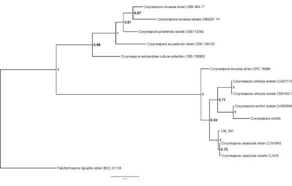
Figure 1 Bayesian phylogram generated using ITS sequence of 13 reference fungus as mentioned previously.The tree is rooted withFalciformispora lignatilisstrain BCC21118 as outgroup. The number on the nodes indicate Bayesian posterior probability based on sampling frequency of 100 for a total of 500,000 generations.
of the UM 591 isolate was confirmed by PCR amplification of the ITS gene region and ITS-based phylogeny. Homology search of UM 591ITS1sequence in UNITE database return a total of 15 alignments that matchesCorynespora cassiicola. The top three alignments belong toCorynespora cassiicolastrain 6M (Genbank:JX087444, 99% identical, bit score: 1063), Corynespora cassiicolaisolate YP59 (Genbank:FJ852716, 100% identical, bit score: 1007), andCorynespora cassiicolaisolate YP42 (Genbank:FJ852714, 100% identical, bit score: 1007). ITS1sequence ofC. citricolaCBS169.77,C. citricolaCABI211585,C. endiandrae CBS138902, C. leucadendriCBS135133, C. olivaceaCBS484.77,C. olivaceaCBS291.74, C. proliferata CBS112393, C. smithii, C. smithii CABI5649b, C. torulosa CPC15989, C. cassiicola CLN16, and C. cassiicola CLN16R2 were used to construct a phylogram withFalciformispora lignatilisstrain BCC21118 as an out-group (Fig. 1). The result from the phylogenetic analysis shows that UM 591 falls into the cluster ofC. cassiicolaspecies, within the same clade asC. smithiiandC. citricola(Fig. 1). The phylogenetic analysis has confirmed that the UM 591 belongs to the speciesC. cassiicola.
Genome feature of Corynespora cassiicolaUM 591
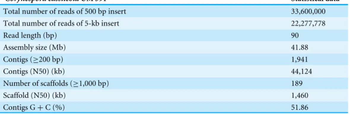
Table 1 Genome statistics ofCorynespora cassiicolaUM 591.
Corynespora cassiicolaUM 591 Statistical data
Total number of reads of 500 bp insert 33,600,000
Total number of reads of 5-kb insert 22,277,778
Read length (bp) 90
Assembly size (Mb) 41.88
Contigs (≥200 bp) 1,941
Contigs (N50) (kb) 44,124
Number of scaffolds (≥1,000 bp) 189
Scaffold (N50) (kb) 1,460
Contigs G+C (%) 51.86
Genome and gene functional annotation
The UM 591 genome contains 13,531 coding genes (>99 bp) with average gene length of 1,420 bp. The gene density in this genome is 3.23 genes per 10 kb sequences with an average of 2.79 exons per gene. A total of 33 rRNAs and 164 tRNAs were predicted in the genome. Protein sequence homology searches in NCBI (nt), Swiss-Prot, and Interpro databases return total of 8,418, 1,692, and 9,824 homologous genes, respectively. Out of the total of 8,418 genes, 6,093 genes encode for hypothetical protein based on the top hit matches in NCBI.Figure 2Asummarises the distribution of 7,962 genes in UM 591 according to classes in Gene Ontology (GO).
All the predicted proteins were subjected to KOG database to assign functional classification, which result in 7,399 matches. Top eight classifications with the highest gene counts are: [O] Posttranslational modification, protein turnover, chaperones (576), [Q] Secondary metabolites biosynthesis, transport and catabolism (555); [I] Lipid transport and metabolism (472); [C] Energy production and conversion (449); [G] Carbohydrate transport and metabolism (427); [T] Signal transduction mechanisms (423); [E] Amino acid transport and metabolism (372); and [J] Translation, ribosomal structure and biogenesis (353). These categories are mainly involved in metabolism, cellular processes and signalling, and information storage and processing (Fig. 2B). However, 1,682 genes were poorly characterised, and these genes were distributed under category [R] General function prediction only, [S] Unknown Function, and [X] Unnamed protein (Fig. 2B).
UM 591 genes are subjected to KEGG database to assign molecular pathways, resulting in the assignment of 3,100 genes to known pathways. Top five KEGG pathway with the most abundant gene distributions are carbohydrate metabolism (727), amino acid metabolism (535), lipid metabolism (389), nucleotide metabolism (277), and xenobiotics biodegradation and metabolism (264) (Fig. 2B).
BUSCO-based quality assessment
a total 1,430 (99.5%) genes, of which 62 (4.6%) were duplicated and 59 (4.4%) were fragmented. Benchmarking on the genes predicted for UM 591 showed that, of 1,438 queried, 1,222 (85.0%) present in single copy, 177 (12.3%) were duplicated while 36 (2.5%) were fragmented. The overall results indicated that the genome assembly and gene prediction of UM 591 are of high degree of completeness (only 8 and 3 BUSCO genes missing in genome and gene set, respectively).
Virulence genes
Virulence factor is briefly defined as any factors that increases the virulence of a pathogen to invade a host (Hogan, Klein & Levitz, 1996). In this predictive study, we performed homology search using PHI-base database on UM 591 predicted genes to identify genes that could be potential virulence factor. The homology search returns 483 matches to known homologs in the database.
We subjected these homology data to KOG to assign functional classification, resulting in 536 annotated genes, of which 82 genes did not fit any functional classes. The top five KOG classifications are: [X] Unnamed protein (82), [R] General function prediction only (52), [T] Signal transduction mechanisms (48), [I] Lipid transport and metabolism (28), and [O] Posttranslational modification, protein turnover, chaperones (25). Eighty-two percent (438/536) of the annotated genes are assigned into single class of functions while the remaining eighteen percent (98/536) of the genes are assigned into multiple classes of functions.
Virulence factor is one of the critical elements in evaluating pathogenicity. Thus, we screened for potential virulence factors which fulfil the criteria as follows: (A) annotated by PHI-base; and (B) implicated with reduced virulence, loss of pathogenicity, lethal, or hypervirulence status as decribed by PHI-based; and (C) localised extracellularly (secreted protein). We managed to filter out 177 potential virulent factor genes (Table S2), of which 61 of them are homologous to lethal genes (gene silencing causes death of the pathogen) and 10 are hypervirulence gene (gene silencing causes elevated pathogenicity of the pathogen). Among these genes, there are 71 putative genes that may involve in human infection because the experimental host annotated in these data are animal model (mouse or rabbit), although it is also important to note that evidence of infection in animal model does not reflect that these genes will cause infections in human. These genes are homologous to virulence gene of Aspergillus fumigatus,Cryptococcus neoformans,Exophiala dermatitidis andC. albicans(Table S2).
CAZyme analysis
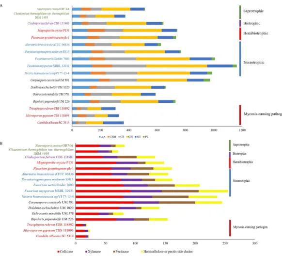
Figure 3 Comparative distribution of CAZymes according to (A) classes of CAZyme module and (B) plant cell wall degrading enzymes.PL, polysaccharide lyases; GT, glycosyl transferases; GH, glycoside hy-drolases; CE, carbohydrate esterases; CBM, carbohydrate-binding module and AA, auxiliary activity.
to CAZyme homology search using dbCAN, resulting in a total of 973 CAZymes predicted. The predicted CAZymes distribution is summarized as follows according to respective classes and gene count: auxiliary activity (AA)—189; carbohydrate esterases (CE)—193, glycoside hydrolases (GH)—354, glycosyl transferases (GT)—115, carbohydrate-binding module (CBM)—75, and polysaccharide lyases (PL)—47. We observed that CE10 (92) CAZymes are the most abundant in UM 591, followed by AA7 (66), CE1 (49), AA3(44), AA9 (41), and GH43 (28). These enzymes are involved in hydrolysis of carbohydrate and non-carbohydrate substrates (CEs and GHs), and oxidative degradation of lignin-based components of plants’ cell wall (AA) (Correia, 2010;Levasseur et al., 2013).
nutritional strategy. CAZyme distribution in UM 591 is comparable to necrotrophic and hemibiotrophic fungi (Fig. 3A). Total pectate lyases, pectinases, and hemicellulose or pectin side chains enzymes (Figs. 3Aand4B) are higher among these fungi compared to saprotrophic and biotrophic fungi. This enrichment is suggestive of UM 591, necrotrophic, and hemibiotrophic fungi in this study possess greater capacity to digest pectin. These fungi also are more enriched in glycoside hydrolases compared to the usual polysaccharide-digestive saprotrophic and biotrophic fungi (Gan et al., 2016;Glass et al., 2013;Ohm et al., 2012). Enrichment in glycoside hydrolases is not surprising in plant pathogens, but the expansion in the modular size of this class of enzyme is suggestive of exceptional plant invasive capability. The most abundant (count of >18 modules) CAZyme module in UM 591 are CE10 (92), AA7 (66), CE1 (49), AA3 (44), AA9 (41), GH3 (25), GH5 (22), GH16 (21), and GT2 (20) (Tables S4andS5). This CAZyme profile is comparable to three species of necrotrophic fungi (Fusarium oxysporum,Fusarium verticillioides, andFusarium solani teleomorphic form -Nectria haematococca) (Fig. 3AandTable S3).Figure 3Balso shows that these four species are more enriched in cell wall degrading enzymes. Our data suggest that UM 591 adopts a necrotrophic lifestyle.
There are 43 genes in UM 591 that are predicted with multiple CAZyme modules per gene (Table S6). These predicted multi-modular CAZymes may suggestive of CAZymes that are capable of more than one enzymatic digestion processes and will require actual experiment to ascertain this finding. Recently, one multi-functional recombinant GH26 enzyme derived from Mehsani buffalo rumen metagenome that is capable of mannase, xylanase, endo-glucanase, and esterase activity has been described (Patel et al., 2016). Although rare, similar multi-functional enzymes also have been reported (Bao et al., 2011; Ko et al., 2011;Zhao et al., 2010).
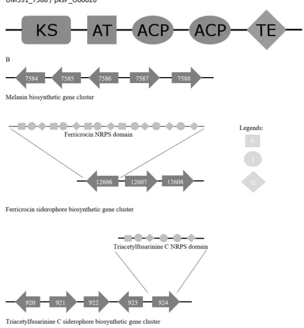
Figure 4 (A) Melanin biosynthesis PKS domain distribution ofA. fumigatusAF293 (Uniprot accession no.:O60026) and UM591_5788. (B) Representation of putative melanin and siderophore biosynthetis cluster. KS, Ketosynthase; ACP, Acyl carrier protein; AT, Acyltransferase; TE, Thioesterase; A, Adenyla-tion; T, ThiolaAdenyla-tion; C, Condensation domain.
comprehensive distribution of CAZyme modules in all reference fungal species used in this study is available inTable S4while a summary of some CAZymes of interest is available in Table S5.
detected by plant’s surface chitin receptor and impairs the plant’s immunogenic responses (Kombrink & Thomma, 2013).
Secreted CAZymes are potential virulence factors especially for plants, but to be a valid virulence factor, actual experimentation is required to validate and affirms the designation. In this section, we attempted to screen for potential virulence factors in UM 591’s CAZymes by performing homology search in PHI-base database using the predicted CAZyme genes. In addition, we pay more attention to secreted CAZymes because these enzymes are more likely to be potential virulence factors in a predictive study. Overall, nine and forty-nine CAZymes homologs that were tested on animal and plant models, respectively, were identified. Out of the nine homologs that are tested on animal models, eight genes are homologous to genes of A. fumigatus, Exophiala dermatitidis, and C. albicansthat are implicated with reduced virulence or lethal status in gene silencing experiments. Only UM591_8532 is a putative secreted GH72 CAZyme and it is homologous toA. fumigatus GEL2 virulence gene. On the other hand, only 19 homologs that were tested on plant models are implicated with reduced virulence, loss of pathogenicity, and hypervirulence status in gene silencing experiments. These homologs originate fromF.oxysporum,M. oryzae, Mycosphaerella graminicola,Monilinia fructicola,Nectria haematococca,Colletotrichum gloeosporioides,Colletotrichum coccodes,Trichoderma virens, and Cladosporium fulvum. From these nineteen genes, eleven are secreted CAZymes, of which four are predicted with high-cysteine content. The three genes that are homologous to the hypervirulence genes of Monilinia fructicolaare UM591_2724, UM591_6311, and UM591_7102. These gene encodes for CE5 cutinases which have been discussed in CAZyme section previously. A comprehensive description of virulence-related CAZymes is available inTable S2.
Secreted peptidase and lipases
Secreted peptidases play important roles in signalling, nutrition procurement, and degradation of host tissue (Ohm et al., 2012). Putative secreted peptidases are identified in two steps. First, UM 591 genes are screened for secreted peptides using SignalIP and subsequently, the secreted peptide genes are subjected to homology search using MEROPS
(Table S7). A total of 216 peptidases are annotated in UM 591, of which 69 are secreted
Huang et al., 2015). Subtilisin (SUB1 to SUB7), leucine aminopeptidase (LAP1 and LAP2), and dipeptidyl peptidase (DPPIV and DPPV) are few of the peptidases that are selectively produced (Chen et al., 2010;Huang et al., 2015). In UM 591, UM591_4361 is homologous to LAP1 (81% identical, Swiss-Prot) while UM591_11691 is homologous to LAP2 (54% identical, Swiss-Prot). Both homologs had been annotated by MEROPS as Mername-AA063 peptidase ofA. fumigatusandPseudomonas-type secreted aminopeptidase ofPseudomonas aeruginosa, respectively. Carboxypeptidases (M14A, S10) and aminopeptidases (M28A) are commonly studied in human pathogenic fungi as well and these peptidases are also identified in UM 591 (Monod et al., 2002;Sriranganadane, 2011) (Table S7). Myroilysin (UM591_9269) which digest elastin is annotated. This peptidase belongs to M10A family and it is homologous to myroilysin of deep sea bacterium Myroides profundi(Yang et al., 2015). Interestingly, UM591_9269 also matched to nematode astacin 35 peptidase (UM591_9269) of M12A family. This peptidase has been proposed to digest cuticular collagen (Park et al., 2010). Sulfite efflux pump encoded by SSU1 gene had been proposed to aid keratin digestion by transporting sulfite to keratin for cleavage of cysteine-disulfide bridges prior to keratinase digestion (Chinnapun, 2015). In UM 591, we identified two SSU1 genes (UM591_737 and UM591_10590).
Other than peptidases, two classes of peptidase inhibitors are annotated by MEROPS, which are peptidase A inhibitor 1 of I09 family (UM591_11255 and 7170) and serine carboxypeptidase Y inhibitor of I51 (UM591_2665 and 3685) family. Peptidase A inhibitor 1 inhibits subtilisin family peptidases while serine carboxypeptidase Y inhibitor inhibits some of serine endopeptidase of S1 family. Interestingly, peptidase encoded by UM591_11255 was predicted to have a subtilisin domain, which may harbour proteolytic as well as inhibiting activities. A comprehensive description of peptidases is available inTable S7.
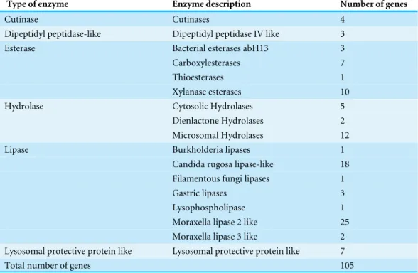
In order to identify putative lipases, UM 591 genes are subjected to homology search using Lipase Engineering database, returning with a total of 105 genes which are homologous to six types of lipases, three types of hydrolases, four types of esterases, cutinase, dipeptidyl peptidase IV-like, lysophospholipase and lysosomal protective peptide-like enzymes (Table 2).
Table 2 Summary of lipases in UM 591 retrieved from homology search using Lipase Engineering database.A comprehensive description of lipases is available inTable S8.
Type of enzyme Enzyme description Number of genes
Cutinase Cutinases 4
Dipeptidyl peptidase-like Dipeptidyl peptidase IV like 3
Esterase Bacterial esterases abH13 3
Carboxylesterases 7
Thioesterases 1
Xylanase esterases 10
Hydrolase Cytosolic Hydrolases 5
Dienlactone Hydrolases 2
Microsomal Hydrolases 12
Lipase Burkholderia lipases 1
Candida rugosa lipase-like 18 Filamentous fungi lipases 1
Gastric lipases 3
Lysophospholipase 1
Moraxella lipase 2 like 25 Moraxella lipase 3 like 2 Lysosomal protective protein like Lysosomal protective protein like 7
Total number of genes 105
on pathogen-host interaction description annotated by PHI-base and criteria (secreted protein and PHI-base virulence status) previously described in virulence gene section. Five secreted lipases are identified in UM 591 (Table S8). Three cutinase genes (UM591_2724, UM591_6311, and UM591_7102) are homologous toMfCUT1hypervirulence genes (52%, 50% and 52% identical, respectively; PHI: 2383) ofMonilinia fructicola. A hypothetical secreted Moraxella lipase 3-like gene (UM591_6979) are homologous to TMPL of Alternaria brassicola(55% identical, PHI: 2296). This gene is transmembrane protein that regulate redox homeostasis and is an important component for development of conidia in A. brassicicola(Kim et al., 2009). A putative secreted GH10 family xylanase esterase (UM591_7944) is homologous to endo-1, 4-beta-xylanase gene ofM. oryzae(63% identical, PHI: 2204).
Secondary metabolites
identified using the SMURF tool for analysis. A total of 38 secondary metabolite gene clusters were predicted based on the predicted PKS and NRPS backbone genes. Potential cluster of genes is screened manually with reference to the predicted backbone genes via descriptive searching with annotations from NCBI, Swiss-Prot, and Interpro databases. In addition to that, secondary metabolites genes which are descriptively annotated but not predicted as one of the gene clusters are investigated as well. Here, we identified putative melanin, siderophore, terpene (ent-kaurene and lycopene), and toxins (sterigmatocystin, cercosporin, HC-toxin, and gliotoxin) biosynthesis genes and clusters as summarized in Table S9.
Melanin biosynthesis
Melanin has been reported to protect fungi from UV, oxidative, and chemical stresses (Jacobson, 2000). Melanin is an important virulence factor that aid in host penetration by the generation of turgor pressure to exert pressure on plant cell wall for penetrative purpose (Howard & Ferrari, 1989). With reference toCochliobolus heterostrophusPKS18 (Eliahu et al., 2007) (NCBI accession no.:AAR90272) and three critical genes involved in melanin biosynthesis that was described inAlternaria alternata(Kimura & Tsuge, 1993), we managed to identify a putative melanin biosynthesis gene cluster in UM 591 (Table S9). The cluster (UM591_5784 to UM591_5788) consist of two unknown genes (UM591_7586 and UM591_7587) and two critical components (melanin PKS and tetrahydroxynaphthalene reductase) out of the usual six-gene cluster which has been well studied in other fungi (Eliahu et al., 2007;Pihet et al., 2009;Woo et al., 2010). The important third component, scytalone dehydratase, is predicted (UM591_76 and UM591_10618). Gene distribution in the cluster are comparatively similar to that ofMagnaporthe grisea(Eliahu et al., 2007) and PKS domain motif is the same as Pksp/Alb1 ofA. fumigatus(Fig. 4). Thus, melanin is postulated to be biosynthesized using DHN-melanin biosynthesis pathway in UM 591.
Siderophore biosynthesis
The amino acids sequences of UM591_924 and sidD (UniProt accession no:Q4WF53) (Schrettl et al., 2007) are subjected to InterproScan to elucidate the NRPS motif, and the result shows same domain pattern and distribution in both proteins.
Terpene biosynthesis
Sesquiterpenes are commonly produced by fungi and its gene distributions in the biosynthesis cluster may vary from having one core synthases to multiple synthases or having multiple cytochrome P450 genes (Wawrzyn, Bloch & Schmidt-Dannert, 2012). The backbone gene is identified by Interpro domain search for terpene cyclase conserved domain, and the gene cluster was identified by searching for presence of nearby P450 gene and neighbourhood genes that could be made up of transporter, regulatory, other synthase, and other biosynthetic genes (Wawrzyn, Bloch & Schmidt-Dannert, 2012). UM 591 genome encodes a great span of genes with P450 domain (223 genes) distanced from one to another by up to 300 genes. This allows great flexibility of non-clustered terpene/terpenoid synthesis if relevant synthase is present in the vicinity.
We identified a putativeent-kaurene and a lycopene biosynthesis cluster by screening for terpene cyclase and synthase domains. Putativeent-kaurene cluster (UM591_10047 to UM591_10055) consists of two synthase genes (UM591_10052 and UM591_10054)
(Table S9). Both genes could possibly encode for either copalyl diphosphate synthase
(CPS) or ent-kaur-16-ene synthase (KS). Copalyl diphosphate is an intermediate component for biosynthesis ofent-kaurene which, in turn, is an intermediate component of gibberellin biosynthesis (Hedden & Kamiya, 1997). Gibberellin biosynthesis pathway requires additional gene of gibberellic acid (GA4) desaturase (des) and four P450 classes of oxidases (GA14synthase (P450-1), GA20oxidase (P450-2), and C13-oxidase (P450-3)) (Rim et al., 2013) aside from genes found in anent-kaurene gene cluster.des, P450-1, P450-2, and P450-3 genes are not specifically annotated in UM 591 genome data, but there is one P450 gene (UM591_10049) within the putativeent-kaurene cluster. As discussed previously, UM 591 have a great span of P450 gene across the whole genome. Hence, the function of the other three P450 components (P450-1, P450-2, and P450-3) may be easily substituted. P450-4 and geranylgeranyl pyrophosphate (GGPP) synthase were identified about 3,000 genes downstream from the cluster (UM591_13501 and UM591_10561, respectively), which suggestive that this putative gibberellin/ent-kaurene biosynthesis may involve more than one cluster of genes. Interestingly, there is one transcription factor at each end of the cluster, suggesting that gene transcription may initiate from either direction. Based on the arrangement of genes in the putativeent-kaurene cluster (Lengeler et al., 2000), transcription initiated from the regulator at downstream is more likely whereby copalyl diphosphate is first produced from geranyl pyrophosphate catalysed by CPS followed by production ofent-kaurene catalysed by KS and subsequently, oxidation by P450 oxidases.
CarX was proposed to catalyse similar aldehyde dehydrogenation asNeurospora crassa’s ylo-1 enzyme to produce neurosporaxanthin (Avalos, Prado-Cabrero & Estrada, 2012;Jin, Lee & Lee, 2010). A light-sensing rhodopsin (carO) (Thewes et al., 2005) is identified within the cluster butcarT which is usually found in the carotenoid biosynthesis cluster (Jin, Lee & Lee, 2010;Prado-Cabrero et al., 2007) could be encoded by UM591_3196 (44% identical, Uniprot accession no:A1KQY4). This gene is identified based on amino acid identity and carotenoid domain (IPR004294) as annotated by Interpro.
Toxin biosynthesis
We predicted a great range of toxin biosynthesis pathway in the UM591 genome (Table S9). Aflatoxin is commonly affiliated to food contamination (Hammami et al., 2014) and has been extensively studied inAspergillusspp. (Yu et al., 2004). Aflatoxin and sterigmatocystin biosynthesis pathways may share majority of the biosynthesis protein since sterigmatocystin is the precursor to produce aflatoxin (Yu et al., 2004). We managed to identify majority of the aflatoxin biosynthesis pathway components including omtB/ aflO/ stcP, which produces sterigmatocystin, and ordA/ aflQ, which produces the final product aflatoxins (Yu et al., 2004). We also identified putative omtA/aflP in UM 591 by amino acid sequence identity and domain search with reference to omtA (UniProt accession no: P55790) of Aspergillus flavus, showing that UM 591 may be able to synthesize aflatoxin aside from sterigmatocystin.
A putative cercosporin cluster (UM591_7760 to UM591_7765) is identified, but the two usual cercosporin biosynthesis pathway components, CTB6 and CTB7, could not be found within this cluster of genes (Chen et al., 2007). The closest matching gene for CTB7, a FAD/FMN-dependent oxidoreductase, is UM591_1286 with reference to the known domain of CTB7 (UniProt accession no:A0ST45). CTB6 encodes for putative NADPH-dependent oxidoreductase, but there are no specific matches based on protein nomenclature, similar keywords search (such as CTB6, Cercospora, and cercosporin), or by domain matching because this protein is commonly conserved in living organisms.
The majority of core gliotoxin biosynthesis pathway components are identified in UM 591 except for P450 oxidoreductase gliF, glutathione S-transferase gliG, dipeptidase gliJ, and o-methyltransferase gliM (Gardiner & Howlett, 2005). Alternatively, we screened for possible candidate genes for these missing components by domain search with reference to gli gene cluster data fromA. fumigatus(Cramer et al., 2006;Gardiner & Howlett, 2005) and fromAspergillusand Aspergillosis Website (Atherton, 2014). Other than transcription factor, gliotoxin biosynthesis also is regulated by laeA and gipA (Dolan et al., 2015). UM591_507 (80% identical) encodes for a laeA-like peptide ofC. heterostrophus, but gipA is not found. gliZ, gliC, gliA, gliN, and gliP are identified in a cluster (UM591_12893, UM591_12898, UM591_12902, UM591_12903, and UM591_12905, respectively;Table S9) as predicted by SMURF. The missing gliF could be functionally substituted either by a probable monooxygenase (UM591_12904, 49% identical, NCBI nt accession no:
GAA90272) in the cluster or by wide ranges of P450 oxidoreductases/monooxygenases
HC-toxin was well described particularly inCochliobolusandAlternariaspecies (Wight, Labuda & Walton, 2013). Here, we predicted three probable genes encoding for HC-toxin synthetases (UM591_6434, UM591_11373, and UM591_13673). The majority of HC-toxin biosynthesis components are identified by domain search with reference to TOXD, TOXF, and TOXG (Uniprot accession no:P54006,Q9Y885, andQ9UW18, respectively). However, no match for TOXC is found, and the only fatty acid synthase beta subunit gene in UM 591 is UM591_668, which is one of the predicted components in aflatoxin biosynthesis. However, the domain annotated by Interpro for this gene is missing the starter unit ACP transacylase (IPR032088) domain that is found in TOXC/AjTOXC (Uniprot accession
no:Q92215andS5FIF0), but two additional domains, PKS acyl transferase (IPR020801)
and aldolase-type TIM barrel (IPR013785), are found in UM591_668, suggesting that this protein may serve more than one functions.
CONCLUSION
ADDITIONAL INFORMATION AND DECLARATIONS
Funding
This study was supported by High Impact Research MoE Grant UM.C/625/1/HIR/MOHE /MED/31 (Account no. H-20001-00-E000070) from the Ministry of Education Malaysia. The funders had no role in study design, data collection and analysis, decision to publish, or preparation of the manuscript.
Grant Disclosures
The following grant information was disclosed by the authors: Ministry of Education Malaysia: UM.C/625/1/HIR/MOHE/MED/31.
Competing Interests
Authors Yung-Chie Tan, Pei-Sin Chong, Jia-shiun Khoo, and Wai-Yan Yee are employed by Codon Genomics SB. There are no patents, products in development or marketed products to declare.
Author Contributions
• Hong Keat Looi analyzed the data, wrote the paper, prepared figures and/or tables.
• Yue Fen Toh and Su Mei Yew conceived and designed the experiments, performed the experiments, contributed reagents/materials/analysis tools.
• Shiang Ling Na performed the experiments, contributed reagents/materials/analysis tools.
• Yung-Chie Tan, Pei-Sin Chong, Jia-Shiun Khoo and Wai-Yan Yee analyzed the data, contributed reagents/materials/analysis tools, reviewed drafts of the paper.
• Kee Peng Ng reviewed drafts of the paper.
• Chee Sian Kuan conceived and designed the experiments, performed the experiments, contributed reagents/materials/analysis tools, reviewed drafts of the paper.
DNA Deposition
The following information was supplied regarding the deposition of DNA sequences: Whole Genome Shotgun project at NCBI/Genbank under the accession
JAQF00000000.1.
Data Availability
The following information was supplied regarding data availability: The raw data has been supplied as aSupplementary File.
Supplemental Information
Supplemental information for this article can be found online athttp://dx.doi.org/10.7717/
REFERENCES
Ahmed FA, Alam N, Khair A. 2014.Incidence and Biology ofCorynespora cassiicola (Berk. & Curt.) Wei. disease of okra in Bangladesh.Bangladesh Journal of Botany
42:265–272.
Al-Fakih AA. 2014.Overview on the fungal metabolites involved in mycopathy.Open Journal of Medical Microbiology4:38–63DOI 10.4236/ojmm.2014.41006.
Arrach N, Fernández-Martín R, Cerdá-Olmedo E, Avalos J. 2001.A single gene for lycopene cyclase, phytoene synthase, and regulation of carotene biosynthesis in Phycomyces.Proceedings of the National Academy of Sciences of the United States of America98:1687–1692DOI 10.1073/pnas.98.4.1687.
Atherton G. 2014.Cluster 27. Gliotoxin biosynthetic cluster.Available athttp:// www.
aspergillus.org.uk(accessed on 5 April 2016).
Avalos J, Limón MC. 2015.Biological roles of fungal carotenoids.Current Genetics
61:309–324DOI 10.1007/s00294-014-0454-x.
Avalos J, Prado-Cabrero A, Estrada AF. 2012. Neurosporaxanthin production by NeurosporaandFusarium. In: Barredo J-L, ed.Microbial carotenoids from fungi: methods and protocols. Totowa: Humana Press, 263–274.
Băguţ ET, Baldo A, Mathy A, Cambier L, Antoine N, Cozma V, Mignon B. 2012.
Subtilisin Sub3 is involved in adherence ofMicrosporum canisto human and animal epidermis.Veterinary Microbiology160:413–419DOI 10.1016/j.vetmic.2012.06.011.
Baldo A, Tabart J, Vermout S, Mathy A, Collard A, Losson B, Mignon B. 2008.Secreted subtilisins ofMicrosporum canisare involved in adherence of arthroconidia to feline corneocytes.Journal of Medical Microbiology57:1152–1156
DOI 10.1099/jmm.0.47827-0.
Bao L, Huang Q, Chang L, Zhou J, Lu H. 2011.Screening and characterization of a cellulase with endocellulase and exocellulase activity from yak rumen metagenome. Journal of Molecular Catalysis B: Enzymatic73:104–110
DOI 10.1016/j.molcatb.2011.08.006.
Barhoom S, Sharon A. 2004.cAMP regulation of ‘‘pathogenic’’ and ‘‘saprophytic’’ fungal spore germination.Fungal Genetics and Biology 41:317–326
DOI 10.1016/j.fgb.2003.11.011.
Bender J, Flieger A. 2010. Lipases as pathogenicity factors of bacterial pathogens of humans. In: Timmis KN, ed.Handbook of hydrocarbon and lipid microbiology. Berlin, Heidelberg: Springer Berlin Heidelberg, 3241–3258.
Benoit I, Culleton H, Zhou M, DiFalco M, Aguilar-Osorio G, Battaglia E, Bouzid O, Brouwer CP, El-Bushari HB, Coutinho PM. 2015.Closely related fungi employ diverse enzymatic strategies to degrade plant biomass.Biotechnology for Biofuels
8:107–121DOI 10.1186/s13068-015-0285-0.
Blatzer M, Schrettl M, Sarg B, Lindner HH, Pfaller K, Haas H. 2011.SidL, anAspergillus fumigatustransacetylase involved in biosynthesis of the siderophores ferricrocin and hydroxyferricrocin.Applied and Environmental Microbiology77:4959–4966
Cantarel BL, Coutinho PM, Rancurel C, Bernard T, Lombard V, Henrissat B. 2009.
The Carbohydrate-Active EnZymes database (CAZy): an expert resource for glycogenomics.Nucleic Acids Research37:D233–D238DOI 10.1093/nar/gkn663.
Casadevall A. 2007.Determinants of virulence in the pathogenic fungi.Fungal Biology Reviews21:130–132DOI 10.1016/j.fbr.2007.02.007.
Chen H, Lee MH, Daub ME, Chung KR. 2007.Molecular analysis of the cercosporin biosynthetic gene cluster inCercospora nicotianae.Molecular Microbiology
64:755–770DOI 10.1111/j.1365-2958.2007.05689.x.
Chen J, Yi J, Liu L, Yin S, Chen R, Li M, Ye C, Zhang Y-Q, Lai W. 2010.Substrate adaptation ofTrichophyton rubrumsecreted endoproteases.Microbial Pathogenesis
48:57–61DOI 10.1016/j.micpath.2009.12.001.
Chinnapun D. 2015.Virulence factors involved in pathogenicity of dermatophytes. Walailak Journal of Science and Technology12:573–580.
Collado IG, Sánchez AJM, Hanson JR. 2007.Fungal terpene metabolites: biosynthetic relationships and the control of the phytopathogenic fungusBotrytis cinerea.Natural Product Reports24:674–686DOI 10.1039/b603085h.
Conesa A, Götz S, García-Gómez JM, Terol J, Talón M, Robles M. 2005.Blast2GO: a universal tool for annotation, visualization and analysis in functional genomics research.Bioinformatics21:3674–3676DOI 10.1093/bioinformatics/bti610.
Correia MAdS. 2010.Structural and functional insights into the role of Carbohydrate Esterases and Carbohydrate-Binding Modules in plant cell wall hydrolysis. PhD thesis, Universidade Técnica de Lisboa.
Cosgrove DJ. 2005.Growth of the plant cell wall.Nature Reviews Molecular Cell Biology
6:850–861.
Cramer RA, Gamcsik MP, Brooking RM, Najvar LK, Kirkpatrick WR, Patterson TF, Balibar CJ, Graybill JR, Perfect JR, Abraham SN. 2006.Disruption of a nonribo-somal peptide synthetase inAspergillus fumigatuseliminates gliotoxin production. Eukaryotic Cell5:972–980DOI 10.1128/EC.00049-06.
Devescovi G, Bigirimana J, Degrassi G, Cabrio L, LiPuma JJ, Kim J, Hwang I, Venturi V. 2007.Involvement of a quorum-sensing-regulated lipase secreted by a clinical isolate ofBurkholderia glumaein severe disease symptoms in rice.Applied and Environmental Microbiology73:4950–4958DOI 10.1128/AEM.00105-07.
Dixon LJ, Schlub RL, Pernezny K, Datnoff LE. 2009.Host specialization and phyloge-netic diversity ofCorynespora cassiicola.Phytopathology99:1015–1027
DOI 10.1094/PHYTO-99-9-1015.
Dolan SK, O’Keeffe G, Jones GW, Doyle S. 2015.Resistance is not futile: gliotoxin biosynthesis, functionality and utility.Trends in Microbiology23:419–428
DOI 10.1016/j.tim.2015.02.005.
Eddine AN, Hannemann F, Schafer W. 2001.Cloning and expression analysis of NhL1, a gene encoding an extracellular lipase from the fungal pea pathogenNectria haema-tococcaMP VI (Fusarium solanif. sp. pisi) that is expressed in planta.Molecular Genetics and Genomics265:215–224DOI 10.1007/s004380000410.
Eliahu N, Igbaria A, Rose MS, Horwitz BA, Lev S. 2007.Melanin biosynthesis in the maize pathogenCochliobolus heterostrophusdepends on two mitogen-activated protein kinases, Chk1 and Mps1, and the transcription factor Cmr1.Eukaryotic Cell
6:421–429DOI 10.1128/EC.00264-06.
Gan P, Narusaka M, Kumakura N, Tsushima A, Takano Y, Narusaka Y, Shirasu K. 2016.Genus-wide comparative genome analyses of Colletotrichum species reveal specific gene family losses and gains during adaptation to specific infection lifestyles. Genome Biology and Evolution8:1467–1481DOI 10.1093/gbe/evw089.
Gardiner DM, Howlett BJ. 2005.Bioinformatic and expression analysis of the putative gliotoxin biosynthetic gene cluster ofAspergillus fumigatus.FEMS Microbiology Letters248:241–248DOI 10.1016/j.femsle.2005.05.046.
Glass NL, Schmoll M, Cate JH, Coradetti S. 2013.Plant cell wall deconstruction by ascomycete fungi.Annual Review of Microbiology67:477–498
DOI 10.1146/annurev-micro-092611-150044.
Haas H, Eisendle M, Turgeon BG. 2008.Siderophores in fungal physiology and viru-lence.Annual Review of Phytopathology46:149–187
DOI 10.1146/annurev.phyto.45.062806.094338.
Hammami W, Fiori S, Al Thani R, Kali NA, Balmas V, Migheli Q, Jaoua S. 2014.
Fungal and aflatoxin contamination of marketed spices.Food Control37:177–181
DOI 10.1016/j.foodcont.2013.09.027.
Hedden P, Kamiya Y. 1997.GIBBERELLIN BIOSYNTHESIS: enzymes, genes and their regulation.Annual Review of Plant Physiology and Plant Molecular Biology
48:431–460DOI 10.1146/annurev.arplant.48.1.431.
Hogan LH, Klein BS, Levitz SM. 1996.Virulence factors of medically important fungi. Clinical Microbiology Reviews9:469–488.
Howard RJ, Ferrari MA. 1989.Role of melanin in appressorium function.Experimental Mycology13:403–418DOI 10.1016/0147-5975(89)90036-4.
Howlett BJ. 2006.Secondary metabolite toxins and nutrition of plant pathogenic fungi. Current Opinion in Plant Biology9:371–375DOI 10.1016/j.pbi.2006.05.004.
Hsueh P-R. 2011. Corynespora. In: Liu D, ed.Molecular detection of human fungal pathogens. CRC Press.
Huang Y, Busk PK, Herbst F-A, Lange L. 2015.Genome and secretome analyses provide insights into keratin decomposition by novel proteases from the non-pathogenic fungusOnygena corvina.Applied Microbiology and Biotechnology99:9635–9649
DOI 10.1007/s00253-015-6805-9.
Huang H-K, Liu C-E, Liou J-H, Hsiue H-C, Hsiao C-H, Hsueh P-R. 2010.Subcutaneous infection caused byCorynespora cassiicola, a plant pathogen.Journal of Infection
Jacobson ES. 2000.Pathogenic roles for fungal melanins.Clinical Microbiology Reviews
13:708–717DOI 10.1128/CMR.13.4.708-717.2000.
Jin J-M, Lee J, Lee Y-W. 2010.Characterization of carotenoid biosynthetic genes in the ascomyceteGibberella zeae.FEMS Microbiology Letters302:197–202
DOI 10.1111/j.1574-6968.2009.01854.x.
Jones P, Binns D, Chang H-Y, Fraser M, Li W, McAnulla C, McWilliam H, Maslen J, Mitchell A, Nuka G. 2014.InterProScan 5: genome-scale protein function classification.Bioinformatics30:1236–1240DOI 10.1093/bioinformatics/btu031.
Kim K-H, Willger SD, Park S-W, Puttikamonkul S, Grahl N, Cho Y, Mukhopadhyay B, Cramer Jr RA, Lawrence CB. 2009.TmpL, a transmembrane protein required for intracellular redox homeostasis and virulence in a plant and an animal fungal pathogen.PLOS Pathogens5:e1000653DOI 10.1371/journal.ppat.1000653.
Kimura N, Tsuge T. 1993.Gene cluster involved in melanin biosynthesis of the filamen-tous fungusAlternaria alternata.Journal of Bacteriology175:4427–4435
DOI 10.1128/jb.175.14.4427-4435.1993.
Ko K-C, Han Y, Choi JH, Kim G-J, Lee S-G, Song JJ. 2011.A novel bifunctional endo-/exo-type cellulase from an anaerobic ruminal bacterium.Applied Microbiology and Biotechnology89:1453–1462DOI 10.1007/s00253-010-2949-9.
Kobayashi H, Debeaupuis J, Bouchara J, Latge J. 1993.An 88-kilodalton antigen secreted byAspergillus fumigatus.Infection and Immunity61:4767–4771.
Kohler A, Kuo A, Nagy LG, Morin E, Barry KW, Buscot F, Canbäck B, Choi C, Cichocki N, Clum A. 2015.Convergent losses of decay mechanisms and rapid turnover of symbiosis genes in mycorrhizal mutualists.Nature Genetics47:410–415
DOI 10.1038/ng.3223.
Kõljalg U, Nilsson RH, Abarenkov K, Tedersoo L, Taylor AF, Bahram M, Bates ST, Bruns TD, Bengtsson-Palme J, Callaghan TM. 2013.Towards a unified paradigm for sequence-based identification of fungi.Molecular Ecology 22:5271–5277
DOI 10.1111/mec.12481.
Kombrink A, Thomma BP. 2013.LysM effectors: secreted proteins supporting fungal life.PLOS Pathogens9:e1003769DOI 10.1371/journal.ppat.1003769.
Krogh A, Larsson B, Von Heijne G, Sonnhammer EL. 2001.Predicting transmembrane protein topology with a hidden Markov model: application to complete genomes. Journal of Molecular Biology305:567–580 DOI 10.1006/jmbi.2000.4315.
Kuan CS, Yew SM, Toh YF, Chan CL, Ngeow YF, Lee KW, Na SL, Yee W-Y, Hoh C-C, Ng KP. 2015.Dissecting the fungal biology ofBipolaris papendorfii: from phylogenetic to comparative genomic analysis.DNA Research22:219–232
DOI 10.1093/dnares/dsv007.
Lagesen K, Hallin P, Rødland EA, Stærfeldt H-H, Rognes T, Ussery DW. 2007.
RNAmmer: consistent and rapid annotation of ribosomal RNA genes.Nucleic Acids Research35:3100–3108DOI 10.1093/nar/gkm160.
Lakshmanan P, Jeyarajan R, Vidhyasekaran P. 1990.A boll rot of cotton caused byCorynespora Cassiicolain Tamil Nadu, India.Phytoparasitica18:171–173
Leite R, Barreto R. 2000.Petal spotting of hydrangea flowers caused byCorynespora cassiicola: old pathogen—new disease.Mycologist 14:80–83
DOI 10.1016/S0269-915X(00)80010-5.
Lengeler KB, Davidson RC, D’souza C, Harashima T, Shen W-C, Wang P, Pan X, Waugh M, Heitman J. 2000.Signal transduction cascades regulating fungal development and virulence.Microbiology and Molecular Biology Reviews64:746–785
DOI 10.1128/MMBR.64.4.746-785.2000.
Levasseur A, Drula E, Lombard V, Coutinho PM, Henrissat B. 2013.Expansion of the enzymatic repertoire of the CAZy database to integrate auxiliary redox enzymes. Biotechnology for Biofuels6:41–55DOI 10.1186/1754-6834-6-41.
Li M, Liang X, Rollins JA. 2012.Sclerotinia sclerotiorumγ-glutamyl transpeptidase
(Ss-Ggt1) is required for regulating glutathione accumulation and development of sclerotia and compound appressoria.Molecular Plant-Microbe Interactions
25:412–420DOI 10.1094/MPMI-06-11-0159.
Lowe TM, Eddy SR. 1997.tRNAscan-SE: a program for improved detection of transfer RNA genes in genomic sequence.Nucleic Acids Research25:955–964
DOI 10.1093/nar/25.5.0955.
Lv GX, Ge YP, Shen YN, Li M, Zhang X, Chen H, Deng S, De Hoog GS, Liu WD. 2011.
Phaeohyphomycosis caused by a plant pathogen,Corynespora cassiicola.Medical Mycology49:657–661DOI 10.3109/13693786.2011.553635.
Mahgoub E. 1969.Corynespora cassiicola, a new agent of maduromycetoma.Journal of Tropical Medicine and Hygiene72:218–221.
Monod M, Capoccia S, Léchenne B, Zaugg C, Holdom M, Jousson O. 2002.Secreted proteases from pathogenic fungi.International Journal of Medical Microbiology
292:405–419DOI 10.1078/1438-4221-00223.
Morgenstern I, Powlowski J, Tsang A. 2014.Fungal cellulose degradation by oxida-tive enzymes: from dysfunctional GH61 family to powerful lytic polysaccharide monooxygenase family.Briefings in Functional Genomics13:471–481
DOI 10.1093/bfgp/elu032.
Nghia NA, Kadir J, Sunderasan E, Abdullah MP, Malik A, Napis S. 2008. Morpho-logical and inter simple sequence repeat (ISSR) markers analyses ofCorynespora cassiicolaisolates from rubber plantations in Malaysia.Mycopathologia166:189–201
DOI 10.1007/s11046-008-9138-8.
O’Connell RJ, Thon MR, Hacquard S, Amyotte SG, Kleemann J, Torres MF, Damm U, Buiate EA, Epstein L, Alkan N. 2012.Lifestyle transitions in plant pathogenic Colletotrichumfungi deciphered by genome and transcriptome analyses.Nature Genetics44:1060–1065DOI 10.1038/ng.2372.
Onesirosan P, Mabuni C, Durbin R, Morin R, Rich D, Arny D. 1975.Toxin pro-duction byCorynespora cassiicola.Physiological Plant Pathology5:289–295
DOI 10.1016/0048-4059(75)90095-8.
Park J-O, Pan J, Möhrlen F, Schupp M-O, Johnsen R, Baillie DL, Zapf R, Moerman DG, Hutter H. 2010.Characterization of the astacin family of metalloproteases in C. elegans.BMC Developmental Biology10:1DOI 10.1186/1471-213X-10-1.
Patel AB, Patel AK, Shah MP, Parikh IK, Joshi CG. 2016.Isolation and characterization of novel multifunctional recombinant family 26 glycoside hydrolase from Mehsani buffalo rumen metagenome.Biotechnology and Applied Biochemistry63:257–265
DOI 10.1002/bab.1358.
Petersen TN, Brunak S, Von Heijne G, Nielsen H. 2011.SignalP 4.0: discriminating signal peptides from transmembrane regions.Nature Methods8:785–786
DOI 10.1038/nmeth.1701.
Pihet M, Vandeputte P, Tronchin G, Renier G, Saulnier P, Georgeault S, Mallet R, Chabasse D, Symoens F, Bouchara J-P. 2009.Melanin is an essential component for the integrity of the cell wall ofAspergillus fumigatusconidia.BMC Microbiology
9:1DOI 10.1186/1471-2180-9-1.
Prado-Cabrero A, Estrada AF, Al-Babili S, Avalos J. 2007.Identification and biochem-ical characterization of a novel carotenoid oxygenase: elucidation of the cleavage step in theFusariumcarotenoid pathway.Molecular Microbiology 64:448–460
DOI 10.1111/j.1365-2958.2007.05665.x.
Qi Y-X, Zhang X, Pu J-J, Liu X-M, Lu Y, Zhang H, Zhang H-Q, Lv Y-C, Xie Y-X. 2011.Morphological and molecular analysis of genetic variability within isolates ofCorynespora cassiicolafrom different hosts.European Journal of Plant Pathology
130:83–95DOI 10.1007/s10658-010-9734-6.
Quinlan AR, Hall IM. 2010.BEDTools: a flexible suite of utilities for comparing genomic features.Bioinformatics26:841–842DOI 10.1093/bioinformatics/btq033.
Rawlings ND, Barrett AJ, Bateman A. 2012.MEROPS: the database of proteolytic enzymes, their substrates and inhibitors.Nucleic Acids Research40:343–350
DOI 10.1093/nar/gkr987.
Reichard U, Léchenne B, Asif AR, Streit F, Grouzmann E, Jousson O, Monod M. 2006.
Sedolisins, a new class of secreted proteases fromAspergillus fumigatuswith endo-protease or tripeptidyl-peptidase activity at acidic pHs.Applied and Environmental Microbiology72:1739–1748DOI 10.1128/AEM.72.3.1739-1748.2006.
Revankar SG, Sutton DA. 2010.Melanized fungi in human disease.Clinical Microbiology Reviews23:884–928DOI 10.1128/CMR.00019-10.
Rim SO, Yoon HJ, Lee JH, Kim CM, Kim JG. 2013.Characterization of gibberellin biosynthetic gene cluster fromFusarium proliferatum.Journal of Microbiology and Biotechnology23:623–629DOI 10.4014/jmb.1212.12029.
Ronquist F, Huelsenbeck JP. 2003.MrBayes 3: Bayesian phylogenetic inference under mixed models.Bioinformatics19:1572–1574DOI 10.1093/bioinformatics/btg180.
siderophores duringAspergillus fumigatusinfection.PLOS Pathogens3:e128
DOI 10.1371/journal.ppat.0030128.
Seaman W, Shoemaker R, Peterson E. 1965.Pathogenicity ofCorynespora cassiicolaon soybean.Canadian Journal of Botany43:1461–1469DOI 10.1139/b65-154.
Simão FA, Waterhouse RM, Ioannidis P, Kriventseva EV, Zdobnov EM. 2015.BUSCO: assessing genome assembly and annotation completeness with single-copy orthologs. Bioinformatics31(19):3210–3212DOI 10.1093/bioinformatics/btv351.
Smart MG. 1991. The plant cell wall as a barrier to fungal invasion. In: Cole GT, Hoch HC, eds.The fungal spore and disease initiation in plants and animals. Boston: Springer US, 47–66.
Sriranganadane D. 2011.Comparison of proteolytic system secreted in dermatophytes and ‘Aspergillus fumigatus’, used as a reference. PhD thesis, Université de Neuchâtel.
Tatusov RL, Fedorova ND, Jackson JD, Jacobs AR, Kiryutin B, Koonin EV, Krylov DM, Mazumder R, Mekhedov SL, Nikolskaya AN. 2003.The COG database: an updated version includes eukaryotes.BMC Bioinformatics4:1DOI 10.1186/1471-2105-4-1.
Ter-Hovhannisyan V, Lomsadze A, Chernoff YO, Borodovsky M. 2008.Gene pre-diction in novel fungal genomes using an ab initio algorithm with unsupervised training.Genome Research18:1979–1990DOI 10.1101/gr.081612.108.
Thewes S, Prado-Cabrero A, Prado M, Tudzynski B, Avalos J. 2005.Characterization of a gene in the car cluster ofFusarium fujikuroithat codes for a protein of the carotenoid oxygenase family.Molecular Genetics and Genomics274:217–228
DOI 10.1007/s00438-005-0015-6.
Turgeon BG, Bushley K. 2009. Secondary metabolism. In: Borkovich K, Ebbole D, Momany M, eds.Cellular and molecular biology of filamentous fungi. Washington, D.C.: ASM Press, 376–395.
Voigt CA, Schäfer W, Salomon S. 2005.A secreted lipase ofFusarium graminearumis a virulence factor required for infection of cereals.The Plant Journal42:364–375
DOI 10.1111/j.1365-313X.2005.02377.x.
Wang X, Wang W, Lin Z, Wang X, Li T, Yu J, Liu W, Tong Z, Xu Y, Zhang J. 2014.
CARD9 mutations linked to subcutaneous phaeohyphomycosis and TH17 cell deficiencies.The Journal of Allergy and Clinical Immunology133:905–908
DOI 10.1016/j.jaci.2013.09.033.
Wawrzyn GT, Bloch SE, Schmidt-Dannert C. 2012.5 discovery and characterization of terpenoid biosynthetic pathways of fungi.Methods in Enzymology515:83–105
DOI 10.1016/B978-0-12-394290-6.00005-7.
Wight WD, Labuda R, Walton JD. 2013.Conservation of the genes for HC-toxin biosynthesis inAlternaria jesenskae.BMC Microbiology13:165
DOI 10.1186/1471-2180-13-165.
Woo PC, Tam EW, Chong KT, Cai JJ, Tung ET, Ngan AH, Lau SK, Yuen KY. 2010.High diversity of polyketide synthase genes and the melanin biosyn-thesis gene cluster inPenicillium marneffei.FEBS Journal277:3750–3758
Yamada H, Takahashi N, Hori N, Asano Y, Mochizuki K, Ohkusu K, Nishimura K. 2013.Rare case of fungal keratitis caused byCorynespora cassiicola.Journal of Infection and Chemotherapy19:1167–1169DOI 10.1007/s10156-013-0579-8.
Yang J, Zhao H-L, Tang B-L, Chen X-L, Su H-N, Zhang X-Y, Song X-Y, Zhou B-C, Xie B-B, Weiss AS. 2015.Mechanistic insight into the elastin degradation process by the metalloprotease myroilysin from the deep-seaBacterium Myroides profundiD25. Marine Drugs13:1481–1496DOI 10.3390/md13031481.
Yew SM, Chan CL, Lee KW, Na SL, Tan R, Hoh C-C, Yee W-Y, Ngeow YF, Ng KP. 2014.
A five-year survey of dematiaceous fungi in a tropical hospital reveals potential opportunistic species.PLOS ONE9:e104352DOI 10.1371/journal.pone.0104352.
Yi F, Chew F, Lim S, Tan H, Tan T, Lee B. 2000.982 Characterisation of the allergenic components in spores of fungiExserohilum rostratumandCorynespora casiicola [Abstract 982].Journal of Allergy and Clinical Immunology105:S333.
Yin Y, Mao X, Yang J, Chen X, Mao F, Xu Y. 2012.dbCAN: a web resource for automated carbohydrate-active enzyme annotation.Nucleic Acids Research
40:W445–W451DOI 10.1093/nar/gks479.
Yu J, Chang P-K, Ehrlich KC, Cary JW, Bhatnagar D, Cleveland TE, Payne GA, Linz JE, Woloshuk CP, Bennett JW. 2004.Clustered pathway genes in afla-toxin biosynthesis.Applied and Environmental Microbiology70:1253–1262
DOI 10.1128/AEM.70.3.1253-1262.2004.
Zhao Z, Liu H, Wang C, Xu J-R. 2014.Erratum to: comparative analysis of fungal genomes reveals different plant cell wall degrading capacity in fungi.BMC Genomics
15:1DOI 10.1186/1471-2164-15-1.
Zhao S, Wang J, Bu D, Liu K, Zhu Y, Dong Z, Yu Z. 2010.Novel glycoside hydrolases identified by screening a Chinese Holstein dairy cow rumen-derived metagenome library.Applied and Environmental Microbiology 76:6701–6705
![Figure 2 UM 591 gene functions assigned by (A) KOG, and (B) KEGG categories. Legends: [A] RNA processing and modification; [B] Chromatin structure and dynamics; [C] Energy production and con-version; [D] Cell cycle control, cell division, chromosome parti](https://thumb-eu.123doks.com/thumbv2/123dok_br/18142781.326645/8.918.274.837.120.798/functions-categories-processing-modification-chromatin-structure-production-chromosome.webp)