Insight into the Evolution of Variable Chromosomal
Virulence Factors in
Staphylococcus aureus
Shinya Watanabe1¤a, Teruyo Ito1,2*, Takashi Sasaki1, Shanshuang Li1, Ikuo Uchiyama3, Kozue Kishii1¤b, Ken Kikuchi1,2, Robert Leo Skov4, Keiichi Hiramatsu1,2
1Department of Infection Control Science, Graduate School of Medicine, Juntendo University, Bunkyo-ku, Tokyo, Japan,2Department of Bacteriology, School of Medicine, Juntendo University, Bunkyo-ku, Tokyo, Japan,3National Institute for Basic Biology, National Institutes of Natural Sciences, Myodaiji, Okazaki, Japan,4National Center for Antimicrobials and Infection Control, Statens Serum Institut, Copenhagen, Denmark
Abstract
Background: The production of staphylocoagulase (SC) causing the plasma coagulation is one of the important characteristics ofStaphylococcus aureus. Although SCs have been classified into 10 serotypes based on the differences in the antigenicity, genetic bases for their diversities and relatedness to chromosome types are poorly understood.
Methodology/Principal Findings: We compared the nucleotide sequences of 105 SC genes (coa), 59 of which were determined in this study. D1 regions, which contain prothrombin-activating and -binding domains and are presumed to be the binding site of each type-specific antiserum, were classified into twelve clusters having more than 90% nucleotide identities, resulting to create two novel SC types, XI and XII, in addition to extant 10 types. Nine of the twelve SC types were further subdivided into subtypes based on the differences of the D2 or the central regions. The phylogenetical relations of the D1 regions did not correlate exactly with either one ofagr types and multilocus sequence types (STs). In addition, genetic analysis showed that recombination events have occurred in and aroundcoa. So far tested, STs of 126S. aureus strains correspond to the combination of SC type andagrtype except for the cases of CC1 and CC8, which contained two and three different SC types, respectively.
Conclusion: The data suggested that the evolution of coa was not monophyletic in the species. Chromosomal recombination had occurred atcoaand agr loci, resulting in the carriage of the combinations of allotypically different important virulence determinants in staphylococcal chromosome.
Citation:Watanabe S, Ito T, Sasaki T, Li S, Uchiyama I, et al. (2009) Genetic Diversity of Staphylocoagulase Genes (coa): Insight into the Evolution of Variable Chromosomal Virulence Factors inStaphylococcus aureus. PLoS ONE 4(5): e5714. doi:10.1371/journal.pone.0005714
Editor:Christophe Herman, Baylor College of Medicine, United States of America
ReceivedDecember 8, 2008;AcceptedApril 23, 2009;PublishedMay 27, 2009
Copyright:ß2009 Watanabe et al. This is an open-access article distributed under the terms of the Creative Commons Attribution License, which permits unrestricted use, distribution, and reproduction in any medium, provided the original author and source are credited.
Funding:This work was supported by a Grant-in-Aid for 21st Century COE Research and Grant-in-Aid for Scientific Research on Priority Areas (13226114) from The Ministry of Education, Science, Sports, Culture and Technology of Japan. S.W. was supported by a Research Fellowships of the Japan Society for the Promotion of Science for Young Scientists (18-53122). The funders had no role in study design, data collection and analysis, decision to publish, or preparation of the manuscript.
Competing Interests:The authors have declared that no competing interests exist.
* E-mail: [email protected]
¤a Current address: Tuberculosis Research Section, Laboratory of Clinical Infectious Disease, National Institute of Allergy and Infectious Disease, National Institutes of Health, Bethesda, Maryland, United States of America
¤b Current address: Laboratory of Molecular Epidemiology for Infectious Agents, Kitasato Institute for Life Sciences, Kitasato University, Minato-ku, Tokyo, Japan
Introduction
Staphylococcus aureus is a persistent resident of the nasal membrane and skin of warm-blooded animals, and a major causative agent of hospital and community-associated infections. Staphylocoagulase (SC) that causes coagulation of plasma is one of the extracellular virulence factors produced by S. aureus strains, and is regarded as a marker for discriminatingS. aureusfrom other less pathogenetic staphylococci called as coagulase-negative staphylococci.
SC causes the coagulation of plasma without the usual proteolytic cleavages caused by factor Xa. SC binds to prothrombin and the complex of SC and prothrombin induces plasma coagulation by converting fibrinogen into fibrin [1,2].
regions were rather diverse, whereas the central regions were relatively conserved. Since identities of both nucleotide and amino acid in the D2 regions were higher than those in the D1 regions, it has been suggested that the D1 region might be responsible for the antibody recognition site for type specific antiserum [5].
In Japan, SC serotyping has been applied to epidemiological study of S. aureus isolates. However, this method has not been widely used in other countries. It seemed that it was regarded as time-consuming and laborious method or that researchers were unaware of the method. Therefore, we recently developed multiplex PCRs (M-PCRs) to classify SC types simply and rapidly [7]. So far as tested, the results of the M-PCRs correlated well to those of serotyping.
Accessory gene regulator (agr) operon, well known regulatory system inS. aureusstrains, is composed of a large set of genes,agrA,
agrC,agrD,agrB, and RNAIII. Since variation exists in the regions from agrCto agrB, agrtyping can be done by either PCR or by determining nucleotide sequences of agrD. Four agr types are reported, and used for epidemiological classification of S. aureus
isolates [8]. A more extensive typing method, multilocus sequence typing (MLST), has been widely used, which assigns every strain with a sequence type (ST) [9]. By using eBURST program, phylogenet-ically related STs can be grouped as a clonal complex (CC) [10].
In this paper, we studied correlation of the two typing methods with the SC typing by using 105 S. aureus strains with theircoa
sequenced and 21 strains whose SC types were determined by the M-PCRs and serotyping. Analysis for recombination events among coa with their flanking regions suggested that horizontal genetic transfer in and aroundcoahave occurred amongS. aureus
stains.
Materials and Methods
Bacterial strains and culture conditions
A total of 126 methicillin-resistant and susceptible S. aureus
strains (MRSA and MSSA) from various categories were used in this study including 70 MRSA (64 from humans and 6 from cats) and 40 methicillin-susceptibleS. aureus (MSSA) strains (11 from humans and 29 from cats and cows) (table S1) [3,4,7,11–19].S. aureusstrains were cultivated at 37uC in tryptic soy broth or on tryptic soy agar (Becton Dickinson CO., Ltd.), and stored at 280uC in 50% glycerol.
We also used the nucleotide sequences of two coa partially-sequenced strains isolated in Hokkaido in Japan [20] and of fourteen whole genome-sequenced strains [21–31] obtained from the DDBJ/EMBL/GenBank databases.
SC typing of staphylocoagulase
Serotying: The Serotypes of SCs were determined by the inhibition test for coagulation of plasma using commercially available specific antibodies to type I to VIII SCs of S. aureus
(Denka Seiken Co., Ltd., Tokyo, Japan). We did not determine type IX and type X SCs since antisera to the SC types were not commercially available [5].
M-PCRs: The SC types were determined by M-PCRs consisting of two sets of primers identifying types I–VIIIcoa[7]. M-PCR set A and set B contained primers for identifying thecoaof type III, IV, VII and VIII, andfemAas an internal positive and primers identifying thecoaof type I, II, V, and VI, andfemA, respectively. DNA preparation
Chromosomal DNA was extracted using Isoprant (Nippon Gene Co,. Ltd., Tokyo, Japan) [5]. 800mL of the overnight culture in tryptic soy broth was poured into a tube, and cells were collected by centrifugation. The pellet was re-suspended with 400mL of SMM buffer (0.5 M sucrose, 0.1 M disodium maleate, 0.002 M MgCl2: pH6.5), 20mL of 2 mg/ml lysostaphin (Wako Pure Chemical Industries, Ltd, Osaka, Japan) was added, and then the mixture was left at 37uC for more than 20 min until protoplasts were formed. The protoplasts were collected by centrifugation, and their chromosomal DNAs were extracted by Isoparant as recommended by the manufacture.
DNA sequencing ofcoa
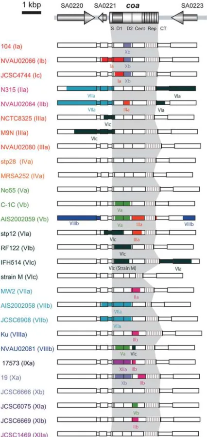
coa was amplified by PCR using chromosomal DNA as a template. DNA fragments were amplified using primer sets: coa-F-1 (59 -TAATGTAGATTGGGCAATTACA-39) and coa2 (59 -ATGCTTTAATTCAGTTAGAAGC-39). The reaction mixtures consisted of 50-mL total volume solutions containing 10 ng of genomic DNA, 5 pmol of each primer, 0.2 U of Ex Taq DNA polymerase (Takara Bio Inc., Shiga, Japan), 5mL of ExTaqBuffer and 10 mM of each deoxynucleotide triphosphate. Amplification was performed with Thermal Cycler Dice (Takara Bio Inc.), and parameters were 30 cycles of 30 s at 94uC, 1 min at 55uC and 2 min at 72uC, and hold at 4uC. The PCR products were purified using a High Pure PCR product purification kit (Roche Molecular System Inc.). Nucleotide sequencing was carried out using BigDye Figure 1. Stracture of SC and its gene locus. A. Domain
organization of SC inS. aureusN315. S, signal sequence; D1, D1 region; D2, D2 region; Cent, central region, Rep, repeat region; CT, C-terminal region.B.Organization of thecoa-flanking region and the loci ofcoa, agroperon and MLST genes inS. aureusN315 genome.
Terminator version 3.1 Cycle Sequencing Kit (Applied Biosys-tems, CA, USA) and 3730 DNA Analyzer (Applied BiosysBiosys-tems, CA, USA and Hitachi, Ltd., Tokyo Japan) according to the manufacturer’s instructions. The flanking regions of coa were determined as described previously [5].
MLST
MLST was preformed as described previously [9]. CC was determined by eBURST analysis [10]. It was defined as a group in which the STs were identical in sequence at six of seven MLST genes (arcC,aroE,glpF,gmk,pta,tpiandyqiL) to at least one other ST in the group.
agr typing
agrtypes were determined using two sets of M-PCRs reported previously [8].
Bioinformatic analysis
Genetic analysis was mainly carried out using programs in GENETYX-MAC Version 13.0.3 (GENETYX Corporation, Tokyo, Japan).The nucleotide sequences of coaor concatenated fragments of MLST genes were aligned using the clustalW Version 1.83 program with 1000 times bootstrapping [32] and neighbor-joining analysis was carried out with Tajima-Nei parameter model. Phylogenetic trees were visualized with TreeView X program [33]. Recombinations in and around coawere detected using the RDP3 Beta 34 software [34]. We used six automated recombination detection methods, RDP [35], Geneconv [36], Bootscanning [37], Maximum Chi Square (MaxChi) [38], Chimaera [39], and Sister Scanning (SiScan) [40]. Results were verified by visual inspection.
Nucleotide sequence accession numbers
The nucleotide sequences of coaand their flanking sequences used in this study have been deposited in the DDBJ/EMBL/ GenBank databases under accession numbers as follows: AB436972-AB436988 (17 entries), AB437138, AB488498-AB488510 (13 entries), and AB489873- AB489901 (29 entries).
Results
Common structures shared among 103 SCs
To understand the overall structures as well as their diversities ofcoainS. aureus, we listed 103 strains and conducted experimental characterization of SCs of 100 strains by serotyping and M-PCRs except for three strains of which we could use sequence data only. By serotyping, 78 strains were classified into either one of eight serotypes, but 7 strains could not be classified into one of eight serotypes and 15 strains could not be tested since coagulation of plasma could not be observed for up to 48 h incubation (table S1). With M-PCRs,coaof 88 strains were classified into one of eight types, leaving that of 12 strains still unclassifiable. We determined nucleotide sequences of coa of 59 strains that contained SC-nontypeable strains by both serotyping and M-PCRs, and that were chosen mostly based on genetic backgrounds distinct from 10 reference strains of SC types and othercoa-sequenced strains. The structures of these 59coawere analyzed together with the extant 44
coa: 30 of coa-sequenced strains and 14 of whole genome-sequenced stains. The overall structures of coa and other characteristics of the 103coa-sequenced strains includingagrtypes and STs are shown in figure 1 and table S1, respectively.
Allcoaare composed of the 6 regions. The 59end of 78-bp and the 39end of 18-bp are identical among all the genes except for a
coa that carried single nucleotide silent mutation in the 59 end.
They carried D1, D2, and central regions, other than the case of strain M, of whichcoahad a 140 amino acids deletion spanning from the D2 region to the central region. Although all of them carried 81-bp tandem repeats at the 39terminus, the numbers of repeats were diverse ranging from one (TSCC26) to nine (NVAU02070, NVAU02080 and Ku). The sizes of coa ranged from the smallest one, 1404 bp (strain M) to the largest one, 2280 bp (Ku). Except forcoaof strain M, the differences in sizes were mostly due to the number of 81-bp tandem repeats.
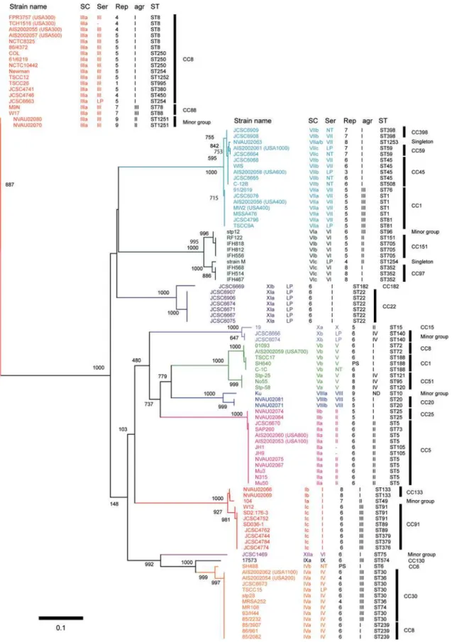
SC types based on the differences among the D1 regions Historically, SCs have been classified into 10 serotypes based on the differences in antigenicity by inhibition test using type-specific antibodies against each type of staphylocoagulase proteins. Since our previous study suggested the D1 regions might be the major antibody recognition site for type-specific antibodies [5], we conducted nucleotide and amino acid sequence comparison among 103 genes and 2 partially-sequenced coa. Phylogenetic trees were created to compare the relationship between serotypes and nucleotide diversities in the D1 regions (figure 2) as well as amino acid diversities (data not shown).
The D1 regions of the 105 coa were classified into 12 major clusters and those belonging to each cluster showed nucleotide identities with more than 90% (figure 2). Since each one of 10 clusters contained one of the 10 SC reference strains, it was inferred that these 10 clusters represented extant 10 serotypes. Consequently, we regarded other two additional clusters as new types and designated them as types XI and type XII SC, respectively. As we describe bellow, a group including seven strains with ST22 or ST182 and withagrtype I had type XI SC, and one strain with ST75 andagrtype I had type XII SC. The average of nucleotide identity among thecoaD1 regions of all the 12 SC types was 67.1%, ranging from the highest (89.0%, type IV vs type IX) to the lowest (58.8%, type III vs type IX) (table 1).
SC subtypes classified based on the differences in either D2 or central region
We previously reported that type VI SCs were classified into three subtypes (types VIa, VIb and VIc) based on the nucleotide differences in the D2 or the central regions [7]. Although the three subtypes of type VI SCs were highly homologous with nucleotide identities of more than 96.6% in the D1 region, they differed considerably in the D2 and the central region (figure 2 and table S2). If we adopt the criteria that an SC type should be assigned with more than 90% nucleotide identities of the D1 regions, and these SC types should be classified further into distinct subtypes with more than 90% nucleotide identities in both the D2 and the central region, nine of 12 SCs were further subdivided into several subtypes. Two SC types (I and VII) can thus be subdivided into three subtypes, and six SC types (II, IV, V, VIII, X and XI) into two subtypes (table S2). The comparisons of nucleotide identities of the D1, the D2, and the central regions of the different subtypes are shown in table S2.
Figure 2. Phylogenetic relationship among the nucleotide sequences of the D1 regions of 105coa.The Neighbor-Joining (NJ) tree was constructed using Clustral W and TreeView. The numbers at nodes refer to bootstrap replicates out of 1000 that support the node. Abbreviations are as follows: SC, the type of stapylocoagulase; Ser, serotype of staphylocoagulase; Rep, a number of 81-bp tandem-repeat units ofcoa;agr,agrtype; ST, multilocus sequence type; LP in Serotype section, Staphylocoagulase production was too low to clot serum within 48 hr; NT in Serotype section, Non typable; ‘‘-’’ in Serotype section, Not tested; PS in Rep section, Partially sequenced; ND inagrsection, Not detected by M-PCR foragr.
In contrast to the phylogenetic tree of the D1 regions, in which strains of the same SC types clustered together, phylogenetic tree of thecoaflanking regions showed that strains harboring thecoaof the same SC types did not always belonged to the same cluster, but
distributed in the different clusters. For example, thecoaflanking regions of N315 (SC type IIa), was split from that of NVAU02064 (type IIb), but closely related to that of stp12 (type VIa). Instead, the coa flanking regions rather phylogenetically correlated to Table 1.Identities of nucleotide sequences of D1 regions ofcoaand their deduced amino acid sequences among 12 SC types.
strain
name SC type Ia IIa IIIa IVa Va VIa VIIa VIIIa IXa Xa XIa XIIa
104 Ia 53.7% 51.7% 57.7% 56.4% 57.7% 57.0% 60.4% 58.4% 57.0% 50.3% 57.7%
N315 IIa 64.4% 51.0% 50.3% 59.0% 51.4% 51.7% 61.7% 51.0% 51.7% 60.3% 55.7%
NCTC8325 IIIa 63.8% 63.1% 53.0% 41.6% 48.3% 53.0% 45.0% 55.0% 44.3% 53.7% 55.7%
stp28 IVa 67.6% 64.2% 65.5% 54.4% 49.7% 50.3% 55.0% 85.2% 45.6% 48.3% 70.5%
No55 Va 65.8% 70.5% 58.8% 66.9% 54.7% 46.3% 66.4% 54.4% 52.3% 53.4% 53.0%
stp12 VIa 67.6% 67.7% 66.4% 64.7% 66.3% 54.4% 59.1% 52.3% 51.0% 58.8% 49.7%
MW2 VIIa 69.1% 61.7% 64.4% 63.3% 63.5% 68.8% 51.7% 48.3% 54.4% 51.0% 50.3%
Ku VIIIa 71.1% 70.5% 62.5% 66.6% 72.9% 69.5% 65.5% 56.4% 57.0% 59.7% 55.0%
17573 IXa 70.3% 66.2% 67.0% 89.0% 66.2% 68.6% 63.7% 69.6% 47.7% 49.7% 73.8%
19 Xa 66.2% 67.0% 64.9% 61.6% 66.0% 66.1% 67.2% 70.0% 63.8% 53.7% 46.3%
JCSC6671 XIa 65.0% 70.4% 67.8% 62.2% 68.0% 71.2% 68.2% 69.3% 64.4% 69.6% 55.0%
JCSC1469 XIIa 67.6% 65.8% 64.9% 76.7% 68.2% 63.5% 63.3% 66.3% 77.8% 64.2% 65.3%
Identities of nucleotide sequences of D1 regions ofcoaand their deduced amino acid sequences among 12 SC types.
Nucleotide identities are shown in cells in the bottom left half of the table and amino acid identities are shown in cells in the upper right half of the table. doi:10.1371/journal.pone.0005714.t001
Figure 3. Phylogenetic relationship among thecoaflanking regions of 29S. aureusstrains.The sequences of the regions spanning from ORF encoding hypothetical protein corresponding to glycerophosphodiester phosphodiesterase (SA0220) to ORF corresponding to acetyl-CoA acetyltransferase homologue (SA0223) apart fromcoaare used. The numbers at nodes refer to bootstrap replicates out of 1000 that support the node. Abbreviations are as follows: SC, stapylocoagulase (SC) type;agr,agrtype; ST, multilocus sequence type; ND inagrsection, Not detected by M-PCR foragr.
housekeeping genes examined by MLST as shown below, suggesting that coa evolved independently from other genes in the genome.
Evidences for recombination amongcoaand its flanking regions
The D1 region ofcoapresented much diversity, so did in a less degree the D2 and central regions. To know whether recombi-nation played a role in the generation of the diversity, we sought evidence for recombination at thecoalocus using RDP3 Beta 34 software. With the data set of 28 nucleotide sequences of the region spanning from SA0220 to SA0223, from which nucleotide sequences of the repeat regions were removed, 38 recombination events were predicted by the program. Among them, we listed 22 putative recombination events that were detected by more than 3 of six programs with statistical significance P,1025 and were verified by visual inspection (figure 4). The DNA regions derived from a minor parent were identified by RDP3 and the data were confirmed by visual inspection, too. However, there are still possibilities that the relations between a minor parent and a recombinant might be replaced as suggested by the program.
The data clearly indicated that recombination have occurred in or aroundcoaloci because all of them showed very low P values and were detected by most of the six recombination-detecting program. For example, the case of strain 17573 that showed the lowest P-value (6.93610267 in RDP, 2.52610217 in Geneconv, 2.76610227in Bootscan, 1.34610225in MaxChi, 7.33610229in Chimaera and 2.96610219 in SiScan) among those of all predicted recombination events. The 796-bp region spanning from D1 region to D2 region in 17573 (SC type, IX) was replaced by that of JCSC1469 (SC type, XII). That is consistent with the fact that the D1 region of 17573 was relatively similar to that of JCSC1469 (figure 2) although MLST allele of 17573 was phylogenetically distinct from that of JCSC1469 (figure 5).
Most of the recombination events have occurred withincoaloci. In some case, the recombination regions involving the D1 regions were replaced those of other background and the recombination seems to create new SC subtypes. For example, the 1005-bp region spanning from SA0221 to the D1 regions ofcoaof NVAU02066 (SC type Ib) and the 413-bp region involving the D1 region of JCSC4744 (SC type Ic) were replaced by that of 104 (SC type Ia). The recombination might contribute to the variation ofcoa. Similar events might have occurred in SC V, VI, VII and X strains (figure 4).
The relationship between SC types and MLST allelic profiles
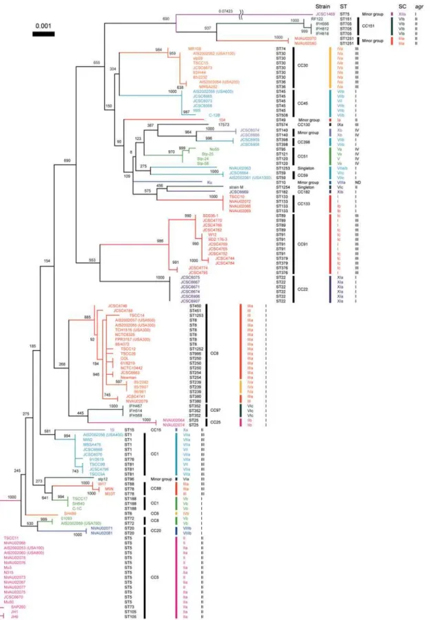
To gain an insight into the relatedness between SC types and MLST allelic profiles, we created a phylogenetic tree based on concatenated sequences of seven housekeeping genes used for MLST and compared their relations to SC types (figure 5). We used a total of 126 S. aureus strains (table S1), which were composed of 105 coa-sequenced strains described above and 21 strains whose SC types were determined by the M-PCRs [7].
Most of strains belonging to a given CC carried acoa of the same SC type except for the cases of two CCs. The strains belonging to CC1 carried two SC types, VIIa and Vb and those belonging to CC8 carried three SC types, IIIa, IVa and Vb. However, when looking at the relationship between ST and SC type, we found strains of a given ST type carried a specified SC type: ST8, type IIIa; ST239, type IVa; and ST72, Vb.
Although each cluster composed of a single CC was occupied by strains of the same SC type, strains of the same SC type but different subtype did not always show high phylogenetical
relatedness to MLST genes (figure 5). In other words, some strains with a given D1-based SC type belonged to phylogenet-ically distinct CCs which were remote in the phylogenetic tree, e.g., SC type IIa belonged to CC5 and SC type IIb belonged to CC25. It suggested that multiple recombination includingcoaloci have occurred inS. aureuschromosome.
Strain JCSC1469, which was isolated in Australia [17], was considered to be an orphan isolate on the basis of the MLST tree (ST75), and did not cluster with any strains used in this study. It was assigned with a novel SC type XIIa, which was moderately related to SC type IX with 77.8% of the nucleotides identities in the D1 regions.
Comparing phylogenetic correlation between MLST genes and
coaflanking regions, we found that they were relatively similar to each other. Some pairs of strains with different SC types were closely related in both trees of MLST genes and coa flanking regions, for example JCSC6075 (SC XIa) and JCSC4744 (SC Ic), AIS2002059 (SC Vb) and NVAU02081 (SC VIIIb), RF122 (SC VIb) and NVAU02080 (SC IIIa), JCSC6908 (SC VIIb) and JCSC6666 (SC Xb), and JCSC6669 (SC XIb) and Ku (SC VIIIa) (figure 3 and 5).
The relationship amongagrtypes, SC types and ST alleles Furthermore,agr types of the 126 strains were determined by M-PCRs [8] to investigate relations amongagrtypes, SC types and ST alleles. One hundred and twenty five strains could be classified into one of the four alleles ofagr, leaving only one untypeable strain Ku. Similar to the case with SC typing, all the strains of a given CC were classified into the sameagrgroup. Although each
agrtype was not assembled in the MLST tree, the data confirmed the previously described findings [41]. However, when we have examined the phylogenetic relationship betweenagrtype and SC type, we found that strains belonging to a given agr type were classified into several SC types. In other words, a SC type did not always correlate to a specificagrtype (figure 5 and Table S1). The
agrtype I strains harbored multiple SC types such as I–VIII, XI and XII. Theagrtype II strains contained such SC types as I, II, III VI and X,agrtype III strains contained strains of SC types I, III, IV, VI, VII and IX, andagrtype IV strains contained strains of SC types, V and X, respectively. Our data showed that a given CC belong to a SC type and anagrgroup so far tested, suggesting that a CC could be inferred by the combination of SC type andagrtype that can be determined by PCRs rather easily.
Discussion
We ascertained the common structure ofcoaby analysis of the 105coasequences. The clustering analysis of the D1 regions of the
coashowed that they were classified into 12 clusters. Ten of the 12 clusters were correlated with extant 10 serotypes and the strains belonging to a cluster were assigned with a single serotype except for a case of strain JCSC1469, verifying that the D1 region contains the binding sites of antibodies used for serotyping. Identification of two novel types showed further variability ofcoa. SC types have been used as a marker in epidemiological study and the relationship between SC type and chromosome type have been suggested. In this study, we investigated relationship between SC type and ST supposed to represent the entire chromosome genotype by comparing the phylogenetic trees based on thecoa
Figure 4. Predicted recombination sites identified in and aroundcoaof 28S. aureusstrains.The nucleotide sequences from SA0220 to SA0223, from which the repeat sequences located at 39end ofcoawere removed, were used for this analysis. Recombination events and their breakpoints were detected by RDP3 Beta 34 software and those with significant P value,1025in at least three of six recombination-detecting programs are listed. Recombinant (daughter) sequences were indicated with the recombination sites derived from minor parent sequences highlighted in the color represented by each SC type.
Figure 5. Phylogenetic relationship of core regions represented by concatenated sequences of seven housekeeping genes fragments used for MLST and their correlations to SC types andagrtypes.The NJ tree was constructed using Clustral W and visualized by TreeView. The numbers at nodes refer to bootstrap replicates out of 1000 that support the node. Abbreviations are as follows: SC, stapylocoagulase (SC) type;agr,agrtype; ST, multilocus sequence type; ND inagrsection, Not detected by M-PCR foragr. SC subtypes are indicated only in the cases of thecoa-sequenced strains. SCs of the other strains, of which SC types were determined with M-PCR, were not subtyped.
To date, genomes of fourteen S. aureus strains have been sequenced [21–31]. Comparison of the genomes has revealed that the S. aureus genome consists of core genes and accessory genes [42,43]. Most of the accessory genes were carried by mobile genetic elements, which have been acquired from intra- or interspecies genetic transfer. Such exchange of genetic information by horizontal gene transfer is a very important process forS. aureus
to evolve its genome and adapt to environmental change [42]. Lindsay et al identified the core genes common to all strains by microarray analysis [43]. Some regions in the core genome are exceptionally variable in sequence between lineages and these genes were termed core-variable (CV) genes. Many CV genes encode virulence factors for extracellular or cell surface proteins such ascoa,fnbAB,ebh,sasAGandsdrDE. The genes inagrcluster are also known as CV genes and they have four major variants [44,45]. These CV genes are scattered throughout the genome and make up approximately 10 to 12% of any genome [43].
We analyzed two CV genes or gene allele,coaand agr, in this study. The nucleotide polymorphisms of these loci have been used for strain typing inS. aureus. However, we found here that they were not phylogenetically related to each other. By MLST, strains with the same SC type were scattered in the MLST tree (figure 5). This indicates thatcoacan be laterally transferred among different lineages. Similar tocoa, all strains of a given CC were classified into the sameagrgroup although eachagrtypes was not assembled in the MLST tree. This is the same results reported previously that
agrallele is correlated with CC [41].
coaandagrgenes are located on the chromosome (figure 1) and neither of these genes reside on mobile genetic elements as judged from the genome sequences of 14S. aureusstrains. The nucleotide comparison of coa flanking regions suggested that coa and the flanking regions have not coevolved. Assuming that recombination events or mutations occurred more frequently at coaloci than at flanking regions, we tried to detect recombination event using RDP3 program with sets of 28 nucleotide sequences of approx-imately 5.7 kb DNA region. Among the predicted recombination events, we listed in figure 5 only events whose crossover points were clearly seen and in which relations between parents and daughter were indicated. The data show that recombination events have occurred at the region in or around coa loci. Identification of putative crossovers suggested that the recombination between S. aureusstrains with different genetic backgrounds have occurred. We reported previously thatcoabelong to genomic islets and the direct repeat sequences identified immediate proximity ofcoamight have served as the recombination points to acquire an alternative coa
allele [42]. However, in this study, such sequences could not be identified as recombination break points. Introduction ofcoaallele in the cell presumably by phage transduction or transformation and recombination between the twocoamight have occurred in the past. The frequency of such the recombination might be not so frequent as genetic transfer caused by any mobile genomic elements, because SC type were conserved in the same CC except for a little exceptional case so far tested.
Most of all strains belonging to the same CC defined by MLST had the same SC type and agr type (figure 5). It indicates that determination of both SC andagrtypes could be predicted by the type of lineage of theS. aureus strains except for CC8 and CC1 strains. One of the reasons why this is seen in the CC8 group is because coa located in the large chromosome recombination regions, which was described previously [5,46]. Combination of M-PCRs forcoaandagrwill be an useful strain typing method for understanding the changing epidemiology of MRSA infections and for evaluating the efficacy of outbreak intervention and prevention strategies in clinical setting.
In this study, we showed the differences incoawere observed mostly in the D1 regions among all SCs. Crystal structural analysis of complexes of active 1–325 aa fragment of SC with thrombin and prethrombin-2 revealed that the D1 region of SC contains prothrombin-activating and -binding domains [6]. Therefore, it seems thatS. aureusstrains have changed their antigenecities of SC by altering the amino acids to evade the host immune system. It was reported that pooled normal human gamma-globulin inhibits the clotting activity of staphylocoagulase produced by 24 different staphylococcal bacteriophage propagating strains [47]. Although clotting activities of SCs from 19 strains of them were inhibited for at least 24 hr by pooled normal human gamma-globulin, with the 5 remaining strains, gamma-globulin also inhibited clotting but for a shorter period of time (4–5 hr). The data indicate that humans actually have anti-staphylocoagulase antibodies and they couldn’t effectively inhibit the staphylocoagulse activity of all SC types.
It has also been speculated that these variation in D1 regions might be useful to adapt to the different SC-attachment sites in prothrombin among mammalian species [5]. In this study, we analyzed S. aureus strains isolated from three different species, human, cow and cat. We also analyzed two strains from humans with infection of isolates of ST398, which have been related to pigs. We could not find the animal species-specific SC types. However, there are some differences of SC types between the host species. We previously reported that 65 SC type VI strains isolated from cow were classified into two subtypes, type VIb and VIc, whereas most of the SC type VI strains isolated from humans were subtyped into VIa [7]. It was also described that a fully active SC partial fragment, SC-(1–325) of SC type I strain 104, activates bovine prothrombin, but the SC-(1–325)?bovine prothrombin complex is about 5,800-fold less active than its human counterpart [48]. Structural analysis showed the less activity is mainly due to structural collision of N-terminal insertion into the bovine (pro) thrombin Ile16 pocket, which is an important step to activate prothrombin. Strain 104 was isolated from human. It is interesting whether type VIb and VIc SC?human prothrombin complex is similarly less active than its bovine counterpart for activation of bovine prothrombin and vice versa. If so this could indicate that SC is host specific. In this study, 29 strains isolated from cats were analyzed by M-PCRs and eighteencoaof them were sequenced. Seven of eighteen sequenced strains from cats had different SC subtypes (Ib, IIb, VIIa/b and VIIIb) from those of human isolates. Twocoaof strains isolated from patients associated with pigs were sequenced and typed SC type VIIb. However, type VIIb strains were also found from isolates of ST45 and ST508, the ST45 strains are well established human pathogens and the ST508 strains were isolated from cat.
Besides S. aureus, six species of other coagulase positive staphylococci have been reported,S. intermedius,S. pseudintermedius,
S. schleiferisubsp.coagulans,S. hyicus,S. lutraeandS. delphini[49–51]. Similar toS. aureusthey colonize the skin and mucosal membrane of warm-blood animals, and more frequently cause animal infections than coagulase-negative staphylococci [52]. It is interesting that coagulase-positive staphylococci tend to have host specificity [52,53]. We could not detectcoain the six species by PCR with primers designed based on thecoasequences ofS. aureus
sequences ofcoaare diverged. In turn, possession of different type of coagulase and the presumed diversity of thecoasequences in the non-aureus strains could imply that SC plays a role in host specificity. Alternatively, one might expect that orthologous loci in other species could be diverse regardless of their role in host specificity.
Here we showed evidence for the recombination events between differentcoaalleles. The diversification of SC may be a key strategy forS. aureusto circumvent humoral immunity elicited by the host immune systems.
Supporting Information
Table S1 Characteristics of the 126S. aureusstrains examined in this study
Found at: doi:10.1371/journal.pone.0005714.s001 (0.02 MB PDF)
Table S2 Nucleotide and amino acid identities on D1, D2 and central regions among same SC types Identities of nucleotide sequences of D1, D2 and central regions ofcoaand their deduced
amino acid sequences among each same SC type. Nucleotide identities are shown in cells in the bottom left half of the table and amino acid identities are shown in cells in the upper right half of the table.
Found at: doi:10.1371/journal.pone.0005714.s002 (0.04 MB PDF)
Acknowledgments
We thank Nobuyuki Kobayashi of Sapporo Medical University for kindly providing us withcoasequences of strains; SH640 and SH488. We also thank Hideo Igarashi for the kind gift of 3S. aureusstrains; Stp-24, Stp-25 and Stp-58.
Author Contributions
Conceived and designed the experiments: SW TI KH. Performed the experiments: SW TI TS SL KK. Analyzed the data: SW TI IU KH. Contributed reagents/materials/analysis tools: TS KK KK RS. Wrote the paper: SW TI RS KH.
References
1. Hemker HC, Bas BM, Muller AD (1975) Activation of a pro-enzyme by a stoichiometric reaction with another protein. The reaction between prothrombin and staphylocoagulase. Biochim Biophys Acta 379: 180–188.
2. Kawabata S, Morita T, Miyata T, Iwanaga S, Igarashi H (1986) Isolation and characterization of staphylocoagulase chymotryptic fragment. Localization of the procoagulant- and prothrombin-binding domain of this protein. J Biol Chem 261: 1427–1433.
3. Kanemitsu K, Yamamoto H, Takemura H, Kaku M, Shimada J (2001) Relatedness between the coagulase gene 39-end region and coagulase serotypes amongStaphylococcus aureusstrains. Microbiol Immunol 45: 23–27.
4. Ushioda H, Terayama T, Sakai S, Zen-Yoji H, Nishiwaki M, et al. (1981) Coagulase typing ofStaphylococcus aureusand its application in routine work. In: Jeljaszewiez J, ed. Staphylococci and staphylococcal infections, Zentbl Bakteriol Suppl 10. Stuttgart, Germany: Gustav Fischer Verlag. pp 77–83.
5. Watanabe S, Ito T, Takeuchi F, Endo M, Okuno E, et al. (2005) Structural comparison of ten serotypes of staphylocoagulases in Staphylococcus aureus. J Bacteriol 187: 3698–3707.
6. Friedrich R, Panizzi P, Fuentes-Prior P, Richter K, Verhamme I, et al. (2003) Staphylocoagulase is a prototype for the mechanism of cofactor-induced zymogen activation. Nature 425: 535–539.
7. Sakai F, Takemoto A, Watanabe S, Aoyama K, Ohkubo T, et al. (2008) Multiplex PCRs for the Assignments of Staphylocoagulase Types and Subtypes of Type VI Staphylocoagulase. J Microbiol Methods 75: 312–317.
8. Shopsin B, Mathema B, Alcabes P, Said-Salim B, Lina G, et al. (2003) Prevalence ofagrspecificity groups amongStaphylococcus aureusstrains colonizing children and their guardians. J Clin Microbiol 41: 456–459.
9. Enright MC, Day NP, Davies CE, Peacock SJ, Spratt BG (2000) Multilocus sequence typing for characterization of resistant and methicillin-susceptible clones ofStaphylococcus aureus. J Clin Microbiol 38: 1008–1015. 10. Feil EJ, Li BC, Aanensen DM, Hanage WP, Spratt BG (2004) eBURST:
inferring patterns of evolutionary descent among clusters of related bacterial genotypes from multilocus sequence typing data. J Bacteriol 186: 1518–1530. 11. Scott AC (1969) A capsulateStaphylococcus aureus. J Med Microbiol 2: 253–260. 12. Ito T, Katayama Y, Asada K, Mori N, Tsutsumimoto K, et al. (2001) Structural comparison of three types of staphylococcal cassette chromosomemecintegrated in the chromosome in methicillin-resistant Staphylococcus aureus. Antimicrob Agents Chemother 45: 1323–1336.
13. Utsui Y, Yokota T (1985) Role of an altered penicillin-binding protein in methicillin- and cephem-resistant Staphylococcus aureus. Antimicrob Agents Chemother 28: 397–403.
14. Kishii K, Ito T, Watanabe S, Okuzumi K, Hiramatsu K (2008) Recurrence of heterogeneous methicillin-resistant Staphylococcus aureus (MRSA) among the MRSA clinical isolates in a Japanese university hospital. J Antimicrob Chemother 62: 324–328.
15. Berglund C, Ma X, Ikeda M, Watanabe S, So¨derquist B, et al. (2009) Genetic diversity of methicillin-resistantStaphylococcus aureuscarrying type IV SCCmecin O¨ rebro County and the western region of Sweden. J Antimicrob Chemother 63: 32–41.
16. Hisata K, Kuwahara-Arai K, Yamanoto M, Ito T, Nakatomi Y, et al. (2005) Dissemination of methicillin-resistant staphylococci among healthy Japanese children. J Clin Microbiol 43: 3364–3372.
17. Okuma K, Iwakawa K, Turnidge JD, Grubb WB, Bell JM, et al. (2002) Dissemination of new methicillin-resistantStaphylococcus aureus clones in the community. J Clin Microbiol 40: 4289–4294.
18. McDougal LK, Steward CD, Killgore GE, Chaitram JM, McAllister SK, et al. (2003) Pulsed-field gel electrophoresis typing of oxacillin-resistantStaphylococcus aureusisolates from the United States: establishing a national database. J Clin Microbiol 41: 5113–5120.
19. Lewis HC, Molbak K, Reese C, Aarestrup FM, Selchau M, et al. (2008) Pigs as source of methicillin-resistantStaphylococcus aureusCC398 infections in humans, Denmark. Emerg Infect Dis 14: 1383–1389.
20. Kinoshita M, Kobayashi N, Nagashima S, Ishino M, Otokozawa S, et al. (2008) Diversity of staphylocoagulase and identification of novel variants of staphylo-coagulase gene in Staphylococcus aureus. Microbiology and Immunology 52: 334–348.
21. Baba T, Bae T, Schneewind O, Takeuchi F, Hiramatsu K (2008) Genome sequence of Staphylococcus aureusstrain Newman and comparative analysis of staphylococcal genomes: polymorphism and evolution of two major pathoge-nicity islands. J Bacteriol 190: 300–310.
22. Baba T, Takeuchi F, Kuroda M, Yuzawa H, Aoki K, et al. (2002) Genome and virulence determinants of high virulence community-acquired MRSA. Lancet 359: 1819–1827.
23. Diep BA, Gill SR, Chang RF, Phan TH, Chen JH, et al. (2006) Complete genome sequence of USA300, an epidemic clone of community-acquired meticillin-resistantStaphylococcus aureus. Lancet 367: 731–739.
24. Gill SR, Fouts DE, Archer GL, Mongodin EF, Deboy RT, et al. (2005) Insights on evolution of virulence and resistance from the complete genome analysis of an early methicillin-resistantStaphylococcus aureusstrain and a biofilm-producing methicillin-resistantStaphylococcus epidermidisstrain. J Bacteriol 187: 2426–2438. 25. Gillaspy AF, Worrell V, Orvis J, Roe BA, Dyer DW, et al. (2006)Staphylococcus
aureusNCTC8325 genome. In: Fischetti V, Novick R, Ferretti J, Portnoy D, Rood J, eds. Gram-positive pathogens. Washington, DC.: ASM Press. pp 381–412.
26. Herron-Olson L, Fitzgerald JR, Musser JM, Kapur V (2007) Molecular correlates of host specialization inStaphylococcus aureus. PLoS ONE 2: e1120. 27. Highlander SK, Hulten KG, Qin X, Jiang H, Yerrapragada S, et al. (2007)
Subtle genetic changes enhance virulence of methicillin resistant and sensitive
Staphylococcus aureus. BMC Microbiol 7: 99.
28. Holden MT, Feil EJ, Lindsay JA, Peacock SJ, Day NP, et al. (2004) Complete genomes of two clinical Staphylococcus aureus strains: evidence for the rapid evolution of virulence and drug resistance. Proc Natl Acad Sci U S A 101: 9786–9791.
29. Kuroda M, Ohta T, Uchiyama I, Baba T, Yuzawa H, et al. (2001) Whole genome sequencing of meticillin-resistant Staphylococcus aureus. Lancet 357: 1225–1240.
30. Mwangi MM, Wu SW, Zhou Y, Sieradzki K, de Lencastre H, et al. (2007) Tracking the in vivo evolution of multidrug resistance inStaphylococcus aureusby whole-genome sequencing. Proc Natl Acad Sci U S A 104: 9451–9456. 31. Neoh HM, Cui L, Yuzawa H, Takeuchi F, Matsuo M, et al. (2008) Mutated
response regulatorgraRis responsible for phenotypic conversion ofStaphylococcus aureusfrom heterogeneous intermediate resistance to vancomycin-intermediate resistance. Antimicrob Agents Chemother 52: 45–53.
32. Thompson JD, Higgins DG, Gibson TJ (1994) CLUSTAL W: improving the sensitivity of progressive multiple sequence alignment through sequence weighting, position-specific gap penalties and weight matrix choice. Nucleic Acids Res 22: 4673–4680.
34. Martin DP, Williamson C, Posada D (2005) RDP2: recombination detection and analysis from sequence alignments. Bioinformatics 21: 260–262.
35. Martin D, Rybicki E (2000) RDP: detection of recombination amongst aligned sequences. Bioinformatics 16: 562–563.
36. Padidam M, Sawyer S, Fauquet CM (1999) Possible emergence of new geminiviruses by frequent recombination. Virology 265: 218–225.
37. Martin DP, Posada D, Crandall KA, Williamson C (2005) A modified bootscan algorithm for automated identification of recombinant sequences and recom-bination breakpoints. AIDS Res Hum Retroviruses 21: 98–102.
38. Smith JM (1992) Analyzing the mosaic structure of genes. J Mol Evol 34: 126–129.
39. Posada D, Crandall KA (2001) Evaluation of methods for detecting recombination from DNA sequences: computer simulations. Proc Natl Acad Sci U S A 98: 13757–13762.
40. Gibbs MJ, Armstrong JS, Gibbs AJ (2000) Sister-scanning: a Monte Carlo procedure for assessing signals in recombinant sequences. Bioinformatics 16: 573–582.
41. Robinson DA, Monk AB, Cooper JE, Feil EJ, Enright MC (2005) Evolutionary genetics of the accessory gene regulator (agr) locus in Staphylococcus aureus. J Bacteriol 187: 8312–8321.
42. Baba T, Takeuchi F, Kuroda M, Ito T, Yuzawa H, et al. (2004) The
Staphylococcus aureusgenome. In: Ala’Aldeen D, Hiramatsu K, eds. STAPHY-LOCOCCUS AUREUS Molecular and Clinical Aspects. Chichester, United Kingdom: Horwood Publishing. pp 66–153.
43. Lindsay JA, Moore CE, Day NP, Peacock SJ, Witney AA, et al. (2006) Microarrays reveal that each of the ten dominant lineages ofStaphylococcus aureus
has a unique combination of surface-associated and regulatory genes. J Bacteriol 188: 669–676.
44. Jarraud S, Lyon GJ, Figueiredo AM, Gerard L, Vandenesch F, et al. (2000) Exfoliatin-producing strains define a fourthagrspecificity group inStaphylococcus aureus. J Bacteriol 182: 6517–6522.
45. Ji G, Beavis R, Novick RP (1997) Bacterial interference caused by autoinducing peptide variants. Science 276: 2027–2030.
46. Robinson DA, Enright MC (2004) Evolution ofStaphylococcus aureusby large chromosomal replacements. J Bacteriol 186: 1060–1064.
47. Streitfeld MM, Sallman B, Shoelson SM (1959) Staphylocoagulase inhibition by pooled human gamma-globulin. Nature 184(Suppl 21): 1665–1666. 48. Friedrich R, Panizzi P, Kawabata S, Bode W, Bock PE, et al. (2006) Structural
basis for reduced staphylocoagulase-mediated bovine prothrombin activation. J Biol Chem 281: 1188–1195.
49. Devriese LA, Vancanneyt M, Baele M, Vaneechoutte M, De Graef E, et al. (2005)Staphylococcus pseudintermediussp. nov., a coagulase-positive species from animals. Int J Syst Evol Microbiol 55: 1569–1573.
50. Freney J, Kloos WE, Hajek V, Webster JA, Bes M, et al. (1999) Recommended minimal standards for description of new staphylococcal species. Subcommittee on the taxonomy of staphylococci and streptococci of the International Committee on Systematic Bacteriology. Int J Syst Bacteriol 49 Pt 2: 489–502. 51. Foster G, Ross HM, Hutson RA, Collins MD (1997)Staphylococcus lutraesp. nov.,
a new coagulase-positive species isolated from otters. Int J Syst Bacteriol 47: 724–726.
52. Fitzgerald JR, Penades JR (2008) Staphylococci of Animals. In: Lindsay JA, ed. Staphylococcus: Molecular Genetics. Norwich, UK: Caister Academic Press. pp 255–269.
53. Sasaki T, Kikuchi K, Tanaka Y, Takahashi N, Kamata S, et al. (2007) Reclassification of phenotypically identifiedStaphylococcus intermediusstrains. J Clin Microbiol 45: 2770–2778.
54. Komori Y, Iimura N, Yamashita R, Sugihara H, Nikai T (2001) Character-ization of coagulase fromStaphylococcus intermedius. J Nat Toxins 10: 111–118. 55. Igarashi H, Shingaki H, Ushioda H, Fujikawa T, Terayama T, et al. (1985)