Genetics of
Progressive
Supranuclear Palsy
Sun Young Im,1 Young Eun Kim,1 Yun Joong Kim1,2,3
1Department of Neurology, Hallym University College of Medicine, Anyang, Korea 2ILSONG Institute of Life Science, Hallym University, Anyang, Korea
3Hallym Institute of Translational Genomics & Bioinformatics, Anyang, Korea
Received: August 15, 2015 Revised: August 21, 2015 Accepted: August 24, 2015 Corresponding author: Yun Joong Kim, MD, PhD, ILSONG Institute of Life Science, Hal-lym University, 22 Gwanpyeong-ro 170beon-gil, Dongan-gu, Anyang 14068, Korea Tel: +82-31-380-1666 Fax: +82-31-388-3427 E-mail: [email protected]
ABSTRACT
Progressive supranuclear palsy (PSP) is a neurode-generative syndrome that is clinically characterized by progressive postural instability, supranuclear gaze palsy, parkinsonism and cognitive decline. Pathologically, diagnosis of PSP is based on char-acteristic features, such as neurofibrillary tangles, neutrophil threads, tau-positive astrocytes and their processes in basal ganglia and brainstem, and the accumulation of 4 repeat tau protein. PSP is gener-ally recognized as a sporadic disorder; however, un-derstanding of genetic background of PSP has been expanding rapidly. Here we review relevant publica-tions to outline the genetics of PSP. Although only small number of familial PSP cases have been re-ported, the recognition of familial PSP has been in-creasing. In some familial cases of clinically proba-ble PSP, PSP pathologies were confirmed based on NINDS neuropathological diagnostic criteria. Sev-eral mutations in MAPT, the gene that causes a form of familial frontotemporal lobar degeneration with tauopathy, have been identified in both spo-radic and familial PSP cases. The H1 haplotype of
MAPT is a risk haplotype for PSP, and within H1, a sub-haplotype (H1c) is associated with PSP. A re-cent genome-wide association study on autopsy-proven PSP revealed additional PSP risk alleles in
STX6 and EIF2AK3. Several heredodegenerative parkinsonian disorders are referred to as PSP-look-alikes because their clinical phenotype, but not their pathology, mimics PSP. Due to the fast develop-ment of genomics and bioinformatics, more genet-ic factors related to PSP are expected to be dis-covered. Undoubtedly, these studies will provide a better understanding of the pathogenesis of PSP and clues for developing therapeutic strategies.
Key Words
Progressive supranuclear palsy; Genetics; MAPT; Microtubule-associated protein tau;
Familial progressive supranuclear palsy.
http://dx.doi.org/10.14802/jmd.15033 / J Mov Disord 2015;8(3):122-129 pISSN 2005-940X / eISSN 2093-4939
Genetics of PSP Im SY, et al.
INTRODUCTION
Since the irst description of progressive neurode-generative cases with supranuclear gaze palsy and bulbar palsy, progressive supranuclear palsy (PSP; formerly, Richardson’s syndrome) has been estab-lished as a neurodegenerative disorder with charac-teristic clinicopathological features.1 Clinically PSP
is diagnosed according to the NINDS-SPSP crite-ria,2 where falls and supranuclear gaze palsy are the
most important features. Deinitive diagnosis of PSP is based on pathological criteria.3 Accumulation of
4 repeat tau protein in neurons and glia in basal gan-glia and the brainstem is a characteristic feature of PSP.4 he clinical phenotypic spectrum of PSP has
broadened during the last decade.5-11 Ater a
semi-nal article published by Williams et al.5 introduced
the concept of PSP-parkinsonism and PSP-Richard-son’s syndrome, subtypes of PSP have been recog-nized in pathologically conirmed PSP cases.12 hese
include pure akinesia with gait freezing,13
cortico-basal syndrome,6 progressive non-luent aphasia,14
primary lateral sclerosis,8 and a behavioral variant
of frontotemporal dementia.15 Diferential burden
and distribution of tau pathologies between cortices, and basal ganglia and brainstem are related to atyp-ical phenotypes.4 Likewise, although PSP has been
generally recognized as a sporadic disorder, under-standing of the genetic background of PSP has been expanded.
his review will cover the genetic aspects of PSP, focusing on family history, single gene mutation causes of PSP, common risk alleles or haplotypes and PSP-mimicking disorders with known caus-ative genes.
FAMILIAL PSP
Traditionally, PSP has been considered as a spo-radic condition. he prevalence of PSP is reportedly 5–6 cases/100,000.16-19 he incidence of PSP
increas-es steeply with age, from 1.7 at 50 to 59 years to 14.7 at 80 to 99 years.20 It was not long ago that family
history of PSP and other neurodegenerative condi-tions was investigated systematically in PSP.21 Using
a structured questionnaire studying family history, Donker Kaat et al.21 found that 57 (33%) out of 172
patients with PSP had at least one irst-degree rela-tive who had dementia or parkinsonism, which is
higher compared to those for controls from the Rot-terdam study (25%) [odds ratio (OR) 1.5, 95% con-fidence interval (CI) 1.02–2.13]. In patients with PSP, irst-degree relatives with parkinsonism were more commonly observed compared to controls (OR 3.9, 95% CI 1.99–7.61). Seven percent of the pa-tients with PSP fulilled the criteria for an autoso-mal dominant mode of transmission.
Recently, Fujioka et al.32 reviewed 59 cases out of
19 families with familial PSP.21-31 While not all
re-ported patients with familial PSP fulilled the NI-NDS-SPSP criteria for the diagnosis of clinically pro-bable PSP in which supranuclear ophthalmoplegia was observed in 76.3% of the cases and falls were in reported in 44.1% of the cases, there were patholog-ically conirmed cases.32 In a retrospective study
re-viewing autopsy results and medical records of 375 pathologically conirmed PSP cases, excluding those carrying a MAPT mutation, family history of PSP was observed in 3% of the cases, whereas family his-tory of PSP, parkinsonism or dementia was observed in 15% of the cases.33 PSP cases with a family history
showed atypical features more oten, and the H1 ha-plotype was overrepresented in familial PSP com-pared to sporadic PSP.
SINGLE-GENE MUTATION AS
A CAUSE OF PSP
Cases of familial or sporadic PSP caused by sin-gle-gene mutation, which includes the MAPT and
LRRK2 genes, have been reported.
MAPT
mutations in PSP
Tau protein, which is encoded by MAPT, is a mi-crotubule-binding protein that is believed to be in-volved in assembly and stabilization of microtu-bules.34,35 Neuropathological changes in tau protein
accumulation is observed in Alzheimer’s disease, PSP, corticobasal degeneration (CBD), Pick’s dis-ease and argyrophilic grain disdis-ease.36 here are
J Mov Disord 2015;8(3):122-129
JMD
of exon 10 leads to the production of 3-repeat forms, whereas its inclusion leads to 4-repeat iso-forms. he expression levels of 3-repeat and 4-re-peat isoforms are similar in normal adult human ce-rebral cortex, whereas those may change in neuro-degenerative tauopathies.35
Mutation of MAPT was identiied as a causative gene for familial tauopathies, which have several dif-ferent clinicopathological diagnoses.37-39 hese
famil-ial tauopathies caused by mutations of MAPT (FT-DP-17T/MAPT) are characterized by frontal lobe dysexecutive symptoms, disinhibition, dementia, pa-rkinsonism, amyotrophy or supranuclear gaze pal-sy40 and are highly variable within or between
fami-lies. FTDP-17T/MAPT may present with a variety of clinical syndromes such as frontotemporal lobar degeneration with parkinsonism, a behavioral vari-ant of frontotemporal dementia, Pick disease, PSP-like, CBD-PSP-like, or neurodegeneration with overlap-ping features. Biochemical analysis of insoluble tau is also variable, which shows predominance of 3-re-peat, or 4-re3-re-peat, or 3- and 4-repeat at a similar level.35
The first MAPT mutation found in PSP was a p. R5L mutation in a sporadic case whose brain show-ed globose neuroibrillary tangles, neutrophil thread, and tuted astrocytes, which were stained with anti-tau antibodies in basal ganglia and brainstem.41
Bio-chemical analysis of insoluble aggregates from sub-cortical regions showed predominance for four-re-peat tau, similar to PSP. Additionally, a functional study revealed that the p.R5L allele of MAPT alters the ability of tau to promote microtubule assembly. Although pathological confirmation was not per-formed in all published cases of familial or sporadic PSP with a MAPT mutation, there are cases of PSP with a MAPT mutation diagnosed either clinically or pathologically.21,31 Because the number of PSP
cases included are small and studies were not syste-matically performed,21,41-43 the prevalence of a MAPT
mutation in PSP cannot be estimated.
Currently, more than 40 diferent MAPT muta-tions have been shown to cause FTDP-17T/MAPT
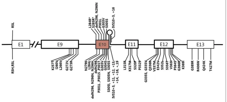
in individuals from over 100 families, while in PSP, 10 diferent MAPT mutations have been reported (Figure 1).44-47 Interestingly, except for the R5L
mu-tation, all other variants (p.L284R, p.S285R, p. delN296, p.N296N, p.P301L, p.G303V, p.S305S, IVS10+3, and IVS10+6) are located in exon 10 and its splicing region. Seven mutations are in coding regions and the other two mutations are in the splicing region of exon 10. Patients with p.L284R or p.S285R MAPT mutations presented with only the PSP phenotype; however, both PSP and FTDP-17T/
MAPT phenotypes were recognized in the other
Figure 1. A schematic diagram showing exons of MAPT and locations of mutations. Mutations discovered in patients with progressive su-pranuclear palsy (PSP) or PSP-like phenotypes were marked in the upper half of the diagram and those with frontotemporal lobar degener-ation presentdegener-ation were marked in the lower half. E1 to E13 refer to number of exons. Note all mutdegener-ations except for p.R5L are located in exon 10 or in stem-loop structure at the boundary between exon 10 and the intron following exon 10. Two mutations (p.L284R and p.S285R; marked with asterisk) were found only in patients with PSP phenotype.
Genetics of PSP Im SY, et al.
mutations. Given that PSP is 4-repeat tauopathy, mutations in MAPT exon 10 or its splicing regions may enhance production of 4 repeat tau isoforms48,49
or alter conformation of tau protein and affect its degradation process.
LRRK2
mutations and PSP
Patients with levodopa-responsive parkinsonism in western Nebraska kindred in whom a LRRK2
(PARK8) R1441C mutation was discovered, pleo-morphic pathology was reported in the autopsied brain.50 Within this family, afected members
show-ed Lewy body Parkinson’s disease, diffuse Lewy body disease, nigral degeneration without distinc-tive histopathology, or PSP-like pathology. he af-fected family members with PSP-like pathology had supranuclear palsy, neuronal loss in the substantia nigra, and tau pathologies (globose neuroibrillary tangles, tufted astrocytes, and oligodendroglial coiled bodies) without Lewy body or Lewy neuritis. In an analysis of the LRRK2 mutation in familial or sporadic PD from Crete by Spanaki et al.,51 two
parkinsonian brothers were identiied with a R1441H mutation in LRRK2. In both siblings, parkinsonism began as typical levodopa-responsive parkinsonism followed by a relatively rapid deterioration after a decade. he brothers later showed postural instabil-ity, supranuclear gaze palsy, bulbar dysfunction, de-mentia and other symptoms/signs, which are typi-cal for PSP. However, neuropathologitypi-cal studies of these cases were not reported.
Contrary to these two families suggesting that mutations in LRRK2 may be a genetic cause of PSP, other studies failed to detect a LRRK2 mutation in patients with PSP, although only small number of PSP cases were included for a LRRK2 mutation sc-reen. Screening for LRRK2 mutations were negative in clinically diagnosed PSP patients of Caucasian origin52,53 or of Asian origin.54 A similar study of
def-inite PSP cases in whom diagnosis was conirmed by neuropathology also reported negative results.55,56
Taken together, these results suggest that mutations of LRRK2 in patients with PSP are rare.
Other locus for familial PSP
Currently, only one PSP locus has been identiied by linkage analysis. Ros et al.28 identiied a PSP locus
from a linkage analysis of a large Spanish family in which PSP was inherited in autosomal dominant
fashion.26-28 Clinical phenotypes were typical of PSP,
although a minority of patients showed atypical fe-atures. Pathological diagnosis of PSP was conirmed according to the NINDS pathological criteria.27
Haplotype analysis around a chromosomal region with a maximum multipoint LOD score identiied a 3.4 cM candidate disease locus between markers D1S238 and D1S2823 on chromosome 1q31.1.28
However, no further progress has been published on the gene responsible.57 Recently, familial PSP with
a deinite PSP pathology but negative for a MAPT
mutation has been published, but no locus has been reported.21,31
GENETIC RISK FACTORS
ASSOCIATED WITH PSP
MAPT
haplotypes and sub-haplotypes
Conrad et al.58 identiied an association of an
in-tronic dinucleotide repeat polymorphic marker in
MAPT with PSP, but not with other tauopathies such as Alzheimer’s disease or parkinsonism-dementia complex of Guam. Single nucleotide polymorphisms (SNPs) in an extended haplotype of 1.3 Mbp cover-ing MAPT were found to be in complete linkage dis-equilibrium. Moreover, a common haplotype (H1 or MAPT H1-clade) was significantly over-repre-sented in patients with PSP.59-61 he identiication of
the H1 haplotype conferring a risk for PSP was con-sistently replicated in Caucasian populations. Al-though underlying molecular mechanisms conferred by the H1 haplotype have not been identiied, sup-porting evidence in cell models is that the H1 hap-lotype enhanced MAPT expression.62 he H2
hap-lotype that is present exclusively in Caucasians was found to be inversion polymorphism.63 Using
hap-lotype tagging SNPs spanning MAPT, Pittman et al.64 identified sub-haplotypes within H1. They
found that a common sub-haplotype (MAPT H1c) on the background of the MAPT H1-clade is over-represented in PSP. MAPT H1c was also found over-represented in patients with PSP-parkinsonism com-pared to controls.9 The MAPT H1-clade was also
found associated with an increased risk of PD, how-ever, MAPT H1c is not involved.65,66MAPT H1c was
J Mov Disord 2015;8(3):122-129
JMD
Genome-wide association study in PSP
To find additional PSP risk variants other than the MAPT H1 haplotype, a genome-wide associa-tion (GWA) study was performed using a pooled-DNA approach in 288 PSP patients from the US or Canada diagnosed based on neuropathology and 344 age- and sex-matched normal controls.68 Byas-sessing differences in allelic frequency, based on ranking differences in probe intensities for more than 500,000 SNPs between two cohorts, this study confirmed the MAPT H1 haplotype as a risk for PSP. A second major locus on chromosome 11p12-p11 showed evidence of association at allelic (p < 0.001), genotypic (p < 0.001), and haplotypic (p < 0.0001) levels. he DNA damage-binding protein-2 and lysosomal acid phosphatase-2 genes located in a single haplotype block of this locus were suggest-ed as candidates for conferring risk of PSP.
Höeglinger et al.69 carried out a GWA study in
two stages. Stage 1, a discovery cohort consists of 1,114 autopsy-conirmed PSP cases and 3,247 con-trols of European ancestry. SNPs that passed a cut-of (p≤ 10-3) in Stage 1 were genotyped in the
sec-ond stage (an independent set of validation cohort), which consisted of 1,051 PSP cases that were diag-nosed clinically and 3,560 controls. Joint analysis of stage 1 and 2 revealed that SNPs in STX6, MOBP, MAPT, and EIF2AK3 were associated with PSP. In the MAPT H1 region, ater controlling H1/H2, some SNPs remained associated with the maximum fall-ing in MAPT (rs242557). STX6 encodes syntaxin 6, a SNARE class protein that is involved in intracel-lular membrane traicking along endocytic and se-cretory transport pathways.70 A recent study showed
that the GWA SNP rs1411478 in STX6 is a strong expression quantitative trait locus with signiicantly lower expression of STX6 in the white matter of carriers of the risk allele.71EIF2AK3 encodes PERK,
a component of the endoplasmic reticulum (ER) unfolded protein response (UPR). When excess un-folded proteins accumulate in the ER, PERK is acti-vated by dimerization and trans-autophosphoryla-tion, which in turn phosphorylates eukaryotic tr-anslation-initiation factor 2α (eIF2α). eIF2α inhibits global translation, which allows ER to clear unfold-ed proteins. In the afectunfold-ed regions in PSP, the UPR was activated.72MOBP encodes MOBP protein, which
is produced by oligodendrocytes and is present in the major dense line of central nervous system
my-elin. Regions of high MOBP expression overlap with those afected in PSP, suggesting that dysfunction of myelin or oligodendrocytes is involved in the PSP pathogenesis. Genotype-expression correlation an-alysis showed that SNP alleles across the entire H1/ H2 inversion and lanking regions show strong cor-relation with MAPT expression. However, ater con-trolling for H1/H2, all signiicant genotype-expres-sion correlation for MAPT disappeared.69 CBD is a
neurodegenerative disorder with tauopathy that mimics PSP. Recently, a GWA study for CBD was reported to share common risk variants with PSP, which included rs1768208 at MOBP and rs242557 in MAPT H1c.73
HEREDITARY
NEURODEGENERATIVE
DISORDERS MIMICKING PSP:
PSP-LOOK-ALIKES
he expression of “look-alikes” in neurodegener-ative disorders was irst coined by Dr. Marsden in describing ive cases whose symptoms and signs on clinical grounds were compatible with typical CBD, but who turned out not to have CBD, but rather, other disorders, such as periventricular ischemic lesion with multiple infarction, sudanophilic leurko-dystrophy, Pick disease and PSP in postmortem ex-amination.74 As current understanding of the
ge-netics and molecular pathology of neurodegener-ative disorders has been advancing, the complicated relationship of convergence and divergence among clinical manifestation, pathology and causative genes has been recognized.75,76 Diferentiation of a
heredi-tary neurodegenerative disease from a similar phe-notypic syndrome may be important with regard to diferent prognoses and genetic counseling, as well as potential therapeutic and research implications.77,78
“PSP-look-alikes” refer to hereditary neurodegen-erative disorders that clinically mimic PSP, showing parkinsonism and early supranuclear gaze palsy, but that are not compatible with neuropathology of PSP. Recently, PSP-look-alikes were extensively re-viewed by Stamelou et al. 79 PSP-look-alike disorders
Genetics of PSP Im SY, et al.
(mutations in GBA), mitochondrial disorders, and familial Creutzfeldt-Jakob disease (mutations in
PRNP).
CONCLUSIONS
Our understanding of the genetic background of PSP has been expanding rapidly. This knowledge will not only help improve accurate diagnosis of PSP and other PSP-look-alike disorders, but will also provide insight into the pathogenesis, toward the eventually development of therapeutic strategies.
Conflicts of Interest
he authors have no inancial conlicts of interest.
Acknowledgments
This research was supported by the basic Science Research Program through the National Research Foundation of Korea (NRF) funded by the Ministry of Education, Science and Tech-nology (NRF-2013R1A1A4A01007783).
REFERENCES
1. Steele JC, Richardson JC, Olszewski J. Progressive supra-nuclear palsy. A heterogeneous degeneration involving the brain stem, basal ganglia and cerebellum with vertical gaze and pseudobulbar palsy, nuchal dystonia and dementia. Arch Neurol 1964;10:333-359.
2. Litvan I, Agid Y, Calne D, Campbell G, Dubois B, Duvoisin RC, et al. Clinical research criteria for the diagnosis of pro-gressive supranuclear palsy (Steele-Richardson-Olszewski syndrome): report of the NINDS-SPSP international work-shop. Neurology 1996;47:1-9.
3. Hauw JJ, Daniel SE, Dickson D, Horoupian DS, Jellinger K, Lantos PL, et al. Preliminary NINDS neuropathologic cri-teria for Steele-Richardson-Olszewski syndrome (progres-sive supranuclear palsy). Neurology 1994;44:2015-2019. 4. Dickson DW, Ahmed Z, Algom AA, Tsuboi Y, Josephs KA.
Neuropathology of variants of progressive supranuclear palsy. Curr Opin Neurol 2010;23:394-400.
5. Williams DR, de Silva R, Paviour DC, Pittman A, Watt HC, Kilford L, et al. Characteristics of two distinct clinical phenotypes in pathologically proven progressive supranu-clear palsy: Richardson’s syndrome and PSP-parkinsonism. Brain 2005;128(Pt 6):1247-1258.
6. Tsuboi Y, Josephs KA, Boeve BF, Litvan I, Caselli RJ, Cavi-ness JN, et al. Increased tau burden in the cortices of pro-gressive supranuclear palsy presenting with corticobasal syndrome. Mov Disord 2005;20:982-988.
7. Josephs KA, Boeve BF, Dufy JR, Smith GE, Knopman DS, Parisi JE, et al. Atypical progressive supranuclear palsy un-derlying progressive apraxia of speech and nonluent apha-sia. Neurocase 2005;11:283-296.
8. Josephs KA, Katsuse O, Beccano-Kelly DA, Lin WL, Uitti RJ, Fujino Y, et al. Atypical progressive supranuclear palsy with corticospinal tract degeneration. J Neuropathol Exp Neurol 2006;65:396-405.
9. Williams DR, Pittman AM, Revesz T, Lees AJ, de Silva R. Genetic variation at the tau locus and clinical syndromes associated with progressive supranuclear palsy. Mov
Dis-ord 2007;22:895-897.
10. Han HJ, Kim H, Park JH, Shin HW, Kim GU, Kim DS, et al. Behavioral changes as the earliest clinical manifestation of progressive supranuclear palsy. J Clin Neurol 2010;6:148-151.
11. Josephs KA, Eggers SD, Jack CR Jr, Whitwell JL. Neuro-anatomical correlates of the progressive supranuclear palsy corticobasal syndrome hybrid. Eur J Neurol 2012;19:1440-1446.
12. Williams DR, Lees AJ. Progressive supranuclear palsy: clinicopathological concepts and diagnostic challenges. Lancet Neurol 2009;8:270-279.
13. Williams DR, Holton JL, Strand K, Revesz T, Lees AJ. Pure akinesia with gait freezing: a third clinical phenotype of progressive supranuclear palsy. Mov Disord 2007;22:2235-2241.
14. Josephs KA, Petersen RC, Knopman DS, Boeve BF, Whit-well JL, Dufy JR, et al. Clinicopathologic analysis of fron-totemporal and corticobasal degenerations and PSP. Neu-rology 2006;66:41-48.
15. Hassan A, Parisi JE, Josephs KA. Autopsy-proven progres-sive supranuclear palsy presenting as behavioral variant frontotemporal dementia. Neurocase 2012;18:478-488. 16. Golbe LI, Davis PH, Schoenberg BS, Duvoisin RC.
Preva-lence and natural history of progressive supranuclear palsy. Neurology 1988;38:1031-1034.
17. Schrag A, Ben-Shlomo Y, Quinn NP. Prevalence of pro-gressive supranuclear palsy and multiple system atrophy: a cross-sectional study. Lancet 1999;354:1771-1775. 18. Nath U, Ben-Shlomo Y, homson RG, Morris HR, Wood
NW, Lees AJ, et al. he prevalence of progressive supranu-clear palsy (Steele-Richardson-Olszewski syndrome) in the UK. Brain 2001;124(Pt 7):1438-1449.
19. Kawashima M, Miyake M, Kusumi M, Adachi Y, Nakashi-ma K. Prevalence of progressive supranuclear palsy in Yo-nago, Japan. Mov Disord 2004;19:1239-1240.
20. Bower JH, Maraganore DM, McDonnell SK, Rocca WA. Incidence of progressive supranuclear palsy and multiple system atrophy in Olmsted County, Minnesota, 1976 to 1990. Neurology 1997;49:1284-1288.
21. Donker Kaat L, Boon AJ, Azmani A, Kamphorst W, Bre-teler MM, Anar B, et al. Familial aggregation of parkinson-ism in progressive supranuclear palsy. Neurology 2009;73: 98-105.
22. David NJ, Mackey EA, Smith JL. Further observations in progressive supranuclear palsy. Neurology 1968;18:349-356. 23. Mata M, Dorovini-Zis K, Wilson M, Young AB. New form
of familial Parkinson-dementia syndrome: clinical and pathologic indings. Neurology 1983;33:1439-1443. 24. Ohara S, Kondo K, Morita H, Maruyama K, Ikeda S,
Yanag-isawa N. Progressive supranuclear palsy-like syndrome in two siblings of a consanguineous marriage. Neurology 1992; 42:1009-1014.
25. Brown J, Lantos P, Stratton M, Roques P, Rossor M. Familial progressive supranuclear palsy. J Neurol Neurosurg Psy-chiatry 1993;56:473-476.
26. de Yébenes JG, Sarasa JL, Daniel SE, Lees AJ. Familial pro-gressive supranuclear palsy. Description of a pedigree and review of the literature. Brain 1995;118 (Pt 5):1095-1103. 27. Rojo A, Pernaute RS, Fontán A, Ruíz PG, Honnorat J,
Lynch T, et al. Clinical genetics of familial progressive su-pranuclear palsy. Brain 1999;122(Pt 7):1233-1245. 28. Ros R, Gómez Garre P, Hirano M, Tai YF, Ampuero I, Vidal
2005;57:634-J Mov Disord 2015;8(3):122-129
JMD
641.
29. Gazeley S, Maguire JA. Familial progressive supranuclear palsy. Clin Neuropathol 1996;15:215-220.
30. Tetrud JW, Golbe LI, Forno LS, Farmer PM. Autopsy-proven progressive supranuclear palsy in two siblings. Neu-rology 1996;46:931-934.
31. Fujioka S, Sanchez Contreras MY, Strongosky AJ, Ogaki K, Whaley NR, Tacik PM, et al. hree sib-pairs of autopsy-confirmed progressive supranuclear palsy. Parkinsonism Relat Disord 2015;21:101-105.
32. Fujioka S, Van Gerpen JA, Uitti RJ, Dickson DW, Wszolek ZK. Familial progressive supranuclear palsy: a literature review. Neurodegener Dis 2014;13:180-182.
33. Fujioka S, Algom AA, Murray ME, Strongosky A, Soto-Or-tolaza AI, Rademakers R, et al. Similarities between famil-ial and sporadic autopsy-proven progressive supranuclear palsy. Neurology 2013;80:2076-2078.
34. Goedert M. Neuroibrillary pathology of Alzheimer’s dis-ease and other tauopathies. Prog Brain Res 1998;117:287-306.
35. Goedert M, Jakes R. Mutations causing neurodegenerative tauopathies. Biochim Biophys Acta 2005;1739:240-250. 36. Lee VM, Goedert M, Trojanowski JQ. Neurodegenerative
tauopathies. Annu Rev Neurosci 2001;24:1121-1159. 37. Poorkaj P, Bird TD, Wijsman E, Nemens E, Garruto RM,
Anderson L, et al. Tau is a candidate gene for chromosome 17 frontotemporal dementia. Ann Neurol 1998;43:815-825. 38. Hutton M, Lendon CL, Rizzu P, Baker M, Froelich S, Houl-den H, et al. Association of missense and 5’-splice-site mu-tations in tau with the inherited dementia FTDP-17. Nature 1998;393:702-705.
39. Spillantini MG, Murrell JR, Goedert M, Farlow MR, Klug A, Ghetti B. Mutation in the tau gene in familial multiple system tauopathy with presenile dementia. Proc Natl Acad Sci U S A 1998;95:7737-7741.
40. Park HK, Chung SJ. New perspective on parkinsonism in frontotemporal lobar degeneration. J Mov Disord 2013;6:1-8. 41. Poorkaj P, Muma NA, Zhukareva V, Cochran EJ, Shannon KM, Hurtig H, et al. An R5L tau mutation in a subject with a progressive supranuclear palsy phenotype. Ann Neurol 2002;52:511-516.
42. Kim HJ, Jeon BS, Yun JY, Seong MW, Park SS, Lee JY. Screening for MAPT and PGRN mutations in Korean pa-tients with PSP/CBS/FTD. Parkinsonism Relat Disord 2010;16:305-306.
43. Ogaki K, Li Y, Takanashi M, Ishikawa K, Kobayashi T, Nonaka T, et al. Analyses of the MAPT, PGRN, and C9orf72 mutations in Japanese patients with FTLD, PSP, and CBS. Parkinsonism Relat Disord 2013;19:15-20.
44. Goedert M, Spillantini MG. Tau mutations in frontotempo-ral dementia FTDP-17 and their relevance for Alzheimer’s disease. Biochim Biophys Acta 2000;1502:110-121. 45. Rohrer JD, Paviour D, Vandrovcova J, Hodges J, de Silva R,
Rossor MN. Novel L284R MAPT mutation in a family with an autosomal dominant progressive supranuclear palsy syndrome. Neurodegener Dis 2011;8:149-152.
46. Goedert M, Ghetti B, Spillantini MG. Frontotemporal de-mentia: implications for understanding Alzheimer disease. Cold Spring Harb Perspect Med 2012;2:a006254. 47. Cruts M, Theuns J, Van Broeckhoven C. Locus-specific
mutation databases for neurodegenerative brain diseases. Hum Mutat 2012;33:1340-1344.
48. Skoglund L, Viitanen M, Kalimo H, Lannfelt L, Jönhagen ME, Ingelsson M, et al. The tau S305S mutation causes frontotemporal dementia with parkinsonism. Eur J Neurol
2008;15:156-161.
49. Stanford PM, Halliday GM, Brooks WS, Kwok JB, Storey CE, Creasey H, et al. Progressive supranuclear palsy pa-thology caused by a novel silent mutation in exon 10 of the tau gene: expansion of the disease phenotype caused by tau gene mutations. Brain 2000;123(Pt 5):880-893.
50. Zimprich A, Biskup S, Leitner P, Lichtner P, Farrer M, Lin-coln S, et al. Mutations in LRRK2 cause autosomal-domi-nant parkinsonism with pleomorphic pathology. Neuron 2004;44:601-607.
51. Spanaki C, Latsoudis H, Plaitakis A. LRRK2 mutations on Crete: R1441H associated with PD evolving to PSP. Neurol-ogy 2006;67:1518-1519.
52. Hernandez D, Paisan Ruiz C, Crawley A, Malkani R, Wer-ner J, Gwinn-Hardy K, et al. he dardarin G 2019 S muta-tion is a common cause of Parkinson’s disease but not other neurodegenerative diseases. Neurosci Lett 2005;389:137-139. 53. Madzar D, Schulte C, Gasser T. Screening for LRRK2
R1441 mutations in a cohort of PSP patients from Germa-ny. Eur J Neurol 2009;16:1230-1232.
54. Tan EK, Skipper L, Chua E, Wong MC, Pavanni R, Bon-nard C, et al. Analysis of 14 LRRK2 mutations in Parkin-son’s plus syndromes and late-onset ParkinParkin-son’s disease. Mov Disord 2006;21:997-1001.
55. Ross OA, Whittle AJ, Cobb SA, Hulihan MM, Lincoln SJ, Tot M, et al. Lrrk2 R1441 substitution and progressive su-pranuclear palsy. Neuropathol Appl Neurobiol 2006;32:23-25.
56. Gaig C, Ezquerra M, Martí MJ, Valldeoriola F, Muñoz E, Lladó A, et al. Screening for the LRRK2 G2019S and co-don-1441 mutations in a pathological series of parkinsonian syndromes and frontotemporal lobar degeneration. J Neu-rol Sci 2008;270:94-98.
57. Pastor P. Familial neurodegeneration in progressive supra-nuclear palsy: more frequent than expected? Neurology 2009;73:86-87.
58. Conrad C, Andreadis A, Trojanowski JQ, Dickson DW, Kang D, Chen X, et al. Genetic evidence for the involve-ment of tau in progressive supranuclear palsy. Ann Neurol 1997;41:277-281.
59. Baker M, Litvan I, Houlden H, Adamson J, Dickson D, Perez-Tur J, et al. Association of an extended haplotype in the tau gene with progressive supranuclear palsy. Hum Mol Genet 1999;8:711-715.
60. Pastor P, Ezquerra M, Perez JC, Chakraverty S, Norton J, Racette BA, et al. Novel haplotypes in 17q21 are associated with progressive supranuclear palsy. Ann Neurol 2004;56: 249-258.
61. Pittman AM, Myers AJ, Duckworth J, Bryden L, Hanson M, Abou-Sleiman P, et al. he structure of the tau haplotype in controls and in progressive supranuclear palsy. Hum Mol Genet 2004;13:1267-1274.
62. Kwok JB, Teber ET, Loy C, Hallupp M, Nicholson G, Mel-lick GD, et al. Tau haplotypes regulate transcription and are associated with Parkinson’s disease. Ann Neurol 2004; 55:329-334.
63. Rademakers R, Melquist S, Cruts M, heuns J, Del-Favero J, Poorkaj P, et al. High-density SNP haplotyping suggests altered regulation of tau gene expression in progressive su-pranuclear palsy. Hum Mol Genet 2005;14:3281-3292. 64. Pittman AM, Myers AJ, Abou-Sleiman P, Fung HC,
Genetics of PSP Im SY, et al.
65. Vandrovcova J, Pittman AM, Malzer E, Abou-Sleiman PM, Lees AJ, Wood NW, et al. Association of MAPT haplotype-tagging SNPs with sporadic Parkinson’s disease. Neurobiol Aging 2009;30:1477-1482.
66. Ezquerra M, Pastor P, Gaig C, Vidal-Taboada JM, Crucha-ga C, Muñoz E, et al. Diferent MAPT haplotypes are asso-ciated with Parkinson’s disease and progressive supranu-clear palsy. Neurobiol Aging 2011;32:547.e11-e16. 67. Myers AJ, Pittman AM, Zhao AS, Rohrer K, Kaleem M,
Marlowe L, et al. he MAPT H1c risk haplotype is associat-ed with increasassociat-ed expression of tau and especially of 4 repeat containing transcripts. Neurobiol Dis 2007;25:561-570. 68. Melquist S, Craig DW, Huentelman MJ, Crook R, Pearson
JV, Baker M, et al. Identiication of a novel risk locus for progressive supranuclear palsy by a pooled genomewide scan of 500,288 single-nucleotide polymorphisms. Am J Hum Genet 2007;80:769-778.
69. Höglinger GU, Melhem NM, Dickson DW, Sleiman PM, Wang LS, Klei L, et al. Identiication of common variants inluencing risk of the tauopathy progressive supranuclear palsy. Nat Genet 2011;43:699-705.
70. Jung JJ, Inamdar SM, Tiwari A, Choudhury A. Regulation of intracellular membrane traicking and cell dynamics by syntaxin-6. Biosci Rep 2012;32:383-391.
71. Ferrari R, Ryten M, Simone R, Trabzuni D, Nicolaou N, Hondhamuni G, et al. Assessment of common variability and expression quantitative trait loci for genome-wide as-sociations for progressive supranuclear palsy. Neurobiol Aging 2014;35:1514.e1-e12.
72. Stutzbach LD, Xie SX, Naj AC, Albin R, Gilman S; PSP Ge-netics Study Group, et al. he unfolded protein response is activated in disease-affected brain regions in progressive supranuclear palsy and Alzheimer’s disease. Acta Neuro-pathol Commun 2013;1:31.
73. Kouri N, Ross OA, Dombroski B, Younkin CS, Serie DJ, So-to-Ortolaza A, et al. Genome-wide association study of cor-ticobasal degeneration identiies risk variants shared with progressive supranuclear palsy. Nat Commun 2015;6:7247. 74. Bhatia KP, Lee MS, Rinne JO, Revesz T, Scaravilli F, Davies
L, et al. Corticobasal degeneration look-alikes. Adv Neurol 2000;82:169-182.
75. Klein C, Schneider SA, Lang AE. Hereditary parkinson-ism: Parkinson disease look-alikes--an algorithm for clini-cians to “PARK” genes and beyond. Mov Disord 2009;24: 2042-2058.
76. Karageorgiou E, Miller BL. Frontotemporal lobar degener-ation: a clinical approach. Semin Neurol 2014;34:189-201. 77. Halliday GM, Song YJ, Creasey H, Morris JG, Brooks WS,
Kril JJ. Neuropathology in the S305S tau gene mutation. Brain 2006;129(Pt 3):E40.
78. Armstrong MJ, Litvan I, Lang AE, Bak TH, Bhatia KP, Bor-roni B, et al. Criteria for the diagnosis of corticobasal de-generation. Neurology 2013;80:496-503.