C U R R E N T T O P I C UDC: 616-072.1::618.1-006-089 DOI: 10.2298/VSP1309861M
Laparoscopy in gynecologic oncology: A review of literature
Laparoskopija u ginekološkoj onkologiji – pregled literature
Aljoša Mandiü, Andrija Goluboviü, Ivan Majdevac
Oncology Institute of Vojvodina, Sremska Kamenica, Serbia
Key words:
genital neoplasms, female; laparoscopy; gynecologic surgical procedures; lymph node excision.
Kljuÿne reÿi:
polni organi, ženski, neoplazme; hirurgija, laparoskopska; hirurgija, ginekološka, procedure; limfadenektomija.
Introduction
Introducing laparoscopy in gynecology during the 1960s in Europe placed gynecology as pioneer in this less invasive approach in surgery. At that time, gynecological laparoscopy was predominantly used in diagnostics by means of direct visualization of pelvis minor. During the 1970s the gynecolo-gists shifted the use of endoscopic technology from diagnostic to surgical purposes such as tubal ligation. From that time, laparoscopy has become a leading technique in diagnostics and surgical treatment of benign lesions in gynecology 1. By using laparoscopy in detection of bladder and prostate carci-noma spreading into the lymph nodes urologists have become the pioneers of the use of laparoscopy in the field of oncol-ogy 2. During the late 1980s, the initiators of endoscopic on-cology began with laparoscopic evaluations of the condition and spreading of the malignant disease. The first reports were available at the beginning of 1990s, and with them came the controversial opinions about the usefulness of laparoscopy and minimal invasive surgery in gynecologic oncology. Disagree-ments were mainly associated with medicolegal aspects of possible consequences resulted from inadequate surgical treatment of malignant diseases.
The trends of laparoscopy use in gynecologic surgery
There has been an increasing trend in the use of mini-mal invasive techniques for resection and/or staging of ma-lignancies in gynecology during the last ten years. Many studies report the advantages of these procedures, their effi-cacy, safety and adequacy in surgical treatment of gyneco-logic malignancies 3–6. A survey conducted in 2004 and 2007 among the members of the Society of Gynecologic Oncolo-gist (SGO) in the USA showed a significant increase in the
use of laparoscopy. Forty-six percent of 850 SGO members responded to the survey. In 2004 survey, laparoscopic sur-gery was indicated in 84% of cases while in 2007 survey it was increased to 91%. The following laparoscopic proce-dures were most often indicated 7: laparoscopically assisted vaginal hysterectomy (LAVH) or total laparoscopic hyster-ectomy (TLH) and staging of endometrial carcinoma (43%); diagnostics of adnexal masses (39%); prophylactic salpingo-oophorectomy in case of women at high risk for ovarian can-cer (11%).
These procedures are accepted as the most convenient for use in endoscopic surgery. The question regarding the conversion from laparoscopy to laparotomy was answered by 90% of the survey participants. In 2004, 25% of them did the conversion, while in 2007 only 3% of conversions were re-ported by 94% of surveyed SGO members 7. The mentioned data and numerous papers published in Europe and Asia point to the increasing trend of using laparoscopy in gyne-cologic oncology (Table 1).
The launch of minimally invasive surgery reduces the operation-induced trauma, provides a faster recovery, short-ens the hospitalization and lowers the total costs of treat-ment. The purpose of using laparoscopy in gynecologic sur-gery is to confirm therapeutic efficacy compared with stan-dard surgical procedures and to reduce the appearance of side effects. Still open are dilemmas regarding the results of treatment after laparoscopic surgery in oncology and intra-operative complications such as injuries of intestines, larger blood vessels and tumor cells dissemination.
treat-ment in oncology is the complete removal of malignant tu-mor and interruption of its spreading, and control and alle-viation of disease symptoms. If the aim can be achieved by minimally invasive laparoscopic procedures than their use is justified but not at any price and if they are harmful to pa-tients’ health 8. Nevertheless, laparoscopic surgery in gyne-cologic oncology has become a standard procedure in the majority of medical institutions in developed countries. Modern medical technology, acquired experience, and better surgical training with modern endoscopy equipment have been the main reasons for that.
The surgeons have agreed that laparoscopic techniques are associated with extremely gradual process of learning, which starts with small and simple procedures and goes up to more complex and comprehensive laparoscopic operations. It should be mentioned that laparoscopic operations in gyne-cologic oncology could be performed only by surgeons who have already mastered the techniques of classic surgery and are skilled to manage the complications. The learning curve starts with classic surgery procedures in gynecologic oncol-ogy and continues with learning the basics of laparoscopic operations and skills under supervision. The next step is to have sufficiently enough training after which come the actual performance of laparoscopic procedures and operations in the treatment of gynecologic oncology patients 8.
The use of robotics in laparoscopy
Robotic assisted surgery is a new aspect in gynecologic oncology which eliminates the basic ergonomic problem for a surgeon and the most important long learning curve. In ad-dition, it gives a 3-D vision and magnification: the surgeon controls the camera, the image is directly projected, the movements are intuitive, the instruments are articulated and ergonomic, the tremor is eliminated. The first surgical robots were presented during the 1980s. The development of ro-botic surgery made possible broader applications for surgical indications. ROBODOC was the first surgical robot ap-proved by the United States Food and Drug Administration FDA. The next were Automatic Endoscopic System for Op-timal Positioning (AESOP) in 1994 and ZEUS, a second-generation robotic system in 1998 9. The da Vinci surgical
system is the most sophisticated of the surgical robotic sys-tems. Based upon the first reports made by Advincula and Reynolds on the use of robot for myomectomies, FDA ap-proved the use of the da Vinci in gynecologic procedures in April 2005 10–13. At the annual SGO meeting in February 2006, Boggess 14 did a live demonstration of radical hyster-ectomy and reported on 13 previously performed operations. Since then, the use of robotic surgery in gynecologic oncol-ogy has constantly been improved in world centers.
Laparoscopy in endometrial carcinoma
Laparoscopic approach in treatment of endometrial car-cinoma implies laparoscopic determination of the stage of the disease combined with laparoscopically assisted vaginal (LAVH) or laparoscopic hysterectomy and bilateral ad-nexectomy. In the initial FIGO stage I of endometrial carci-noma, which is limited only to the uterus, laparoscopically assisted vaginal hysterectomy with bilateral adnexectomy should be applied whenever it is technically possible 15, 16. Zullo et al. 17 conducted a randomized study to compare laparoscopy vs laparotomy in patients with early stages of endometrial carcinoma. The authors showed that the safety and efficiency of laparoscopy was the same as in the open approach, pointing out the benefit of laparoscopy in relation to the quality of life during the first 6 months after the sur-gery. Tozzi et al. 18 reported the first results of the survival of patients with endometrial carcinoma who were operated laparoscopically in comparison to those patients who under-went open surgery. Based on the average follow-up of 44 months of patients with endometrial carcinoma FIGO stage I, they found that a disease-free interval among laparoscopi-cally operated patients was 91% compared to 94% among patients treated with classic surgery. Overall survival was 86% compared to 90% in patients with laparotomy. Malur et al. 19 presented 70 patients with stage I-III of endometrial carcinoma: 37 patients had laparoscopically assisted vaginal hysterectomy and 33 underwent open surgery. Comparative analysis of the removed lymphatic nodes and duration of surgery did not show a statistically significant difference. The recurrence-free interval did not show statistically sig-nificant difference between the laparoscopy group (97%) and
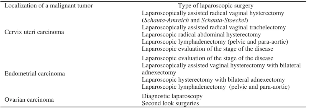
Table 1 Laparoscopic procedures that are frequently applied in gynecological oncology
Localization of a malignant tumor Type of laparoscopic surgery
Cervix uteri carcinoma
Laparoscopically assisted radical vaginal hysterectomy (Schauta-Amreichand Schauta-Stoeckel)
Laparoscopically assisted radical vaginal trachelectomy Laparoscopic radical abdominal hysterectomy
Laparoscopic lymphadenectomy (pelvic and para-aortic) Laparoscopic evaluation of the stage of the disease
Endometrial carcinoma
Laparoscopic evaluation of the stage of the disease
Laparoscopically assisted vaginal hysterectomy with bilateral adnexectomy
Laparoscopic hysterectomy with bilateral adnexectomy Laparoscopic lymphadenectomy (pelvic and para-aortic)
Ovarian carcinoma Diagnostic laparoscopy
the laparotomy group (93%). Similar results were presented in relation to overall survival, 84% in the laparoscopic group, and 91% in the laparotomy group.
Ju et al. 20 included 5 prospective and 8 retrospective studies in a meta-analysis. The comparison of the laparo-scopic approach to open surgery in endometrial carcinoma did not confirm a statistically significant difference for the overall survival and the recurrence of the disease, while the number of complications was lower in the group with the laparoscopic approach. Furthermore, the analysis of 5 studies dealing with the number of lymphatic nodes in tested groups did not confirm any statistically significant difference. In the study of Janda et al. 21, the quality of life after total laparo-scopic hysterectomy (TLH), n = 190, and total abdominal hysterectomy (TAH), n = 142, was examined. In the early phase of the recovery period, the improvement of the quality of life was more pronounced in patients with TLH. Better quality of life continued its trend in the TLH group even 6 months after the surgery. Longer duration of the surgery was statistically significant in the TLH group (138 ± 43 min), when compared with the TAH group (109 ± 34 min; p = 0.001). Intraoperative complications were similarly present in both of the groups (TAH 8/142, 5.6%, and TLH 14/190, 7.4%;p = 0.55). During the postoperative period, two times more adverse events occurred in the TAH group than in the TLH patients (33/142, 23.2%, and 22/190, 11.6%, respec-tively; p = 0.004). Serious postoperative complications were more frequent in the TAH patients (27/142, 19.0%) than in the TLH group (15/190, 7.9%; p = 0.002). The advantages of the laparoscopic approach, together with the vaginal hyster-ectomy imply less percentage of postoperative complica-tions, shorter recovery period at the hospital, even in the group of obese patients 22.
Laparoscopy in cervix uteri carcinoma
Vaginal radical hysterectomy as a method of choice in the treatment of cervix uteri carcinoma was the initial idea for performance of the first laparoscopic lymphadenectomy in the treatment of early invasive cervix uteri carcinoma. The greatest advantage of the vaginal operative procedure is the possibility of closure of the cervix at the very beginning of the surgery and the reduction of chances for tumor dissemi-nation. Dargent performed the laparoscopic lymphadenec-tomy after the Schauta’s vaginal radical hystereclymphadenec-tomy in 1986 in Lion, and that was the first step of implementation of laparoscopy in combination with already familiar radical vaginal surgery. Dargent published the first results after the surgeries of 51 patients, where a three-year long survival was registered in 95%. At the beginning, laparoscopy had extra-peritoneal approach, but since 1992, Querleu has been pro-moting a transumbilical transperitoneal laparoscopic dissec-tion of lymphatic nodes 23. After more than 10 years of im-plementation in surgical practice, the analysis showed that the laparoscopic lymphadenectomy is an equally safe and re-liable method as laparotomy, with the great advantage of the minimally invasive approach. Laparoscopic lymphadenec-tomy can also be performed as a diagnostic procedure in
pa-tients with the early stage of the cervix uteri carcinoma be-fore making the decision on the selection of the therapeutic procedure 24, 25. More frequent implementation of laparos-copy created conditions for performance of laparoscopic radical abdominal hysterectomy and laparoscopically as-sisted radical vaginal trachelectomy (LVRT) 26–29. In 1994, Daniel Dargent presented the concept of radical vaginal trachelectomy, where the body of uterus, ovaries, and the fallopian tubes were preserved and the vaginal approach radically removed the cervix uteri, the upper third of the va-gina and a part of parametrium. This procedure is combined with the laparoscopically assisted lymphadenectomy in an identical way as in the case of laparoscopically assisted radi-cal vaginal hysterectomy 23. These surgeries are reserved only for the early stages of the cervix uteri carcinoma with the aim of preservation of the patient’s fertility 30. Imple-mentation of laparoscopy in the advanced disease is limited to lymphadenectomy, which is performed in some centers, because it was proved that the patients who had their bulky lymph nodes removed before the therapy had better sur-vival 8. Introduction of robotic surgery has also widened the indication of laparoscopic robotic surgery implementation in the treatment of cervical carcinoma 31. Magrina et al. 32 pre-sented the comparison of robotic laparoscopic radical hyster-ectomy with classical laparoscopic approach and laparotomy. The average durations of robotic procedure, laparoscopy and laparotomy were 189.6 min, 220.4 min and 166.8 min, re-spectively, with loss of blood of 133 mL, 208 mL, and 443.6 mL, respectively. The average number of removed lymphatic nodes was 25.9 in robotic laparoscopy, 25.9 in laparoscopy and 27.7 in laparotomy with hospital stay of 1.7, 2.4 and 3.6 days, respectively. There were no differences in intra- and postoperative complications among the tested groups. During the follow-up of all the three groups in duration of 31.1 months, no disease recurrence was registered 32.
Laparoscopy in ovarian carcinoma
There is a general consensus that implementation of laparoscopy as a surgical approach in treatment of benign adnexal masses is entirely justified due to reduced loss of blood, shorter hospital stay, less complications and pain and reduced treatment expenses in comparison to the open ap-proach33.
The role of laparoscopy in the treatment of ovarian car-cinoma remains in the domain of discussion and insuffi-ciently clear directives. In the majority of cases, laparoscopy imposed itself as a method of surgical staging of the disease and deciding on further treatment of ovarian carcinoma.
When compared, the results of laparoscopic surgical staging, in relation to laparotomy, showed reduced loss of blood, shorter hospital stay and less complications with longer operative time. The numbers of lymphatic glands and survival were not significantly statistically different 34, 35.
Conclusion
Implementation of laparoscopy, ie minimally invasive surgery (MIS) in gynecological oncology represents today a significant therapeutic modality of treatment without
com-promising basic oncological principles. Using such a medical technology in oncology patients in Serbia imposes discussion on a new approach in education and organization of centers with adequate equipment where this type of surgery could be implemented with a sufficient number of cases.
R E F E R E N C E S
1. Gordon AG, Magos AL. The development of laparoscopic sur-gery. Bailliere’s Clin Obstet Gynaecol 1989; 3(3): 429–49. 2. Hald T, Rasmussen F. Extraperitoneal pelvioscopy: a new aid
in staging of lower urinary tract tumors. A preliminary report. J Urol 1980; 124(2): 245–8.
3. Walker JL, Piedmonte M, Spiros N. Phase III trial of laparos-copy versus laparotomy for surgical resection and compre-hensive surgical staging of uterine cancer: a Gynecologic On-cology Group study funded by the National Cancer Institute. Gynecol Oncol 2006; 101(suppl 1): S11–2.
4. Frumovitz M, dos Reis R, Sun CC, Milam MR, BeversMW, Brown J, et al. Comparison of total laparoscopic and abdominal radi-cal hysterectomy for patients with early stage cerviradi-cal cancer. Obstet Gynecol 2007; 110(1): 96–102.
5. Leblanc E, Querleu D, Narducci F, Occelli B, Papageorgiou T, Sonoda Y. Laparoscopic restaging of early stage invasive ad-nexal tumors: a 10-year experience. Gynecol Oncol 2004; 94(3): 624î9.
6. Abu-Rustum NR, Sonoda Y. Transperitoneal laparoscopic staging with aortic and pelvic lymph node dissection for gy-necologic malignancies. Gynecol Oncol 2007; 104(2) :5–8. 7. Mabrouk M, Frumovitz M, Greer M, Sharma S, Schmeler KM,
Soliman PT, et al. Trends in laparoscopic and robotic surgery among gynecologic oncologists: A survey update. Gyneco-logic Oncology 2009; 112(3): 501–5.
8. đurĀeviý S, VidakoviýS. Primena laparoskopije u ginekološkoj onkologiji,. In: đurĀeviý S, Kesiý V, editors. Ginekološka on-kologija. Novi Sad: Faculty of Medicine; 2009. p. 336î40. 9. Mendivil A, Holloway RW, Boggess JF. Invited Review:
Emer-gence of robotic assisted surgery in gynecologic oncology: American perspective. Gynecol Oncol 2009; 114(2): S24îS31.
10. Advincula AP, Reynolds RK. The use of robot-assisted laparo-scopic hysterectomy in the patient with a scarred or obliter-ated anterior cul-de-sac. JSLS 2005; 9(3): 287–91.
11. Advincula AP, Song A, BurkeW, ReynoldsRK. Preliminaryexperi-encewithrobot-assisted laparoscopic myomectomy. J Am As-soc Gynecol Laparosc 2004; 11(4): 511–8.
12. Reynolds RK, Advincula AP. Robot-assisted laparoscopic hys-terectomy: technique and initial experience. Am J Surg 2006; 191(4): 555–60.
13. Reynolds RK, Burke WM, Advincula AP. Preliminary experience with robot-assisted laparoscopic staging of gynecologic ma-lignancies. JSLS 2005; 9(2): 149–58.
14. Boggess JF. Robotic-assisted radical hysterectomy for cervical cancer: National Library of Medicine Archives. 2006 [updated 2006; cited 2008 August 11]. Available from: http://www.nlm.nih.gov/medlineplus/surgeryvideos.html. 15. Cho YH, Kim DY, Kim JH, Kim YM, Kim YT, Nam JH.
Lapa-roscopic management of early uterine cancer: 10-year experi-ence in Asan Medical Center. Gynecol Oncol 2007; 106(3): 585–90.
16. Barakat RR, Lev G, Hummer AJ, Sonoda Y, Chi DS, Alektiar KM, et al. Twelve-year experience in the management of en-dometrial cancer: a change in surgical and postoperative ra-diation approaches. Gynecol Oncol 2007; 105(1): 150–6.
17. Zullo F, Palomba S, Russo T, Falbo A, Costantino M, Tolino A, et al. A prospective and randomized comparison between lapa-roscopic and laparotomic approaches in women with early stage endometrial cancer: a focus on the quality of life. Am J Obstet Gynecol 2005; 193: 1344–52.
18. Tozzi R, Malur S, Koehler C, Schneider A. Laparoscopy versus laparotomy in endometrial cancer: first analysis of survival of a randomized prospective study. J Minim Invasive Gynecol 2005; 12(2): 130–6.
19.Malur S, Possover M, Michels W, Schneider A. Laparoscopic-assisted vaginal versus abdominal surgery in patients with en-dometrial cancer-a prospective randomized trial. Gynecol Oncol 2001; 80(2): 239î44.
20. Ju W, Myung SK, Kim Y, Choi HJ, Kim SC. Comparison of Laparoscopy and Laparotomy for Management of Endome-trial Carcinoma. A Meta-analysis. Int J Gynecol Cancer 2009; 19(3): 400î6.
21. Janda M, Gebski V, Brand A, Hogg R, Jobling TW, Land R, et al. Quality of life after total laparoscopic hysterectomy versus total abdominal hysterectomy for stage I endometrial cancer (LACE): a randomised trial. Lancet Oncol 2010; 11(8): 772î80.
22. Devaja O, Samara I, Papadopoulos AJ. Laparoscopically Assisted Vaginal Hysterectomy (LAVH) Versus Total Abdominal Hysterectomy (TAH) in Endometrial Carcinoma Prospective Cohort Study. Int J Gynecol Cancer 2010; 20(4): 570î5. 23. Mandic A, Novakovic P, Nincic D. Surgical approaches towards
fertility preservation in young patients with early invasive cer-vical carcinoma. J BUON2009; 14(4): 581î6.
24. Leblanc F, Narducci F, Chevalier A, Taieb S, Castelain B, Querleu D. Pretherapeutic laparoscopic staging of locally advanced cervical carcinomas: technique and results. Gynecol Oncol 2005; 99(3 Suppl 1): S157–8.
25. Fagotti A, Fanfani F, Longo R, Legge F, Mari A, Gagliardi ML, et al. Which role for pre-treatment laparoscopic staging? Gy-necol Oncol 2007; 107(1 Suppl 1): S101î5.
26. Chen DM, Lim PC, Spirtos NM. Laparoscopic radical hyster-ectomy. CME J Gynecol Oncol 2001; 6(1): 117î28.
27. Dargent D. Laparoscopic assisted vaginal radical hysterectomy – evolution of a concept. CME J Gynecol Oncol 2001; 6(1): 102î9.
28. Kadar N, Reich H. Laparoscopically asisted radical Schauta hysterectomy and bilateral laparoscopic pelvic lymphadenec-tomy for the treatment og bulky stage I B carcinoma of the cervix. Gynecol Endosc 1993; 2: 135î42.
29. Canis M, Mage G, Wattiez A, Pouly JL, Manhes H, Bruhat MA. Does endoscopic surgery have a role in radical surgery of cancer of the cervix uteri? J Gynecol Obstet Biol Reprod (Paris) 1990; 19(7): 921. (French)
30. Leblanc E, Narducci F, Ferron G, Querleu D. Indications and teaching fertility preservation in the surgical management of gynecologic malignancies: European perspective. Gynecol Oncol 2009; 114(1): 32–6.
32. Magrina JF, Kho RM, Weaver AL, Montero RP, Magtibay PM. Robotic radical hysterectomy: Comparison with laparoscopy and laparotomy. Gynecol Oncol 2008; 109(1): 86–91. 33. Medeiros LR, Stein AT, Fachel J, Garry R, Furness S.
Laparos-copy versus laparotomy for benign ovarian tumor: a system-atic review and meta-analysis. Int J Gynecol Cancer 2008; 18(3): 387–99.
34. Ghezzi F, Cromi A, Uccella S, Bergamini V, Tomera S, Franchi M, et al. Laparoscopy versus laparotomy for the surgical
man-agement of apparent early stage ovarian cancer. Gynecol On-col May 2007; 105(2): 409–13.
35. Park JY, Bae J, Lim MC, Lim SY, Seo SS, Kang S, et al. Laparo-scopic and laparotomic staging in stage I epithelial ovarian cancer: a comparison of feasibility and safety. Int J Gynecol Cancer 2008; 18(6): 1202–9.