hedgehog
Leads to Morphological Changes during Fin
Development
Koji Sakamoto1., Koh Onimaru1., Keijiro Munakata1., Natsuno Suda1
, Mika Tamura1, Haruki Ochi2¤, Mikiko Tanaka1*
1Graduate School of Bioscience and Biotechnology, Tokyo Institute of Technology, Nagatsuta-cho, Midori-ku, Yokohama, Japan,2Institute of Neuroscience, University of Oregon, Eugene, Oregon, United States of America
Abstract
We explored the molecular mechanisms of morphological transformations of vertebrate paired fin/limb evolution by comparative gene expression profiling and functional analyses. In this study, we focused on the temporal differences of the onset ofSonic hedgehog(Shh) expression in paired appendages among different vertebrates. In limb buds of chick and mouse,Shhexpression is activated as soon as there is a morphological bud, concomitant withHoxd10expression. In dogfish (Scyliorhinus canicula), however, we found thatShhwas transcribed late in fin development, concomitant with Hoxd13 expression. We utilized zebrafish as a model to determine whether quantitative changes inhoxexpression alter the timing ofshhexpression in pectoral fins of zebrafish embryos. We found that the temporal shift of Shh activity altered the size of endoskeletal elements in paired fins of zebrafish and dogfish. Thus, a threshold level ofhoxexpression determines the onset ofshhexpression, and the subsequent heterochronic shift of Shh activity can affect the size of the fin endoskeleton. This process may have facilitated major morphological changes in paired appendages during vertebrate limb evolution.
Citation:Sakamoto K, Onimaru K, Munakata K, Suda N, Tamura M, et al. (2009) Heterochronic Shift inHox-Mediated Activation ofSonic hedgehogLeads to Morphological Changes during Fin Development. PLoS ONE 4(4): e5121. doi:10.1371/journal.pone.0005121
Editor:Vincent Laudet, Ecole Normale Supe´rieure de Lyon, France
ReceivedDecember 11, 2008;AcceptedMarch 12, 2009;PublishedApril 13, 2009
Copyright:ß2009 Sakamoto et al. This is an open-access article distributed under the terms of the Creative Commons Attribution License, which permits unrestricted use, distribution, and reproduction in any medium, provided the original author and source are credited.
Funding:This work was supported by a Grant-in-Aid for Young Scientists (A) from the Ministry of Education, Science, Sports and Culture of Japan, by a Tokyo Tech Award for Challenging Research and by a grant from the Hayashi Memorial Foundation for Female Natural Scientists. The funders had no role in study design, data collection and analysis, decision to publish, or preparation of the manuscript.
Competing Interests:The authors have declared that no competing interests exist.
* E-mail: [email protected]
¤ Current address: Graduate School of Biological Sciences, Nara Institute of Science and Technology, Takayama, Ikoma, Nara, Japan
.These authors contributed equally to this work.
Introduction
There has been considerable debate regarding the fundamental mechanisms that direct morphological transformations from fins into limbs with respect to the expression patterns of 59-locatedHox genes and subsequent Shh expression [1,2,3]. It is generally accepted, however, that, the enlargement of the fin endoskeleton along the proximal-distal axis within the lineage of basal sarcopterygians (lobe-finned fishes) results from changes in the heterochronic folding of the apical fin fold [4]; other possibilities have scarcely been discussed. Here we have investigated the genetic basis of morphological transitions of the vertebrate fin endoskeleton primarily via comparative gene expression profiling and functional analyses, focusing especially on the temporal onset ofShh expression. Because two paired appendages are unique to gnathostomes—and cartilaginous fish occupy the earliest branch of the gnathostome lineage—the study of the cartilaginous dogfish may provide insight into how animals have acquired morpholog-ically diverse paired appendages. Although the developmental mechanisms of such morphological changes are still under debate, the evolutionary acquisition of Shh function in growing paired appendages might have been a crucial step in implementing morphological innovations of paired appendages.
[14,15]. Similarly, a requirement for shh acitivity in cell proliferation in the zebrafish pectoral fin bud has also been suggested [16]. These results raise the possibility that the duration of exposure to Shh activity may have been critical for the morphological evolution of paired appendages.
To investigate the possibility that the duration of exposure to Shh activity may have been critical for the morphological evolution of paired appendages, we analyzed fin development in embryos of the cartilaginous dogfish Scyliorhinus canicula. In limb buds of chick and mouse, Shh expression is activated as soon as there is a morphological bud, whereas in S. canicula fin buds, consistent with reported data in other cartilaginous fishes [17],Shh is transcribed late in fin development. Several molecular triggers that activateShhexpression have been proposed, including Hand2 and Fibroblast growth factor (Fgf) [18,19]. In pectoral fins ofS. canicula, Hand2 transcripts localize posteriorly at a much earlier stage thanShh transcripts, and it is therefore unlikely thatHand2 correlates directly with the late onset ofShhtranscription [20]. In vertebrate limb buds, Fgfs are secreted from the apical ectodermal ridge that rims the distal edge of the buds, and these Fgfs play pivotal roles in limb bud initiation and outgrowth, at least in part by inducing and maintaining the expression of Shh in the underlying mesenchyme [19,21,22,23,24]. Hoxa and Hoxd have also been demonstrated to drive Shh expression in mouse limb buds [2,3,25]. Furthermore, recent experiments have shown that Hox proteins bind to a conserved regulatory region ofShh, thereby promoting Shh expression within developing mouse limb buds [26]. In our current study, we show that a threshold level ofhox expression is essential for the onset ofshhexpression and that the subsequent heterochronic shift of Shh activity leads to changes in the size of pectoral fins. These results imply that a quantitative change inhoxexpression could have involved a heterochronic shift of shh expression and subsequent morphological changes of endoskeleton during limb evolution.
Materials and Methods
Animals
S. caniculaeggs were incubated at 12,16uC in sea water and staged according to Ballard et al. (1993). The gross duration of incubation described in Ballard et al. (1993) was as follows: stage 27 (42–46 days), stage 29 (49–53 days), stage 32 (75–125 days). Because duration of stage 32 is long, we subdivided stage 32 into ‘‘early stage 32’’ (75–100 days) and ‘‘late stage 32’’ (101–125 days). Wild-type (TL strain and AB/Tu¨bingen strain) zebrafish (Danio rerio) were maintained at 28.5uC and staged using standard morphological criteria [27].
Identification of S. caniculagene homologs
We identified fragments ofS. canicula(Sc) Fgf8(296 bp), Meis1 (357 bp), Hoxa11 (357 bp), Hoxa13 (389 bp), Hoxd11 (534 bp), Hoxd13 (296 bp), Pbx2 (653 bp), Ptc2 (1157 bp) and GAPDH (230 bp) from cDNA pools prepared from stage 24–30 embryos using degenerate primers. The degenerate primers were designed to anneal to coding regions containing the following amino acid
sequences: ScFgf8, TYQLYSRT and VHFMKRL; ScMeis1,
CDNFCHR and GIFPKVA; ScHoxa11, QVQPVRE and
AATSSS; ScHoxa13, AYTSSEV and PMESYQP; ScHoxd11,
CQMTFPYS and PYTKYQIR; ScHoxd13, PVEKYMDV and
IWFQNRRV; ScPbx2, QQIMTIT and PYPSEEA; ScPtc2,
IHAFSTT and QFKYFSFYNF; ScGAPDH, ASCTTN and
VIPELN. S. canicula ScHoxd10 (785 bp) and ScHoxd12 cDNAs (316 bp) were amplified by PCR using the following primers which hybridized to the indicated published sequence: ScHoxd10,
GenBank accession number DQ659105, 59
-GGGAACATACG-GAATGCAGACC-39 and
59-GTAAGAGCGTGAATCTGAC-CG-39; ScHoxd12, GenBank accession number DQ659106,
59-CCCTTCTATTTCGCCAACCTG-39 and
59-CCCAAGTGA-TACCAGCATCC-39. The nucleotide sequences of the ScFgf8, ScHoxd13,ScMeis1,ScHoxd11,ScHoxa11, ScHoxa13,ScPbx2,ScPtc2 andScGAPDH cDNAs were deposited in the GenBank database
under the accession numbers: DQ647321–DQ647323,
DQ854846, EU005549–EU005551, EU814484 and EU826015, respectively.
Whole-mount in situ hybridization and immunohistochemistry
S. canicula embryos were removed from their egg casings and dissected from the yolk mass. Whole-mountin situhybridization of S. caniculaand immunostaining ofS. caniculaembryos were carried out as described [20]. Whole-mount in situ hybridization of zebrafish was performed as described [28]. Probes for zebrafish hoxd10a,hoxd11aandhoxd13awere amplified by reverse transcrip-tion-polymerase chain reaction (RT-PCR) using primers derived from published sequences (www.ensembl.org). For whole-mount immunostaining, embryos were prepared as described [29]. The monoclonal antibody against human Fgf4 (R & D Systems) was used at a 1:300 dilution.
Microinjection
For mRNA injection, the full-length cDNAs encodinghoxa13a, hoxd10a,hoxd13a,hoxd4andpbx2were individually cloned into the pCS2+ vector and the corresponding mRNAs were synthesized using the MEGAscript kit (Ambion). The mRNAs were dissolved in endotoxin-free H2O to a final concentration of 20 mg/ml. Morpholino antisense oligonucleotides (MOs) were obtained from Gene Tools, Inc. The followinghoxd10aandhoxd13aMOs targeted the boundary between exon 1 and intron 1 of each respective gene
(Gene Tools, Inc.): MO-hoxd10a,
CCGTTTATTGTACC-CACCTTTGCCT; MO-hoxd13a,
CAGAGCTGAGGTCT-TACCTGTTAAT. The pbx2 MO was used as described [30].
The standard control MO obtained from Gene Tools, Inc. was used as an injection control. MOs were dissolved in sterile H2O at concentrations of 1, 2.5 or 5 mg/ml and phenol red was added to the solution. Approximately 1 nl of mRNA or MO was injected at the one-cell stage using a microinjector (IM30, Narishige).
To test the efficiency of thehoxd10a-MO and thehoxd13a-MO, RT-PCR was performed using total RNA from 30 embryos at 24 hpf to detect spliced and unsplicedhoxd10aor hoxd13amRNAs. The following PCR primers forhoxd10aandhoxd13awere used for
amplification:hoxd10a, 59-TGTCCACCTGCACATTTTCAC-39
and 59-CTTGTCTGTCAGTCAGGTTGACGC-39;hoxd13a,
59-GAGATCTTAGACATGAGACTTG-39 and
59-CCTCTTTG-AATTCGAGATTCTC-39. Amplification ofeif4atranscripts was used as a control [31].
Semi-quantitative and quantitative expression analysis Lateral plate mesoderm overlying the yolk of zebrafish embryos and pectoral fin buds of dogfish embryos were isolated by dissection. Total RNA was extracted from dissected embryos using the RNeasy Mini kit (Qiagen). To remove genomic DNA, each RNA sample was treated with RNase-free DNase (Qiagen). The RNA was used as a template for synthesizing cDNA using AMV Reverse Transcriptase (Promega). The following PCR primers for ScFgf8 were used for amplification: 59
-AGATTAACGCAAA-GGCGGAGG-39and 59-GAATCAATGCTACTGCTGAAG-39.
hoxd11a and ScShh transcripts were amplified with the following
primers: hoxd10a, 59-CCAAAGTCAGCACGCTGGAG-39 and
59-CTCCCGAGTCAGATACATGTTG-39; hoxd11a, 59
-ACAC-CGTGGAGGAGGAATCC-39 and 59
-CGTTCAAGTTCTCG-GATCTGG-39; ScShh, 59-CTGACAGGCTGATGACACAG-39
and 59-ATCCCGTACTTGGTTCGGTC-39. To determine
relative transcript levels of functionalhoxd10a,hoxd11a, and ScShh RT-PCR products were subjected to agarose gel electrophoresis, soaked in a 1mg/ml ethidium bromide solution, and the intensity of each band was measured using the ImageJ program (National Institute of Health, Bethesda, MD). For quantitative real-time RT-PCR, we used the 7300 real-time PCR System (Applied Biosystems) with SYBR Green I. hoxd11a, shh, ScHoxd10, ScHoxd11, ScHoxd12 and ScHoxd13 transcripts were amplified with the following primers: hoxd11a, 59-
CCGTT-TCAACCTGCGATGAAG -39 and 59-
CGTTCAAGTTCT-CGGATCTGG -39; shh,
59-TTGACTGGGTCTATTACG-AGTCC-39 and 59-GGTTCAGGTCCTTCACGGCCTTC -39;
ScHoxd10, 59- GAACTATCGGACAATGAGAC -39 and 59
-CGGTCAGATTCACGCTCTTAC -39; ScHoxd11, 59-
TCGG-ACACCTCTAACTATGAAC -39 and 59-
ACACTGTTAC-CGGAGGACTC -39; ScHoxd12, 59-
CCCTTCTATTTCG-CCAACCTG -39and 59- TGATGGAGACTGAGTTGCTG -39;
ScHoxd13, 59- ACTGACGAGGTGTCATCCAG -39 and 59-TGCATCGCAGGTTAGTGGATAG -39.
The relative expression level of each gene was normalized to gapdh expression [32] for zebrafish andScGAPDH expression for dogfish embryos. Each standard deviation was calculated using data from three independent experiments.
Cyclopamine and SAG treatment
To investigate the effect of hedgehog (hh) signaling on pectoral fin buds, zebrafish embryos were treated from 23 hpf to 27 or 57 hpf with either 0.6% (v/v) ethanol in fish water (vehicle) [28] or with 60mM cyclopamine (Biomol), a hh signaling antagonist, dissolved in vehicle. Incubation with cyclopaminewas terminated by washing in fish water, and embryos were incubated until fixation. To examine the effect of hh signaling on adaxial cells, zebrafish embryos were treated from the 1-cell-stage to the 8-somite-stage with either 1.0% (v/v) ethanol in fish water (vehicle) [28], 100mM SAG (Alexis), a hh signaling agonist, or 100mM cyclopamine in vehicle.
Dogfish embryos were treated for 4 days from stage 28 with cyclopamine or 6 days from stage 30 with SAG. Briefly, 50ml of 10 mM cyclopamine dissolved in ethanol or 25ml of 100mM SAG dissolved in ethanol was injected into the dogfish egg case, which then was reared in seawater. For SAG treatment, 25ml of 100mM SAG was added 3 days after the first day of treatment. Control embryos were reared in seawater. Incubation with cyclopamine or with SAG was terminated by washing in seawater several times, and embryos were reared in seawater until fixation.
Cartilage staining
Cartilage staining was conducted as described [33].
Results
Shhis transcribed late inS. caniculadevelopment, concomitant withHoxd13 expression
The evolutionary acquisition of Shh function into growing paired appendages might have been crucial in implementing the morphological evolution of tetrapod appendages. We previously reported thatShhexpression could not be detected in the fin buds of dogfish (Scyliorhinus canicula) embryos at stage 27 [20] and further
studies have confirmed this finding (Fig. 1A). In addition, however, when we examined fin buds at much later stage 29, we detected posteriorShhexpression (Fig. 1A). By early stage 32,Shhexpression became downregulated in fin buds (Fig. 1A), as confirmed by RT-PCR analysis (Fig. S1)[20]. In contrast,Shhexpression in chick and mouse is activated as soon as there is a morphological bud and persists at least until the distal region that will give rise to digits is produced [8]. This suggests that temporal shifts in the Shh expression during vertebrate limb evolution might have led to major morphological innovations and diversification in paired appendages. To explore this possibility further, we investigated several genetic components that may have contributed to acquisition of Shh expression in fins at this late stage of development in dogfish. We first examined whether Fgf signalling inS. caniculafins is reduced and/or delayed, leading to a delay in Shhexpression in fin buds. Although the distal edge ofS. caniculafin buds has an ectodermal structure called the apical fin fold that is similar to the apical ridge of limb buds of higher vertebrates, it is not known whether the apical fin fold produces Fgf. It is possible that Fgf is not produced at a time that would influence Shh expression. Therefore we isolated cDNA fragments ofFgf8fromS. caniculaembryos and examined their expression patterns at stages 27–32 (Fig. S1B–D).In situhybridization experiments showed that Fgf8was expressed in the developing gill filaments and nasal pits of stage 27S. caniculaembryos (Fig. S1B). In contrast,Fgf8transcripts could not be detected in the apical fin folds at any stage examined (Fig. S1C–E). We also investigated production of Fgfs using an antibody against Fgf4. We found that anti-Fgf4 antibody-positive cells were distributed in the apical ectodermal fold at stage 27 (Fig. S1F). Wnt signaling induces Fgf expression via a b-catenin -dependent pathway in limb bud–forming regions in vertebrates. To test the probe efficacy in the apical fin fold ofS. caniculafins, we isolatedb-catenincDNA fragments and examined their expression pattern. InS. caniculafins at stages 27 (not shown) to 32 (Fig. S1G), abundant b-catenin transcripts were observed, including in the apical fin fold (arrows in Fig. S1G), demonstrating probe efficacy. These results indicated that signaling by Fgfs occurs at early fin bud stages inS. caniculaand may be involved in fin patterning and outgrowth. We therefore concluded that the late onset of Shh transcription in fin buds is not due to a delay inFgf expression during the early bud stages.
The Hox genes have recently been shown to regulate Shh transcription in developing mouse limb buds. In higher verte-brates, ectopicHoxexpression leads toShhtranscription, whereas functional ablation of Hox genes leads to distal limb truncations caused by the absence of Shh expression [2]. To investigate whether the late onset ofShhtranscription in fin buds ofS. canicula is regulated byHoxgenes, we isolated cDNA fragments of the 59-locatedHoxa and Hoxd genes, such as Hoxa11, Hoxa13, Hoxd10, Hoxd11,Hoxd12 andHoxd13 fromS. caniculaand examined their expression patterns. Very weak hybridization signal was seen for Hoxa11in the posterior fin buds and muscle buds at stage 27, but this signal intensified in later stages (Fig. 1B).Hoxa13 expression appeared at stage 27 in the distal region and persisted in the same region at least until early stage 32 (Fig. 1C). Thus, expression of 59 -locatedHoxagenes in the developing pectoral fins in S. canicula was greater at stage 29 than at stage 27 and remained nested and overlapping throughout development in a manner remarkably similar to that seen in zebrafish [1] andPolydon spathula[34].
pectoral fins of stage 27 embryos (Fig. 1G). By stage 29, whenShh expression is turned on, Hoxd10–12 expression had increased (Fig. 1D–F), andHoxd13expression appeared in the posterior part of the pectoral fin buds (Fig. 1G). At early stage 32, Hoxd10 expression persisted in the posterior fins, but expression ofHoxd11– 13had decreased (Fig. 1D–G). Thus,Shhexpression was transcribed at stage 29 concomitantly withHoxd13expression in pectoral fins of S. caniculaembryos (Fig. 1I). To quantify the expression levels ofS. canicula Hoxd10–13(ScHoxd10–13) in pectoral fin buds of embryos, we performed quantitative real-time PCR using total RNA from pectoral fin buds at stages 26, 27 and 29 (Fig. 1H).ScHoxd10–13 mRNA levels in pectoral fin buds had dramatically increased by stage 29 (Fig. 1H). These results suggested that the temporal expression ofHoxin the pectoral fins may correlate with the late onset ofShhtranscription inS. caniculaembryos.
The level ofhoxtranscripts is critical for the onset ofshh
expression in pectoral fin primordia of zebrafish embryos In the dogfishS. caniculapectoral fins,Shh, which is transcribed at a late stage in fin development, was expressed at the same time as Hoxd13 (Fig. 1I). In contrast, shh expression in zebrafish occurred at 24 hours post-fertilization (hpf) and was concomitant withhoxd10aexpression in pectoral fin primordia (Figs. 2A and C, Fig. S2). To address whether expression of the 59-hoxgenes could shift the onset ofshh transcription in pectoral fin primordia, we manipulated the expression levels of specifichoxtranscripts in the zebrafish model system (Figs. 2 and 3).
We used an antisense morpholino oligonucleotide (MO) to change the levels ofhoxd10aorhoxd13atranscripts. The MOs were designed to inhibit splicing of hoxd10a or hoxd13a pre-mRNA, leading to the knockdown of hoxd10a or hoxd13a function. Unspliced hoxd10a transcripts were detectable by RT-PCR in embryos injected with 7.5 ng of thehoxd10aMO (1333-bp band in Fig. 2B, lower panel), whereas in embryos injected with the control MO, spliced hoxd10a mRNAs were detected (618-bp band in Fig. 2B, lower panel). We also detected unspliced hoxd13a transcripts in embryos injected with 7.5 ng of the hoxd13aMO, (316-bp band in Fig. 2B, lower panel), whereas no band was detected in embryos injected with 5 ng of the control MO (Fig. 2B, lower panel). These results demonstrated that the MOs targeting hoxd10aand hoxd13aefficiently blocked production of the mature hoxd10aandhoxd13aspliced transcripts.
We then examined the pectoral fins of hoxd10a or hoxd13a zebrafish morphants with those of control morphants at 24 and 25.5 hpf. Expression ofshhwas first observed in pectoral fin primordia of 24 hpf embryos injected with 5 ng control MO (91.2% of morphants, n = 34, Figs. 2C and D, Fig. S4). When 5 ng ofhoxd10a MO was used, however,shhexpression was initiated in only 28.1% of 24 hpf embryos (n = 32); by 25.5 hpf,shhwas expressedin 72.7% of morphants (n = 33, Figs. 2C and D, Fig. S4). This delay in the onset of shh expression was also observed in 70.0% of embryos
injected with 2.5 ng of hoxd10a MO (n = 30, Fig. 2D, Fig. S4). However, injection of a lower concentration ofhoxd10aMO (1 ng) did not cause a delay in onset ofshhexpression in any morphants (n = 30, Fig. 2D, Fig. S4). In zebrafish,hoxd13aexpression appeared in pectoral fin primordia at a much later stage (28 hpf, Fig. S2) than shh(24 hpf, Figs 2C). When we injected 5 nghoxd13aMO into eggs, shh expression was observed in pectoral fin primordia of 24 hpf morphants (81.8%, n = 22, Figs. 2C and D, Fig. S4), similar to that for embryos injected with 5 ng control MO at 24 hpf (91.4%, n = 34, Figs. 2C and D, Fig. S4). Semi-quantitative RT-PCR showed that injection of increasing amounts of hoxd10a MO efficiently reduced the amount of spliced, functional hoxd10a transcripts in a dose-dependent manner from the lateral plate mesoderm of zebrafish morphants at 24 hpf (Fig. 2E). Transcription ofshhalso was first observed at 24 hpf in the pectoral fin primordia of embryos injected with 1 ng ofhoxd10aMO (Fig. 2D), although the amount of splicedhoxd10atranscripts was reduced to 50% of that of control embryos (Fig. 2E). In contrast,shhexpression was not observed in the pectoral fin primordia of zebrafish embryos injected with 2.5 nghoxd10aMO (Fig. 2D), in which the amount of spliced hoxd10atranscripts was reduced to 15% of that of control embryos (Fig. 2E). Transcripts of functionalhoxd11awere barely detectable in pectoral fin primordia of zebrafish embryos injected with either control MO or 2.5 nghoxd10a MO. Expression of shh could be detected byin situhybridization (Fig. 2C) by 25.5 hpf, whenhoxd11a expression was detected inhoxd10amorphants (Fig. 2E), although the amount of functional hoxd10a transcripts was still effectively reduced. Results from the real-time quantitative RT-PCR analyses confirmed these observations (Fig. S3).
Because the onset of shh expression in hoxd10a morphants coincided with the onset ofhoxd11aexpression (Fig. 2C and 4), it is possible thatshhis transcribed only when a certain threshold level of accumulatedhoxis present in zebrafish pectoral fin primordia. To test this hypothesis, we injected hoxd10a mRNA or hoxd13a mRNA into embryos and investigated whether excess amounts of hoxdmRNA could accelerate the timing of onset ofshhexpression in pectoral fin primordia. Although control embryos did not express shh in pectoral fin primordia at 22.5 hpf (0%, n = 32, Fig. 3A, Fig. S4), 88% of embryos injected with 20 pg hoxd10a mRNA expressedshhin pectoral fin primordia at 22.5 hpf (n = 25, Fig. 3, Fig. S4). In embryos injected withhoxd10amRNA, the onset of shh expression was accelerated in a dose-dependent manner (Fig. 3B, Fig. S4). These observations were confirmed by real-time quantitative RT-PCR analyses (Fig. S3D). Likewise,shhtranscripts appeared at 22.5 hpf in 82.6% of embryos injected with 20 pg hoxd13amRNA (n = 23, Fig. 3, Fig. S4). Thus, expression levels of hoxdare crucial for the timing ofshh expression in zebrafish fin primordia. In mouse limb buds,Hoxagenes, as well asHoxdgenes, are involved in regulation of Shh expression [3]. We therefore investigated whether the onset ofshhexpression in fin primordia could also be triggered by a threshold level ofhoxa. At 22.5 hpf,shh Figure 1.Shhexpression commences late inS. canicula(Sc) fin development, concomitant withHoxd13expression.(A–G) Pectoral fin buds. Anterior is to the left. (A)ScShhexpression at stages 27, 29 and early stage 32. Transcripts were present in the posterior region (arrowheads) at stage 29 but absent at stages 27 and early stage 32. (B, C) Expression ofScHoxa11(B) andScHoxa13(C).ScHoxa11transcripts were first detected in the posterior region and in the muscle buds. By early stage 32, transcripts were restricted to the posterior-distal region.ScHoxa13transcripts were restricted to the distal part of the fin buds throughout fin development. Arrowheads indicate limits ofScHoxaexpression. (D–G) Expression of
ScHoxd10(D),ScHoxd11(E),ScHoxd12(F) andScHoxd13(G). TheScHoxdgenes were expressed collinearly at early stages.ScHoxd10–d12transcripts were apparent at stage 27, whereasScHoxd13transcripts were first observed in the posterior mesenchyme at stage 29. Arrowheads indicate the anterior limits ofScHoxdexpression. (H) Quantitative PCR analysis to determine the expression levels ofScHoxd10–13in the pectoral fins of stage 26, 27 and 29 dogfish embryos. Relative expression was normalized againstScGAPDHtranscripts. Note that levels ofScHoxd10–13transcript expression increased at stage 29. Expression ofScHoxd10–d13in stage 26 pectoral fins, or expression ofScHoxd13in stage 27 pectoral fins, was not detectable. (I) Schematic representation of temporalHoxdexpression andShhexpression during pectoral fin development in S. canicula.Shh was expressed concomitantly withHoxd13.
Figure 2. Timing of shh expression in zebrafish embryo fin primordia depends onhox transcript accumulation. (A) Schematic representation of temporalhoxandshhexpression in the pectoral fin primordia of zebrafish embryos.shhwas expressed at 24 hpf concomitantly with hoxd10aexpression. (B) RT-PCR analysis to determine the efficiency of thehoxd10aor hoxd13a splice-blocking morpholino (MO). In the schematics, arrows represent forward (F) and reverse (R) primers, and the short red bars represent thehoxd10aMO andhoxd13aMO. Lower panel, analysis of RT-PCR products by agarose gel electrophoresis. Products of 618 bp and 1333 bp represent spliced and unsplicedhoxd10amRNA, respectively. The 316-bp RT-PCR product represents splicedhoxd13amRNA. Amplification ofeif4acDNA was used as a control. (C) Whole-mountin situhybridization to detectshhexpression in the pectoral fin primordia ofD. rerioembryos injected with 5 ng control MO (top panels), 5 nghoxd10a
MO (middle panels) or 5 nghoxd13aMO (bottom panels) at the indicated hpf. Red ovals highlight the pectoral fin primordia. Note thatshh
expression was first observed at 24 hpf in the fin primordia of embryos injected with control (top) orhoxd13aMO (bottom), whereasshhtranscripts became detectable at 25.5 hpf in the primordia of most embryos injected withhoxd10aMO (middle). (D) Percentages of embryos with detectable or undetectable levels ofshhexpression observed at 22.5, 24, and 25.5 hpf following injection of control MO,hoxd10aMO orhoxd13aMO (see also Figure S4). A representative image depicting the detectable or undetectable levels ofshhexpression in the pectoral fin primordia is shown at the left. Insets show high magnification views of pectoral fin primordia. (E) Semi-quantitative RT-PCR analysis to determine the expression levels of 59hoxd
whenshhis transcribed in pectoral fin buds. The relative levels ofhoxd10aandhoxd11atranscripts in the lateral plate mesoderm of morphants were quantified. Relative expression was normalized againstgapdhtranscripts.
doi:10.1371/journal.pone.0005121.g002
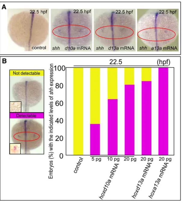
Figure 3.hoxtranscript accumulation is critical for the onset ofshhexpression in fin development.(A) Expression ofshhin pectoral fin primordia ofD. rerioembryos injected with 5 ng control MO, 20 pghoxd10amRNA, 20 pghoxd13amRNA or 20 pghoxa13amRNA at 22.5 hpf. Red ovals highlight the pectoral fin primordia. Note that transcripts ofshhbecame detectable at 22.5 hpf in the fin primordia of embryos injected with
hoxd10a,hoxd13aorhoxa13amRNA. (B) The percentage of embryos with the indicated level ofshhexpression at 22.5 hpf following injection of control MO,hoxd10amRNA,hoxd13amRNA orhoxa13amRNA is shown in the bottom panel (see also Figure S4). A representative image depicting the detectable or undetectable levels ofshhexpression in the pectoral fin primordia is shown at the left.
expression was seen in pectoral fin primordia in 100% of zebrafish embryos injected with 20 pghoxa13amRNA (n = 27, Fig. 3, Fig. S4). Thus, expressing a threshold level ofhoxacould also triggershh expression in pectoral fin primordia (Fig. 4). Our results indicate that specific threshold levels ofhoxgene products likely trigger the heterochronic shift ofshhexpression in pectoral fin primordia.
Temporal shift of Shh activity leads to morphological changes in endoskeletal elements of pectoral fins in zebrafish and dogfish
We next investigated whether a change in the timing of onset of shhexpression induced by injection ofhoxd10aMO could lead to a
change in the zebrafish pectoral fin morphology (Fig. 5A and B). Zebrafish pectoral fins consist of an scapulocoracoid, a post-coracoid process, an endoskeletal disc, and actinotrichs at 5 days post-fertilization (dpf) [36]. Embryos were fixed and stained with Alcian Blue. Measurement of the endoskeletal discs of embryos injected withhoxd10aMO revealed that the total length of the disc along the proximal-distal axis was 8.41% shorter (P,0.001) compared with controls (control embryos, n = 8; hoxd10a MO injected embryos, n = 16; Fig. 5B).
To confirm that a change in the timing of Shh activity during fin development could modify fin size, we treated embryos between 23 and 27 hpf with 60mM cyclopamine, a steroidal alkaloid that inhibits hh signal transduction (Fig. 5C and D).
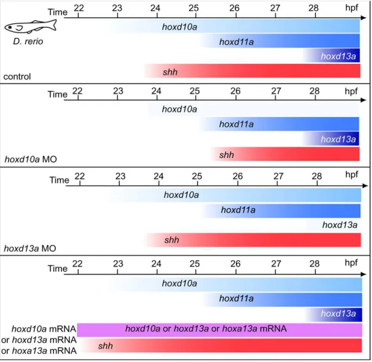
Figure 4. Schematic representation of temporalhoxandshhexpression in pectoral fin primordia of zebrafish embryos.Expression of
shhwas observed at 24 hpf and was concomitant withhoxd10aexpression. The onset ofshhexpression inhoxd10amorphants was concomitant with the onset ofhoxd11aexpression, whereasshhexpression was not delayed inhoxd13amorphants. In embryos injected withhoxd10a,hoxd13aor
Control embryos showed expression of ptc1, a marker for the primary targets of hh signaling, in the posterior margin of fin primordia at 27 hpf and 30 hpf (n = 4 and 5, respectively, Fig. 5D). Expression of ptc1 in cyclopamine-treated embryos was barely detectable in the fin primordia at 27 hpf (n = 5, Fig. 5D), whereas posterior activation ofptc1was readily detectable by 30 hpf (n = 9, Fig. 5D), indicating that cyclopamine treatment efficiently blocked hh signaling through 27 hpf. Thus, stimulation of an artificial heterochronic shift of Shh activity in the pectoral fin primordia was successful. Expression levels ofshh, which are upregulated by a feedback loop of hh signaling, were normal in fin primordia of either ethanol- or cyclopamine-treated embryos at 30 hpf, indicating that Shh activity itself is not required for maintenance of shh expression between 23 and 30 hpf. Taken together, the results indicate thatshhsignal transduction was efficiently blocked in fin primordia of embryos treated with cyclopamine until 27 hpf, but signaling was recovered at least by 30 hpf. To examine the fin morphology at 5 dpf, embryos were fixed and stained with Alcian Blue. Measurements of the cyclopamine-treated endoskeletal discs revealed that the total length of the disc along the proximal-distal axis was 10.6% shorter than those of controls (ethanol-treated embryos, n = 7; cyclopamine-treated embryos, n = 9). The differ-ence in the length between the ethanol- and cyclopamine-treated discs was significant at 0.05 levels by Student’s t-test with Welch’s correction (Fig. 5D). A longer exposure with cyclopamine until 57 hpf resulted in a more severe reduction (19.4%) in the length of the endoskeletal disc (ethanol-treated embryos, n = 7; cyclopamine-treated embryos, n = 9; Fig. S5). This reduction seemed to be depend on both the apical fold activity and shh activity [16]. These results indicate that the temporal shift of the onset ofshhexpression in pectoral fin primordia can lead to a change in the size of the endoskeletal discs along the proximal-distal axis in zebrafish embryos.
We then investigated whether hh signaling can be manipulated in pectoral fins of dogfish embryos (Fig. 5E–H). Prior to the treatment of dogfish embryos with SAG, agonists of smoothened [37], we tested whether SAG is applicable in live embryos using zebrafish and confirmed that we could manipulate hh activity by treatment with SAG (Fig. S5C). We then treated dogfish embryos with cyclopamine or SAG to test whether such treatment could modify hh signaling in developing dogfish embryos. At stage 29, Ptc2expression was observed in the posterior margin of pectoral fins of control embryos, whereas noPtc2transcripts were detected in pectoral fins of cyclopamine-treated embryos (Fig. 5E). On the other hand, treatment with SAG resulted in extensive Ptc2 expression in pectoral fins at stage 31 (Fig. 5E). These data
demonstrated that hh signaling could be directly manipulated in dogfish embryos during fin development.
To examine whether the heterochronic shift of hh activity could alter the morphology of dogfish pectoral fins, we reared SAG-treated dogfish embryos for 11 to 12 weeks and then stained them with Alcian Blue. For SAG-treated embryos (n = 8), the width of the metapterygium was 19.8% greater compared with control embryos (n = 6; Fig. 5G, H). The difference in the metapterygium width between the control- and SAG-treated discs was significant by the Student’s t-test with Welch’s correction (P,0.005; Fig. 5H). Taken together, our results indicate that altering the threshold levels of hox transcripts can trigger a heterochronic shift of shh expression in pectoral fin primordia, and the subsequent temporal shift of Shh activity causes changes in the size of the fin endoskeleton.
Discussion
Our investigation of the genetic basis of vertebrate morpholog-ical evolution has yielded the following findings. (1)Shhexpression appears as soon as there is a morphological bud in mouse and chick embryos (concomitant with Hoxd10), whereas Shh is transcribed very late (concomitant withHoxd13) in pectoral fin buds of dogfish (S. canicula). (2) A threshold level of accumulatedhox transcripts is critical for the timing ofshhexpression; specifically, if the amount ofhoxd10atranscripts is below a threshold level,shh expression does not appear untilhoxd11ais expressed in zebrafish. (3) A quantitative change ofhoxtranscripts leads to changes in the size of the zebrafish endoskeleton. (4) A temporal shift in Shh activation in paired fins leads to a change in endoskeleton size in both dogfish and zebrafish.
Heterochronic shift ofShhtranscriptional onset depends on the quantity ofHox
Examination of collinear 59-locatedHoxaand Hoxdexpression revealed that Shh expression was turned on when Hoxd13 expression appeared, concomitant with a further increase in 59-located Hoxa and Hoxd expression. These results raise the possibility that the late onset ofShh transcription in the pectoral fins ofS. caniculaembryos might correlate with either specificHox transcripts or the overall expression level ofHoxtranscripts. Using zebrafish embryos, which allowed us to alter the levels of specific hox transcripts, we showed that the onset of shh expression is controlled by a certain threshold level of accumulated 59-located hox transcripts. A recent study using Hoxa/Hoxd double mutant mice showed that there is a boundary betweenHoxd9, the lastHox Figure 5. Temporal shift of Shh activity leads to changes in pectoral fin morphology.(A)shhexpression appears at 25.5 hpf in pectoral fin primordia ofD. rerioembryos injected with 5 ng ofhoxd10aMO. (B) At 5 dpf, pectoral fins of embryos injected with control MO or withhoxd10aMO were stained with Alcian Blue (left). Cleithrum (cl), scapulocoracoid (sc), postcoracoid process (pop), endoskeletal disc (ed) and actinotrichs (ac) are indicated. Scale bars: 100mm. The relative lengths of the endoskeletal disc are presented in the graph (right).*P,0.001, as assessed by Student’s
t-test. (C)shhexpression appears at 24 hpf, concomitantly withhoxd10a, in pectoral fin primordia ofD. rerio. Hedgehog signaling was blocked by treatment with 60mM cyclopamine from 23 to 27 hpf, resulting in ablation ofptc1expression until at least 27 hpf.ptc1expression was recovered by
30 hpf in pectoral fin primordia of cyclopamine-treated embryos. (D)ptc1andshhexpression were examined in control or cyclopamine-treated embryos at the indicated stages (left). At 5 dpf, pectoral fins of control or cyclopamine-treated embryos were stained with Alcian Blue (middle). Scale bars: 200mm. The relative lengths of the endoskeletal disc are represented in a graph (right).*P,0.05, as assessed by Student’s t-test with Welch’s correction. (E)ShhandPtc2expression disappeared before stage 31 in pectoral fin buds ofS. caniculaembryos. Hedgehog signaling was extended by treatment with SAG for 6 days from stage 30 to 31, resulting in extension ofPtc2expression until at least stage 31. (F)Ptc2expression was examined in control or SAG-treated embryos at stage 31 (5 days after the initial treatment). (G) Pectoral fins of control or SAG-treated embryos were stained with Alcian Blue. Anterior is to the left. Proximal is to the top. Insets show magnified views of the pectoral fin metapterygium. Note that the width of the metapterygium (arrows) of SAG-treated embryos was significantly increased. Scale bars: 1 mm. (H) Comparison of the size of the pectoral fin endoskeleton between control and SAG-treatedS. canicula embryos. The table shows the total body length (TL), metapterygium length (ML), metapterygium width (MW), width across the base of pectoral fin endoskeleton (WPF), and length of pectoral fin endoskeleton (LPF) of control and SAG-treated embryos. The metapterygium lengths are represented in the bar graph.*P,0.05, as assessed by Student’s t-test.
unable to elicit Shh transcription, and Hoxd10, the first Hox to activateShh[3] —that is, between the genes expressed throughout the limb bud and those excluded from the anterior region. The authors proposed that the limb anterior-posterior polarity arises from the co-option of the collinearHoxgene expression across the main body axis [3]. Importantly, our experiments in dogfish showed thatShhtranscripts do not appear until the onset ofHoxd13 expression regardless of the nested posterior expression ofHoxd10– 12. In other words, theShhdoes not always initiate its expression even when the three penultimate Hoxd genes have already expressed posteriorly in paired appendages. The combination of experiments using both dogfish and zebrafish embryos has demonstrated that 59-located Hox transcripts may not always reach the threshold levels required to stimulate Shh expression, even when the last four Hox paralog groups are expressed posteriorly. Absolute quantification of Hox gene transcripts necessary for Shh activation in mouse limb buds and in dogfish fin buds would allow us to further characterize the mechanisms by whichHoxgene expression thresholds contribute to the evolution of vertebrate paired appendages. Although currently threre are no cartilaginous fishes amenable to transgenics manipulation or MO/ mRNA injection, the prospective manipulation ofHoxexpression levels in these primitive gnathostomes should provide direct insight into our hypothesis of paired appendage evolution.
Hoxand co-factors in heterochronic shift ofShh
activation
During vertebrate evolution, quantitative changes in Hox expression, Hox cofactors, and/or other unknown factors, could have shifted the onset ofShhexpression, leading to changes in the morphology of endoskeleton.Hoxgenes act partially through the aid of co-factors, such as Meis and Pbx [38]. Although 59-located Hox genes have been shown to act throughMeis, we found that only Pbx2expression overlapped with Shh expression in pectoral fins in dogfish embryos (Fig. S6). Furthermore, manipulation of the level of pbx2 expression in zebrafish embryos resulted in a change in the timing of the onset ofshhexpression in pectoral fin primordia in a low percentage of embryos (see Fig. S4 and S6). This may be due to a low level of hoxin pectoral fin primordia. Alternatively, Pbx may make a smaller contribution than Hox to the activation of Shh expression. Biochemical approaches that address the roles of Hox co-factors in the onset ofShhexpression will provide new insights into vertebrate limb evolution.
Signalling pathways that controlHoxexpression levels Signalling that regulatesHoxtranscriptional activation has been studied intensively. Retinoic acid is one of the factors thought to play key roles in controllingHoxgene transcription [39,40,41]. In zebrafish, a lack of retinoic acid in the pectoral fin buds results in the downregulation ofshh,hoxd11andhoxd12[42]. In mice lacking retinoic acid-synthesizing enzyme gene–retinaldehyde dehydrogenase 2 (Raldh2),Shhexpression is greatly reduced in the limb buds and seen along the distal margin, whereas Hoxd11 and Hoxd12 are ectopically expressed in early limb buds [43]. Hox genes are differentially activated by retinoic acid in a concentration-dependent manner and in a sequential order that is collinear with their 39 to 59 arrangement in the cluster [44]. It would be interesting to explore whether retinoic acid reaches levels sufficient to activate 59Hoxdgenes at different times in the posterior paired appendages between dogfish and other tetrapods.
The zinc finger transcriptional factor GLI3 is another protein known to modulateHoxexpression. In early limb buds of mouse embryos, GLI3 negatively regulates the expression of 59-located Hoxdgenes [45,46]. In mouse and chick embryos,Gli3expression
is excluded from the posterior part of the limb buds, whenHand2 expression appears in the posterior region.Gli3, in turn, restricts Hand2expression in the posterior limb buds [47]. Such reciprocal antagonism seems to have been established in cartilaginous fishes, asHand2expression is restricted to the posterior part of pectoral fins in S. canicula [20], indicating GLI3 may be involved in regulatingHoxexpression in the posterior region of dogfish fins. In addition, GLI3 physically interacts with HOXD12 during digit patterning [48]. In this regard, comparative analysis of the expression and function of Gli3 with respect toHox expression would enhance our understanding of the evolution of genetic networks involved in regulatingShhexpression.
Heterochronic shift of Shh onset in vertebrate fin evolution
Our results provide new clues for understanding the sequential events of vertebrate fin/limb evolution, especially with respect to the molecular mechanisms that change the onset ofshhexpression and lead to morphological changes in endoskeletal components (Fig. 6). It has been proposed that paired appendages adopted collinear expression ofHoxfrom the main body axis concomitant with their emergence in the body wall [3,49,50] (Fig. 6). Our results suggest that if threshold levels of accumulated 59 Hox transcripts were not reached, Shh expression may have been delayed or silent in ancestral fin buds. Quantitative changes in accumulated 59 Hox may have led to altered onset of Shh expression, resulting in enlargement of endoskeletal elements during fin evolution (Fig. 6).
Endoskeletal components of paired appendages during the transformation from fins into limbs have been throughly discussed. Comparison of the paired appendages in fossils and in living primitive sarcopterygian fishes (lobe-finned fishes including lungfish and coelacanths) showed that endoskeletal elements of the paired appendages increased in size prior to the acquisition of the digital plates. Thus, the transition from fins to limbs seems to have required at least two major events, namely the enlargement of proximal endoskeletal elements with subsequent acquisition of digital plates. It has been proposed that the transformation of the apical fin fold into the short, apical ectodermal ridge may have promoted endoskeletal proliferation [4]. Here, we demonstrated that a temporal shift in Shh activity could have also led to changes in the size of the endoskeletal elements along the proximal-distal axis.
with Fgfs promote overproliferation of the posterior mesenchymal cells, leading to asymmetric growth of the limb [20,51,52]. Furthermore, recent studies revealed that Shh signalling controls not only the specification of digit progenitors but also cell proliferation in limb buds of chick embryos [14]. Thus, the temporal shift of Shh expression during vertebrate fin/limb evolution could have acted independently of, and/or synergisti-cally with, Fgf signals from the apical fold, which also shift the timing of folding and promote cell proliferation, thereby contributing to the formation of the endoskeleton.
Because zebrafish larval pectoral fins are later remodeled to form the adult pectoral fins, it is difficult to speculate which endoskeletal components of paired fins among primitive fishes may have been affected by temporal changes inshhexpression during evolution. Furthermore, the metapterygium was lost in the teleost lineage. Therefore, examination of these features in the paired appendages of the primitive cartilaginous dogfish is highly informative. Although dogfish embryos did not survive beyond 2 weeks after cyclopamine treatment (presumably due to the multiple malformations; data not shown), we have succeeded in keeping them alive for 12 weeks after treatment with SAG (Fig. 5G, H). We showed that extension of Shh activity using SAG could enlarge the metapterygium of dogfish pectoral fins. The metapterygium, a proximal component of the dogfish fin, has been considered to have persisted in sarcopterygian fishes and was the ancestral structure from which the tetrapod limb evolved. Enlargement of the dogfish fin metapterygium by extending Shh activity indicates that Shh could have promoted the proliferation of cells that formed the proximal structures among ancestral species. We propose that a heterochronic shift of the onset ofShh expression could have been mediated by changes in the level of Hox (and Hox co-factors) and that such transcriptional heteroch-rony could have influenced the proliferation of cells that contributed to the formation of endoskeletal components during vertebrate paired appendage evolution (Fig. 6). It would not be
surprising if such a system controls the morphological diversifica-tion of paired appendages in different lineages (including lineages of cartilaginous fishes). It will be interesting to characterize these features of the body plan among different vertebrates having various types of paired appendages.
Supporting Information
Figure S1 Expression of Shh and Fgfs during S. canicula fin development. (A) RT-PCR ofScShhin stage 29 and 32S. canicula pectoral fin buds (left); results for stage 27S. caniculaembryos have been published [20]. The right panel shows semi-quantitative analysis ofScShhmRNA expression in pectoral fins relative to the ScGAPDH mRNA level. (B) Frontal view of the facial region at stage 27. (C–D, FG) Pectoral fin buds. Anterior is to the left. (B–D) ScFgf8 expression at stage 27 (B, C) and 32 (D). Although transcripts were observed in nasal pits (np) and gill filaments (gf), no transcripts were detected in the apical fin fold (aff). (E) RT-PCR of ScFgf8 in head (Head) and pectoral fins (Pec) of S. canicula embryos. (F) Staining of anti-Fgf4 antibody at stage 27. Arrowheads indicate anti-Fgf4-positive cells in the apical fin fold. (G) Scb-catenin expression at stage 32. Abundant Scb-catenin transcripts in pectoral fins including the apical fin fold (arrow-heads) demonstrates probe efficacy.
Found at: doi:10.1371/journal.pone.0005121.s001 (8.68 MB TIF)
Figure S2 Expression ofhoxd10a,hoxd11aandhoxd13aduringD. rerio pectoral fin development. Dorsal view of embryos injected with 5 ng of the control morpholino (MO) at 24, 25.5, 27 and 28 hpf. Red ovals highlight the pectoral fin primordia. Expression of hoxd10awas initially detected at 24 hpf,hoxd11aat 25.5 hpf, and hoxd13aat 28 hpf.
Found at: doi:10.1371/journal.pone.0005121.s002 (8.08 MB TIF)
Figure S3 Quantitative PCR analyses of hoxd11a and shh expression in the lateral plate mesoderm of zebrafish embryos. Figure 6. Diagram representing the effect ofShhexpression heterochrony on vertebrate paired appendage evolution.A model suggesting that the early fin buds may have acquired low levels ofHoxexpression by co-option of collinearHoxexpression in the main body axis [53]. Changes in accumulatedHoxcould have led to altered onset ofShhexpression, resulting in enlargement of the endoskeletal elements during fin evolution.
Levels of hoxd11a(A) and shh(B–D) mRNAs in the lateral plate mesoderm of embryos were quantified relative to thegapdhmRNA level. (A–D) Expression levels ofhoxd11a(A) andshh(B, C) in the lateral plate mesoderm of 24 hpf (B) and 25.5 hpf (A, C) embryos injected with 5 ng control, 1 nghoxd10a, or 2.5 nghoxd10aMO. (D) Quantitative PCR analyses to determine the expression levels ofshhin the lateral plate mesoderm of embryos injected with 5 ng
control MO, 5 pg hoxd10a mRNA, or 20 pg hoxd10a mRNA.
Expression of shhwas undetectable by quantitative PCR in 22.5 hpf injected with 5 ng control MO.
Found at: doi:10.1371/journal.pone.0005121.s003 (2.32 MB TIF)
Figure S4 Onset of shh expression in zebrafish embryo fin primordia primarily depends onhoxexpression. The percentage of D. rerioembryos expressing the indicated level ofshhtranscript at 22.5, 24, or 25.5 hpf following injection of the indicated amount of control MO,hoxd10aMO,hoxd13aMO,hoxd10amRNA,hoxd13a
mRNA, hoxa13amRNA, pbx2MO or pbx2 mRNA is shown. A
representative image depicting the detectable or undetectable levels of shh expression in the pectoral fin primordia is shown in Figure 2D.
Found at: doi:10.1371/journal.pone.0005121.s004 (0.44 MB EPS)
Figure S5 Treatment of zebrafish embryos with cyclopamine or SAG. (A) Hedgehog signaling was blocked by treatment with 60mM cyclopamine from 23 to 57 hpf, resulting in ablation ofptc1 expression until at least 60 hpf.ptc1expression recovered by 72 hpf in pectoral fin primordia of cyclopamine-treated embryos. (B)ptc1 expression was examined in control or cyclopamine-treated embryos at the indicated stages (left). At 5 dpf, pectoral fins of control (n = 7) or cyclopamine-treated embryos (n = 9) were stained with Alcian Blue (middle). The relative lengths of the endoskeletal disc are presented in the graph (right). *P,1026
, as assessed by Student’s t-test. Cleithrum (cl), scapulocoracoid (sc), postcoracoid process (pop), endoskeletal disc (ed) and actinotrichs (ac) are indicated. Scale bars: 200mm. (C) Zebrafish embryos were treated with SAG or cyclopamine, and ptc1 expression was
examined in adaxial cells. The specification of adaxial cells is known to depend on Hh signaling [1]. Panels show the dorsal view ofptc1expression in an 8-somite-stage control embryo (left), in a SAG-treated embryo (middle), and in a cyclopamine-treated embryo (right). In control embryo, adaxial cells are indicated by brackets. Note thatptc1expression is expanded in the SAG-treated embryo (brackets), whereas it is undetectable in the cyclopamine-treated embryo (right). 1. Wolff C, Roy S, Ingham PW (2003) Multiple muscle cell identities induced by distinct levels and timing of hedgehog activity in the zebrafish embryo. Curr Biol 13: 1169– 1181.
Found at: doi:10.1371/journal.pone.0005121.s005 (7.33 MB TIF)
Figure S6 The hox co-factor pbx makes a lesser contribution than hox to the onset ofshh expression. (A) Expression ofMeis1 andPbx2in the pectoral fin ofS. caniculaembryos at the indicated stages (top panels). Anterior is to the left. Arrowheads indicate transcripts in the proximal region. (B) Left: representative images depicting the detectability of shh expression in the pectoral fin primordia. Right: the percentage of D. rerio embryos with the indicated level ofshhexpression observed at 22.5, 24, and 25.5 hpf following injection of control MO,pbx2MO, orpbx2mRNA. Found at: doi:10.1371/journal.pone.0005121.s006 (9.69 MB TIF)
Acknowledgments
We thank C. Tickle for her generous support and helpful comments. We also thank A. A. W. Tweedale for collectingS. caniculaembryos, E. Tiecke and A. Bain for fixing embryos, Y. Murata, Y. Aita and K. Yoshida for technical assistance, S. Hirose, M. Okabe, K. Shirahige and N. Okada for allowing us to use their facilities, and K. Hoshijima for technical advice.
Author Contributions
Conceived and designed the experiments: MT. Performed the experi-ments: KS KO KM NS MT HO MT. Analyzed the data: KS KO KM NS MT HO MT. Contributed reagents/materials/analysis tools: KS KO KM NS MT HO MT. Wrote the paper: MT.
References
1. Sordino P, van der Hoeven F, Duboule D (1995) Hox gene expression in teleost fins and the origin of vertebrate digits. Nature 375: 678–681.
2. Kmita M, Tarchini B, Zakany J, Logan M, Tabin CJ, et al. (2005) Early developmental arrest of mammalian limbs lacking HoxA/HoxD gene function. Nature 435: 1113–1116.
3. Tarchini B, Duboule D, Kmita M (2006) Regulatory constraints in the evolution of the tetrapod limb anterior-posterior polarity. Nature 443: 985–988. 4. Thorogood P (1991) The development of the Teleost fin and implications for our
understanding of tetrapod limb evolution. In: Hinchliffe JR, Hurle JM, Summerbell D, eds. Developmental Patterning of the Vertebrate Limb. New York: Plenum Pub Corp.
5. Saunders JW Jr, Gasseling MT (1968) Ectoderm-mesenchymal interaction in the origin of wing symmetry. In: Fleischmajer R, Billingham RE, eds. Epithelial– Mesenchymal Interactions. Baltimore: Williams and Wilkins. pp 78–97. 6. Tickle C, Summerbell D, Wolpert L (1975) Positional signalling and
specification of digits in chick limb morphogenesis. Nature 254: 199–202. 7. Tickle C (1981) The number of polarizing region cells required to specify
additional digits in the developing chick wing. Nature 289: 295–298. 8. Riddle RD, Johnson RL, Laufer E, Tabin C (1993) Sonic hedgehog mediates the
polarizing activity of the ZPA. Cell 75: 1401–1416.
9. Echelard Y, Epstein DJ, St-Jacques B, Shen L, Mohler J, et al. (1993) Sonic hedgehog, a member of a family of putative signaling molecules, is implicated in the regulation of CNS polarity. Cell 75: 1417–1430.
10. Krauss S, Concordet JP, Ingham PW (1993) A functionally conserved homolog of the Drosophila segment polarity gene hh is expressed in tissues with polarizing activity in zebrafish embryos. Cell 75: 1431–1444.
11. Yang Y, Drossopoulou G, Chuang PT, Duprez D, Marti E, et al. (1997) Relationship between dose, distance and time in Sonic Hedgehog-mediated regulation of anteroposterior polarity in the chick limb. Development 124: 4393–4404.
12. Harfe BD, Scherz PJ, Nissim S, Tian H, McMahon AP, et al. (2004) Evidence for an expansion-based temporal Shh gradient in specifying vertebrate digit identities. Cell 118: 517–528.
13. Scherz PJ, McGlinn E, Nissim S, Tabin CJ (2007) Extended exposure to Sonic hedgehog is required for patterning the posterior digits of the vertebrate limb. Dev Biol 308: 343–354.
14. Towers M, Mahood R, Yin Y, Tickle C (2008) Integration of growth and specification in chick wing digit-patterning. Nature 452: 882–886.
15. Zhu J, Nakamura E, Nguyen MT, Bao X, Akiyama H, et al. (2008) Uncoupling Sonic hedgehog control of pattern and expansion of the developing limb bud. Dev Cell 14: 624–632.
16. Neumann CJ, Grandel H, Gaffield W, Schulte-Merker S, Nusslein-Volhard C (1999) Transient establishment of anteroposterior polarity in the zebrafish pectoral fin bud in the absence of sonic hedgehog activity. Development 126: 4817–4826.
17. Dahn RD, Davis MC, Pappano WN, Shubin NH (2007) Sonic hedgehog function in chondrichthyan fins and the evolution of appendage patterning. Nature 445: 311–314.
18. Charite J, McFadden DG, Olson EN (2000) The bHLH transcription factor dHAND controls Sonic hedgehog expression and establishment of the zone of polarizing activity during limb development. Development 127: 2461–2470. 19. Crossley PH, Minowada G, MacArthur CA, Martin GR (1996) Roles for FGF8
in the induction, initiation, and maintenance of chick limb development. Cell 84: 127–136.
20. Tanaka M, Munsterberg A, Anderson WG, Prescott AR, Hazon N, et al. (2002) Fin development in a cartilaginous fish and the origin of vertebrate limbs. Nature 416: 527–531.
21. Maruoka Y, Ohbayashi N, Hoshikawa M, Itoh N, Hogan BL, et al. (1998) Comparison of the expression of three highly related genes, Fgf8, Fgf17 and Fgf18, in the mouse embryo. Mech Dev 74: 175–177.
22. Niswander L, Martin GR (1992) Fgf-4 expression during gastrulation, myogenesis, limb and tooth development in the mouse. Development 114: 755–768.
24. Sun X, Mariani FV, Martin GR (2002) Functions of FGF signalling from the apical ectodermal ridge in limb development. Nature 418: 501–508. 25. Zakany J, Kmita M, Duboule D (2004) A dual role for Hox genes in limb
anterior-posterior asymmetry. Science 304: 1669–1672.
26. Capellini TD, Di Giacomo G, Salsi V, Brendolan A, Ferretti E, et al. (2006) Pbx1/Pbx2 requirement for distal limb patterning is mediated by the hierarchical control of Hox gene spatial distribution and Shh expression. Development 133: 2263–2273.
27. Kimmel CB, Ballard WW, Kimmel SR, Ullmann B, Schilling TF (1995) Stages of embryonic development of the zebrafish. Dev Dyn 203: 253–310. 28. Westerfield M (2000) A guide for the laboratory use of zebrafish (Danio rerio). The
zebrafish book. Eugene: University of Oregon Press.
29. Horigome N, Myojin M, Ueki T, Hirano S, Aizawa S, et al. (1999) Development of cephalic neural crest cells in embryos of Lampetra japonica, with special reference to the evolution of the jaw. Dev Biol 207: 287–308.
30. Waskiewicz AJ, Rikhof HA, Moens CB (2002) Eliminating zebrafish pbx proteins reveals a hindbrain ground state. Dev Cell 3: 723–733.
31. Thisse B, Heyer V, Lux A, Alunni V, Degrave A, et al. (2004) Spatial and temporal expression of the zebrafish genome by large-scale in situ hybridization screening. Methods Cell Biol 77: 505–519.
32. Covassin L, Amigo JD, Suzuki K, Teplyuk V, Straubhaar J, et al. (2006) Global analysis of hematopoietic and vascular endothelial gene expression by tissue specific microarray profiling in zebrafish. Dev Biol 299: 551–562.
33. Kimmel CB, Miller CT, Kruze G, Ullmann B, BreMiller RA, et al. (1998) The shaping of pharyngeal cartilages during early development of the zebrafish. Dev Biol 203: 245–263.
34. Davis MC, Dahn RD, Shubin NH (2007) An autopodial-like pattern of Hox expression in the fins of a basal actinopterygian fish. Nature 447: 473–476. 35. Freitas R, Zhang G, Cohn MJ (2007) Biphasic Hoxd gene expression in shark
paired fins reveals an ancient origin of the distal limb domain. PLoS ONE 2: e754.
36. Grandel H, Schulte-Merker S (1998) The development of the paired fins in the zebrafish (Danio rerio). Mech Dev 79: 99–120.
37. Chen JK, Taipale J, Young KE, Maiti T, Beachy PA (2002) Small molecule modulation of Smoothened activity. Proc Natl Acad Sci U S A 99: 14071–14076.
38. Burglin TR (1997) Analysis of TALE superclass homeobox genes (MEIS, PBC, KNOX, Iroquois, TGIF) reveals a novel domain conserved between plants and animals. Nucleic Acids Res 25: 4173–4180.
39. Serpente P, Tumpel S, Ghyselinck NB, Niederreither K, Wiedemann LM, et al. (2005) Direct crossregulation between retinoic acid receptor {beta} and Hox genes during hindbrain segmentation. Development 132: 503–513.
40. Gould A, Itasaki N, Krumlauf R (1998) Initiation of rhombomeric Hoxb4 expression requires induction by somites and a retinoid pathway. Neuron 21: 39–51.
41. Huang D, Chen SW, Langston AW, Gudas LJ (1998) A conserved retinoic acid responsive element in the murine Hoxb-1 gene is required for expression in the developing gut. Development 125: 3235–3246.
42. Gibert Y, Gajewski A, Meyer A, Begemann G (2006) Induction and prepatterning of the zebrafish pectoral fin bud requires axial retinoic acid signaling. Development 133: 2649–2659.
43. Niederreither K, Vermot J, Schuhbaur B, Chambon P, Dolle P (2002) Embryonic retinoic acid synthesis is required for forelimb growth and anteroposterior patterning in the mouse. Development 129: 3563–3574. 44. Simeone A, Acampora D, Arcioni L, Andrews PW, Boncinelli E, et al. (1990)
Sequential activation of HOX2 homeobox genes by retinoic acid in human embryonal carcinoma cells. Nature 346: 763–766.
45. Buscher D, Bosse B, Heymer J, Ruther U (1997) Evidence for genetic control of Sonic hedgehog by Gli3 in mouse limb development. Mech Dev 62: 175–182. 46. Zuniga A, Zeller R (1999) Gli3 (Xt) and formin (ld) participate in the positioning of the polarising region and control of posterior limb-bud identity. Development 126: 13–21.
47. te Welscher P, Fernandez-Teran M, Ros MA, Zeller R (2002) Mutual genetic antagonism involving GLI3 and dHAND prepatterns the vertebrate limb bud mesenchyme prior to SHH signaling. Genes Dev 16: 421–426.
48. Chen Y, Knezevic V, Ervin V, Hutson R, Ward Y, et al. (2004) Direct interaction with Hoxd proteins reverses Gli3-repressor function to promote digit formation downstream of Shh. Development 131: 2339–2347.
49. Coates MI (1993) Hox genes, fin folds and symmetry. Nature 364: 195–196. 50. Thorogood P, Ferretti P (1993) Hox gene, fin folds and symmetry. Nature 364:
196.
51. Niswander L, Jeffrey S, Martin GR, Tickle C (1994) A positive feedback loop coordinates growth and patterning in the vertebrate limb. Nature 371: 609–612. 52. Laufer E, Nelson CE, Johnson RL, Morgan BA, Tabin C (1994) Sonic hedgehog and Fgf-4 act through a signaling cascade and feedback loop to integrate growth and patterning of the developing limb bud. Cell 79: 993–1003.