Submitted10 August 2015 Accepted 5 January 2016 Published25 January 2016
Corresponding author Piotr S. Fudalej,
Academic editor Satheesh Elangovan
Additional Information and Declarations can be found on page 11
DOI10.7717/peerj.1625
Copyright 2016 Von Böhl et al.
Distributed under
Creative Commons CC-BY 4.0
OPEN ACCESS
Age-related changes of dental pulp tissue
after experimental tooth movement in
rats
Martina Von Böhl1, Yijin Ren2, Anne M. Kuijpers-Jagtman1,
Piotr S. Fudalej3,4and Jaap C. Maltha1
1Department of Orthodontics and Craniofacial Biology, Radboud University Nijmegen, Nijmegen,
The Netherlands
2Department of Orthodontics, University of Groningen, University Medical Centre Groningen,
Groningen, Griningen, The Netherlands
3Department of Orthodontics and Dentofacial Orthopedics, University of Bern, Bern, Switzerland 4Department of Orthodontics, Institute of Dental Science, Palacký University Olomouc, Olomouc,
Czech Republic
ABSTRACT
It is generally accepted that the effect of orthodontic tooth movement on the dental pulp in adolescents is reversible and that it has no long-lasting effect on pulpal physiology. However, it is not clear yet if the same conclusion is also valid for adult subjects. Thus, in two groups of rats, aged 6 and 40 weeks respectively, 3 molars at one side of the maxilla were moved together in a mesial direction with a standardized orthodontic appliance delivering a force of 10 cN. The contralateral side served as a control. Parasagittal histological sections were prepared after tooth movement for 1, 2, 4, 8, and 12 weeks. The pulp tissue was characterized for the different groups, with special emphasis on cell density, inflammatory cells, vascularity, and odontoblasts. Dimensions of dentin and the pulpal horns was determined and related with the duration of orthodontic force application and age ware evaluated. We found that neither in young nor in adult rats, force application led to long-lasting or irreversible changes in pulpal tissues. Dimensional variables showed significant age-related changes. In conclusion, orthodontic tooth movement per se has no long-lasting or irreversible effect on pulpal tissues, neither in the young nor in the adult animals.
SubjectsZoology, Dentistry, Histology
Keywords Dental pulp, Orthodontics, Wear, Ageing, Rats, Tooth movement
INTRODUCTION
Conflicting results have been presented on the putative adverse effects of orthodontic tooth movement on the dental pulp. Some claim permanent damage (Hargreaves, Goodis & Seltzer, 2002), but others find no significant long-lasting effects (Unsterseher et al., 1987). It is well known that the application of orthodontic forces induces the release of proinflammatory cytokines in the dental pulp, leading to a temporal acute inflammation and hyperemia (Raiden et al., 1998). Part of this reaction is an upregulation of 1α, IL-1β, IL-3, IL-6, and TNFα(Bletsa, Berggreen & Brudvik, 2006;Yamaguchi et al., 2004) and apoptosis (Perinetti et al., 2004;Perinetti et al., 2005;Shigehara, Matsuzaka & Inoue, 2006;
Studies in adult rats have shown that the vascular reaction shows biphasic characteris-tics. An initial decrease for approximately 30 min is followed by a temporary increase in blood flow for the subsequent 24–72 h (Santamaria et al., 2007;Santamaria et al., 2006). Others, however, reported a reversible increase in the number of blood vessels during the first three to seven days of force application (Abi-Ramia et al., 2010;Nixon et al., 1993;
Shigehara, Matsuzaka & Inoue, 2006). However, after 6 weeks the vascularity of the pulp had returned to normal, even in cases with severe root resorption (Tripuwabhrut et al., 2010). Studies on isolated human pulp cells derived from premolars extracted during orthodontic tooth movement have shown that increase in vascularity might be caused by a stimulation of angiogenesis through an upregulation of VEGF, FGF2, PDGF, and TGFβ (Derringer & Linden, 1998;Derringer & Linden, 2003;Derringer & Linden, 2004).
Such temporal changes in pulpal blood flow are not only found during tipping movements in rats, but also during intrusion and extrusion in human adults (Barwick & Ramsay, 1996;Sano et al., 2002) and adolescents (Raiden et al., 1998;Ramazanzadeh et al., 2009;Subay et al., 2001).
Temporal vacuolization of the odontoblasts or disruption of the odontoblastic layer in the dental pulp is often described in adolescent humans after intrusion or extrusion (Ramazanzadeh et al., 2009;Stenvik & Mjor, 1971) and in rats and dogs during orthodon-tic tooth movement (Abi-Ramia et al., 2010;Anstendig & Kronman, 1972) or during intrusion (Abi-Ramia et al., 2010;Konno et al., 2007;Santamaria et al., 2007) . However, others reported no effects in rats during tipping movement (Abi-Ramia et al., 2010;
Santamaria et al., 2007).
The above mentioned studies have been performed in adolescent humans or in young adult animals. They suggest that pulpal reactions, evoked by orthodontic interventions are reversible and have no long-lasting effect on pulpal physiology. However, an ever-growing number of adult and elderly individuals seek orthodontic treatment and the question arises whether pulpal reactions on orthodontic therapy change with age.
It is well known that canal and chamber volume is inversely proportional to age. Reparative dentin resulting from restorative procedures, trauma, attrition, and recurrent caries also contributes to decrease of canal and chamber size (Hargreaves & Cohen, 2011). Other age-related changes in the pulp are compromised circulation and innervation, fat droplet deposition, odontoblastic vacuolization, reticular atrophy, pulpal fibrosis, hyaline degeneration, mucoid degeneration, and diffuse calcification (Bernick & Nedelman, 1975;
Morse, 1991).
MATERIAL AND METHODS
Ethical permission for the study was obtained according to the guidelines of the Board for Animal Experiments of the Radboud University Nijmegen.
Two groups of 30 male Wistar rats, one aged 6 weeks (150–250 g) and the other 40 weeks (400–550 g) respectively, were acclimatized for 2 weeks before the start of the experiment. They were housed under normal laboratory conditions and fed powdered rat chow (Sniff, Soest, The Netherlands) and water ad libitum. A standard 12-hour light-dark cycle was maintained. The age of 6 weeks in rats corresponds with a circumpubertal age in humans (i.e., 12–15 years), and the age of 40 weeks corresponds with adult age in humans (Sengupta, 2013).
A split-mouth design was used with the experimental side randomly chosen and the contralateral side as the control. Power analysis with the following assumptions:α=0.05, β=0.2,d=5, ands=3, demonstrated thatN =6 per group (i.e., 6 experimental and 6 control sides for each time point) is sufficient for the analyses with a power of 0.8.
An orthodontic appliance was placed only on the experimental side under general anesthesia (FFM-mix 2.8 ml/kg intraperitoneally, containing fluanisone 6.8 mg/kg, fentanyl 0.1 mg/kg, and midazolam 3.4 mg/kg.) (Janssen, Beerse, Belgium). The appliance has been described extensively elsewhere (Ren, Maltha & Kuijpers-Jagtman, 2004;Ren et al., 2003). In brief, a transverse hole was drilled through the middle root level of the upper incisors and alveolar bone and a stainless steel ligature wire was put through it. A preformed ligature wire (80.20 mm) enclosing all three upper molars was bonded (Clearfil SE Bond, Kuraray Europe GmbH, Dusseldorf, Germany) to the buccal side of the molars. By this the three molars could be moved mesially as one unit by a SentalloyR closed coil spring (10 cN,8 0.22 mm, eyelet80.56 mm, GAC, NY, USA) that was attached to the ligature through the upper incisors at one end and the ligature around the three molars at the other end (Fig. 1). This appliance has two major advantages. First, the molar unit is almost bodily moved related to the relative large mesiodistal dimensions of the molar block that is to be moved, and second, the force per molar is comparable to normally used forces in the clinical situation (Ren, Maltha & Kuijpers-Jagtman, 2004;Ren et al., 2003) . After a force application for 1, 2, 4, 8, or 12 weeks, 6 rats from the ‘‘young’’ and 6 animals of the ‘‘old’’ groups were killed.
Histological procedure
The rats received a lethal dose of anesthetic before they were killed. They were then perfused with 4% paraformaldehyde solution in 0.1 M PBS at 37 ◦C. The maxillae were dissected and immersed in 4% paraformaldehyde for 24 h at 4 ◦C, then they were rinsed in 0.1 M
phosphate buffered saline (PBS). After decalcification in 10% ethylenediaminetetraacetate (EDTA) and paraffin embedding, serial parasagittal 7µm sections were cut. Every 25th
section was collected and stained with hematoxylin and eosin.
Measurements
Figure 1 Schematic drawing of the orthodontic appliance.The Sentalloy spring delivers a continuous force of 10 cN on all three molars together.
ipsilateral incisor at the gingival gingival level (I-M distance) at the following time points 0, 1, 2, 4, 8, and 12 weeks. The experimental tooth movement was calculated as the changes in the difference between the I-M distances at the experimental and the conrol side. By this method, possible confounding factors as anchorage loss from the incisors and physiological distal drift of the molars is compensated for.
From each rat three sections were selected from both the experimental and the control side, in which at least one root was completely present including the pulp chamber and the apical foramen. From these sections, the histological features of the dental pulp were described, with special emphasis on odontoblastic layer, blood vessels, cell density, and inflammatory cells in the pulp chamber.
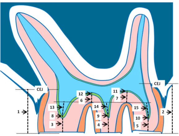
Figure 2 Schematic drawing of a parasagittal section of a rat molar.The measured parameters are indi-cated. For explanation see text.
Statistical analysis
The differences between the amount of tooth movement between the young and the old animals was analyzed by Studentst tests. The data of the dimensional measurements on the molars from the two age groups were initially analyzed by a two-way ANOVA on the raw data of all variables, with the group (experimental or control), and the duration of force application (1, 2, 4, 8, or 12 weeks) as independent factors. As this analysis showed no significant group effect for any of the variables, the data from the experimental and the control sides were pooled. Subsequently, the time effects were analyzed by one-way ANOVA on all dimensional variables in both age groups separately with the experimental period as independent variable.
A separate one-way ANOVA was performed after the data from both age groups were combined to study the long term changes in these parameters with the real age of the animals as independent factor. A linear regression analysis was performed and the explained variance was calculated for all parameters in the two age groups and in the combined age group.
RESULTS
Experimental tooth movement
Figure 3 Histological sections of young (A, B, C) and old rats (D, E, F).The most clear difference be-tween the control and experimental pulpal tissue is that the latter show some more and wider blood ves-sels. The most prominent difference between the young and old animals is that in the latter, the odonto-blasts are more well-organized and active, and the predentin layer is more pronounced. The low-power pictures show the dramatic difference in wear between the young and old animals. All sections H & E staining.
1.05±0.41mm/w for the young and 0.38±0.28 mm/w for the old animals. The rate of tooth movement slowed down to about 0.05 mm/w for the young and 0.02 mm/w for the old animals at the end of the experimental period. The total tooth movement over the experimental period of 12 weeks was 2.69±0.62 mm for the young and 1.23±0.56 mm for the old animals, and the tooth movement curves can best be described by logarithmic equations, showing an R2 of 0.996 and 0.965 for the young and the old group, respectively.
Histological dental pulp tissue survey Control side in young rats
No histological differences were encountered between animals, which were for a short (1–2 weeks) or long period (8–12 weeks) in the experiment. The pulps in the control teeth showed only very few inflammatory cells and small blood vessels (Fig. 3A). The odontoblastic layer and cementum surface showed an irregular morphology. At the roof of the pulp chamber and in the pulpal horns just a thin predentine layer was seen (Fig. 3A). The tips of the cusps showed very little or no wear (Fig. 3C).
Experimental side in young rats
The pulp of the experimental teeth showed a lower cell density than the controls, and inflammatory cells were scarce. The number of blood vessels was increased compared to the controls and in general their diameter was larger (Fig. 3B). The odontoblastic layer and cementum surface were irregular. The predentin layer at the top of the pulp chamber appeared to be slightly thicker than in the controls (Fig. 3B). Similar to the control animals, the tips of the cusps showed very little or no wear.
Control side in old rats
No substantial histological differences were found between animals that stayed for a short (1–2 weeks) or long period (8–12 weeks) in the experiment. In the control teeth from the old group, the pulpal cell density was less than in the young animals. Only few inflammatory cells were present. A limited number of small blood vessels were present throughout the pulp (Fig. 3D).
The odontoblastic layer and predentin were well organized. At the top of the pulp chamber the predentin layer tended to be somewhat thicker than in the young animals (Fig. 3D). The tips of the cusps showed severe wear (Fig. 3F).
Experimental side in old rats
Again, no substantial histological differences were found between animals that stayed for a short (1–2 weeks) or long period (8–12 weeks) in the experiment. The number of inflammatory cells in the dental pulp was larger than in the control teeth. The number of blood vessels was larger and they showed a larger diameter compared to the controls (Fig. 3E). The odontoblastic cell layer and the predentin surface were organized in a regular way. The predentin layer in the pulpal horns and the roof of the pulp chamber was thicker than in the young animals and similar to the control animals in the old group (Fig. 3E). The tips of the cusps showed severe wear, similar to the controls. The amount of wear increased during the experimental period.
Dimensional parameters
Figs. 3Cand3Fillustrate the general morphology of the first molars in the young and the old group.
Two-way ANOVA with the group (experimental or control), and duration of force application (1, 2, 4, 8, or 12 weeks) as independent factors showed no significant group effect for any of the variables. Therefore, the data from the experimental and the control groups were pooled (Table 1).
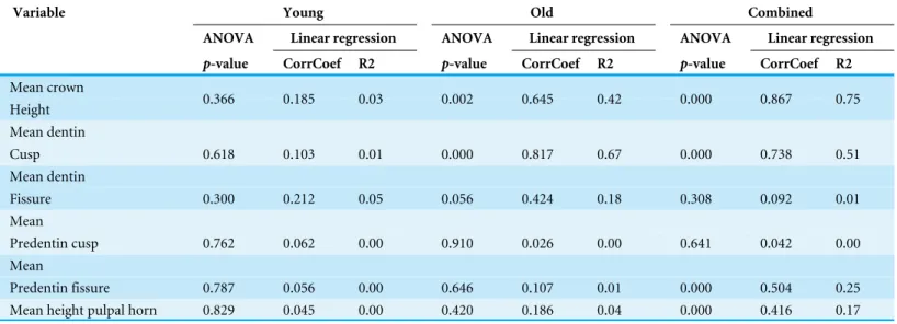
The one-way ANOVA for none of the mean variables in the young group showed any significant time effect, low correlation coefficients (R≤0.2) and a very small explained variance (R2≤0.05) (Table 2). In the old group only the mean crown height showed a significant decrease, and the mean thickness of the dentin in the cusps a significant increase over time. The explained variance (R2) was 0.42 and 0.67 respectively (Table 2).
Table 1 Means and standard deviations (sd) for the combined data of the dimensional parameters in the young and the adult groups.
Age (weeks) Crown Dentin Predentin Pulpal horn
Height Cusp Fissure Cusp Fissure Height
Mean sd Mean sd Mean sd Mean sd Mean sd Mean sd
Young group
7 33.3 2.5 16.5 2.5 12.4 1.9 0.8 0.2 0.1 0.2 2.5 3.4
8 30.0 2.5 14.4 2.2 13.1 2.2 0.8 0.3 0.1 0.1 3.1 2.7
10 29.4 4.0 16.5 1.9 13.3 2.4 1.0 0.3 0.1 0.2 1.5 2.2
14 31.0 2.7 16.0 2.5 14.3 2.6 1.0 0.1 0.1 0.1 2.7 2.4
18 33.4 3.7 15.2 1.3 14.7 2.6 0.8 0.2 0.1 0.1 3.2 1.8
Old group
41 17.8 3.1 6.2 2.5 14.3 0.8 0.9 0.2 0.4 0.2 −0.3 1.5
42 18.4 2.7 6.6 2.4 15.2 1.1 0.8 0.3 0.2 0.2 −1.2 1.1
44 20.6 2.8 7.0 3.0 10.8 2.8 0.7 0.1 0.5 0.3 1.6 2.8
48 18.2 3.4 8.1 3.3 10.4 4.4 0.8 0.2 0.4 0.4 0.6 2.1
52 8.3 4.1 13.4 4.8 14.1 2.0 1.1 0.4 0.5 0.3 1.6 1.8
Table 2 Statistical analysis of the effect of experimental period (in the young and the old group) and real age (in the combined data) for the di-mensional measurements.
Variable Young Old Combined
ANOVA Linear regression ANOVA Linear regression ANOVA Linear regression
p-value CorrCoef R2 p-value CorrCoef R2 p-value CorrCoef R2
Mean crown
Height 0.366 0.185 0.03 0.002 0.645 0.42 0.000 0.867 0.75
Mean dentin
Cusp 0.618 0.103 0.01 0.000 0.817 0.67 0.000 0.738 0.51
Mean dentin
Fissure 0.300 0.212 0.05 0.056 0.424 0.18 0.308 0.092 0.01
Mean
Predentin cusp 0.762 0.062 0.00 0.910 0.026 0.00 0.641 0.042 0.00
Mean
Predentin fissure 0.787 0.056 0.00 0.646 0.107 0.01 0.000 0.504 0.25
Mean height pulpal horn 0.829 0.045 0.00 0.420 0.186 0.04 0.000 0.416 0.17
Notes.
CorrCoef, Correlation coefficient.
the dentin layer in cusps (Fig. 4B). Both variables showed a high explained variance (R2) of 0.745 and 0.506 respectively. Both other variables, showing a significant time dependency show far smaller explained variances, namely 0.247 for the mean predentin thickness in the fissure (Fig. 4C) and 0.172 for the mean height of the pulpal horn (Fig. 4D).
Figure 4 Scatter plots of the combined measurement data from the animals from the young and the old group.Linear regression s lines and 95% CI are given.
in the young group than in the old one, and finally the height of the pulpal horn in the young group is larger than in the old group (p-values for all these variables < 0.05).
DISCUSSION
present study only minor effects of orthodontic force application on pulpal tissues became apparent.
Another reason that no clear effects on pulpal tissues have been found in the present study might be that our appliance induced an almost bodily movement of the molar block (Ren, Maltha & Kuijpers-Jagtman, 2004;Ren et al., 2003), while others used tipping movements of the first molar only (Abi-Ramia et al., 2010;Bletsa, Berggreen & Brudvik, 2006;Grunheid, Morbach & Zentner, 2007;Santamaria et al., 2007;Shigehara, Matsuzaka & Inoue, 2006;Tripuwabhrut et al., 2010), intrusion (Barwick & Ramsay, 1996;Ikawa et al., 2001;Konno et al., 2007;Raiden et al., 1998;Ramazanzadeh et al., 2009;Stenvik, 1971;
Veberiene et al., 2009), or extrusion (Ramazanzadeh et al., 2009;Stenvik, 1971;Subay et al., 2001).
The only pulpal change that was consistently found was a small increase in the vascularity in both the young and the old tooth movement group. This is in agreement with several other authors (Nixon et al., 1993;Raiden et al., 1998;Sano et al., 2002;Shigehara, Matsuzaka & Inoue, 2006;Wong et al., 1999). A drawback of the chosen measurement protocol in which the first histological evaluation is performed after one week is that short-lasting changes in the pulpal tissue, as described in the literature will be missed (Barwick & Ramsay, 1996;Bletsa, Berggreen & Brudvik, 2006;Brodin, Linge & Aars, 1996;Ikawa et al., 2001;Nixon et al., 1993;Santamaria et al., 2007;Santamaria et al., 2006). However, this is not a serious problem as short-lasting pulpal changes are of less clinical importance than persisting ones.
The finding that the dimensional variables in the young animals did not show any significant time effect can be explained in two ways. The first is that they do not change at all over the 12-week period; the other is that the changes are very slow and variable. As the changes in the dimensional variables are all related to occlusal wear of the dental tissues, it seems reasonable to suppose that their changes over time will be very slow as long as the crown is covered by enamel. During the 12-week experimental period in the old animals, a significant decrease with a high explained variance is found for the mean crown height and the mean thickness of the dentin in the cusps. This suggests a faster wear in the absence of occlusal enamel, which was probably worn by normal physiological processes before the animals were included in the study at an age of 40 weeks. As a consequence, secondary dentin deposition in the pulpal horns was stimulated, leading to a significant increase in its thickness, a decrease in the heights of the pulpal horns, and a decrease in the volume of the pulp chamber. The analysis of the combined data from the young and the old animals points in the same direction.
CONCLUSION
experimental data from an animal study in rats to human outcome is difficult and it is not surprising that severe wear as seen in rodents, is seldom encountered in adult orthodontic patients. Therefore, these findings have only very limited clinical implications.
ADDITIONAL INFORMATION AND DECLARATIONS
Funding
The authors received no funding for this work.
Competing Interests
The authors declare there are no competing interests.
Author Contributions
• Martina Von Böhl and Jaap C. Maltha conceived and designed the experiments, performed the experiments, analyzed the data, wrote the paper, prepared figures and/or tables, reviewed drafts of the paper.
• Yijin Ren and Piotr S. Fudalej analyzed the data, wrote the paper, reviewed drafts of the paper.
• Anne M. Kuijpers-Jagtman conceived and designed the experiments, analyzed the data, wrote the paper, reviewed drafts of the paper.
Animal Ethics
The following information was supplied relating to ethical approvals (i.e., approving body and any reference numbers):
Board for Animal Experiments of the Radboud University Nijmegen—refernce number: KUNDEC 2001-07.
Data Availability
The following information was supplied regarding data availability: The raw data has been supplied in theSupplemental Information.
Supplemental Information
Supplemental information for this article can be found online athttp://dx.doi.org/10.7717/ peerj.1625#supplemental-information.
REFERENCES
Abi-Ramia LB, Stuani AS, Stuani AS, Stuani MB, Mendes Ade M. 2010.Effects of
low-level laser therapy and orthodontic tooth movement on dental pulps in rats.Angle Orthodontist80:116–122DOI 10.2319/120808-619.1.
Anstendig HS, Kronman JH. 1972.A histologic study of pulpal reaction to orthodontic
tooth movement in dogs.Angle Orthodontist 42:50–55.
Barwick PJ, Ramsay DS. 1996.Effect of brief intrusive force on human pulpal blood
flow.American Journal of Orthodontics and Dentofacial Orthopedics110:273–279
Bernick S, Nedelman C. 1975.Effect of aging on the human pulp.Journal of Endodontics 1:88–94DOI 10.1016/S0099-2399(75)80024-0.
Bletsa A, Berggreen E, Brudvik P. 2006.Interleukin-1alpha and tumor necrosis
factor-alpha expression during the early phases of orthodontic tooth movement in rats. Eu-ropean Journal of Oral Sciences114:423–429DOI 10.1111/j.1600-0722.2006.00400.x.
Brodin P, Linge L, Aars H. 1996.Instant assessment of pulpal blood flow after
orthodontic force application.Journal of Orofacial Orthopedics57:306–309
DOI 10.1007/BF02197551.
Derringer KA, Linden RW. 1998.Enhanced angiogenesis induced by diffusible
angio-genic growth factors released from human dental pulp explants of orthodontically moved teeth.European Journal of Orthodontics20:357–367DOI 10.1093/ejo/20.4.357.
Derringer KA, Linden RW. 2003.Angiogenic growth factors released in human
dental pulp following orthodontic force.Archives of Oral Biology48:285–291
DOI 10.1016/S0003-9969(03)00008-6.
Derringer KA, Linden RW. 2004.Vascular endothelial growth factor, fibroblast growth
factor 2, platelet derived growth factor and transforming growth factor beta
released in human dental pulp following orthodontic force.Archives of Oral Biology 49:631–641DOI 10.1016/j.archoralbio.2004.02.011.
Grunheid T, Morbach BA, Zentner A. 2007.Pulpal cellular reactions to experimental
tooth movement in rats.Oral Surgery, Oral Medicine, Oral Pathology, Oral Radiology, and Endodontology104:434–441DOI 10.1016/j.tripleo.2007.03.022.
Hargreaves KM, Cohen S. 2011.Cohen’s pathways to the pulp. St Louis: Mosby.
Hargreaves KM, Goodis HE, Seltzer S. 2002.Seltzer and Bender’s dental pulp. Chicago:
Quintessence Pub Co.
Ikawa M, Fujiwara M, Horiuchi H, Shimauchi H. 2001.The effect of short-term tooth
intrusion on human pulpal blood flow measured by laser Doppler flowmetry.
Archives of Oral Biology 46:781–787DOI 10.1016/S0003-9969(01)00049-8.
Konno Y, Daimaruya T, Iikubo M, Kanzaki R, Takahashi I, Sugawara J, Sasano T. 2007. Morphologic and hemodynamic analysis of dental pulp in dogs after molar intrusion with the skeletal anchorage system.American Journal of Orthodontics and Dentofacial Orthopedics132:199–207DOI 10.1016/j.ajodo.2005.07.029.
Morse DR. 1991.Age-related changes of the dental pulp complex and their
relation-ship to systemic aging.Oral Surgery, Oral Medicine, Oral Pathology72:721–745
DOI 10.1016/0030-4220(91)90019-9.
Nixon CE, Saviano JA, King GJ, Keeling SD. 1993.Histomorphometric study of
dental pulp during orthodontic tooth movement.Journal of Endodontics19:13–16
DOI 10.1016/S0099-2399(06)81034-4.
Perinetti G, Varvara G, Festa F, Esposito P. 2004.Aspartate aminotransferase activity
in pulp of orthodontically treated teeth.American Journal of Orthodontics and Dentofacial Orthopedics125:88–92DOI 10.1016/j.ajodo.2003.02.006.
Perinetti G, Varvara G, Salini L, Tete S. 2005.Alkaline phosphatase activity in dental
Raiden G, Missana L, Santamaria de Torres E, Kozuszko S, Pedroso R. 1998.Pulpal response to intrusive orthodontic forces.Acta Odontol Latinoam11:49–54.
Ramazanzadeh BA, Sahhafian AA, Mohtasham N, Hassanzadeh N, Jahanbin A, Shakeri
MT. 2009.Histological changes in human dental pulp following application of
intrusive and extrusive orthodontic forces.Journal of Oral Science51:109–115
DOI 10.2334/josnusd.51.109.
Ren Y, Maltha JC, Kuijpers-Jagtman AM. 2004.The rat as a model for orthodontic
tooth movement–a critical review and a proposed solution.European Journal of Orthodontics26:483–490DOI 10.1093/ejo/26.5.483.
Ren Y, Maltha JC, Van’t Hof MA, Kuijpers-Jagtman AM. 2003.Age effect on
orthodontic tooth movement in rats.Journal of Dental Research82:38–42
DOI 10.1177/154405910308200109.
Sano Y, Ikawa M, Sugawara J, Horiuchi H, Mitani H. 2002.The effect of continuous
intrusive force on human pulpal blood flow.European Journal of Orthodontics 24:159–166DOI 10.1093/ejo/24.2.159.
Santamaria Jr M, Milagres D, Iyomasa MM, Stuani MB, Ruellas AC. 2007.Initial pulp
changes during orthodontic movement: histomorphological evaluation.Brazilian Dental Journal18:34–39.
Santamaria Jr M, Milagres D, Stuani AS, Stuani MB, Ruellas AC. 2006.Initial changes in
pulpal microvasculature during orthodontic tooth movement: a stereological study.
European Journal of Orthodontics28:217–220DOI 10.1093/ejo/cji117.
Sengupta P. 2013.The Laboratory rat: relating its age with human’s.International Journal
of Preventive Medicine4:624–630.
Shigehara S, Matsuzaka K, Inoue T. 2006.Morphohistological change and expression
of HSP70, osteopontin and osteocalcin mRNAs in rat dental pulp cells with orthodontic tooth movement.The Bulletin of Tokyo Dental College47:117–124
DOI 10.2209/tdcpublication.47.117.
Stenvik A. 1971.The effect of extrusive orthodontic forces on human pulp and dentin.
Scandinavian Journal of Dental Research79:430–435.
Stenvik A, Mjor IA. 1971.The effect of experimental tooth intrusion on pulp and
dentine.Oral Surgery, Oral Medicine, Oral Pathology32:639–648
DOI 10.1016/0030-4220(71)90331-8.
Subay RK, Kaya H, Tarim B, Subay A, Cox CF. 2001.Response of human pulpal
tissue to orthodontic extrusive applications.Journal of Endodontics27:508–511
DOI 10.1097/00004770-200108000-00003.
Tripuwabhrut P, Brudvik P, Fristad I, Rethnam S. 2010.Experimental orthodontic
tooth movement and extensive root resorption: periodontal and pulpal changes. Eu-ropean Journal of Oral Sciences118:596–603DOI 10.1111/j.1600-0722.2010.00786.x.
Unsterseher RE, Nieberg LG, Weimer AD, Dyer JK. 1987.The response of human pulpal
Veberiene R, Smailiene D, Danielyte J, Toleikis A, Dagys A, Machiulskiene V. 2009. Effects of intrusive force on selected determinants of pulp vitality.Angle Orthodontist 79:1114–1118DOI 10.2319/110408-563R.1.
Wong VS, Freer TJ, Joseph BK, Daley TJ. 1999.Tooth movement and vascularity of the
dental pulp: a pilot study.Australian Orthodontic Journal15:246–250. Yamaguchi M, Kojima T, Kanekawa M, Aihara N, Nogimura A, Kasai K. 2004.
Neuropeptides stimulate production of interleukin-1 beta, interleukin-6, and tumor necrosis factor-alpha in human dental pulp cells.Inflammation Research53:199–204