Mabel Rodrigues Alves Esmeraldo(a)
Maria Goretti Freire de Carvalho(a)
Rejane Andrade de Carvalho(a) Rennaly de Freitas Lima(b) Edja Maria Melo de Brito Costa(b)
(a) Departamento de Patologia, Faculdade de Odontologia, Universidade Potiguar - UnP, Natal, RN, Brazil.
(b) Departamento de Odontopediatria, Faculdade de Odontologia, Universidade Estadual da Paraíba - UEPB, Campina Grande, PB, Brazil.
Corresponding Author: Edja Maria Melo de Brito Costa E-mail: [email protected]
Inflammatory effect of green propolis
on dental pulp in rats
Abstract: Pulpotomy in deciduous teeth is a controversial issue, especial-ly with regard to alternative materials used for the direct pulp capping of the root canal pulp tissue. The aim of the present study was to perform a histological analysis of the initial reaction of the root canal pulp tissue in rats, following pulpotomy and pulp capping with (1) green propolis extract, (2) iodoform paste, (3) green propolis extract + iodoform and (4) calcium hydroxide paste with saline solution. Analyses were performed after 24 hours, 72 hours and 7 days. The substances containing green propolis extract and iodoform led to the production of an intense inlam-matory iniltrate and necrosis in the root canal pulp tissue throughout the analyses. In the calcium hydroxide group, inlammatory iniltrate only prevailed at the 72-hour evaluation. Among the substances tested, calcium hydroxide paste induced the lowest intensity of inlammatory re-sponse in the root canal pulp tissue. Longer studies should be carried out to analyze the pulp repair process following pulpotomy and pulp capping with the compounds analyzed.
Descriptors: Pulpotomy; Propolis; Dental Materials.
Introduction
Root canal treatment in the deciduous teeth is one of the most widely discussed subjects in pediatric dentistry. The main focus of discussion is the protection of the remaining pulp following pulpotomy.1 Direct pulp
capping is performed to protect the pulp tissue from bacterial agents and induce a local tissue response, thereby maintaining its vitality.2 A variety
of materials have been employed for pulp capping in deciduous teeth, the most common of which is calcium hydroxide, despite its limitations.3
The ideal material for the protection of the remaining pulp tissue should be bactericidal and biocompatible with the pulp and adjacent structures. It should also promote a tissue repair process and not inter-fere with physiological root resorption. However, the best treatment for the deciduous dentition has not yet been deined.4 In this regard,
stud-ies have been carried out on the biocompatibility of different capping materials,5,6 including the analysis of natural products with therapeutic
properties.7
Propolis is a natural derivative with anti-inlammatory and antimi-crobial properties, and has been used as a pulp capping material in hu-man teeth, demonstrating results that are comparable to those obtained with mineral trioxide aggregate and calcium hydroxide.2 Green propolis Declaration of Interests: The authors
certify that they have no commercial or associative interest that represents a conflict of interest in connection with the manuscript.
Submitted: Dec 04, 2012
is a Brazilian variety that has been widely used ow-ing to its different pharmacological properties,8 and
that has a complex chemical composition including phenolic compounds, terpenes and essential oils.9
The aim of the present study was to assess the initial response of the dental pulp in rats, following pulpotomy and pulp capping with substances con-taining green propolis.
Methodology
An in vivo experimental study was carried out with histological evaluations and a descriptive anal-ysis of the data.
Eighteen male Wistar rats (Rattus norvegicus al-binus) at an approximate age of 90 days and weigh-ing between 250 and 300 g were obtained from the
Universidade Potiguar animal lodging facility, Na-tal, RN, Brazil, following approval from the Animal Research Ethics Committee of the institution. Sev-enty-two teeth (upper and lower irst molars) were divided into groups based on capping material and evaluation time (24 hours, 72 hours and 7 days). Each group was initially made up of six teeth.
The animals received general anesthesia through an intramuscular injection of tiletamine and zo-lazepam (Zoletil-50, Virbac do Brasil, Indústria e Comércio Ltda., São Paulo, Brazil; 50 mg / 1 kg of body weight), with 1 g of anesthetic powder diluted in 5 mL of sterile water. The animals were immo-bilized on an appropriate operating table in dorsal decubitus with their mouth kept open for access to the pulp of the upper and lower teeth.
Surgical access to the pulp chamber was per-formed on the occlusal surface with a sterile high-speed FG 1/4 round bur (KG Sorensen, São Paulo, Brazil) under water cooling. Irrigation was then per-formed with saline solution, and the pulp tissue was dried with a sterile absorbent paper cone. Pulp cap-ping was performed with the following materials:
1. aqueous solution of green propolis extract with 12% active ingredient (Propomax, Apis Flora,
Ribeirão Preto, Brazil);
2. iodoform paste: iodoform + camphorated para-monochlorophenol (CPMC) + Rifocort (Merrel
Lepetit, São Paulo, Brazil);
3. green propolis extract paste with iodoform; and
4. calcium hydroxide paste with saline solution.
The chambers were sealed with provisional CAVIT cement (Espe, Seefeld, Germany). The
aqueous green propolis extract was inserted into the pulp chamber with the aid of a fragment of sterile, endodontic absorbent paper cone, which remained within the chamber.
The animals were kept in cages under adequate environmental conditions with free access to bal-anced, pasty feed and water throughout the experi-ment, until the day of sacriice.
The animals were sacriiced in a carbon dioxide chamber and decapitated at the pre-established times following the clinical procedure (24 hours, 72 hours and 7 days), followed by dissection of their maxilla and mandibles. Macroscopic and microscopic anal-yses were made of the dissected parts with the aid of a light microscope (Olympus CX31, Olympus, To-kyo, Japan) coupled to an Olympus digital camera under 40×, 100× and 400× magniication to ascer-tain the presence of a coronal seal or lack thereof. Teeth that had lost their seal were discarded. Thus, among the 72 teeth that underwent the procedure, 58 were selected for histological analysis. The parts were ixed in a 10% formalin solution for 24 hours, followed by decalciication in 7.5% nitric acid for 24 to 36 hours. Decalciication was considered satisfac-tory when the part offered no resistance to perfora-tion with an insulin needle. The material was then cleaved and sent for histological processing follow-ing routine laboratory methodology:
• dehydration in alcohol,
• clearing in xylol and
• embedment in parafin.
Next, 3 µm slices were prepared on a microtome, placed on slides and stained with hematoxylin and eosin. The reading of the slides was performed with the aid of a light microscope. The analysis of tissue phenomena involved inlammatory change and ne-crosis assessment.
the substances tested, evaluation times and histo-logical alterations. Swelling and vascular conges-tion were common in all groups, with no signiicant morphological differences. In many cases, vascular congestion was observed at more distant sites from the compromised pulp.
Discussion
The histological evaluation of the pulp tissue re-vealed that aqueous green propolis extract led to an increase in the intensity of the inlammatory inil-trate at seven days (Figure 1). Since the teeth in this
1. absence or insigniicant presence of inlamma-tory iniltrate / necrosis;
2. inlammatory iniltrate / necrosis close to the pulp medication, reaching up to one third of the root canal pulp tissue;
3. inlammatory iniltrate / necrosis involving up to two thirds of the root canal pulp tissue; and
4. inlammatory iniltrate / necrosis involving more than two thirds of the root canal pulp tissue.
Results
Tables 1 and 2 show the indings according to
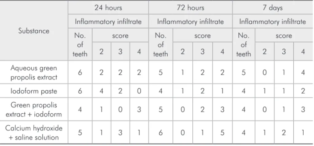
Substance
24 hours 72 hours 7 days
Necrosis Necrosis Necrosis
No. of teeth
Score No.
of teeth
Score No.
of teeth
Score
1 2 3 4 1 2 3 4 1 2 3 4
Aqueous green
propolis extract 6 0 2 4 0 5 0 2 2 1 5 0 2 2 1
Iodoform paste 6 0 1 3 2 4 1 2 0 1 4 1 3 0 0
Green propolis
extract + iodoform 4 1 2 0 1 5 0 3 2 0 4 0 2 2 0
Calcium hydroxide
+ saline solution 5 0 3 1 1 6 0 2 3 1 4 1 2 0 1
*Among the 72 pulpotomized teeth, 14 were excluded, reducing the sample size to 58 teeth. Score 1 = absence or insignificant presence of necrosis; score 2 = necrosis close to the pulp medication, reaching up to one third of the root canal pulp tissue; score 3 = necrosis involving up to two thirds of the root canal pulp tissue; score 4 = necrosis involving more than two thirds of the root canal pulp tissue.
Table 2 - Distribution of number of teeth according to substances tested, evaluation time and presence of necrosis.*
Substance
24 hours 72 hours 7 days
Inflammatory infiltrate Inflammatory infiltrate Inflammatory infiltrate No.
of teeth
score No.
of teeth
score No.
of teeth
score
2 3 4 2 3 4 2 3 4
Aqueous green
propolis extract 6 2 2 2 5 1 2 2 5 0 1 4
Iodoform paste 6 4 2 0 4 1 2 1 4 1 1 2
Green propolis
extract + iodoform 4 1 0 3 5 0 2 3 4 0 1 3
Calcium hydroxide
+ saline solution 5 1 3 1 6 0 1 5 4 1 2 1
*Among the 72 pulpotomized teeth, 14 were excluded, reducing the sample size to 58 teeth. Score 1 = ab-sence or insignificant preab-sence of inflammatory infiltrate; score 2 = inflammatory infiltrate close to the pulp medication, reaching up to one third of the root canal pulp tissue; score 3 = inflammatory infiltrate involving up to two thirds of the root canal pulp tissue; score 4 = inflammatory infiltrate involving more than two thirds of the root canal pulp tissue.
group were protected with a fragment of absorbent paper soaked with the solution, further studies are needed to determine whether the increase in inlam-mation stemmed from a reaction to the foreign body (absorbent paper) or was a response induced by the propolis itself. On the other hand, the inlammatory reaction may indeed have been caused by the sub-stance, and may indicate a positive tissue response promoting cellular reorganization and repair of the exposed pulp.
Previous studies with a longer evaluation time10,2
report satisfactory results with green propolis,
com-parable to those achieved with calcium hydroxide.11
Moreover, an alcohol extract of propolis was found to induce the formation of collagen bridges and den-tin after 28 days.12 Other studies have demonstrated
that propolis has a low irritating potential13,14 and
induces a repair process in both epithelial tissue15
and pulp tissue.16 The free-radical- and
superoxide-neutralizing components of propolis are believed to be responsible for its main regenerative mecha-nisms.17 Longer studies should be carried out to
ana-lyze the behavior of the dental pulp exposed to this substance.
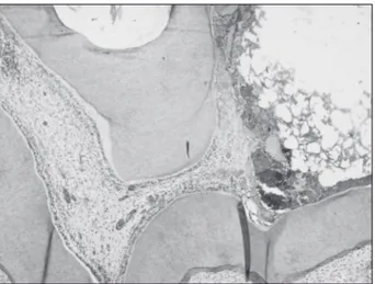
Figure 4 - Photomicrograph of a rat tooth 7 days after pulpotomy and pulp capping with calcium hydroxide show-ing light inflammatory infiltrate (HE, 100×).
Figure 1 - Photomicrograph of a rat tooth 7 days after pulp-otomy and pulp capping with green propolis extract show-ing significant necrosis and mild inflammatory infiltrate (HE, 100×).
Figure 2 - Photomicrograph of a rat tooth 24 hours after pulpotomy and pulp capping with iodoform paste showing light presence of inflammatory infiltrate (HE, 100×).
The iodoform paste induced a small degree of inlammatory iniltrate in the irst 24 hours (Fig-ure 2); however, two of the four teeth analyzed on Day 7 exhibited a signiicant degree of inlamma-tory iniltrate, with a predominance of neutrophils. This paste contains Riforcort, which is a corticoid
(prednisolone) associated to an antibiotic (rifampi-cin). Prednisolone must be the main ingredient re-sponsible for inhibiting the initial inlammatory iniltrate in the conjunctive tissue, insofar as this substance is capable of inhibiting vasodilatation and the inlow of leukocytes.18
The paste containing green propolis extract and iodoform induced a signiicant inlammatory reac-tion in the pulp tissue at all three evaluareac-tion times (Figure 3). Studies found in the literature have re-ported that iodoform is a tissue irritant; this may have contributed to the histological indings in this group. Despite the substantial presence of inlam-matory iniltrate, necrosis ranged from mild to moderate (scores 2 and 3). Iodoform stimulates cell proliferation by producing an initial inlammatory reaction and tissue necrosis and attracting defense cells to the site, especially polymorphonuclear cells, which are rapidly absorbed and replaced with nor-mal conjunctive tissue.19
In the calcium hydroxide group, signiicant in-lammatory iniltrate only prevailed in the 72-hour evaluation and was less marked on Day 7 (Figure 4). Necrosis in the specimens ranged from mild to mod-erate (scores 2 and 3) at the three evaluation times, in most cases. Previous studies have reported that calcium hydroxide induces a lesser degree of
inlam-matory iniltrate in the initial hours, progressing to a moderate degree after longer periods, and induc-ing subsequent tissue repair.20 The necrosis seen in
pulp tissue following contact with calcium hydrox-ide is the result of its alkalinity. This alkalinity ac-tually has a beneicial effect on the injured tissue, insofar as it causes mild irritation and stimulates the conjunctive tissue to defend and repair itself, initiat-ing an inlammatory reaction to control and elimi-nate the irritating agent.21
It was not the intention of the present study to criticize the endodontic pastes used in the treatment of the pulp of deciduous teeth, but rather to provide information on biocompatible materials that may be used as direct pulp capping methods. Longer studies are needed to determine whether the intensity of the inlammatory response observed in the pulp tissue after applying propolis extract is beneicial to the repair process. A number of studies are currently underway to investigate the composition of propolis collected from different regions of Brazil, and ana-lyze its biological activity against oral pathogenic microorganisms. Moreover, further studies are needed to deine eficient methods for using propolis in pulp therapy.
Conclusion
Green propolis induced an inlammatory reaction in rat dental pulp following pulpotomy. This reac-tion was more intensive when the extract was com-bined with iodoform. Studies conducted with a lon-ger evaluation time are needed to analyze the effect of green propolis on the pulp tissue repair process.
References
1. Pilipili CM, Vanden Abbeele A, van den Abbeele K. Pulp-otomy of deciduous teeth. Rev Belge Med Dent (1984). 2004;59(3):156-62. French.
2. Parolia A, Kundabala M, Rao NN, Acharya SR, Agrawal P, Mohan M, et al. A comparative histological analysis of human pulp following direct pulp capping with Propolis, mineral trioxide aggregate and Dycal. Aust Dent J. 2010 Mar;55(1):59-64.
3. Dammaschke T, Leidinger J, Schäfer E. Long-term evaluation of direct pulp capping--treatment outcomes over an average period of 6.1 years. Clin Oral Investig. 2010 Oct;14(5):559-67.
4. Sonmez D, Sari S, Cetinbaş T. A comparison of four pulp-otomy techniques in primary molars: a long-term follow-up. J Endod. 2008 Aug;34(8):950-5.
5. Briso AL, Rahal V, Mestrener SR, Dezan Junior E. Biological response of pulps submitted to different capping materials. Braz Oral Res. 2006 Jul-Sep;20(3):219-25.
7. Horn RC, Vargas VMF. Antimutagenic activity of extracts of natural substances in the Salmonella/microsome assay. Mu-tagenesis. 2003 Mar;18(2):113-8.
8. Dornelas CA, Fechine-Jamacaru FV, Albuquerque IL, Maga-lhães HI, Souza AJ, Alves LA, et al. Chemoprevention with green propolis extracted in L-lysine versus carcinogenesis promotion with L-lysine in N-Butyl-N-[4-hydroxybutyl] ni-trosamine (BBN) induced rat bladder cancer. Acta Cir Bras. 2012 Feb;27(2):185-92.
9. Pagliarone AC, Orsatti CL, Búfalo MC, Missima F, Bachiega TF, Araújo Júnior JP, et al. Propolis effects on pro-inflam-matory cytokine production and Toll-like receptor 2 and 4 expression in stressed mice. Int Immunopharmacol. 2009 Oct;9(11):1352-6.
10. Sabir A, Tabbu CR, Agustiono P, Sosroseno W. Histological analysis of rat dental pulp tissue capped with propolis. J Oral Sci. 2005 Sep;47(3):135-8.
11. Schuurs AH, van Joost T, van Loon LA. Cutaneous and mu-cosal reactions to dental materials. Ned Tijdschr Tandheelkd. 1999 Sep;106(9):334-9. Deutch.
12. Scheller S, Luciak M, Tustanowski J, Koziol M, Obuszko Z, Kurylo B. Biological properties and clinical application of propolis. XI. Histopathological analysis after intravenous application of ethanol extract of propolis (EEP). Arzneimit-telforschung. 1978;28(9):1594-5.
13. Silva FB, Almeida JM, Sousa SM. Natural medicaments in endodontics: A comparative study of anti-inflammatory ac-tion. Braz Oral Res. 2004 Apr-Jun;18(2):174-9.
14. Arruda CMF, Marchini LC, Moreti ACCC, Otsuk IP, So-dré GS. Características físico-químicas de méis da Chapada
do Araripe/Santana do Cariri-Ceará. Acta Sci Animal Sci. 2005 Jan-Mar;27(1):171-6.
15. Magro Filho O, Carvalho AC. Application of propolis to den-tal sockets and skin wounds. J Nihon Univ Sch Dent. 1990 Mar;32(1):4-13.
16. Bretz WA, Chiego Jr DJ, Marcucci MC, Cunha I, Custódio A, Schneider LG. Preliminary report on the effects of propolis on wound healing in the dental pulp. Z Naturforsch C. 1998 Nov-Dec;53(11-12):1045-8.
17. Krol W, Scheller S, Czuba Z, Matsuno T, Zydowicz G, Shani J, et al. Inhibition of neutrophils’ chemiluminescence by etha-nol extract of propolis (EEP) and its pheetha-nolic components. J Ethnopharmacol. 1996 Dec;55(1):19-25.
18. Cerqueira DF, Mello-Moura AC, Santos EM, Guedes-Pinto AC. Cytotoxicity, histopathological and microbiological, and clinical aspects of an endodontic iodoform-based paste used in pediatric dentistry: a review. J Clin Pediatr Dent. 2008 Winter;32(2):105-10.
19. Friend LA, Browne RM. Tissue reactions to some root filling materials implanted in the bone of rabbits. Arch Oral Biol. 1969 Jun;14(6):629-38.