Adipokine Imbalance in the Pericardial Cavity
of Cardiac and Vascular Disease Patients
Atlanta G. I. M. Elie1, Pia S. Jensen2, Katrine D. Nissen1, Ilvy M. E. Geraets1,3, Aimin Xu4, Erfei Song4, Maria L. Hansen5,6, Akhmadjon Irmukhamedov5,6, Lars M. Rasmussen2,6, Yu Wang4, Jo G. R. De Mey1,3,5,6*
1Dept. Cardiovascular and Renal Research, Institute of Molecular Medicine, University of Southern Denmark, Odense, Denmark,2Dept. Clinical Biochemistry and Pharmacology, Odense University Hospital, Odense, Denmark,3Cardiovascular Research Institute Maastricht, Maastricht, the Netherlands,4State Key Laboratory of Pharmaceutical Biotechnology and Dept. Pharmacology and Pharmacy, Li Ka Shing Faculty of Medicine, The University of Hong Kong, Hong Kong, China,5Dept. Cardio-Thoracic and Vascular Surgery, Odense University Hospital, Odense, Denmark,6Centre for Individualized Medicine in Arterial Diseases (CIMA), Odense University Hospital, Odense, Denmark
Abstract
Aim
Obesity and especially hypertrophy of epicardial adipose tissue accelerate coronary athero-genesis. We aimed at comparing levels of inflammatory and atherogenic hormones from adipose tissue in the pericardial fluid and circulation of cardiovascular disease patients.
Methods and Results
Venous plasma (P) and pericardial fluid (PF) were obtained from elective cardiothoracic sur-gery patients (n = 37). Concentrations of leptin, adipocyte fatty acid-binding protein (A-FABP) and adiponectin (APN) were determined by enzyme-linked immunosorbent assays (ELISA). The median concentration of leptin in PF (4.3 (interquartile range: 2.8–9.1)μg/L)
was comparable to that in P (5.9 (2.2–11)μg/L) and these were significantly correlated to
most of the same patient characteristics. The concentration of A-FABP was markedly higher (73 (28–124) versus 8.4 (5.2–14)μg/L) and that of APN was markedly lower (2.8 (1.7–4.2)
versus 13 (7.2–19) mg/L) in PF compared to P. APN in PF was unlike in P not significantly related to age, body mass index, plasma triglycerides or coronary artery disease. PF levels of APN, but not A-FABP, were related to the size of paracardial adipocytes. PF levels of APN and A-FABP were not related to the immunoreactivity of paracardial adipocytes for these proteins.
Conclusion
In cardiac and vascular disease patients, PF is enriched in A-FABP and poor in APN. This adipokine microenvironment is more likely determined by the heart than by the circulation or paracardial adipose tissue.
a11111
OPEN ACCESS
Citation:Elie AGIM, Jensen PS, Nissen KD, Geraets IME, Xu A, Song E, et al. (2016) Adipokine Imbalance in the Pericardial Cavity of Cardiac and Vascular Disease Patients. PLoS ONE 11(5): e0154693. doi:10.1371/journal.pone.0154693
Editor:Alexander G Obukhov, Indiana University School of Medicine, UNITED STATES
Received:January 15, 2016
Accepted:April 18, 2016
Published:May 3, 2016
Copyright:© 2016 Elie et al. This is an open access article distributed under the terms of theCreative Commons Attribution License, which permits unrestricted use, distribution, and reproduction in any medium, provided the original author and source are credited.
Data Availability Statement:All relevant data are within the paper and its Supporting Information file.
Funding:This work was supported by the Odense University Hospital, the University of Southern Denmark, the Danish Council for Independent Research [DFF-4092-00098], the Direktør Kurt Bønnelycke og hustru fru Grethe Bønnelyckes Fond and by grants from the Hong Kong Research Grant Council [HKU780410M, HKU781311M,
Introduction
Obesity promotes atherosclerosis [1,2]. Hypertrophic chronically inflamed adipose tissues produce less signaling molecules with vascular protective actions such as adiponectin (APN) [3,4] and more hormones with inflammatory and atherogenic effects such as leptin [5] and adipocyte fatty acid-binding protein (A-FABP) [6]. The circulating hormonal levels of these adipokines are likely dominated by the large subcutaneous and abdominal visceral adipose tis-sue depots [7].
Local accumulation of fat on the outer surface of the heart around the coronary arteries (epicardial adipose tissue (EAT) [8,9]) is associated with coronary artery disease (CAD) in not only obese but also non-obese patients [10–15]. This indicates local paracrine effects of adipo-kines on coronary arteries [8,16,17]. Paired comparisons of EAT with subcutaneous or abdominal visceral adipose tissue biopsies revealed differences in fatty acid composition [18], expression and release of chemokines and inflammatory cytokines along with retention of inflammatory cells [15,19] and in expression and secretion of adipokines [14,15,17,20].
The composition of the pericardial fluid (PF) surrounding the heart has been proposed to be comparable to that of the interstitium of the heart [21,22]. Several angiogenic growth fac-tors, cytokines, natriuretic peptides and classical vasoactive hormones are present at higher concentration in the PF than in the circulating plasma (P) of patients with ischemic heart dis-ease or heart failure [21,23–25].
Here we tested the hypothesis that the pericardial cavity of cardiovascular disease (CVD) patients is a unique adipokine microenvironment. To this end we compared the levels of leptin, A-FABP and APN in the PF and P of cardiothoracic surgery patients.
Material and Methods
Subjects and sample collection
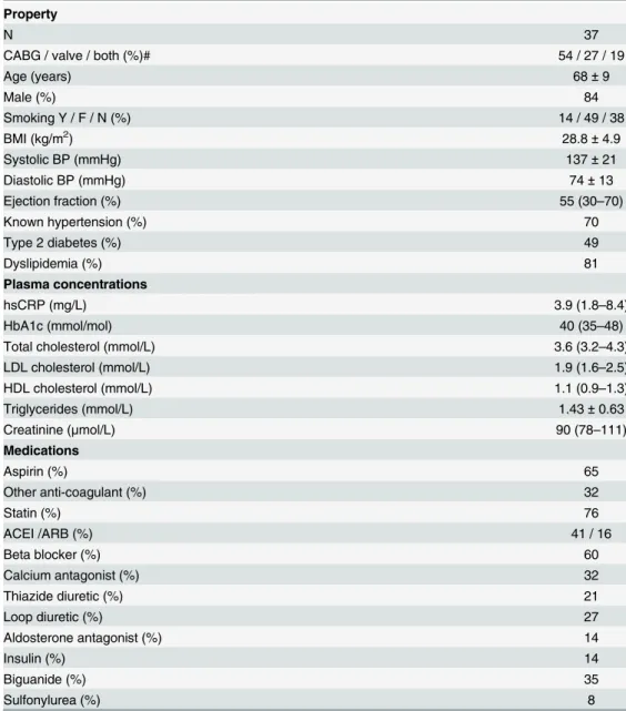
The study was performed in accordance with institutional guidelines and was approved by the Medical Ethical Committee of the Region of Southern Denmark (S-20100044). Investiga-tions were performed conform the principles outlined in the Declaration of Helsinki and informed written consent was obtained from all patients. Between September 2013 and Janu-ary 2014 consecutive patients who underwent elective coronJanu-ary artery bypass grafting (CABG) or cardiac valve replacement surgeries were enrolled. Clinical information was obtained from the patients’medical records. On the day before surgery, peripheral venous blood was drawn into pyrogen-free tubes with K2EDTA as an anticoagulant. Pericardial fluid (2–5 mL in K2EDTA and aprotinin) and a biopsy from the parietal sheet of the pericardium (2 x 2 cm) were obtained after median sternotomy and section of the pericardium at outset of the heart surgeries. The blood and pericardial fluid samples were centrifuged (3220 g for 10 min at 4°C) and the supernatants were stored at -80°C until analysis. The pericardial tissue biopsies were fixed in neutral buffered formalin for 48 hours at room temperature. Patients with haemolytic pericardial fluid samples were not further considered. Analyses were limited to the plasma, pericardial fluid and pericardial tissue biopsy from the same individual patients (N = 37). Routine plasma analyses for HbA1c, lipids, creatinine and high sensitivity C-reactive protein (hsCRP) were performed at the Odense University Hospital clinical bio-chemistry laboratory. Characteristics of the patients were disclosed to the researchers of the pericardial samples after they finalized their data collection. These characteristics are sum-marized inTable 1.
funders had no role in study design, data collection and analysis, decision to publish, or preparation of the manuscript.
Measurements of adipokine concentrations
The levels of APN and A-FABP in P and PF were measured by the respective sandwich ELISA kits (Antibody and Immunoassay Services (AIS), The University of Hong Kong, Hong Kong, China) as previously described [26,27]. P and PF levels of leptin were determined with an ELISA kit from Biovendor Laboratories (Brno, Czech Republic) as previously described [27]. All samples were determined in duplicate. For one patient, plasma A-FABP was below the detection limit.
Table 1. Patient properties, plasma characteristics and prescribed medications. Property
N 37
CABG / valve / both (%)# 54 / 27 / 19
Age (years) 68±9
Male (%) 84
Smoking Y / F / N (%) 14 / 49 / 38
BMI (kg/m2) 28.8±4.9
Systolic BP (mmHg) 137±21
Diastolic BP (mmHg) 74±13
Ejection fraction (%) 55 (30–70)
Known hypertension (%) 70
Type 2 diabetes (%) 49
Dyslipidemia (%) 81
Plasma concentrations
hsCRP (mg/L) 3.9 (1.8–8.4)
HbA1c (mmol/mol) 40 (35–48)
Total cholesterol (mmol/L) 3.6 (3.2–4.3)
LDL cholesterol (mmol/L) 1.9 (1.6–2.5)
HDL cholesterol (mmol/L) 1.1 (0.9–1.3)
Triglycerides (mmol/L) 1.43±0.63
Creatinine (μmol/L) 90 (78–111)
Medications
Aspirin (%) 65
Other anti-coagulant (%) 32
Statin (%) 76
ACEI /ARB (%) 41 / 16
Beta blocker (%) 60
Calcium antagonist (%) 32
Thiazide diuretic (%) 21
Loop diuretic (%) 27
Aldosterone antagonist (%) 14
Insulin (%) 14
Biguanide (%) 35
Sulfonylurea (%) 8
Categorical data are shown as % of the study group, normally distributed continuous variables as mean±SD, non-normally distributed continuous variables as median value (interquartile range).
#
, type of surgery. CABG, coronary artery bypass surgery; BMI, body mass index; BP, blood pressure; hsCRP, high sensitivity C-reactive protein; ACEI/ARB, inhibitor of angiotensin converting enzyme or antagonist of angiotensin AT1 receptors.
Western blotting
For detection of different oligomeric complexes of APN, P and PF samples were incubated with nonreducing sample buffer (1% SDS, 5% glycerol, and 10 mM Tris-HCl (pH 6.8)) at room temperature for 10 min, separated by 4–20% gradient SDS-PAGE, transferred to polyvinyli-dene difluoride membranes, and immunoblotted with affinity-purified rabbit anti-human APN IgG (AIS; 0.2μg/mL) as we described previously [28]. Based on the ELISA results,
sam-ples containing the same amount of total APN (20 ng) were analyzed by non-reducing SDS-PAGE and Western blotting for detection of the distribution of oligomers of APN. Quan-tification was performed by densitometric analysis using ImageJ software (National Institutes of Health, Bethesda, MA, USA). Increasing concentrations of recombinant human Flag-tagged APN protein purified from mammalian cells were used to generate the standard curves and to establish the specificity of the experimental approach in comparison to P and PF samples from 3 randomly selected patients (S1 Fig). Abundance of APN oligomers with molecular
weight>200 kDa was expressed as % of total APN.
Immunohistochemistry
Presence and distribution of A-FABP and APN in pericardial tissue biopsies were determined by immunohistochemical (IHC) staining. Paraffin sections (5μm) of formalin-fixed tissue
were rehydrated and endogenous peroxidase activity was quenched with H2O2in methanol. Microwave treatment was used for heat induced antigen retrieval using citrate (for APN) or Tris-ethylene glycol (TEG, for A-FABP). Sections were incubated overnight at 4°C with pri-mary monoclonal antibody against APN (1:500, ab22554, ABCAM, Cambridge, UK) or poly-clonal antibody against A-FABP (1:1200, 11030, AIS, Hong Kong, China) followed by incubation with goat anti-mouse or goat anti-rabbit secondary antibody, respectively (both 1:1000, P0447 and P0448 resp. Dako, Denmark) for 45 minutes. Colorimetric detection was completed with 3.3’-diaminobenzidine (DAB). Sections were counterstained with Mayer’s hematoxylin. Sections without primary antibody served as negative controls. Digital images were obtained with a microscope (Olympus BX51, Japan) and imaging software (cellSens Entry, Olympus Corporation, version 1.7). To estimate the size and adipokine content of adi-pocytes, histological sections were viewed at 10 x magnification and analyzed with ImageJ soft-ware (version 2.0.0-rc-23; NIH, Bethesda, MD, USA). Adipocyte cross-sectional area was determined as described by Chen and Farese [29]. The percentage of adipocytes staining for APN or A-FABP was evaluated in 3–50 square fields (60000 pixels) depending on the thickness of the paracardial adipose tissue.
Statistics
Results
Patient characteristics
Table 1summarizes demographic, clinical and plasma characteristics of the study group.
Patients were undergoing CABG (54%), cardiac valve replacement (27%) or both (19%) indi-cating that 73% were suffering from ischemic heart disease due to CAD. As expected, the majority were elderly overweight males (84%) with a high prevalence of systemic inflammation (high plasma hsCRP levels), dyslipidemia (81%), hypertension (70%) and type 2 diabetes (49%) but not overt heart failure or renal failure as judged from ejection fraction and plasma creatinine. Most had been prescribed a variable combination of anti-coagulant, cholesterol low-ering, renin angiotensin system inhibitory, anti-hypertensive and anti-diabetic medications.
Pericardial fluid levels of leptin, A-FABP and APN
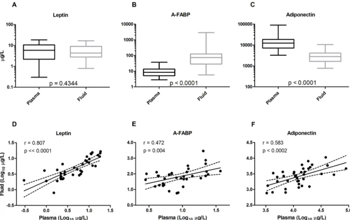
The concentrations of leptin in PF were comparable to those in the circulating P (4.3 (2.8–9.1) versus 5.9 (2.2–11)μg/L,p= 0.4344;Fig 1A). A-FABP, on the other hand, was 9 times more
abundant in PF than P (73 (28–124) versus 8.4 (5.2–14)μg/L,p<0.0001;Fig 1B) while total
APN was 5 times less concentrated in PF than P (2.8 (1.7–4.2) versus 13 (7.2–19) mg/L,
p<0.0001;Fig 1C;Table 2). As a consequence, the leptin/APN ratio was significantly larger in
PF than P (2.2 (1.2–3.1) versus 0.44 (0.15–0.82)μg/mg; 5 fold). For A-FABP the imbalance was
even more marked. The A-FABP/APN ratio was on average more than 30 times larger in PF
Fig 1. Venous plasma and pericardial fluid concentrations of leptin (left), adipocyte fatty acid-binding protein (A-FABP, middle) and adiponectin (right) in cardiac and vascular disease patients (n = 36–37).Top, box plots illustrating median values, interquartile
range and range of concentrations compared by Wilcoxon matched-pairs signed rank test. Bottom, double logarithmic plots by Pearson’s correlation illustrating the relationship between pericardial fluid concentrations and circulating levels of the adipokines.
than in venous P (24 (9.4–70) versus 0.69 (0.35–1.5)μg/mg). Despite marked differences
between the levels of especially A-FABP and APN in PF and P, the concentrations of each of leptin, A-FABP and APN in PF were positively correlated to those in the venous plasma (Fig
1D–1F).
Western blotting was used to determine the relative abundance of multimers of APN. Typi-cal results for PF and P from the same individual patients are shown inFig 2. The contribution of high molecular weight APN (molecular weight>200 kDa) to total APN did not differ
sig-nificantly between PF and venous P (29.1 ± 7.5 and 27.4 ± 6.8%, respectively).
Paracardial adipose tissue properties
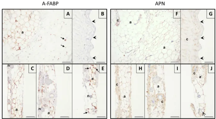
Fig 3illustrates structural features and distribution of A-FABP and APN in the parietal sheet of
the pericardium. Mesothelial cells that line the pericardial cavity lay on a thick collagen band. Abluminally from this structure, a vascular network [30] and paracardial adipose tissue are located. The thickness of this adipose tissue differed markedly between patients. The median cross sectional area of paracardial adipocytes was 12 (8.0–17) 102μm2. With IHC staining,
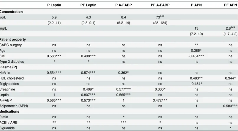
Table 2. Plasma (P) and pericardial fluid (PF) levels of three adipokines and univariate analyses of their interrelationships and associations with patient properties.
P Leptin PF Leptin P A-FABP PF A-FABP P APN PF APN
Concentration
μg/L 5.9 4.3 8.4 73###
(2.2–11) (2.8–9.1) (5.2–14) (28–124)
mg/L 13 2.8###
(7.2–19) (1.7–4.2)
Patient property
CABG surgery ns ns ns ns ** ns
Age ns ns ns ns 0.386* ns
BMI 0.588*** 0.498*** ns ns -0.454*** ns
Type 2 diabetes * * ns ns ns ns
Plasma (P)
HbA1c 0.554*** 0.574*** 0.362* ns ns ns
HDL cholesterol ns ns ns ns 0.483** 0.344*
Triglycerides ns ns ns ns -0.454* ns
Creatinine ns 0.408* 0.577*** 0.330* ns ns
Leptin 1 0.807*** 0.565*** ns ns ns
A-FABP 0.565*** 0.573*** 1 0.472*** ns ns
Adiponectin (APN) ns ns ns ns 1 0.583***
Medications
Statin ns ns * ns ns ns
ACEI / ARB ** ** *** * ns ns
Biguanide ns ns ns ns ns *
A-FABP, adipocyte fatty acid-binding protein; APN, total adiponectin; ns, not statistically significant; other abbreviations as inTable 1. Concentrations are shown as median (interquartile range);
###:
p<0.001 versus plasma by Wilcoxon matched-pairs signed rank test. *,p<0.05;
**,p<0.01 and
***,p<0.001 indicate statistically significant correlations to categorical or continuous variables. For continuous variables the Pearson’s correlation coefficient is shown as well.
APN was found in parts of the adipose tissue, vascular endothelium (some patients only), and in collagen-rich regions. A-FABP was located in some of the adipocytes, nerves, arterioles and capillaries. Mesothelium and arterial smooth muscle cells did not stain for A-FABP or APN. Presence of detectable A-FABP and APN in adipocytes displayed marked regional heterogene-ity (Fig 2). The percentage of paracardial adipocytes that was immunoreactive for A-FABP or APN varied considerably between patients (43 (0.0–94) and 63 (14–100) %), respectively).
Interrelationships and associations
In PF, there were no statistically significant relationships between the levels of the adipokines investigated while in P the levels of leptin and A-FABP were positively correlated (Table 2). As expected [1,31], the circulating level of leptin increased with increasing BMI while the circulat-ing concentration of APN was inversely correlated to BMI (Table 2). Also in PF, leptin levels were positively correlated to BMI but the concentrations of APN in PF displayed no significant relation to BMI.
Table 2further summarizes associations between the concentrations of adipokines and
patient characteristics listed inTable 1. The concentrations of leptin in both P and PF were each related to type 2 diabetes, BMI, circulating levels of glycated hemoglobin and to treatment with an inhibitor of the renin-angiotensin system. For A-FABP in P and PF there were fewer similarities. This discrepancy between the adipokine concentrations in the two compartments was even more marked for APN. APN in P, but not PF, was inversely related to CAD, BMI, and plasma triglycerides and positively correlated to the age of the patients (Table 2).
Fig 2. Representative Western blot images of APN multimers in pericardial fluid (left) and venous plasma (right) of cardiac and vascular disease patients.The Western blots illustrate the composition of oligomeric complexes of APN in pericardial fluid and plasma samples from the same 7 individual patients, indicated by numbers 4–10. The contribution of high molecular APN species (>200 kDa) to the total concentration of APN was calculated as a percentage as described in the methods section.
The size of the paracardial adipocytes was significantly larger in type 2 diabetic patients (13.7 (11.1–18.0) versus 10.2 (6.6–13.1) x 102μm2,p= 0.038) and in CAD patients (13.6
(10.7–18.0) versus (6.7 (5.9–9.9) x 102μm2,p= 0.0004) but was not correlated to BMI
(p= 0.642,r= 0.0839), not related to the PF level of A-FABP (p= 0.735,r= 0.0611) and
inversely related to the PF level of APN (p= 0.0364,r= -0.366). Paracardial adipocyte
immu-noreactivity for A-FABP or APN were significantly correlated (p= 0.0469,r= 0.3720) but not
significantly related to adipocyte size (p= 0.352,r= -0.179 andp= 0.223,r= -0.218
respec-tively) or to the PF concentration of A-FABP or APN (p= 0.507,r= 0.124 andp= 0.460, r= 0.133, respectively).
Discussion
In pericardial fluid of cardiothoracic surgery patients, the concentration of leptin was compara-ble but the level of A-FABP markedly higher and that of APN considerably lower than in the circulating plasma. Leptin levels in pericardial fluid and plasma were correlated to most of the same patient characteristics while APN in pericardial fluid was unlike in the plasma not corre-lated to age, body mass index, type 2 diabetes, plasma triglycerides and CAD. The concentra-tion of APN but not A-FABP in pericardial fluid was related to the size of paracardial adipocytes, whereas both adipokine concentrations in pericardial fluid were not related to the immunoreactivity of the adipocytes in the paracardial pericardium. These findings indicate that the pericardial cavity of cardiovascular disease patients is a pro-inflammatory atherogenic
Fig 3. Representative images of immunohistochemical stainings of adipocyte fatty acid-binding protein (A-FABP; left) and adiponectin (APN; right) in the parietal pericardium.Top, adjacent sections of the adipose tissue part (A and F) and of the luminal (mesothelial) side (B and G) from the same biopsy. Bottom, adjacent sections of the biopsy from another patient focusing on the sub-mesothelial vasculature and its peri-vascular adipose tissue (C-E and H-J). Positive immunoreactivity in the different structures are indicated as follows: a = adipocytes, n = nerves, c = collagen rich regions, open arrow head = endothelium, small arrows = arterioles and capillaries. The thick arrow heads indicate mesothelial cells. Scale bars, 100μm.
adipokine microenvironment determined by components of the heart rather than by the circu-lating hormones or paracardial adipose tissue.
Increased risk for metabolic and cardiovascular diseases associated with increased adiposity [1,2] stimulated interests in signaling molecules secreted by adipose tissues [4–7]. These adipo-kines can reach the cardiovascular system as hormones via the circulation and as paracrine mediators produced by comparatively small adipose tissues that surround the heart and blood vessels. Hyperleptinemia and hypoadiponectinemia in obesity are strongly associated with type 2 diabetes but only moderately with CAD [32,33]. Circulating levels of A-FABP increase with the number of stenotic coronary arteries [34]. On the other hand, the volume of EAT correlates positively with CAD in not only obese but also non-obese patients [8,9,15,17]. Moreover, sev-eral classical vasoactive mediators and angiogenic growth factors are more abundant in the pericardial fluid than in the circulation of ischemic heart disease and heart failure patients [21,
23–25]. For these reasons we performed a small paired cross sectional comparison of
concen-trations of leptin, A-FABP and APN in pericardial fluid and venous plasma. Pericardial fluid was obtained at outset of coronary artery bypass or cardiac valve replacement surgery where the parietal pericardium must be opened to gain access to the heart. The characteristics of the patients that we investigated are comparable to those previously studied with respect to EAT volume and histology [10,11,15,16,18,20] and coronary arteriolar endothelial dysfunction [35,36]. Pericardial fluid was compared to a venous blood sample from the day before surgery. This is justified because earlier studies found no changes in the circulating levels of leptin and APN and an at most 50% increase of plasma A-FABP during the early steps of cardiothoracic surgery [37,38].
Experimental animal evidence (reviewed by Beltowski [5]) links elevations in circulating levels of the 16 kDa protein leptin via loss of leptin function or leptin resistance to atherogene-sis and CAD but findings in human patients are less convincing [32]. In line with earlier reports [5,31,39,40] we observed that in human patients the circulating levels of leptin are positively correlated to BMI, type 2 diabetes and consequences thereof such as elevated HbA1c, but not CAD, age or plasma cholesterol and triglycerides. We found that leptin in pericardial fluid, which to the best of our knowledge was not reported before, is comparable to that in the venous plasma in terms of not only concentration but also relation to most of the same patient characteristics. In sharp contrast to leptin, marked quantitative and qualitative differences were observed for A-FABP and APN. Experimental animal evidence (reviewed by Xu and Van-houtte [6]) links circulating levels of the 15 kDa protein A-FABP that can be secreted by adipo-cytes and macrophages, to cardiovascular diseases and findings in patients link it to the severity of CAD [34] and heart failure [27]. In our paired comparison, A-FABP was nine times more abundant in pericardial fluid than in circulating plasma and while plasma concentrations were significantly related to plasma HbA1c and leptin levels and to treatment with statins, peri-cardial fluid concentrations were not. Experimental evidence (reviewed by Hui et al. [4], Xu and Vanhoutte [6] and Mattu and Randeva [7]) links reductions of the circulating levels of the 90 (trimeric), 180 (hexameric) and>250 kDa protein APN to endothelial dysfunction and
diseases patients differs quantitatively and qualitatively from that in the circulation. For instance, the leptin/APN and the A-FABP/APN ratio’s that are advanced as powerful diagnos-tic markers [41–43] are 5 and more than 30 times larger in the local pericardial fluid than in the circulation, respectively. Although adipokine levels differed markedly between both com-partments, the circulating and pericardial concentrations of leptin, A-FABP and APN were strongly correlated. Thus plasma concentrations may be considered in future longitudinal studies estimating local pericardial changes of adipokines during progression and treatment of cardiac and coronary artery diseases. These will at best be qualitative in nature and underesti-mate imbalances between adipokine species.
The pericardial cavity is considered for local treatment of ischemic heart disease and myo-cardial infarcts by drugs, angiogenic growth factors and stem cells [44,45]. Control of pericar-dial fluid composition is however poorly understood. Some authors consider it a protein poor ultrafiltrate of the plasma [46,47] while others emphasize similarities to the interstitium of the heart [21,22,47]. Our observations of comparable contributions of low and high molecular weight APN isoforms in plasma and pericardial fluid and of different pericardial to plasma ratios for leptin and A-FABP along with earlier findings of elevated levels of lactate dehydroge-nase (140 kDa) in pericardial fluid [46], are not compatible with the former view. We consid-ered contributions of the paracardial adipose tissue lining the outer surface of the parietal pericardium. Paracardial adipocyte size was enlarged in diabetic and CAD patients as previ-ously reported for epicardial adipocytes [48]. Mean paracardial adipocyte immunoreactivities for A-FABP and APN were interrelated but not related to adipocyte size in contrast to the neg-ative associations between the size of subcutaneous and epicardial adipocytes and APN gene expression previously observed by Bambace et al [48]. We could not observe a significant rela-tionship between mean paracardial adipocyte immunoreactivity and pericardial fluid levels of A-FABP and APN. This could indicate that there is no major contribution of paracardial adi-pose tissue to pericardial fluid levels of adipokines. For the two adipokines that we stained in parietal pericardium we observed marked regional heterogeneities. Thus in addition to differ-ences between adipose tissues at different anatomical locations [1,16–20,49], adipokine biol-ogy seems to differ locally within a given adipose tissue. It will be of interest to verify in future investigations whether these regional heterogeneities are correlated to distances from blood vessels, capillaries or macrophages, in order to discriminate between differences from perivas-cular adipose tissue and influences of local oxygen tension and inflammation.
Study Limitations
In this cross sectional investigation of randomly selected cardiothoracic surgery patients a fair number of strong associations could be observed. The causal nature of these could not be addressed. The study was too small to appreciate influences of the more rare aspects in the study population such as female sex and specific medications, or to perform multivariate analy-sis. No pericardial fluid could be sampled from individuals without CVD or cardiovascular drug treatments. This may be partly remedied by studies in a sufficiently large relevant experi-mental animal model [50] and sampling of pericardial fluid from patients undergoing thoracic surgery for non-CVD [51]. Confounding influences of drugs such as statins and PPARγ
diagnostics of our elective cardiothoracic surgery patients. More importantly, we lack informa-tion on epicardial adipose tissue volume [9,11–13,15,16], immunohistochemistry and adipo-kine secretion [14,16,17,19] to propose this adipose tissue compartment as a determinant of pericardial fluid adipokine composition. Finally, whether the pericardial fluid adipokine profile i) represents spillover from the cardiac interstitium with potential for biomarker research of epicardial adipose tissue effects on CAD and ii) might in itself contribute to coronary athero-genesis, coronary microvascular dysfunction, arrhythmogenesis and heart failure, will require a broad variety of dedicated investigations.
Supporting Information
S1 Fig. Standard curve and specificity of adiponectin protein detection by Western blot-ting.
(DOCX)
Acknowledgments
The authors thank Simone Rørdam Preil PhD from the Department of Clinical Biochemistry and Pharmacology (OUH) for assistance with the collection and handling of patient biopsies and characteristics and Inger Nissen from the Department of Cardiovascular and Renal Research (SDU) for assistance with immunohistochemistry.
Author Contributions
Conceived and designed the experiments: LMR YW JGRDM. Performed the experiments: AGIME IMEG KDN AX ES. Analyzed the data: AGIME PSJ KDN IMEG AX ES JGRDM. Con-tributed reagents/materials/analysis tools: AGIME PSJ KDN IMEG AX ES MLH AI LMR YW JGRDM. Wrote the paper: AGIME PSJ KDN IMEG AX ES MLH AI LMR YW JGRDM.
References
1. Hassan M, Latif N, Yacoub M. Adipose tissue: friend or foe? Nat Rev Cardiol. 2012; 9(12):689–702. doi:10.1038/nrcardio.2012.148PMID:23149834.
2. Hubert HB, Feinleib M, McNamara PM, Castelli WP. Obesity as an independent risk factor for cardio-vascular disease: a 26-year follow-up of participants in the Framingham Heart Study. Circulation. 1983; 67(5):968–77. PMID:6219830.
3. Maeda N, Takahashi M, Funahashi T, Kihara S, Nishizawa H, Kishida K, et al. PPARgamma ligands increase expression and plasma concentrations of adiponectin, an adipose-derived protein. Diabetes. 2001; 50(9):2094–9. PMID:11522676.
4. Hui X, Lam KS, Vanhoutte PM, Xu A. Adiponectin and cardiovascular health: an update. Br J Pharma-col. 2012; 165(3):574–90. doi:10.1111/j.1476-5381.2011.01395.xPMID:21457225; PubMed Central PMCID: PMCPMC3315032.
5. Beltowski J. Leptin and atherosclerosis. Atherosclerosis. 2006; 189(1):47–60. doi:10.1016/j. atherosclerosis.2006.03.003PMID:16580676.
6. Xu A, Vanhoutte PM. Adiponectin and adipocyte fatty acid binding protein in the pathogenesis of cardio-vascular disease. Am J Physiol Heart Circ Physiol. 2012; 302(6):H1231–40. doi:10.1152/ajpheart. 00765.2011PMID:22210749.
7. Mattu HS, Randeva HS. Role of adipokines in cardiovascular disease. J Endocrinol. 2013; 216(1):T17–
36. doi:10.1530/JOE-12-0232PMID:23160967.
8. Talman AH, Psaltis PJ, Cameron JD, Meredith IT, Seneviratne SK, Wong DT. Epicardial adipose tis-sue: far more than a fat depot. Cardiovasc Diagn Ther. 2014; 4(6):416–29. doi: 10.3978/j.issn.2223-3652.2014.11.05PMID:25610800; PubMed Central PMCID: PMCPMC4278038.
10. Iwayama T, Nitobe J, Watanabe T, Ishino M, Tamura H, Nishiyama S, et al. Role of epicardial adipose tissue in coronary artery disease in non-obese patients. J Cardiol. 2014; 63(5):344–9. doi:10.1016/j. jjcc.2013.10.002PMID:24230463.
11. Yerramasu A, Dey D, Venuraju S, Anand DV, Atwal S, Corder R, et al. Increased volume of epicardial fat is an independent risk factor for accelerated progression of sub-clinical coronary atherosclerosis. Atherosclerosis. 2012; 220(1):223–30. doi:10.1016/j.atherosclerosis.2011.09.041PMID:22015177.
12. Mahabadi AA, Massaro JM, Rosito GA, Levy D, Murabito JM, Wolf PA, et al. Association of pericardial fat, intrathoracic fat, and visceral abdominal fat with cardiovascular disease burden: the Framingham Heart Study. Eur Heart J. 2009; 30(7):850–6. doi:10.1093/eurheartj/ehn573PMID:19136488; PubMed Central PMCID: PMCPMC3693564.
13. Iacobellis G, Ribaudo MC, Assael F, Vecci E, Tiberti C, Zappaterreno A, et al. Echocardiographic epicardial adipose tissue is related to anthropometric and clinical parameters of metabolic syndrome: a new indicator of cardiovascular risk. J Clin Endocrinol Metab. 2003; 88(11):5163–8. doi:10.1210/jc. 2003-030698PMID:14602744.
14. Grosso AF, de Oliveira SF, Higuchi MeL, Favarato D, Dallan LA, da Luz PL. Synergistic anti-inflamma-tory effect: simvastatin and pioglitazone reduce inflammaanti-inflamma-tory markers of plasma and epicardial adipose tissue of coronary patients with metabolic syndrome. Diabetol Metab Syndr. 2014; 6(1):47. doi:10. 1186/1758-5996-6-47PMID:24684779; PubMed Central PMCID: PMCPMC3974153.
15. Shimabukuro M, Hirata Y, Tabata M, Dagvasumberel M, Sato H, Kurobe H, et al. Epicardial adipose tis-sue volume and adipocytokine imbalance are strongly linked to human coronary atherosclerosis. Arter-ioscler Thromb Vasc Biol. 2013; 33(5):1077–84. doi:10.1161/ATVBAHA.112.300829PMID:23471228.
16. Harada K, Amano T, Kataoka T, Takeshita M, Kunimura A, Takayama Y, et al. Impact of abdominal and epicardial fat on the association between plasma adipocytokine levels and coronary atherosclerosis in non-obese patients. Atherosclerosis. 2014; 237(2):671–6. doi:10.1016/j.atherosclerosis.2014.10.014 PMID:25463104.
17. Payne GA, Kohr MC, Tune JD. Epicardial perivascular adipose tissue as a therapeutic target in obe-sity-related coronary artery disease. Br J Pharmacol. 2012; 165(3):659–69. doi:10.1111/j.1476-5381. 2011.01370.xPMID:21545577; PubMed Central PMCID: PMCPMC3315038.
18. Pezeshkian M, Mahtabipour MR. Epicardial and subcutaneous adipose tissue Fatty acids profiles in diabetic and non-diabetic patients candidate for coronary artery bypass graft. Bioimpacts. 2013; 3 (2):83–9. doi:10.5681/bi.2013.004PMID:23878791; PubMed Central PMCID: PMCPMC3713874.
19. Mazurek T, Zhang L, Zalewski A, Mannion JD, Diehl JT, Arafat H, et al. Human epicardial adipose tis-sue is a source of inflammatory mediators. Circulation. 2003; 108(20):2460–6. doi:10.1161/01.CIR. 0000099542.57313.C5PMID:14581396.
20. Karastergiou K, Evans I, Ogston N, Miheisi N, Nair D, Kaski JC, et al. Epicardial adipokines in obesity and coronary artery disease induce atherogenic changes in monocytes and endothelial cells. Arterios-cler Thromb Vasc Biol. 2010; 30(7):1340–6. doi:10.1161/ATVBAHA.110.204719PMID:20395594.
21. Fujita M, Komeda M, Hasegawa K, Kihara Y, Nohara R, Sasayama S. Pericardial fluid as a new mate-rial for clinical heart research. Int J Cardiol. 2001; 77(2–3):113–8. PMID:11182172.
22. Selmeci L, Antal M, Horkay F, Merkely B, Szokodi I, Bíró L, et al. Enhanced accumulation of pericardial fluid ferritin in patients with coronary artery disease. Coron Artery Dis. 2000; 11(1):53–6. PMID: 10715807.
23. Namiki A, Kubota T, Fukazawa M, Ishikawa M, Moroi M, Aikawa J, et al. Endothelin-1 concentrations in pericardial fluid are more elevated in patients with ischemic heart disease than in patients with nonis-chemic heart disease. Jpn Heart J. 2003; 44(5):633–44. PMID:14587645.
24. Watanabe M, Kawaguchi S, Nakahara H, Hachimaru T. The roles of natriuretic peptides in pericardial fluid in patients with heart failure. Clin Cardiol. 2009; 32(3):159–63. doi:10.1002/clc.20306PMID: 19301292.
25. Fujita M, Ikemoto M, Kishishita M, Otani H, Nohara R, Tanaka T, et al. Elevated basic fibroblast growth factor in pericardial fluid of patients with unstable angina. Circulation. 1996; 94(4):610–3. PMID: 8772678.
26. Wu G, Li H, Fang Q, Jiang S, Zhang L, Zhang J, et al. Elevated circulating lipocalin-2 levels indepen-dently predict incident cardiovascular events in men in a population-based cohort. Arterioscler Thromb Vasc Biol. 2014; 34(11):2457–64. doi:10.1161/ATVBAHA.114.303718PMID:25189569.
27. Liu M, Zhou M, Bao Y, Xu Z, Li H, Zhang H, et al. Circulating adipocyte fatty acid-binding protein levels are independently associated with heart failure. Clin Sci (Lond). 2013; 124(2):115–22. doi:10.1042/ CS20120004PMID:23013043.
formation of its high molecular weight oligomeric complex. J Biol Chem. 2006; 281(24):16391–400. doi: 10.1074/jbc.M513907200PMID:16621799.
29. Chen HC, Farese RV. Determination of adipocyte size by computer image analysis. J Lipid Res. 2002; 43(6):986–9. PMID:12032175.
30. Bloksgaard M, Leurgans TM, Nissen I, Jensen PS, Hansen ML, Brewer JR, et al. Elastin organization in pig and cardiovascular disease patients' pericardial resistance arteries. J Vasc Res. 2015; 52(1):1–
11. doi:10.1159/000376548PMID:25833410.
31. Blüher M, Mantzoros CS. From leptin to other adipokines in health and disease: facts and expectations at the beginning of the 21st century. Metabolism. 2015; 64(1):131–45. doi:10.1016/j.metabol.2014.10. 016PMID:25497344.
32. Sattar N, Wannamethee G, Sarwar N, Chernova J, Lawlor DA, Kelly A, et al. Leptin and coronary heart disease: prospective study and systematic review. J Am Coll Cardiol. 2009; 53(2):167–75. doi:10. 1016/j.jacc.2008.09.035PMID:19130985.
33. Sattar N, Wannamethee G, Sarwar N, Tchernova J, Cherry L, Wallace AM, et al. Adiponectin and coro-nary heart disease: a prospective study and meta-analysis. Circulation. 2006; 114(7):623–9. doi:10. 1161/CIRCULATIONAHA.106.618918PMID:16894037.
34. Rhee EJ, Lee WY, Park CY, Oh KW, Kim BJ, Sung KC, et al. The association of serum adipocyte fatty acid-binding protein with coronary artery disease in Korean adults. Eur J Endocrinol. 2009; 160(2):165–
72. doi:10.1530/EJE-08-0665PMID:19001529.
35. Beleznai T, Feher A, Spielvogel D, Lansman SL, Bagi Z. Arginase 1 contributes to diminished coronary arteriolar dilation in patients with diabetes. Am J Physiol Heart Circ Physiol. 2011; 300(3):H777–83. doi: 10.1152/ajpheart.00831.2010PMID:21217072; PubMed Central PMCID: PMCPMC3064303.
36. Larsen BT, Bubolz AH, Mendoza SA, Pritchard KA, Gutterman DD. Bradykinin-induced dilation of human coronary arterioles requires NADPH oxidase-derived reactive oxygen species. Arterioscler Thromb Vasc Biol. 2009; 29(5):739–45. doi:10.1161/ATVBAHA.108.169367PMID:19213944; PubMed Central PMCID: PMCPMC2727937.
37. Kotulak T, Drapalova J, Lips M, Lacinova Z, Kramar P, Riha H, et al. Cardiac surgery increases serum concentrations of adipocyte fatty acid-binding protein and its mRNA expression in circulating mono-cytes but not in adipose tissue. Physiol Res. 2014; 63(1):83–94. PMID:24182337.
38. Kremen J, Dolinkova M, Krajickova J, Blaha J, Anderlova K, Lacinova Z, et al. Increased subcutaneous and epicardial adipose tissue production of proinflammatory cytokines in cardiac surgery patients: pos-sible role in postoperative insulin resistance. J Clin Endocrinol Metab. 2006; 91(11):4620–7. doi:10. 1210/jc.2006-1044PMID:16895955.
39. Fischer S, Hanefeld M, Haffner SM, Fusch C, Schwanebeck U, Köhler C, et al. Insulin-resistant patients with type 2 diabetes mellitus have higher serum leptin levels independently of body fat mass. Acta Dia-betol. 2002; 39(3):105–10. doi:10.1007/s005920200027PMID:12357293.
40. Correia ML, Haynes WG. Leptin, obesity and cardiovascular disease. Curr Opin Nephrol Hypertens. 2004; 13(2):215–23. PMID:15202616.
41. Jin J, Peng DQ, Yuan SG, Zhao SP, Ning XH, Wang SH, et al. Serum adipocyte fatty acid binding pro-teins and adiponectin in patients with coronary artery disease: the significance of A-FABP/adiponectin ratio. Clin Chim Acta. 2010; 411(21–22):1761–5. doi:10.1016/j.cca.2010.07.031PMID:20688054.
42. Zaletel J, Barlovic DP, Prezelj J. Adiponectin-leptin ratio: a useful estimate of insulin resistance in patients with Type 2 diabetes. J Endocrinol Invest. 2010; 33(8):514–8.
43. Mostowik M, Gajos G, Zalewski J, Nessler J, Undas A. Omega-3 polyunsaturated fatty acids increase plasma adiponectin to leptin ratio in stable coronary artery disease. Cardiovasc Drugs Ther. 2013; 27 (4):289–95. doi:10.1007/s10557-013-6457-xPMID:23584593; PubMed Central PMCID:
PMCPMC3709088.
44. Rubanyi GM. Mechanistic, technical, and clinical perspectives in therapeutic stimulation of coronary collateral development by angiogenic growth factors. Mol Ther. 2013; 21(4):725–38. doi:10.1038/mt. 2013.13PMID:23403495; PubMed Central PMCID: PMCPMC3616542.
45. Núñez García A, Sanz-Ruiz R, Fernández Santos ME, Fernández-Avilés F. "Second-generation" stem cells for cardiac repair. World J Stem Cells. 2015; 7(2):352–67. doi:10.4252/wjsc.v7.i2.352PMID: 25815120; PubMed Central PMCID: PMCPMC4369492.
46. Ben-Horin S, Shinfeld A, Kachel E, Chetrit A, Livneh A. The composition of normal pericardial fluid and its implications for diagnosing pericardial effusions. Am J Med. 2005; 118(6):636–40. doi:10.1016/j. amjmed.2005.01.066PMID:15922695.
48. Bambace C, Telesca M, Zoico E, Sepe A, Olioso D, Rossi A, et al. Adiponectin gene expression and adipocyte diameter: a comparison between epicardial and subcutaneous adipose tissue in men. Cardi-ovasc Pathol. 2011; 20(5):e153–6. doi:10.1016/j.carpath.2010.07.005PMID:20829073.
49. Fitzgibbons TP, Czech MP. Epicardial and perivascular adipose tissues and their influence on cardio-vascular disease: basic mechanisms and clinical associations. J Am Heart Assoc. 2014; 3(2):e000582. doi:10.1161/JAHA.113.000582PMID:24595191; PubMed Central PMCID: PMCPMC4187500.
50. Al-Mashhadi RH, Sørensen CB, Kragh PM, Christoffersen C, Mortensen MB, Tolbod LP, et al. Familial
hypercholesterolemia and atherosclerosis in cloned minipigs created by DNA transposition of a human PCSK9 gain-of-function mutant. Sci Transl Med. 2013; 5(166):166ra1. doi:10.1126/scitranslmed. 3004853PMID:23283366.