Usefulness of ultrasonography in children with
suspected dengue hemorrhagic fever: a literature
review*
Valor da ultrassonografia em crianças com suspeita de febre hemorrágica do dengue: revisão da literatura
Ricardo Villar Barbosa de Oliveira1, Lívia Teresa Moreira Rios2, Maria dos Remédios Freitas Carvalho Branco3, Leônidas Lopes Braga Júnior4, Janílson Moucherek Soares Nascimento5, Gilnara Fontinelle Silva6, Kemuel Pinto Bandeira7
Dengue virus infection is endemic in tropical and subtropical areas. Symptomatic dengue infection is classi-fied into dengue fever or dengue hemorrhagic fever with a tendency to develop shock syndrome. Dengue hemorrhagic fever is characterized by hemorrhagic manifestations, thrombocytopenia and increased capil-lary permeability. Dengue shock syndrome presents findings of dengue hemorrhagic fever with hypotension. Many sonographic findings have been described, including pleural effusion, ascites, gallbladder wall thick-ening and pericardial effusion. The aim of the present review is to describe sonographic findings and to demonstrate the role of ultrasonography in the assessment of children with suspected dengue hemorrhagic fever.
Keywords: Dengue; Dengue hemorrhagic fever; Dengue shock syndrome; Pleural effusion; Ultrasonography.
O dengue é doença endêmica em regiões tropicais e subtropicais. Quando sintomática, classifica-se em fe-bre do dengue e fefe-bre hemorrágica do dengue, com tendência a síndrome do choque do dengue. A fefe-bre hemorrágica do dengue é marcada por manifestações hemorrágicas, trombocitopenia e aumento da per-meabilidade capilar. A síndrome do choque do dengue apresenta os achados de febre hemorrágica do den-gue com hipotensão. Muitos achados ultrassonográficos têm sido descritos, incluindo derrame pleural, as-cite, espessamento da parede da vesícula biliar e derrame pericárdico. O objetivo desta revisão da literatura é descrever os achados ultrassonográficos e demonstrar o papel da ultrassonografia em crianças com sus-peita de febre hemorrágica do dengue.
Unitermos: Dengue; Febre hemorrágica do dengue; Síndrome do choque do dengue; Derrame pleural; Ultras-sonografia.
Abstract
Resumo
* Study developed in the Unit of Pediatrics at Hospital Univer-sitário da Universidade Federal do Maranhão (HU-UFMA), São Luís, MA, Brazil.
1. Master, Coordinator for the Service of Pediatric Imaging, Hospital Universitário da Universidade Federal do Maranhão (HU-UFMA), São Luís, MA, Brazil.
2. Master, Coordinator for the Imaging Center of Service of Obstetrics and Gynecology, Hospital Universitário da Universi-dade Federal do Maranhão (HU-UFMA), São Luís, MA, Brazil.
3. Fellow PhD degree, Associate Professor, Division of Infec-tious and Parasitic Diseases, Course of Medicine, Universidade Federal do Maranhão (UFMA), São Luís, MA, Brazil.
4. MD, Specialist in Pediatrics, Physician at Unit of Pediatrics, Hospital Universitário da Universidade Federal do Maranhão (HU-UFMA), São Luís, MA, Brazil.
5. MD, Specialist in Ultrasonography, Physician at Service of Pediatric Imaging, Hospital Universitário da Universidade Fede-ral do Maranhão (HU-UFMA), São Luís, MA, Brazil.
6. Graduate Student of Medicine, Faculdade de Medicina da Universidade Federal do Maranhão (UFMA), São Luís, MA, Bra-zil.
7. Titular Member of Colégio Brasileiro de Radiologia e Diag-nóstico por Imagem (CBR), MD, Service of Pediatric Imaging, Hospital Universitário da Universidade Federal do Maranhão (HU-UFMA), São Luís, MA, Brazil.
Mailing Address: Dra. Lívia Teresa Moreira Rios. Avenida do
shock syndrome that may be fatal if not ap-propriately treated(1).
In Brazil, DHF incidence is highest in the adult population. A different pattern of distribution by age range has been observed over the last years in the Amazonas state, with highest rates of DHF in children < 15 years of age(2). Approximately 95% of
cases occur in children < 15 years of age, and ≥ 5% in infants(1,3). The most severe
presentations of this disease are more prevalent in children than in adults(4).
Increased capillary permeability is the main feature of DHF, represented by in-creased capillary permeability, with leak-age of albumin out of the vascular space, leading to cavitary effusion and hemocon-centration with increase in the hematocrit
Oliveira RVB, Rios LTM, Branco MRFC, Braga Júnior LL, Nascimento JMS, Silva GF, Bandeira KP. Usefulness of ultrasonog-raphy in children with suspected dengue hemorrhagic fever: a literature review. Radiol Bras. 2010;43(6):401–407.
INTRODUCTION
Dengue fever is an arbovirosis respon-sible for annual epidemics in Brazil. It is caused by one of the four serotypes of den-gue virus (DEN1, DEN2, DEN3 and DEN4), with a clinical spectrum ranging from asymptomatic to severe, life-threaten-ing conditions(1–5).
Symptomatic dengue virus infection presents as a nonspecific febrile condition, as classical dengue, dengue hemorrhagic fever (DHF) with capillary extravasation that may progress to shock, or dengue
Oliveira RVB et al.
levels described as polyserositis(5,6),
classi-fied into mild and severe, according to the World Health Organization criteria(1).
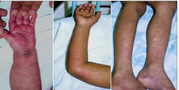
Polyserositis is associated to hemor-rhagic manifestations and thrombocytope-nia (Figure 1). It is well established in the literature that, typically, the hypotension secondary to this plasma leakage occurs up to 48 hours after defervescence, the mo-ment of fever abatemo-ment where the fever decreases to less than 38°C(7).
Despite the nonspecificity of sonog-raphic findings, ultrasonography is useful for the early diagnosis in patients with DHF and for differential diagnosis of other fe-brile diseases(6).
The objective of the present literature review is to describe the main sonographic findings and evaluating the role of ultra-sonography in the assessment of children with suspected dengue hemorrhagic fever.
MAIN SONOGRAPHIC FINDINGS
Ultrasonography is a safe, low-cost im-aging method that do not utilizes ionizing radiation, with high sensitivity to detect early signs of plasma leakage, many times in anticipation of the most critical period of the disease corresponding to the fever abatement to a temperature < 38°C, that is known as defervescence, where the risk for shock is higher. This critical phase extends from the third to the fifth day of febrile disease in children, where gallbladder wall thickening and cavitary effusion are
ob-served in addition to severe abdominal pain, persistent emesis and increased hema-tocrit levels(5–15).
The sonographic signs of plasma leak-age, particularly pleural effusion, may be early identified, up to two days before de-fervescence, preceding changes in hemat-ocrit levels(7).
Sonographic findings express the in-crease in capillary permeability (a sign of plasma leakage) and include cavitary sion (ascites, pleural and pericardial effu-sion), and gallbladder wall thickening present in one third of patients affected by the mild presentation, and in 95% of cases with the severe presentation of DHF. Ad-ditionally, the presence of fluid in the peri-renal space can be visualized(6,8).
Splenomegaly, hepatomegaly and volu-metric increase of the pancreas may also be observed(6).
Balasubramanian et al.(11), in a
compara-tive analysis of extravasation parameters including clinical signs, hemoconcentra-tion > 20%, hypoproteinemia, ultrasonog-raphy and chest radiogultrasonog-raphy, concluded that ultrasonography was the best method for screening DHF, with 91.42% sensitiv-ity and negative predictive value of 84.21%. Ascites, pleural effusion, gallbladder wall thickening and hepatomegaly were re-ported by the authors as predominant sonographic findings.
During a dengue epidemic, the diagno-sis of DHF should be considered as ultra-sonography demonstrates gallbladder wall
thickening, ascites, splenomegaly and pleu-ral effusion in a febrile patient with throm-bocytopenia(9,10).
CAVITARY EFFUSION
There is a correlation between pleural effusion, ascites, presence of fluid in the perirenal space, hepatic subcapsular collec-tion and pericardial effusion with severity in cases of DHF in children(6,7).
Pleural effusion is more commonly ob-served in DHF than in classical dengue, requiring rigorous observation(12).
Pleural effusion is the most frequent sonographic finding in cases of plasma leakage, and is present, even subtlety, in children affected by classical dengue, where this abnormality is usually transitory and of rapid resolution(7). Most frequently,
the onset of pleural effusion occurs imme-diately after defervescence, between the third and seventh day(12). In children,
how-ever, the onset of severe presentations is usually observed at about the third day, but not always is associated with deferves-cence(13). Some studies demonstrate that
pleural effusion may be present up to one day before defervescence in some pa-tients(7). It may be right unilateral or
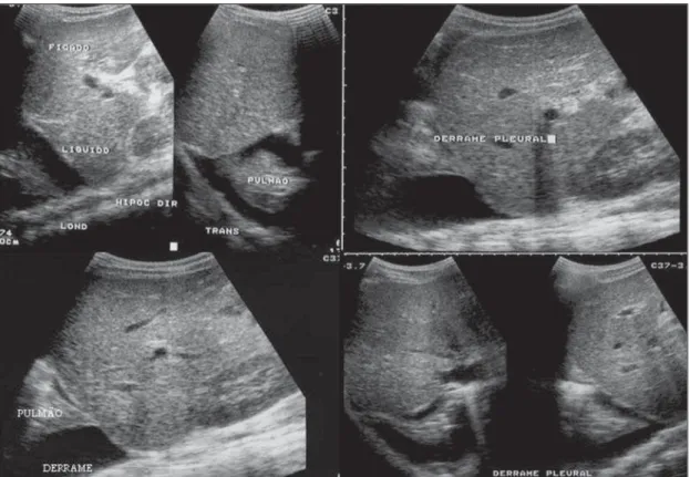
bilat-eral (Figures 2 and 3). It is rarely observed as left unilateral. Conventional chest radi-ography presents lower sensitivity than ul-trasonography in the detection of minor pleural effusions(8,10,11,14). In another study,
however, right lateral decubitus chest
ography performed one day after deferves-cence, has demonstrated higher sensitivity in the detection of pleural effusion as com-pared with ultrasonography. Its disadvan-tage is the higher exposure of the child to ionizing radiation(7).
Pericardial effusion is less frequently found, and may be observed in up to 28.5% of cases in assessments performed between the fifth and seventh febrile days(10,14).
The presence of volumes as large as ap-proximately 1,000 to 1,500 ml is necessary for clinical detection of free fluid, while ultrasonography can identify as little as about 100 ml(16).
Ascites was detected in 26% to 34% of cases with mild DHF, and in 94% to 95% of cases with severe DHF(6,17) (Figure 4).
Hepatic subcapsular fluid is little evi-denced, and its presence is a sign of disease
severity. However, it is a transitory finding (only one to two days) that may be observed at around the fourth to the fifth day after the disease onset(6) (Figure 5).
The presence of fluid in the perirenal space could not be visualized in the cases of mild DHF. However, it could be seen in 77% of patients severely affected by DHF. Thus this is a significant marker for disease severity(6).
GALLBLADDER WALL THICKENING
Gallbladder wall thickening is a non-specific finding frequently observed in other biliary or non-biliary conditions, such as acute cholecystitis, cirrhosis of liver, viral hepatitis, congestive heart failure, chronic kidney disease and hypoalbumin-emia(6–9,17,18).
Although the normal value for gallblad-der wall thickness is still to be well estab-lished in the literature, such finding is con-sidered in cases where the gallbladder wall thickness is > 3.0 mm. A more accurate measurement can be obtained on the ante-rior sub-hepatic wall in a longitudinal sec-tion, avoiding side lobe artifacts caused by
Figure 2. Pleural effu-sion at right, usual find-ing of plasma leakage, present even in a subtle presentation in children with classical dengue. In these cases, this finding is usually transitory and of rapid resolution.
Oliveira RVB et al.
the presence of adjacent intraluminal intes-tinal gas(18-20).
Four distinct gallbladder wall thicken-ing patterns can be identified: a striated pat-tern of multiple hypoechoic layers sepa-rated by echogenic zones; asymmetric pat-tern with echogenic tissue projecting into the gallbladder lumen; a central
hypoecho-genic zone separated by two echohypoecho-genic lay-ers; and a uniform echogenic pattern(21).
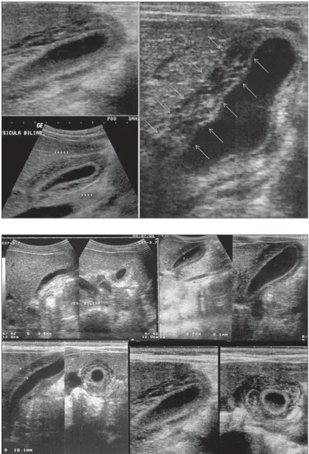
In patients with DHF, the striated pat-tern predominates (Figure 6), as a result of a probable fluid accumulation between the gallbladder wall layers producing stria-tions, as a function of the osmotic intravas-cular pressure(10,21).
Gallbladder wall thickening in children with DHF was first reported in 1991 by Pramuljo & Harun, in a study describing sonographic findings in 29 children with DHF, where 18% of cases presented gall-bladder wall thickening(22).
Gallbladder wall thickening was signifi-cantly associated with severe presentations
Figure 4. Presence of fluid in the abdominal cavity (ascites), in the anterior subhepatic space (A) and in the pelvic region (B).
of dengue, and with thrombocytopenia and hemoconcentration in suspicious cases of dengue. In some studies, this finding has been a relevant marker for clinical
diagno-sis and indicator of severity of DHF in chil-dren(17,21–24) (Figures 7 and 8).
The earliest finding in the subgroup of children with classical dengue was
gall-bladder wall thickening. However, besides resolving faster, it is less frequently de-tected than pleural effusion. There is a higher probability of detection when the
Figure 6. Longitudinal section of gallbladder, demonstrating a striated pattern of parietal thickening with multiple echo-genic layers intermingled with fluid.
Oliveira RVB et al.
US scan is performed at the second or third febrile day(7,13).
Two studies developed in Indonesia by Setiawan et al.(6,17) have found gallbladder
wall thickening > 3.0 mm in one third (32% to 33%) of patients with the mild presen-tation of the disease and in most of those with severe disease (94% to 95%), allow-ing the authors to confirm to positive as-sociation between gallbladder wall thick-ening and the disease severity, and conclud-ing that this findconclud-ing can be utilized in the identification of patients with higher risk for progressing to shock. In the cases of DHF, gallbladder wall thickening > 3.0 mm and < 5.0 mm presents a sensitivity of 93.8% and may be utilized as a criterion for patients’ hospitalization and monitoring. In cases where the gallbladder wall thickness is ≥ 5.0 mm, the specificity achieves 91.7%, a threshold that can be utilized in the selection of patients with higher risk for progressing to shock(17).
Venkata Sai et al.(10) have evaluated 88
children in the age range between two and nine years, with serological tests positive for dengue, and have demonstrated gall-bladder wall thickening in 100% of the patients, evidenced by ultrasonography performed in the period between the sec-ond and seventh febrile day. This finding was followed by pleural effusion, observed
with higher frequency from the fifth febrile day. Then, the authors concluded that, dur-ing an epidemic outbreak, gallbladder wall thickening, either with or without signs of polyserositis in a febrile patient, should suggest the possibility of classical dengue/ DHF.
The value of gallbladder wall thicken-ing as an ancillary factor in the diagnosis and prognosis of children with DHF has also been corroborated by other authors(23– 25). Although this finding is nonspecific and
may be present in other febrile diseases, it is useful in the early diagnosis and predic-tion of severity in cases of DHF, identify-ing the patients with higher risk for pro-gressing to shock.
The sonographic measurement of the gallbladder wall thickness is significantly associated with the severe presentations of dengue, and can also be utilized as a marker for thrombocytopenia and hemoconcentra-tion. Thus, ultrasonography is a relevant method as a prognostic test in the severe presentations of DHF in children.
Chacko & Subramanian, in a study evaluating 59 children with diagnosis of dengue shock syndrome, have reported that the presence of ascites and pleural effusion were the most predictive indicators of shock; on the other hand, gallbladder wall thickening was not associated with the
presence of shock, as reported in previous studies(26).
VOLUMETRIC INCREASE OF ORGANS
Volumetric increase of organs is a non-specific finding, corresponding to enteric findings of dengue, and should be taken into consideration in both clinical and sonographic context of plasma leakage(10).
Hepatomegaly, splenomegaly and, less fre-quently, volumetric increase of the pan-creas have been described in several stud-ies, but these findings are observed with a similar frequency in the mild and severe DHF presentations, with highest incidence of hepatomegaly(6,10,11).
CONCLUSION
In children with suspected dengue hem-orrhagic fever, ultrasonography, although nonspecific, is a relevant ancillary tool for the early diagnosis of plasma leakage signs and for prediction of the disease severity, identifying mild and severe cases of DHF, besides contributing in the differential di-agnosis with other causes of febrile disease.
REFERENCES
trole. São Paulo, SP: Livraria Santos Editora; 2001.
2. Siqueira Júnior JB, Martelli CMT, Coelho GE, et al. Dengue and dengue hemorrhagic fever, Bra-zil, 1981–2002. Emerg Infect Dis. 2005;11:48– 53.
3. Torres EM. Dengue. Rio de Janeiro, RJ: Editora Fiocruz; 2005.
4. Guzmán MG, Kourí G. Dengue: an update. Lan-cet Infect Dis. 2002;2:33–42.
5. Vabo KA, Torres Neto G, Santos AASMD, et al. Achados ultra-sonográficos abdominais em pa-cientes com dengue. Radiol Bras. 2004;37:159– 62.
6. Setiawan MW, Samsi TK, Wulur H, et al. Den-gue haemorrhagic fever: ultrasound as an aid to predict the severity of the disease. Pediatr Radiol. 1998;28:1–4.
7. Srikiatkhachorn A, Krautrachue A, Ratanaprakarn W, et al. Natural history of plasma leakage in dengue hemorrhagic fever: a serial ultrasono-graphic study. Pediatr Infect Dis J. 2007;26:283– 90.
8. Thulkar S, Sharma S, Srivastava DN, et al. Sono-graphic findings in grade III dengue hemorrhagic fever in adults. J Clin Ultrasound. 2000;28:34– 7.
9. Wu KL, Changchien CS, Kuo CH, et al. Early ab-dominal sonographic findings in patients with dengue fever. J Clin Ultrasound. 2004;32:386–8. 10. Venkata Sai PM, Dev B, Krishnan R. Role of
ul-trasound in dengue fever. Br J Radiol. 2005;78: 416–8.
11. Balasubramanian S, Janakiraman L, Kumar SS, et al. A reappraisal of the criteria to diagnose plasma leakage in dengue hemorrhagic fever. Indian Pediatr. 2006;43:334–9.
12. Firmida MC. Derrame pleural na criança com dengue. Acta Scientiae Medica. 2008;1:35–43.
13. Brasil. Ministério da Saúde. Secretaria de Vigi-lância em Saúde. Dengue: diagnóstico e manejo clínico – adulto e criança. 3ª ed. Brasília, DF: Ministério da Saúde; 2007.
14. Pelupessy JM, Allo ER, Jota S. Pericardial effu-sion in dengue haemorrhagic fever. Paediatr Indones. 1989;29:72–5.
15. Quiroz-Moreno R, Méndez GF, Ovando-Rivera KM. Utilidad clínica del ultrasonido en la iden-tificación de dengue hemorrágico. Rev Med Inst Mex Seguro Soc. 2006;44:243–8.
16. Goldberg BB, Goodman GA, Clearfield HR. Evaluation of ascites by ultrasound. Radiology. 1970;96:15–22.
17. Setiawan MW, Samsi TK, Pool TN, et al. Gall-bladder wall thickening in dengue hemorrhagic fever: an ultrasonographic study. J Clin Ultra-sound. 1995;23:357–62.
18. Patriquin HB, DiPietro M, Barber FE, et al. Sonography of thickened gallbladder wall: causes in children. AJR Am J Roentengol. 1983;141:57– 60.
19. Handler SJ. Ultrasound of gallbladder wall
thick-ening and its relation to cholecystitis. AJR Am J Roentgenol. 1979;132:581–5.
20. Laing FC, Federle MP, Jeffrey RB, et al. Ultra-sonic evaluation of patients with acute right up-per quadrant. Radiology. 1981;140:449–55.
21. Teefey SA, Baron RL, Bigler SA. Sonography of the gallbladder: significance of striated (layered) thickening of the gallbladder wall. AJR Am J Roentgenol. 1991;156:945–7.
22. Pramuljo HS, Harun SR. Ultrasound findings in dengue haemorrhagic fever. Pediatr Radiol. 1991; 21:100–2.
23. Gupta S, Singh SK, Taneja V, et al. Gall bladder wall edema in serology proven pediatric dengue hemorrhagic fever: a useful diagnostic finding which may help in prognostication. J Trop Pediatr. 2000;46:179–81.
24. Sehgal A, Gupta S, Tyagi V, et al. Gall bladder wall edema is not pathogenic of dengue infection. J Trop Pediatr. 2002;48:315–6.
25. Colbert JA, Gordon A, Roxelin R, et al. Ultra-sound measurement of gallbladder wall thicken-ing as a diagnostic test and prognostic indicator for severe dengue in pediatric patients. Pediatr Infect Dis J. 2007;26:850–2.