Submitted 26 May 2015 Accepted 4 July 2015 Published15 September 2015
Corresponding author Edward A. Bayer, [email protected]
Academic editor Guo-Qiang Chen
Additional Information and Declarations can be found on page 20
DOI10.7717/peerj.1126
Copyright 2015 Petkun et al.
Distributed under
Creative Commons CC-BY 4.0
OPEN ACCESS
Reassembly and co-crystallization of a
family 9 processive endoglucanase from
its component parts: structural and
functional significance of the
intermodular linker
Svetlana Petkun1, Inna Rozman Grinberg1, Raphael Lamed1, Sadanari Jindou2, Tal Burstein1, Oren Yaniv1, Yuval Shoham3, Linda J.W. Shimon4, Edward A. Bayer5and Felix Frolow1
1Department of Molecular Microbiology and Biotechnology, The Daniella Rich Institute for
Structural Biology, Tel Aviv University, Ramat Aviv, Israel
2Department of Life Sciences, Meijo University, Nagoya, Japan
3Department of Biotechnology and Food Engineering, Technion-Israel Institute of Technology,
Haifa, Israel
4Department of Chemical Research Support, The Weizmann Institute of Science, Rehovot, Israel 5Department of Biological Chemistry, The Weizmann Institute of Science, Rehovot, Israel
ABSTRACT
Non-cellulosomal processive endoglucanase 9I (Cel9I) from Clostridium thermocellumis a modular protein, consisting of a family-9 glycoside hydrolase (GH9) catalytic module and two family-3 carbohydrate-binding modules (CBM3c and CBM3b), separated by linker regions. GH9 does not show cellulase activity when expressed without CBM3c and CBM3b and the presence of the CBM3c was previously shown to be essential for endoglucanase activity. Physical reassociation of independently expressed GH9 and CBM3c modules (containing linker sequences) restored 60–70% of the intact Cel9I endocellulase activity. However, the mechanism responsible for recovery of activity remained unclear. In this work we independently expressed recombinant GH9 and CBM3c with and without their interconnecting linker inEscherichia coli. We crystallized and determined the molecular structure of the GH9/linker-CBM3c heterodimer at a resolution of 1.68 ˚A to understand the functional and structural importance of the mutual spatial orientation of the modules and the role of the interconnecting linker during their re-association. Enzyme activity assays and isothermal titration calorimetry were performed to study and compare the effect of the linker on the re-association. The results indicated that reassembly of the modules could also occur without the linker, albeit with only very low recovery of endoglucanase activity. We propose that the linker regions in the GH9/CBM3c endoglucanases are important for spatial organization and fixation of the modules into functional enzymes.
Subjects Biotechnology
INTRODUCTION
Cellulose is a major component of the plant cell wall, lending structural stability and resilience to an otherwise flaccid material. The propensity of cellulose to form ordered, tightly packed, para-crystalline fibrils hinders its enzymatic degradation. Indeed, the recalcitrant properties of cellulose are such that numerous enzymes are required to act synergistically in achieving its efficient degradation. Many types of bacteria and fungi are capable of degrading cellulose and other plant cell wall polysaccharides in an effective manner, producing a variety of various cellulases and related enzymes, either existing in the free state, or associated with a multi-enzyme complex known as the cellulosome (Bayer et al., 2004;Bayer et al., 2008;Demain, Newcomb & Wu, 2005;Doi & Kosugi, 2004;
Fontes & Gilbert, 2010).Clostridium thermocellumis an anaerobic thermophilic bacterium, known for its efficient degradation of cellulose and other plant cell wall polysaccharides (B´eguin, Millet & Aubert, 1992;Freier, Mothershed & Wiegel, 1988;Garcia-Martinez et al., 1980;Ng, Weimer & Zeikus, 1977;Wiegel, Mothershed & Puls, 1985). The cellulase system of this bacterium includes a remarkable variety of enzymes, some existing in the free state but most associated with a highly efficient multi-enzyme complex, termed cellulosome (Bayer et al., 2004;Lamed, Setter & Bayer, 1983;Lamed et al., 1983;Shoham, Lamed & Bayer, 1999), capable of converting a wide variety of plant-derived polysaccharides directly into soluble sugars and fermentation products (B´eguin & Alzari, 1998;Felix & Ljungdahl, 1993;
Schwarz, 2001;Schwarz, Zverlov & Bahl, 2004). These capabilities renderC. thermocelluma high utility candidate for use in consolidated bioprocessing (CBP) applications (reviewed inAkinosho et al., 2014).
been shown to sequentially hydrolyze cellulose chains and are thus referred to as processive endoglucanases (Gal et al., 1997;Gilad et al., 2003;Irwin et al., 1998;Jeon et al., 2012;Kuusk, Sorlie & Valjamae, 2015;Zverlov, Velikodvorskaya & Schwarz, 2003). Such enzymes appear to possess extended catalytic clefts and the observed processivity appears to require highly coordinated substrate-binding affinities from opposite sides of the cleavage site (Bu et al., 2012;Li, Irwin & Wilson, 2010;Payne et al., 2011).
Cellulase 9I (Cel9I), is a non-cellulosomal family 9 processive endoglucanase from
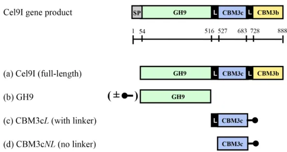
Clostridium thermocellum, which degrades crystalline cellulose (Avicel and filter paper) as well as phosphoric acid-swollen cellulose (PASC) and carboxymethyl cellulose (CMC) (Gilad et al., 2003). This enzyme consists of a catalytic GH9 module at its N terminus, followed by two family 3 carbohydrate-binding modules (CBMs): CBM3c and CBM3b. The three modules are separated by distinctive linker sequences. Such intermodular linker segments were proposed to be important for the physical association of the modules in the space, and to promote intermodular and/or intersubunit protein–protein interactions (Bayer et al., 1998;Bayer et al., 2009;Noach et al., 2008).
The C-terminal CBM3b module, as a classic CBM3, is responsible for targeting the Cel9I enzyme to the planar surface of the crystalline cellulose substrate (Gilad et al., 2003;
Su, Mackie & Cann, 2012;Tormo et al., 1996). It has also been proposed to disrupt the crystalline regions of cellulose, rendering it more accessible to the GH9 catalytic module (Yi et al., 2013) and to contribute to enzyme processivity by preventing the desorption of the catalytic module from cellulose (Telke et al., 2012). The function of the CBM3c is less straightforward. Removal of CBM3c fromC. thermocellumCel9I,C. cellulolyticum
Cel9G andP. BarcinonensisCel9B significantly reduces the enzyme activity (Burstein et al., 2009;Chiriac et al., 2010;Gal et al., 1997). CBM3c modules have been shown to alter the normal function of the GH9 catalytic module ofThermobifida fuscaCel9A from the standard endo-acting mode into a processive endoglucanase (Bayer et al., 1998;Irwin et al., 1998). Thus, Gilad et al., showed in2003that the endoglucanase activity of Cel9I is dependent upon the presence of the CBM3c module and suggested that the fused CBM3c serves an important accessory role for the catalytic domain by altering its character to facilitate processive cleavage of recalcitrant cellulose substrates.
We have previously shown that independently expressed GH9 and linker-containing CBM3c modules of Cel9I readily re-associatein vitroand that this physical reassociation recovers 60–70% of the intact Cel9I endoglucanase activity (Burstein et al., 2009).
We have examined in this work the interaction of the CBM3c with the catalytic module either with or without the intermodular linker in order to better understand the function of the CBM3c in the family-9 enzymes and the role of the linkers regions. The effect of the re-association of the CBM3c with linker (CBM3cL) and the CBM3c without linker (CBM3cNL) on the enzymatic activity of GH9 has been studied by the crystallization and structure determination of the reassembled GH9-CBM3cLcomplex at a resolution of 1.68 ˚A. The results of this study will help us to understand the contribution of ancillary modules in the action of multi-modular glycoside hydrolases.
MATERIALS AND METHODS
Cloning of the GH9, CBM3cLand CBM3cNLproteins
Cloning of the DNA fragments encoding the C-terminally His-tagged CBM3c with the linker and the untagged GH9 module from Cel9I ofC. thermocellum(GenBank accession codeL04735) was described earlier (Burstein et al., 2009;Gilad et al., 2003). C-terminally His-tagged CBM3c without the linker connecting it to the GH9 was amplified using the same procedure and the following primers:F′-5′CCATGGGCGAAGTTCCGGAGGATGAAATA
andR′-5′CTCGAGCGGTTCCCTTCCAAATACCAG. The PCR products were purified and cleaved
with restriction enzymesNcoI andXhoI and inserted into the pET-28a(+)expression vector (Novagen, Madison, WI, USA).
Expression and purification of recombinant proteins
The GH9 and CBM3c modules both with (GH9L, CBM3cL) and without (GH9NLand CBM3cNL) the linker regions were expressed independently by the identical expression procedure.Escherichia colistrain BL21(DE3)RIL harboring the plasmids was aerated at 310 K in 3-liters Terrific Broth supplemented with 25 mg ml−1kanamycin. After 3 h, the culture reached an A600of 0.6; 0.1 mM isopropyl-β-D-1-thiogalactopyranoside was added to induce gene expression, and cultivation was continued at 310 K for an additional 12 h. Cells were harvested by centrifugation (5,000×g for 15 min) at 277 K and were subse-quently re-suspended in 50 mM NaH2PO4, pH 8.0, containing 300 mM NaCl at a ratio of 1 g wet pellet to 4 ml buffer solution. A few micrograms of DNase powder were added prior to the sonication procedure. The suspension was kept on ice during sonication, after which it was centrifuged (20,000×g at 277 K for 20 min), and the supernatant was collected.
subjected to further purification by size-exclusion chromatography. Fast protein liquid chromatography (FPLC) was performed using a Superdex 75pg column and ¨AKTA Prime system (GE Healthcare, Piscataway, New Jersey, USA) to further purify the complex. One peak, corresponding approximately to 70 kDa, matching the predicted molecular weight of the GH9-CBM3c complex, was observed in the chromatogram. The 15 amino-acid linker sequence (about 1.5 kDa) did not significantly affect the elution volume, compared to that of the complex without the linker, presumably due to the limited resolution of the column. The relevant fractions (the purified complexed proteins) were analyzed by 15% sodium dodecyl sulfate-polyacrylamide gel electrophoresis (SDS-PAGE) with Coomassie brilliant blue staining. Two clear bands, of about 52 and 19.5 kDa were observed. The rearranged modules were concentrated to 6 mg ml−1using Centriprep YM-3 centrifugal filter devices YM-3 (Amicon Bioseparation, Millipore Corporation, Bedford, Massachusetts, USA). Protein concentration was determined by measuring UV absorbance at 280 nm.
The full-length Cel9I was purified by affinity chromatography on Avicel as reported earlier (Burstein et al., 2009;Gilad et al., 2003).
Microcalorimetric analysis
Isothermal titration calorimetry (ITC) experiments were carried out using a VP-ITC MicroCalorimeter (MicroCal, LLC, Northampton, Massachusetts, USA) at 298 K. About 300µM solution of CBM3cNLwas injected into a 65µM solution of GH9. The reaction
was performed in a buffer containing 50 mM Tris–HCl, pH 7.5, 150 mM NaCl, 0.05% sodium azide. Heats of dilution of the titrants were subtracted from the titration data, and the corrected data were analyzed using the Origin ITC analysis software package supplied by MicroCal. Thermal titration data were fit to the one binding site model, and enthalpy (ΔH), entropy (ΔS), association constant (Ka) and stoichiometry of binding (N) were determined. In all cases, the calculated stoichiometry (N) was lower than one, most likely due to the fact that the CBM3 proteins lost their native functionality with time. For the analysis, the CBM3 protein concentrations were corrected as to provide a stoichiometry of one. Two titrations were performed to evaluate reproducibility.
Enzyme activity assay
Reactions were performed at 333 K, in 50 mM citrate buffer (pH 6.0). The soluble cellu-lolytic substrate was carboxymethyl cellulose (CMC, Sigma Chem. Co. St. Louis, Missouri, USA). The amount of reducing sugars released from the substrate was determined with the 3,5-dinitrosalicylic acid (DNS) reagent as described by Miller et al. (Miller, 1959). Activity was defined as the amount (micromole) of reducing sugar released after 10 min of reaction.
Crystallization
of the Wizard Crystallization kit from Emerald BioSystems (Rigaku Reagents, Bainbridge Island, Washington, USA). The dyad of GH9 and CBM3cNLdid not yield any crystals. Screening of the GH9-CBM3cLresulted in plate-like crystals that appeared after several days under several conditions, all of which contained PEG 3350 and 4000. The best crystals were obtained in 30% PEG (both 3350 and 4000), 0.2 M magnesium chloride, and 0.1 M Hepes, pH 7.5. Attempts to optimize this condition using microbatch, hanging-drop, and sitting drop methods were unsuccessful, as the crystals remained very thin and fragile. The superfine Eyelash (Ted Pella, Inc, Redding, California, USA) was used to touch these crystals and consequently to streak the sitting drops, composed of 5µl of the protein
solution and 5µl of the precipitating solution (24% PEG 3350, 0.2 M magnesium chloride,
0.1 M Hepes, pH 8.0). After one day, crystals of different morphology, with maximum size of about 0.05 mm, appeared in the drop.
Data collection and crystallographic analysis
The crystals of the GH9-CBM3cLcomplex were harvested from the crystallization drop using a nylon cryo-loop (Hampton Research, Aliso Viejo, California, USA). For data collection, crystals were mounted on the MiTeGen stiffmicro-mount (MiTeGen, Ithaca, New York, USA) made of polyimide and flash-cooled in a nitrogen stream produced by Oxford Cryostream low temperature generator (Cosier & Glazer, 1986) at a temperature of 100 K. Mother-liquor of the crystals served for cryo-protection during the cooling in liquid nitrogen.
Diffraction data from the GH9-CBM3cLcrystals were measured using the ID23-2 beam line at ESRF, Grenoble, France. A MAR CCD 225 area detector and X-ray radiation of 0.873 ˚A wavelength were used. Diffraction data of 480 images with 0.5◦oscillation per image were collected. Data were processed withDENZOand scaled withSCALEPACKas implemented inHKL2000(Otwinowski & Minor, 1997). The crystals diffracted to 1.68 ˚A resolution and belong to the orthorhombic space group P212121, with unit cell parameters
a=70.4,b=88.5,c=106.5 ˚A. There is one GH9-CBM3cLcomplex per asymmetric unit with a Matthews densityVMof 2.37 ˚A3Da−1, corresponding to a solvent content of
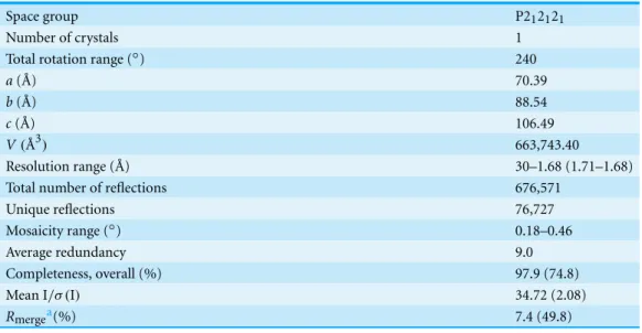
48.15% (Matthews, 1968). The X-ray data analysis statistics are presented inTable 1(Stout & Jensen, 1968).
Molecular replacement was carried out withMOLREP(Vagin & Teplyakov, 1997), using the coordinates of the GH9 and CBM3c modules of endoglucanase 9G from
Table 1 Diffraction data of the GH9-CBM3cin vitroreassembled complex from Cel9I fromC. ther-mocellum.Values shown in parentheses are for the highest resolution cell.
Space group P212121
Number of crystals 1
Total rotation range (◦) 240
a( ˚A) 70.39
b( ˚A) 88.54
c( ˚A) 106.49
V(˚A3) 663,743.40
Resolution range ( ˚A) 30–1.68 (1.71–1.68)
Total number of reflections 676,571
Unique reflections 76,727
Mosaicity range (◦) 0.18–0.46
Average redundancy 9.0
Completeness, overall (%) 97.9 (74.8)
Mean I/σ (I) 34.72 (2.08)
Rmergea(%) 7.4 (49.8)
Notes.
aR
merge=ΣhklΣi|Ii(hkl)− ⟨I(hkl)⟩|/ΣhklΣiIi(hkl), whereΣhkldenotes the sum over all reflections andΣithe sum over
all equivalent and symmetry-related reflections.
building byARP/wARP(Perrakis, Morris & Lamzin, 1999) produced a complete structure withRcrystandRfreeof 0.218 and 0.243, respectively. The model was manually corrected usingCOOT(Emsley & Cowtan, 2004) and refined usingREFMAC5(Murshudov, Vagin & Dodson, 1997). TheRcrystandRfreeimproved to 0.184 and 0.228, respectively. Solvent atoms were built usingARP/warp(Perrakis, Morris & Lamzin, 1999). Refinement of TLS (rigid body translation/libration/screw motions) parameters was performed (Winn, Isupov & Murshudov, 2001;Winn, Murshudov & Papiz, 2003). The model was subjected to several additional cycles of manual rebuilding and refinement. The model converged to finalRcryst andRfreefactors of 0.144 and 0.176, respectively.
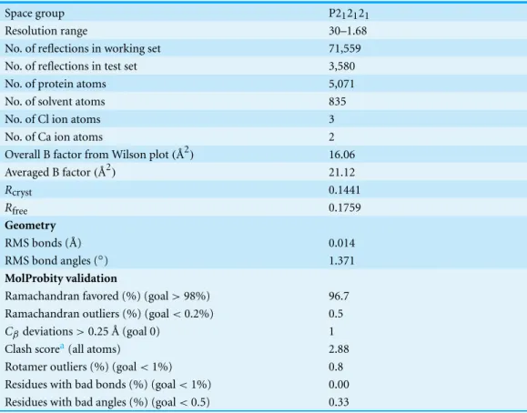
The refinement statistics of the structure are summarized inTable 2. The structure was validated usingMolProbity(Davis et al., 2007).
Protein sequence analysis
Sequence alignments were performed usingCLUSTALW(Larkin et al., 2007) and the coloring of residues (representing degree of conservation) using ProtSkin (Deprez et al., 2005). Sources of the sequences used in this work are as follows:Clostridium thermocellum
Cel9I GH9 module, CBM3c and CBM3b (AAA20892.1);Clostridium cellulolyticumCel9G GH9 module, CBM3c (AAA73868.1);Thermobifida fuscaCel9A GH9 module and CBM3c (AAB42155.1);Cellulomonas fimiCe9A CBM3c (AAA23086.1);Clostridium cellulovorans
EngH CBM3c (AAC38572.2) and CbpA CBM3a (AAA23218.1);Clostridium stercorarium
Table 2 Refinement statistics and results of MolProbity validation for reassembled GH9-CBM3c complex.
Space group P212121
Resolution range 30–1.68
No. of reflections in working set 71,559 No. of reflections in test set 3,580
No. of protein atoms 5,071
No. of solvent atoms 835
No. of Cl ion atoms 3
No. of Ca ion atoms 2
Overall B factor from Wilson plot(˚A2) 16.06
Averaged B factor(˚A2) 21.12
Rcryst 0.1441
Rfree 0.1759
Geometry
RMS bonds ( ˚A) 0.014
RMS bond angles (◦) 1.371
MolProbity validation
Ramachandran favored (%) (goal>98%) 96.7 Ramachandran outliers (%) (goal<0.2%) 0.5
Cβdeviations>0.25 ˚A (goal 0) 1
Clash scorea(all atoms) 2.88
Rotamer outliers (%) (goal<1%) 0.8 Residues with bad bonds (%) (goal<1%) 0.00 Residues with bad angles (%) (goal<0.5) 0.33
Notes.
aClash score is the number of serious steric overlaps (>0.4 ˚A) per 1,000 atoms.
CBM3c (AAG45158.1);Acetivibrio cellulolyticusCel9B CBM3c’ and CBM3b’ (CAI94607.1) and CipV (ScaA) CBM3b (AAF06064.1); Clostridium josui CipA (CipJ) CBM3a
(BAA32429.1);Bacteroides cellulosolvensScaA CBM3b (AAG01230.2);Bacillus subtilis
CelA CBM3b (AAA22307.1);Pectobacterium atrosepticumCelVI CBM3b (X79241.2);
Bacillus licheniformisCelA CBM3b (CAJ70714.1).
RESULTS
Cloning, expression and purification of Cel9I and its modular components
Figure 1 Schematic diagram of the Cel9I gene product and the recombinant proteins (A–D) prepared for this study.The GH9 module alone (B) was prepared with and without an N-terminal His tag (shown schematically in the figure), and the CBM3c’s were prepared with C-terminal His tags. Scale shows the number of amino acid residues and the boundaries of the different regions of the protein.
Recovery of endoglucanase activity upon association of
CBM3cNLand GH9 compared to CBM3cLand GH9
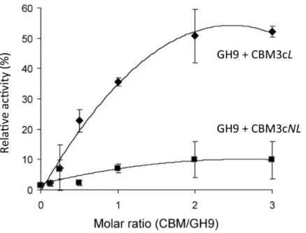
Previous works (Burstein et al., 2009;Gilad et al., 2003) demonstrated that the Cel9I catalytic module alone has no detectable activity on CMC (carboxymethyl cellulose) and that adding the CBM3cLto form the Cel9I-CBM3cLdyad serves to recover up to 70% of the lost activity. To further examine the importance of the linker connecting the GH9 and the CBM3c modules, we tested the ability of CBM3cNLto recover the CMCase activity of GH9. A fixed amount of the catalytic module (70 pmol in 400µl) was mixed
with increasing amounts of CBM3cLor CBM3cNL. The activity of the intact Cel9I enzyme was defined as 100%, and the activity of the reconstituted complexes was measured relative to that of Cel9I. The results indicated that GH9-CBM3cNLexhibit only about 10% of the intact Cel9I activity towards CMC, whereas the reassembled GH9-CBM3cLprovided up to 50% of the activity (Fig. 2). The fact that a higher than one molar ratio was required to obtain maximum activity can be explained by the fact that the CBM protein was only partly functional as was also observed in the ITC experiments described below. Overall, the results suggest that the linker is required for better fitting of the reconstituted CBM3c which results in better recovered activity.
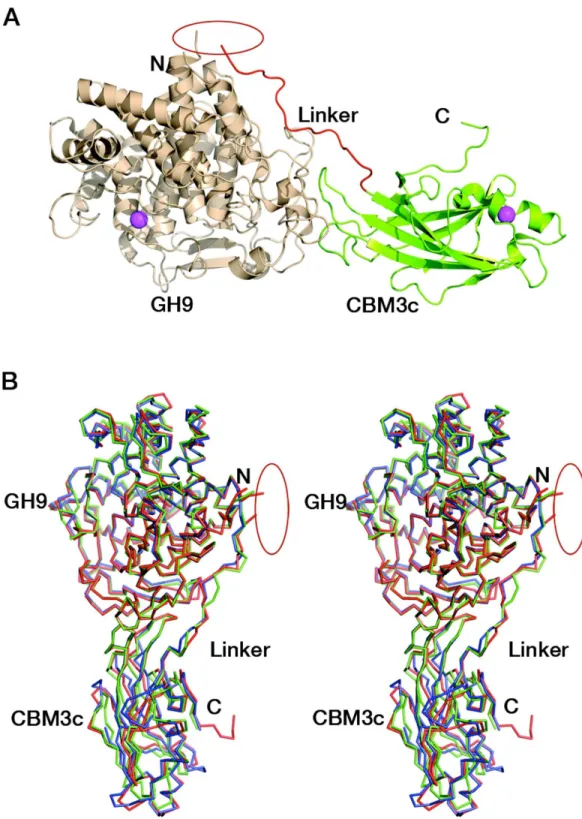
Overall structure of the reassembled GH9-CBM3c
Figure 2 Recovery of activity upon association of CBM3c (with and without linker) and GH9.CMCase activity (µmol reducing sugar released in a 10-min reaction) of His-tagged GH9, mixed either with
CBM3cL(diamonds) or CBM3cNL(squares), was examined. A fixed amount (70 pmol) of the GH9 catalytic module was mixed with increasing amounts of the indicated helper module, and their respective activities were compared to that of the intact Cel9I (GH9-CBM3c-CBM3b, set as 100%).
two domains separately and combined themin vitro. Surprisingly, the combined modules crystallized and formed a structure similar to those of the known GH9 cellulases.
The crystal structure of the reassembledC. thermocellumCel9I GH9-CBM3cLdyad was determined by molecular replacement and the coordinates are deposited in Protein Data Bank with code 2XFG. Data collection and refinement statistics are given inTables 1
and2. The catalytic GH9 and the ancillary CBM3c modules reassembledin vitroto form a dyad (Fig. 3A) similar in structure to the intact tandem GH9-CBM3c modules of the orthologous endoglucanases: Cel9G fromC. cellulolyticum(1G87) and Cel9A (previously termed cellulase E4) fromThermobifida fusca(1TF4), with an RMS deviation of 0.783 ˚A over 468 Cαatoms with Cel9G and 0.757 ˚A with Cel9A (Fig. 3B).
Structure of the GH9 module
The catalytic module of the Cel9I enzyme consists of residues 1–446, comprising 15
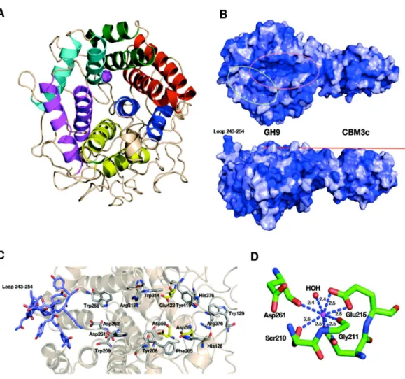
α-helices, whereby the twelve longest ones form the(α/α)6-barrel (Fig. 4A). The hydrophobic core of the GH9 module is formed by 118 hydrophobic and aromatic amino acids, the vast majority of which are also conserved in the GH9 modules from
Figure 4 Structural components of the reassembledC. thermocellumGH9-CBM3c.(A) Structure of the GH9 catalytic module, cartoon representation. Twelveα-helices form an (α/α)6-barrel fold. Pairs of helices, comprising the fold, are emphasized by red, blue, yellow, magenta, cyan and green. (B) Surface representation of the reassembled GH9-CBM3c complex. The residues are shaded according to the extent of their conservation with Cel9G fromC. cellulolyticumand Cel9A fromT. fusca. Darker blue indicates higher conservation. Top, birds-eye view of the catalytic cleft. Bottom, lateral view, showing the flat surface (red bar). Pink ellipse indicates the catalytic cleft, and green ellipse designates terminal portion of the catalytic site. (C) Close-up (same orientation as in B, top) of the catalytic cleft of the Cel9I GH9 module showing functional residues. Carbohydrate-binding residue carbons are colored gray, catalytic residue carbons are colored yellow. Loop 243–254 carbons are colored in light blue. (D) Calcium-binding site of theC. thermocellumCel9I GH9 module. Coordinating residues are shown in stick representation. The calcium ion is colored magenta, and distances to the coordinating atoms are indicated.
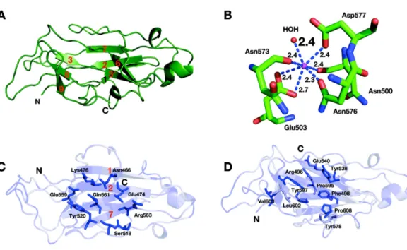
Figure 5 Structure of the CBM3c of Cel9I fromC. thermocellum. C and N termini are indicated (A). Cartoon representation,β-strands are numbered according to the alignment with Cel9G fromC. cellulolyticum, and Cel9A fromT. fusca. (B) Calcium-binding site of the CBM3c. (C) Birds-eye view of the flat surface. Residues are shaded according to their degree of conservation withC. cellulolyticumCel9G andT. fuscaCEL9A. Surface-exposed conserved residues are shown in stick representation. (D) Shallow groove of the CBM3c. Conserved surface residues are shown in stick representation. The residues are colored according to the degree of conservation in CBM3a, CBM3b and CBM3c modules derived from the sequences listed in the Methods section.
bimodular structures, one end of this cleft is blocked by a loop formed by residues 243–254 and the other end is fused with the flat surface of the CBM3c module (Fig. 4B). Details of the catalytic cleft are presented inFig. 4C.
One calcium ion is found near the catalytic cleft of the GH9 module of Cel9I and is coor-dinated by a Ser 210 (OG) 2.6 ˚A, Gly 211 (O) 2.4 ˚A, Asp 261 (O) 2.4 ˚A, Asp 214 bifurcated (OD1, OD2) 2.5 ˚A, and Glu 215 bifurcated (OD1, OD2) 2.5 ˚A (Fig. 4D). Despite some minor changes in the residues of coordination this ion seems to be structurally equivalent to those ofT. fuscaCel9A (RMS deviation 0.160 ˚A over 5 Cαatoms of the coordinating residues), andC. cellulolyticumCel9G (RMS deviation 0.503 ˚A over 4 Cαatoms). In all three cases the calcium ion draws together the N-terminal ends ofα-helixes 8 and 10.
Structure of the CBM3c module
The CBM3c module consists of 150 amino acids arranged in an eightβ-stranded sandwich motif homologous to other known family 3 CBM structures (Gilbert, Knox & Boraston, 2013;Mandelman et al., 2003;Petkun et al., 2010b;Sakon et al., 1997;Shimon et al., 2000;
over 113 atoms with CBM3c fromT. fuscaCel9A). Only 31% of amino acids are located in
β-strands of the CBM3c module from Cel9I; others are found in the loop regions. One calcium ion was found in the upperβ-sheet of the CBM3c molecule (Fig. 5B) and is coordinated by a water molecule and five residues from the upperβ-sheet: Asn 500 (O), Glu 503 bifurcated (OE1, OE2), Asn 573 (O), Asn 576 (OD1), Asp 577 (OD1). This calcium atom is in a similar location as in Cel9A and Cel9G, and probably plays a structural role for most CBM3 modules, as was suggested previously (Tormo et al., 1996).
The lower sheet forms a flat platform conserved between the CBM3c modules and the other two molecular structures. This flat surface is rich in charged and polar conserved surface residues: Asn 466, Glu 474, Lys 476, Ser 518, Tyr 520, Glu 559, Gln 561, and Arg 563 (Fig. 5C). The planar region of the CBM3c modules in all three enzymes is particularly aligned in continuation of the catalytic cleft of the catalytic modules, and has been proposed to bind single chains of cellulose and guide them to the cleft (Mandelman et al., 2003;Sakon et al., 1997).
The CBM3c possesses a very interesting surface structure, formed by theβ-strands on the opposite side of the flat surface, called the “shallow groove” (Shimon et al., 2000;Tormo et al., 1996). The “shallow groove” is lined by four aromatic rings (Phe 498, Tyr 538, Tyr 578 and Tyr 597), two charged or polar residues (Arg 496, and Glu 540), Leu 602, Pro 595 and Pro 608. These residues are also conserved in other CBM3 modules regardless of their subgroup relation (a, b, or c), their cellulose-binding ability and their effect on the activity of the catalytic module.Figure 5Dshows the shallow groove of the CBM3c module from the Cel9I enzyme colored according to the extent of the conservation of the residues in other CBM3a, b and c modules (darker blue represents more conservation). The alignment was performed over 25 CBM3 sequences (11 CBM3c, 12 CBM3b and CBM3b’, and 4 CBM3a). Conservation of this surface structure, regardless of the particular known function of the CBMs, implies that this site has some kind of “generic” function. This shallow groove may serve to bind to single oligosaccharide chains or to peptide chains, such as the intermodular linkers common to cellulases or cellulosomal scaffoldin subunits. There is evidence that the shallow groove interacts with a linker region (Petkun et al., 2010a;Shimon et al., 2000;Yaniv et al., 2012a).
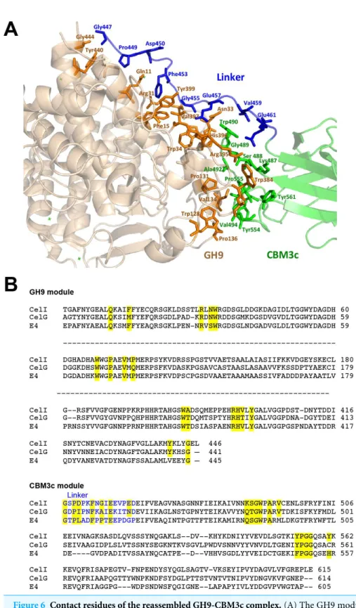
Contact residues between the GH9, linker and CBM3c
Conserved residues of the linker (which spans from Gly447 to Asp462) make numerous contacts with conserved residues of the GH9 module, emphasizing the importance of the linker in this interaction (Fig. 6). A conserved Gly447 of the linker interacts via hydrogen bonds with Gly444 and Tyr440 of the GH9 module. Pro449 forms hydrophobic interactions with Tyr440, and Asp450 forms hydrogen bonds with Gln11. Another linker residue, Phe453, forms hydrophobic and aromatic interactions with two aromatic residues of the GH9 module, i.e., Phe15 and Tyr399. Gly455 forms hydrogen bonds with Asn33 and Arg31. Glu457 forms intricate interactions with a variety of residues of the GH9 module, which include hydrogen bonding with Asn33, Val397, Arg395, as well as hydrogen and ionic interactions with His396. Glu461 exhibits hydrogen and ionic interactions with Arg395. Additionally, linker residues Glu457 and Glu461 form hydrogen bonds and salt bridges with Trp490 and Lys487, respectively. The latter belong to the CBM3c module and are part of a loop (486–498), which protrudes towards and forms interactions with several amino acids of the GH9 module. There are also many hydrogen bonds formed between the neighboring amino acids of the linker, thus contributing to its defined conformation. Altogether the multiple, well-conserved interactions between the linker, the GH9 and the CBM3c modules stabilize the spatial orientation of the modules towards one another and contribute to the structural rigidity of the entire molecule, resulting in an active enzyme structure.
As mentioned above, the mutual spatial orientation of the GH9 and CBM3c modules is very similar to that in the native, intact bimodular pairs from Cel9G and Cel9A leading to the overall similarity in structures. The remarkable conservation of the overall architecture in the reassembledin vitrocomplex together with the striking conservation of the contact residues implies its high functional importance. In all of these structures (Cel9G, Cel9A, and the reassembled GH9-CBM3cLfrom Cel9I), the flat surface of the CBM3c module is aligned in continuation with the catalytic cleft of the GH9 module, making an extended platform (Fig. 4B). This platform is rich in charged and polar surface residues that are highly conserved throughout the family 3 CBMc’s.
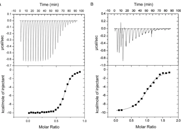
Microcalorimetric analysis of the GH9-CBM3c complex formation The binding constants of GH9 and the CBM3c were obtained by performing isothermal titration calorimetry (ITC) experiments in which a solution of GH9 was titrated with a solution of CBM3c with or without the linker (Fig. 7). Control experiments for each of the components alone were conducted and subtracted from the titration data. In both cases the titration curve could be fitted to a one-site binding model although the calculated stoichiometry was less than one. The low stoichiometry is probably a result of the fact that the soluble CBM module lost its functionality with time and its true active concentration was less than the measured protein concentration. To estimate the binding constants for the two CBM3c forms, the CBM3c concentrations were corrected to provide a stoichiometry of one. In all cases, the binding reactions were enthalpy driven with a negative entropy contribution. CBM3cL provided binding constants(Kd)between
Figure 7 Representative ITC titration of (A) GH9 and CBM3cNL(B) GH9 and CBM3cL.The top panel shows the calorimetric titration and the bottom panel displays the integrated injection heats corrected for control dilution heat. The solid line is the curve of the best fit used to derive the binding parameters, and the fitted data describe an interaction of a one binding site model.
2.9–4.3×10⁻⁷M. Thus, the linker may serve as a mitigating factor for the binding process, ensuring specific binding orientation. This is consistent with the structural data and the activity assays, which emphasizes the important role of the linker in enzyme functioning. In the case of CBM3cNL, the binding process may occur faster in the absence of linker, but may also lead to unspecific binding and aggregation of the modules.
DISCUSSION
through an additional CBM (Kostylev et al., 2012) or by attachment of the enzyme to a CBM-containing scaffoldin via a cohesin-dockerin interaction (Mingardon et al., 2011).
The CBM3c module of Cel9A from theT. fuscahas been proposed to loosely anchor the enzyme to cellulose, to disrupt the hydrogen bonds in crystalline cellulose and to guide a single cellulose strand towards the active site of the GH9 catalytic module (Bayer, Shoham & Lamed, 2006;Li, Irwin & Wilson, 2007). This hypothesis has been supported by molecular docking and molecular dynamics simulation studies (Oliveira et al., 2009). Moreover, double point mutations indicated that high coordination between the substrate affinities of the catalytic module and CBM needs to be precisely controlled (Li, Irwin & Wilson, 2010). Enzyme thermostability was reported to be affected by the presence of the CBM3c probably due to the formation of a compact structure (Chiriac et al., 2010;Su, Mackie & Cann, 2012;Yi et al., 2013).
The previously reported structures of Cel9A fromT. fusca(Sakon et al., 1997) and Cel9G fromC. cellulolyticum(Mandelman et al., 2003) revealed that the catalytic module and the CBM3c are separated by a∼20-residue linker that forms multiple polar and hydrophobic interactions mainly with the GH9 module. In an earlier report, we demonstrated that separately expressed GH9 and CBM3cLfrom Cel9I ofC. thermocelluminteract with one another to form an enzymatically active complex (Burstein et al., 2009). In the current article, we showed further that the GH9 and CBM3c can also be reassembled without the linker, albeit at the expense of catalytic activity, thus emphasizing the importance of the linker in positioning correctly the CBM relative to the GH9 catalytic module.
There is evidence that linkers in multi-modular proteins may serve communication roles between the modules via allosteric mechanisms and variation in their sequences affect enzyme activity (Ma et al., 2011). Linker length and rigidity was shown to play a critical role in the cooperative action of the catalytic module of a cellulase and a CBM (Ting, Makarov & Wang, 2009). Computational studies ofT. fuscaCel9A suggested that thermal contributions to enzyme plasticity and molecular motion at high temperatures may play a role in enhancing CBM and catalytic domain synergy, and the linker may have an important role in this process (Batista et al., 2011). The length of the linkers may also play an important role in protein function and adaptation to the environment (Sonan et al., 2007). Studies in cellulolytic fungi revealed that linkers undergo modifications such as glycoslation and have also been shown to directly bind to the cellulose substrate (Beckham et al., 2012;Payne et al., 2013;Sammond et al., 2012;Srisodsuk et al., 1993). Point mutations in different fungal GH-CBM linkers have also been shown to significantly affect the activity of the enzymes and their stability (Couturier et al., 2013;Lu et al., 2014).
the GH9 module, highlighting the importance of the linker in this interaction. Multiple hydrogen, ionic and hydrophobic bonds between the linker and the functional GH9 and CBM3c stabilize the spatial organization of the modules. The similarity of the reassembled and native intact structures is particularly intriguing, as it suggests that folding of the modular structures and emplacement of the linker during biosynthesis and intermodular recognition during complex formation are governed by the same interactions, which may have distinct functional consequences. In contrast to the GH9-CBM3cL, the re-associated GH9-CBM3cNLcomplex never crystallized, suggesting that the reassembly of the two modules in the absence of linker was somewhat heterogeneous in character.
Single proteins commonly fold into defined structures, wherein their N- and C-terminal ends are in relatively close proximity to one another. If we view the structures of the Theme B1 enzymes, it is evident that their individual modules, the GH9 catalytic module and the CBM3c, are consistent with this rule. The positions of the N- and C-termini of the Theme B1 catalytic module are similar to those of the other GH9 thematic members, including those of Theme A, which lack additional modules. Likewise, the N- and C-termini of CBM3c are essentially the same as all other members of the family 3 CBMs, regardless of their source (i.e., parent cellulase, scaffoldin, etc.). The evolutionary significance of this observation is that, originally, the functional relationship between the two modules was likely a more conventional one, whereby an ancestral CBM3 played a standard targeting role to deliver the GH9 catalytic module to its substrate. During the course of evolution, this relationship changed, and the precise positioning and fusion of a mutated CBM3 with a GH9 catalytic module served to modulate the activity characteristics of the latter. For this purpose, the flat surface of the CBM3c is aligned with the flat surface of the catalytic module, and the appropriate residues that interact with the single cellulose chain are thus aligned with the active site of the GH9 module. As a consequence, the two closely juxtaposed modules can be considered as a single functional entity. The functional positioning and fusion of the two modules, however, are at odds with the inherent locations of the termini of the two modules, such that the C-terminus of the catalytic module is very distant from the N-terminus of the CBM3c. Consequently, nature has provided a very distinctive type of conserved linker, which both connects the two modules and helps secure their required orientation for processive endoglucanase activity.
CONCLUSIONS
Cellulase 9I (Cel9I), a non-cellulosomal family 9 processive endoglucanase from
linker in order to better understand the function of the CBM3c in the family-9 enzymes and the role of the linkers regions.
GH9 catalytic module and CBM3c were able to interact and reassemble both with and without the linker; however, the linker was essential for the endoglucanase catalytic activity. Surprisingly, we were able to crystallize these two separately expressed entities, meaning that their reassembly was very ordered and structurally homogeneous. The molecular structure of the GH9 and CBM3c with the linker region showed that they form a complex similar in structure to the intact tandem GH9-CBM3c modules of the orthologous endoglucanases Cel9G fromC. cellulolyticumand Cel9A fromThermobifida fusca. The flat, conserved surface of the CBM3c module is aligned in continuation with the catalytic cleft of the GH9 module, presumably forming one functional entity, which binds to the planar surface of the cellulose. Conserved residues of the linker make contacts with conserved residues of the GH9 module, highlighting the importance of the linker in this interaction. Overall, our results demonstrate that the linker regions in the GH9/CBM3c endoglucanases are necessary to achieve the right spatial organization of the modules and for the fixation of the modules into functional enzymes.
ACKNOWLEDGEMENTS
This article is dedicated to the memory of Professor Felix Frolow, who passed away on 29 August 2014. We thankfully acknowledge the ESRF for synchrotron beam time and staff scientists of the ID-29 beam line for their assistance.
ADDITIONAL INFORMATION AND DECLARATIONS
Funding
Grant Disclosures
The following grant information was disclosed by the authors: Israel Science Foundation: 293/08, 1349/13, 24/11.
Israel Strategic Alternative Energy Foundation (I-SAEF). Israeli Center of Research Excellence: 152/11.
United States-Israel Binational Science Foundation (BSF). Institute of Science Alternative Energy Research Initiative (AERI). Helmsley Foundation.
Israel Ministry of Science (IMOS).
Competing Interests
Edward Bayer is an Academic Editor for PeerJ.
Author Contributions
• Svetlana Petkun conceived and designed the experiments, performed the experiments, analyzed the data, wrote the paper, prepared figures and/or tables.
• Inna Rozman Grinberg analyzed the data, wrote the paper, reviewed drafts of the paper, prepared figures and/or tables.
• Raphael Lamed conceived and designed the experiments, analyzed the data, wrote the paper, reviewed drafts of the paper.
• Sadanari Jindou performed the experiments, contributed reagents/materials/analysis tools.
• Tal Burstein conceived and designed the experiments, performed the experiments.
• Oren Yaniv conceived and designed the experiments, performed the experiments, analyzed the data.
• Yuval Shoham conceived and designed the experiments, contributed reagents/materials/analysis tools.
• Linda J.W. Shimon analyzed the data, wrote the paper, reviewed drafts of the paper.
• Edward A. Bayer conceived and designed the experiments, analyzed the data, wrote the paper, prepared figures and/or tables, reviewed drafts of the paper.
• Felix Frolow conceived and designed the experiments, analyzed the data, wrote the paper, prepared figures and/or tables.
Data Availability
The following information was supplied regarding data availability: Protein Data Bank, accession code2XFG.
REFERENCES
Akinosho H, Yee K, Close D, Ragauskas A. 2014.The emergence ofClostridium thermocellum
Arai T, Ohara H, Karita S, Kimura T, Sakka K, Ohmiya K. 2001.Sequence of celQ and properties of celQ, a component of theClostridium thermocellumcellulosome.Applied Microbiology and Biotechnology57:660–666DOI 10.1007/s00253-001-0832-4.
Batista PR, De Souza Costa MG, Pascutti PG, Bisch PM, De Souza W. 2011.High temperatures
enhance cooperative motions between CBM and catalytic domains of a thermostable cellulase: mechanism insights from essential dynamics.Physical Chemistry Chemical Physics
13:13709–13720DOI 10.1039/c0cp02697b.
Bayer EA, Belaich J-P, Shoham Y, Lamed R. 2004.The cellulosomes: multi-enzyme machines
for degradation of plant cell wall polysaccharides.Annual Review of Microbiology58:521–554
DOI 10.1146/annurev.micro.57.030502.091022.
Bayer EA, Lamed R, White BA, Flint HJ. 2008.From cellulosomes to cellulosomics.The Chemical
Record8:364–377DOI 10.1002/tcr.20160.
Bayer EA, Morag E, Lamed R, Yaron S, Shoham Y. 1998.Cellulosome structure: four-pronged
attack using biochemistry, molecular biology, crystallography and bioinformatics.
In: Claeyssens M, Nerinckx W, Piens K, eds.Carbohydrases from trichoderma reesei and other microorganisms. London: The Royal Society of Chemistry, 39–65.
Bayer EA, Shoham Y, Lamed R. 2006.Cellulose-decomposing prokaryotes and their enzyme
systems. In: Dworkin M, Falkow S, Rosenberg E, Schleifer K-H, Stackebrandt E, eds.The prokaryotes. Third Edition. New York: Springer-Verlag, 578–617.
Bayer EA, Smith SP, Noach I, Alber O, Adams JJ, Lamed R, Shimon LJW, Frolow F. 2009.Can
we crystallize a cellulosome? In: Sakka K, Karita S, Kimura T, Sakka M, Matsui H, Miyake H, Tanaka A, eds.Biotechnology of lignocellulose degradation and biomass utilization. Tokyo: Ito Print Publishing Division, 183–205.
Beckham GT, Dai Z, Matthews JF, Momany M, Payne CM, Adney WS, Baker SE, Himmel ME. 2012.Harnessing glycosylation to improve cellulase activity.Current Opinion in Biotechnology 23:338–345DOI 10.1016/j.copbio.2011.11.030.
Beckham GT, Stahlberg J, Knott BC, Himmel ME, Crowley MF, Sandgren M, Sorlie M, Payne CM. 2014.Towards a molecular-level theory of carbohydrate processivity in glycoside hydrolases.Current Opinion in Biotechnology27:96–106DOI 10.1016/j.copbio.2013.12.002.
B´eguin P, Alzari PM. 1998.The cellulosome ofClostridium thermocellum.Biochemical Society Transactions26:178–185DOI 10.1042/bst0260178.
B´eguin P, Millet J, Aubert J-P. 1992.Cellulose degradation byClostridium thermocellum: from manure to molecular biology.FEMS Microbiology Letters100:523–528
DOI 10.1111/j.1574-6968.1992.tb05750.x.
Br¨unger TA. 1992.Free R value: a novel statistical quantity for assessing the accuracy of crystal structures.Nature355:472–475DOI 10.1038/355472a0.
Bu L, Nimlos MR, Shirts MR, Stahlberg J, Himmel ME, Crowley MF, Beckham GT. 2012.
Product binding varies dramatically between processive and nonprocessive cellulase enzymes.
Journal of Biological Chemistry287:24807–24813DOI 10.1074/jbc.M112.365510.
Burstein T, Shulman M, Jindou S, Petkun S, Frolow F, Shoham Y, Bayer EA, Lamed R. 2009.
Physical association of the catalytic and helper modules of a processive family-9 glycoside hydrolase is essential for activity.FEBS Letters583:879–884DOI 10.1016/j.febslet.2009.02.013.
Cantarel BL, Coutinho PM, Rancurel C, Bernard T, Lombard V, Henrissat B. 2009.The
Chayen NE, Shaw Stewart PD, Maeder DL, Blow DM. 1990.An automated system for micro-batch protein crystallization and screening.Journal of Applied Crystallography
23:297–302DOI 10.1107/S0021889890003260.
Chiriac AI, Cadena EM, Vidal T, Torres AL, Diaz P, Pastor FI. 2010.Engineering a family 9 processive endoglucanase from Paenibacillus barcinonensis displaying a novel architecture.
Applied Microbiology and Biotechnology86:1125–1134DOI 10.1007/s00253-009-2350-8.
Cosier J, Glazer AM. 1986.A nitrogen-gas-stream cryostat for general X-ray-diffraction studies.
Journal of Applied Crystallography19:105–107DOI 10.1107/S0021889886089835.
Couturier M, Feliu J, Bozonnet S, Roussel A, Berrin JG. 2013.Molecular engineering of fungal
GH5 and GH26 beta-(1,4)-mannanases toward improvement of enzyme activity.PLoS ONE
8:e79800DOI 10.1371/journal.pone.0079800.
Cuskin F, Flint JE, Gloster TM, Morland C, Basle A, Henrissat B, Coutinho PM, Strazzulli A,
Solovyova AS, Davies GJ, Gilbert HJ. 2012. How nature can exploit nonspecific
catalytic and carbohydrate binding modules to create enzymatic specificity.Proceedings of the National Academy of Sciences of the United States of America 109:20889–20894
DOI 10.1073/pnas.1212034109.
Davies G, Henrissat B. 1995.Structures and mechanisms of glycosyl hydrolases.Structure
3:853–859DOI 10.1016/S0969-2126(01)00220-9.
Davis IW, Leaver-Fay A, Chen VB, Block JN, Kapral GJ, Wang X, Murray LW, Arendall 3rd
WB, Snoeyink J, Richardson JS, Richardson DC. 2007.MolProbity: all-atom contacts and
structure validation for proteins and nucleic acids.Nucleic Acids Research35(Web Server
issue):W375–W383DOI 10.1093/nar/gkm216.
Demain AL, Newcomb M, Wu JH. 2005.Cellulase, clostridia, and ethanol.Microbiology and
Molecular Biology Reviews69:124–154DOI 10.1128/MMBR.69.1.124-154.2005.
Deprez C, Lloubes R, Gavioli M, Marion D, Guerlesquin F, Blanchard L. 2005. Solution
structure of theE. coliTolA C-terminal domain reveals conformational changes upon binding to the phage g3p N-terminal domain.Journal of Molecular Biology346:1047–1057
DOI 10.1016/j.jmb.2004.12.028.
Doi RH, Kosugi A. 2004.Cellulosomes: plant-cell-wall-degrading enzyme complexes.Nature
Reviews Microbiology2:541–551DOI 10.1038/nrmicro925.
Emsley P, Cowtan K. 2004.Coot: model-building tools for molecular graphics.ACTA
Crystallo-graphica. Section D: Biological Crystallography60:2126–2132DOI 10.1107/S0907444904019158.
Felix CR, Ljungdahl LG. 1993.The cellulosome—the exocellular organelle of Clostridium.Annual
Review of Microbiology47:791–819DOI 10.1146/annurev.mi.47.100193.004043.
Fontes CM, Gilbert HJ. 2010.Cellulosomes: highly efficient nanomachines designed to deconstruct plant cell wall complex carbohydrates.Annual Review of Biochemistry79:655–681
DOI 10.1146/annurev-biochem-091208-085603.
Freier D, Mothershed CP, Wiegel J. 1988.Characterization ofClostridium thermocellumJW20.
Applied and Environmental Microbiology54:204–211.
Gal L, Gaudin C, Belaich A, Pag`es S, Tardif C, Belaich J-P. 1997.CelG fromClostridium cellulolyticum: a multidomain endoglucanase acting efficiently on crystalline cellulose.Journal of Bacteriology179:6595–6601.
Garcia-Martinez DV, Shinmyo A, Madia A, Demain AL. 1980.Studies on cellulase production
Gilad R, Rabinovich L, Yaron S, Bayer EA, Lamed R, Gilbert HJ, Shoham Y. 2003.CelI, a non-cellulosomal family-9 enzyme fromClostridium thermocellum, is a processive endoglucanase that degrades crystalline cellulose.Journal of Bacteriology 185:391–398
DOI 10.1128/JB.185.2.391-398.2003.
Gilbert HJ, Hazlewood GP. 1993.Bacterial cellulases and xylanases. Journal of General Microbiology139:187–194DOI 10.1099/00221287-139-2-187.
Gilbert HJ, Knox JP, Boraston AB. 2013.Advances in understanding the molecular basis of plant
cell wall polysaccharide recognition by carbohydrate-binding modules.Current Opinion in Structural Biology23:669–677DOI 10.1016/j.sbi.2013.05.005.
Gilkes NR, Henrissat B, Kilburn DG, Miller RCJ, Warren RAJ. 1991.Domains in microbial
b-1,4-glycanases: sequence conservation, function, and enzyme families.Microbiological Reviews55:303–315.
Gruninger RJ, Gong X, Forster RJ, McAllister TA. 2014.Biochemical and kinetic characterization of the multifunctional beta-glucosidase/beta-xylosidase/alpha-arabinosidase, Bgxa1.Applied Microbiology and Biotechnology98:3003–3012DOI 10.1007/s00253-013-5191-4.
Henrissat B. 1991.A classification of glycosyl hydrolases based on amino acid sequence
similarities.Biochemical Journal280:309–316DOI 10.1042/bj2800309.
Henrissat B, Davies G. 1997.Structural and sequence-based classification of glycoside hydrolases.
Current Opinion in Structural Biology7:637–644DOI 10.1016/S0959-440X(97)80072-3.
Henrissat B, Davies GJ. 2000.Glycoside hydrolases and glycosyltransferases. Families, modules, and implications for genomics.Plant Physiology124:1515–1519DOI 10.1104/pp.124.4.1515.
Henrissat B, Romeu A. 1995.Families, superfamilies and subfamilies of glycosyl hydrolases.
Biochemical Journal311:350–351DOI 10.1042/bj3110350.
Irwin D, Shin D-H, Zhang S, Barr BK, Sakon J, Karplus PA, Wilson DB. 1998.Roles of the
catalytic domain and two cellulose binding domains ofThermomonospora fuscaE4 in cellulose hydrolysis.Journal of Bacteriology180:1709–1714.
Jeon SD, Yu KO, Kim SW, Han SO. 2012.The processive endoglucanase EngZ is active in
crystalline cellulose degradation as a cellulosomal subunit of Clostridium cellulovorans.New Biotechnology29:365–371DOI 10.1016/j.nbt.2011.06.008.
Kostylev M, Moran-Mirabal JM, Walker LP, Wilson DB. 2012.Determination of the molecular
states of the processive endocellulase Thermobifida fusca Cel9A during crystalline cellulose depolymerization.Biotechnology and Bioengineering109:295–299DOI 10.1002/bit.23299.
Krissinel E, Henrick K. 2007.Inference of macromolecular assemblies from crystalline state.
Journal of Molecular Biology372:774–797DOI 10.1016/j.jmb.2007.05.022.
Kuusk S, Sorlie M, Valjamae P. 2015.The predominant molecular state of bound enzyme
determines the strength and type of product inhibition in the hydrolysis of recalcitrant polysaccharides by processive enzymes.Journal of Biological Chemistry290:11678–11691
DOI 10.1074/jbc.M114.635631.
Lamed R, Setter E, Bayer EA. 1983.Characterization of a cellulose-binding, cellulase-containing complex inClostridium thermocellum.Journal of Bacteriology156:828–836.
Lamed R, Setter E, Kenig R, Bayer EA. 1983.The cellulosome—a discrete cell surface organelle ofClostridium thermocellumwhich exhibits separate antigenic, cellulose-binding and various cellulolytic activities.Biotechnology and Bioengineering Symposium13:163–181.
Larkin MA, Blackshields G, Brown NP, Chenna R, McGettigan PA, McWilliam H, Valentin F,
Wallace IM, Wilm A, Lopez R, Thompson JD, Gibson TJ, Higgins DG. 2007.ClustalW and
Li Y, Irwin DC, Wilson DB. 2007.Processivity, substrate binding, and mechanism of cellulose hydrolysis byThermobifida fuscaCel9A.Applied and Environmental Microbiology73:3165–3172
DOI 10.1128/AEM.02960-06.
Li Y, Irwin DC, Wilson DB. 2010.Increased crystalline cellulose activity via combinations of amino acid changes in the family 9 catalytic domain and family 3c cellulose binding module ofThermobifida fuscaCel9A.Applied and Environmental Microbiology76:2582–2588
DOI 10.1128/AEM.02735-09.
Lu H, Luo H, Shi P, Huang H, Meng K, Yang P, Yao B. 2014. A novel thermophilic
endo-beta-1,4-mannanase from Aspergillus nidulans XZ3: functional roles of carbohydrate-binding module and Thr/Ser-rich linker region.Applied Microbiology and Biotechnology98:2155–2163DOI 10.1007/s00253-013-5112-6.
Mandelman D, Belaich A, Belaich JP, Aghajari N, Driguez H, Haser R. 2003.X-ray crystal
structure of the multidomain endoglucanase Cel9G fromClostridium cellulolyticumcomplexed with natural and synthetic cello-oligosaccharides.Journal of Bacteriology185:4127–4135
DOI 10.1128/JB.185.14.4127-4135.2003.
Ma B, Tsai CJ, Haliloglu T, Nussinov R. 2011.Dynamic allostery: linkers are not merely flexible.
Structure19:907–917DOI 10.1016/j.str.2011.06.002.
Matthews BW. 1968.Solvent content of protein crystals.Journal of Molecular Biology33:491–497
DOI 10.1016/0022-2836(68)90205-2.
Meekins DA, Raththagala M, Husodo S, White CJ, Guo HF, Kotting O, Vander Kooi CW,
Gentry MS. 2014.Phosphoglucan-bound structure of starch phosphatase Starch Excess4 reveals
the mechanism for C6 specificity.Proceedings of the National Academy of Sciences of the United States of America111:7272–7277DOI 10.1073/pnas.1400757111.
Miller GL. 1959.Use of dinitrosalicylic acid reagent for determination of reducing sugar.Analytical Biochemistry31:426–428.
Mingardon F, Bagert JD, Maisonnier C, Trudeau DL, Arnold FH. 2011.Comparison of family 9
cellulases from mesophilic and thermophilic bacteria.Applied and Environmental Microbiology
77:1436–1442DOI 10.1128/AEM.01802-10.
Mora¨ıs S, Alber O, Barak Y, Hadar Y, Wilson DB, Lamed R, Shoham Y, Bayer EA. 2012.
Functional association of the catalytic and ancillary modules dictates enzymatic activity in glycoside hydrolase family 43β-xylosidase.Journal of Biological Chemistry287:9213–9221
DOI 10.1074/jbc.M111.314286.
Murshudov GN, Vagin AA, Dodson EJ. 1997.Refinement of macromolecular structures by the
maximum-likelihood method.ACTA Crystallographica. Section D: Biological Crystallography
53:240–255DOI 10.1107/S0907444996012255.
Ng TK, Weimer TK, Zeikus JG. 1977.Cellulolytic and physiological properties ofClostridium thermocellum.Archives of Microbiology114:1–7DOI 10.1007/BF00429622.
Noach I, Alber O, Bayer EA, Lamed R, Levy-Assaraf M, Shimon LJW, Frolow F. 2008.
Crystallization and preliminary X-ray analysis ofAcetivibrio cellulolyticuscellulosomal type II cohesin module: two versions having different linker lengths.Acta Crystallographica F64:58–61
DOI 10.1107/S1744309107066821.
Oliveira OV, Freitas LCG, Straatsma TP, Lins RD. 2009.Interaction between the CBM of Cel9A
fromThermobifida fuscaand cellulose fibers.Journal of Molecular Recognition22:38–45
DOI 10.1002/jmr.925.
Otwinowski Z, Minor W. 1997.Processing of X-ray diffraction data collected in oscillation mode.
Payne CM, Bomble YJ, Taylor CB, McCabe C, Himmel ME, Crowley MF, Beckham GT. 2011.Multiple functions of aromatic-carbohydrate interactions in a processive cellulase examined with molecular simulation.Journal of Biological Chemistry286:41028–41035
DOI 10.1074/jbc.M111.297713.
Payne CM, Resch MG, Chen L, Crowley MF, Himmel ME, Taylor 2nd LE, Sandgren M,
Stahlberg J, Stals I, Tan Z, Beckham GT. 2013.Glycosylated linkers in multimodular
lignocellulose-degrading enzymes dynamically bind to cellulose.Proceedings of the National Academy of Sciences of the United States of America110:14646–14651
DOI 10.1073/pnas.1309106110.
Perrakis A, Morris R, Lamzin VS. 1999.Automated protein model building combined with
iterative structure refinement.Nature Structural Biology6:458–463DOI 10.1038/8263.
Petkun S, Jindou S, Shimon LJW, Rosenheck S, Bayer EA, Lamed R, Frolow F. 2010b.Structure
of a family 3b’ carbohydrate-binding module from the Cel9V glycoside hydrolase from
Clostridium thermocellum. Structural diversity and implications for carbohydrate binding.
Acta CrystallogrD66:33–43.
Petkun S, Jindou S, Shimon LJW, Rosenheck S, Bayer EA, Lamed R, Frolow F. 2010a.
Structure of a family 3b’ carbohydrate-binding module from the Cel9V glycoside hydrolase fromClostridium thermocellum: structural diversity and implications for
carbohydrate binding.ACTA Crystallographica. Section D: Biological Crystallography66:33–43
DOI 10.1107/S0907444909043030.
Sakon J, Irwin D, Wilson DB, Karplus PA. 1997.Structure and mechanism of endo/exocellulase
E4 fromThermomonospora fusca.Nature Structural Biology4:810–818
DOI 10.1038/nsb1097-810.
Sammond DW, Payne CM, Brunecky R, Himmel ME, Crowley MF, Beckham GT. 2012.
Cellulase linkers are optimized based on domain type and function: insights from sequence analysis, biophysical measurements, and molecular simulation.PLoS ONE 7:e48615
DOI 10.1371/journal.pone.0048615.
Schwarz WH. 2001.The cellulosome and cellulose degradation by anaerobic bacteria.Applied
Microbiology and Biotechnology56:634–649DOI 10.1007/s002530100710.
Schwarz WH, Zverlov VV, Bahl H. 2004.Extracellular glycosyl hydrolases from Clostridia.
Advances in Applied Microbiology56:215–261.
Shimon LJW, Pag`es S, Belaich A, Belaich JP, Bayer EA, Lamed R, Shoham Y, Frolow F. 2000.
Structure of a family IIIa scaffoldin CBD from the cellulosome ofClostridium cellulolyticumat 2.2 ˚A resolution.ACTA Crystallographica. Section D: Biological Crystallography56:1560–1568
DOI 10.1107/S0907444900012889.
Shoham Y, Lamed R, Bayer EA. 1999. The cellulosome concept as an efficient microbial
strategy for the degradation of insoluble polysaccharides.Trends in Microbiology7:275–281
DOI 10.1016/S0966-842X(99)01533-4.
Sonan GK, Receveur-Brechot V, Duez C, Aghajari N, Czjzek M, Haser R, Gerday C. 2007.The
linker region plays a key role in the adaptation to cold of the cellulase from an Antarctic bacterium.Biochemical Journal407:293–302DOI 10.1042/BJ20070640.
Srisodsuk M, Reinikainen T, Penttila M, Teeri TT. 1993.Role of the interdomain linker peptide of Trichoderma reesei cellobiohydrolase I in its interaction with crystalline cellulose.Journal of Biological Chemistry268:20756–20761.
Su X, Mackie RI, Cann IK. 2012.Biochemical and mutational analyses of a multidomain cellulase/mannanase from Caldicellulosiruptor bescii.Applied and Environmental Microbiology
78:2230–2240DOI 10.1128/AEM.06814-11.
Telke AA, Ghatge SS, Kang SH, Thangapandian S, Lee KW, Shin HD, Um Y, Kim SW. 2012.Construction and characterization of chimeric cellulases with enhanced catalytic activity towards insoluble cellulosic substrates. Bioresource Technology 112:10–17
DOI 10.1016/j.biortech.2012.02.066.
Tina KG, Bhadra R, Srinivasan N. 2007.PIC: protein interactions calculator.Nucleic Acids Research35:W473–W476DOI 10.1093/nar/gkm423.
Ting CL, Makarov DE, Wang ZG. 2009.A kinetic model for the enzymatic action of cellulase.The
Journal of Physical Chemistry B113:4970–4977DOI 10.1021/jp810625k.
Tormo J, Lamed R, Chirino AJ, Morag E, Bayer EA, Shoham Y, Steitz TA. 1996.Crystal structure
of a bacterial family-III cellulose-binding domain: a general mechanism for attachment to cellulose.EMBO Journal15:5739–5751.
Vagin A, Teplyakov A. 1997.MOLREP: an automated program for molecular replacement.The
Journal of Physical Chemistry30:1022–1025.
Venditto I, Najmudin S, Luis AS, Ferreira LM, Sakka K, Knox JP, Gilbert HJ, Fontes CM. 2015.
Family 46 carbohydrate-binding modules contribute to the enzymatic hydrolysis of xyloglucan and beta-1,3-1,4-glucans through distinct mechanisms.Journal of Biological Chemistry
290:10572–10586DOI 10.1074/jbc.M115.637827.
Wiegel J, Mothershed CP, Puls J. 1985.Differences in xylan degradation by various noncellulolytic thermophilic anaerobes andClostridium thermocellum.Applied and Environmental Microbiology 49:656–659.
Wilson DB, Irwin DC. 1999.Genetics and properties of cellulases.Advances in Biochemical
Engineering65:1–21DOI 10.1007/3-540-49194-5 1.
Wilson DB, Kostylev M. 2012.Cellulase processivity.Methods in Molecular Biology908:93–99.
Winn MD, Isupov MN, Murshudov GN. 2001.Use of TLS parameters to model anisotropic
displacements in macromolecular refinement.ACTA Crystallographica. Section D: Biological Crystallography57:122–133DOI 10.1107/S0907444900014736.
Winn MD, Murshudov GN, Papiz MZ. 2003.Macromolecular TLS refinement in REFMAC at
moderate resolution.Methods in Enzymology374:300–321.
Yaniv O, Fichman G, Borovok I, Shoham Y, Bayer EA, Lamed R, Shimon LJ, Frolow F. 2014.
Fine-structural variance of family 3 carbohydrate-binding modules as extracellular biomass-sensing components ofClostridium thermocellumanti-sigmaI factors.ACTA Crystallographica. Section D: Biological Crystallography70:522–534DOI 10.1107/S139900471302926X.
Yaniv O, Frolow F, Levy-Assraf M, Lamed R, Bayer EA. 2012a.Interactions between family
3 carbohydrate binding modules (CBMs) and cellulosomal linker peptides.Methods in Enzymology510:247–259DOI 10.1016/B978-0-12-415931-0.00013-6.
Yaniv O, Petkun S, Shimon LJ, Bayer EA, Lamed R, Frolow F. 2012b. A single mutation
reforms the binding activity of an adhesion-deficient family 3 carbohydrate-binding module.ACTA Crystallographica. Section D: Biological Crystallography 68:819–828
DOI 10.1107/S0907444912013133.
Yaniv O, Shimon LJ, Bayer EA, Lamed R, Frolow F. 2011. Scaffoldin-borne family 3b
Yi Z, Su X, Revindran V, Mackie RI, Cann I. 2013.Molecular and biochemical analyses of CbCel9A/Cel48A, a highly secreted multi-modular cellulase by Caldicellulosiruptor bescii during growth on crystalline cellulose.PLoS ONE8:e84172DOI 10.1371/journal.pone.0084172.
Zhou W, Irwin DC, Escovar-Kousen J, Wilson DB. 2004.Kinetic studies of Thermobifida fusca
Cel9A active site mutant enzymes.Biochemistry43:9655–9663DOI 10.1021/bi049394n.
Zmudka MW, Thoden JB, Holden HM. 2013.The structure of DesR from Streptomyces
venezuelae, a beta-glucosidase involved in macrolide activation.Protein Science22:883–892
DOI 10.1002/pro.2204.
Zverlov VV, Velikodvorskaya GA, Schwarz WH. 2003.Two new cellulosome components
encoded downstream ofcelI in the genome ofClostridium thermocellum: the non-processive endoglucanase CelN and the possibly structural protein CseP.Microbiology149:515–524