DOI: 10.5562/cca2784
Magnetic Properties and Oxygen Defects of Dilute
Metal Doped Tin Oxide Based Semiconductor
Kiyoshi Nomura
Photocatalytic International Research Center, Tokyo University of Science, 2641 Yamazaki, Noda-city, Chiba, 278-8501, Japan Author’s e-mail address: [email protected]
RECEIVED: November 12, 2015 REVISED: December 27, 2015 ACCEPTED: December 28, 2015
THIS PAPER IS DEDICATED TO DR. SVETOZAR MUSIĆ ON THE OCCASION OF HIS 70TH BIRTHDAY
Abstract: Chemical and magnetic states of iron doped tin oxide (SnO2) as a diluted magnetic semiconductor (DMS) at room temperature have
been investigated using 57Fe Mössbauer spectrometry, XRD and magnetometery. The influence of the doping conditions of SnO2 with iron on
the generation of oxygen defects was reviewed and discussed on the basis of ab initio calculations.
The magnetic properties depended on preparation conditions, such as thermal decomposition and sol-gel processing as well as 57Fe and
super-dilute 57Mn implantation. It was shown that Sb codoping in Fe doped SnO2 increases the saturation magnetization. Doping of Fe(Sb)-SnO2
with nonmagnetic Zn ions up to 7 % also increases the magnetization although there is no precipitation of crystalline magnetic phases. The co-doping of two transition metal ions (Fe-Co, Fe-Mn, Fe-Ni and Fe-V) in SnO2 matrix enhanced the magnetization as compared with that of single
metal ion doped samples. It is suggested from different valence states of doped metal ions that double exchange interactions occur through or near the oxygen vacancies in SnO2.
The SnO2 doped with dilute 57Fe may show the intrinsic and/or extrinsic DMS properties. Oxygen vacancies play an important role in the
intrinsic DMS. The intrinsic nature of DMS is supported by both, experimental results and ab initio calculations. The long range interactions between diluted magnetic ions are considered to occur through electrons produced by oxygen vacancies or electrons induced by Sb5+ doping.
Keywords: diluted magnetic semiconductor (DMS), Mössbauer spectrometry, 57Fe doped SnO2 semiconductor, SnO2 co-doped with two metals,
ab initio calculation.
INTRODUCTION
XIDE diluted magnetic semiconductors (DMSs), which possess good semiconducting and magnetic pro-perties at room temperature (RT), are promising materials for spintronic devices. Magnetic alloy semiconductors (II– VI, III–V) such as Mn doped ZnTe, GaAs and InAs, which show ferromagnetism at low temperatures, have already been utilized as spintronic devices.[1] The first oxide DMS
system was reported by Matsumoto et al. (2001). The authors showed that Co doped TiO2 films prepared by laser
ablation show ferromagnetism at RT.[2] T. Dietel et al.[3a]
And Pearton et al.[3b] showed the relationship between
band gap and Curie temperature of semiconductors.[3] In
order to realize RT-DMS, the semiconductor with large band gap is profitable as candidate material.[3] Ogale et al.
reported that transparent Co-doped SnO2 shows the high
temperature ferromagnetism with a giant magnetic
moment.[4] This seems to be due to precipitated magnetic
compounds. SnO2 film with band gap of 3.6 eV is
transparent and electrically conductive. SnO2 doped with
metal ions has been used for a gas sensor working at temperatures of several hundred degrees Celsius.[5]
There remain the questions why the magnetic moment in oxide semiconductors increases by doping them with small amounts of magnetic ions, and why the DMSs show high Curie temperature. The study of the magnetism in diluted magnetic oxides became an interesting issue over the last decade.[6] Coey et al. reported the magnetic
polaron models and the charge transfer band models with impurity band as dilute magnetic mechanism.[7] Which
dilute magnetic oxide semiconductor is fact or fiction is discussed.[8] It is necessary to reveal the chemical and
Croat. Chem. Acta 2015, 88(4), 579–590 DOI: 10.5562/cca2784
57Fe Mössbauer spectrometry provides the
infor-mation of electronic and magnetic states of iron nano-materials. Conversion electron Mössbauer spectrometry and X-ray Mössbauer spectrometry (CEMS and XMS) can be used for characterization of iron included in solid surface and thin layers.[9] The vibration density states (VDOS) of
oxides containing Mössbauer nuclei can be characterized by another Mössbauer technique, the nuclear resonance inelastic scattering (NIS) using synchrotron radiation.[10]
Thus, the valence states, magnetic states, and local VDOS of Fe doped oxide semiconductors can be clarified by using Mössbauer techniques of ca. 1 % 57Fe probes.
Various transparent DMS films have been characterized by 57Fe CEMS. For example, transparent 6 % 57Fe doped TiO2 film, prepared by laser ablation in vacuum
of 1.33×10⁻⁶ Pa, shows Kerr effect (optical ferromagnetism)
at RT. It was clarified by 57Fe CEMS that the magnetic origin
is mainly due to metallic Fe clusters.[11]
In this review article, 57Fe Mössbauer analytical results
and magnetic properties of SnO2 based materials doped with
diluted Fe ions are introduced together with theoretical results obtained by ab initio calculation for configurations of substituted Fe and oxygen defects of Fe doped SnO2.[12]
MAGNETIC PROPERTIES OF Fe
DOPED Sn OXIDE POWDER
Fe Doped SnO
2[13]10 at % Fe doped SnO2 powder was prepared by a sol-gel
method and the thermal decomposition. The relationship between magnetization curves and annealing conditions is shown in Figure 1 (a). The ferromagnetic hysteresis includ-ing a paramagnetic component is observed.
The magnetic hysteresis depends on annealing temperatures. 10 at % Fe doped SnO2 annealed at 600 °C
shows the highest saturation magnetization. The magnetic relaxation peaks are observed in addition to paramagnetic doublets and a minor sextet of Fe2O3 in the Mössbauer
spectrum. The sample annealed at 500 °C shows only two doublets of Fe3+ although the saturation magnetization is
larger than that of the sample annealed at 650 °C, which contains the weak magnetic component of Fe2O3. The
doped Fe species are embedded in so large grains of matrix SnO2 that cannot show the superparamagnetism. However,
there remains the question why the ferromagnetic behavior is obtained.
Figure 1. (a) Magnetic hysteresis of 10 at % Fe doped SnO2 annealed at various temperatures for 2 hours; (b) Mössbauer spectra
DOI: 10.5562/cca2784 Croat. Chem. Acta 2015, 88(4), 579–590 As shown in Figure 2, the saturation magnetization
(Ms) of the sample heated at 500 °C for 2 hours decreases
by annealing at 500 °C further for 4 hours. The oxygen defects are considered to be repaired by annealing for long time. Therefore, the magnetization is related to oxygen defects induced at the initial annealing. Two doublets observed in Fig. 1 (b) are assigned to Fe3+ (isomer shift (IS)
= 0.34 mm s–1, quadrupole splitting (QS)= 0.80 mm s–1) and
Fe3+ (IS= 0.28 mm s–1, QS= 1.60 mm s–1). This suggests that
high spin Fe3+ ions interact as a long range ordering through
electron spin localized at oxygen deficiency under the magnetic fields, that is defect induced ferromagnetism due to overlapped polarons as shown in Figure 3.
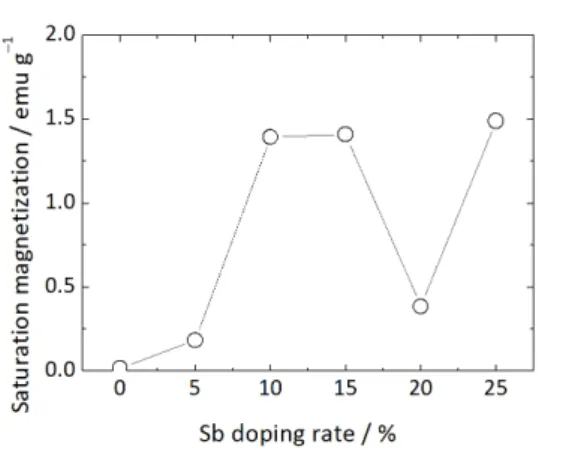
Sb and Fe Codoped SnO
2[14]Sn4+O2 doped with Sb5+ is n-type semiconductor, whereas
SnO2 doped with Fe3+ is p-type semiconductor. The ionic
radii are 0.069 nm for Sn4+, 0.064 nm for Fe3+, and 0.060 nm
for Sb5+, respectively. Doping Sb5+ into SnO2 increases the
electric conductivity and the solubility of Fe3+ doped. The
saturation magnetization of Sn0.8–yFe0.2SbyO2 increases with
the increase of Sb as shown in Figure 4.[14] However, the
magnetization decreases by compensating the induced carrier density by holes formed when the doping rate of Sb becomes almost the same as that of Fe.
Mössbauer spectra of Sn0.8–yFe0.2SbyO2 (y = 0.25)
show major doublets and a minor magnetic sextet at RT. The magnetization is relatively large and the coercivity is small like soft magnetic behavior. When Mössbauer spectra are measured at low temperatures, the area intensity of doublets is relatively transferred into that of a broad sextet with the decrease of the measuring temperature as shown in Figure 5. The hyperfine fields are not so dependent on the measuring temperature. It is considered that the Figure 2. Effect of annealing at 500 °C on magnetization of
10 at % Fe doped SnO2.[13].
Figure 3. Diagram on the formation of polaron. Oxygen
atoms are not displayed. ▯: oxygen deficiency.[7]
Figure 4. Dependence of saturation magnetization on
doping rate of Sb in 20 at % Fe doped SnO2.[14]
Figure 5. Mössbauer spectra of Sn0.55Fe0.2Sb0.25O2,
Croat. Chem. Acta 2015, 88(4), 579–590 DOI: 10.5562/cca2784 doublet peaks obtained at RT are not due to fine particles
of iron oxides, but due to the small domain size, that is the spin-spin interaction. It is to note that a ferromagnetic be-havior including a paramagnetic component is observed in the magnetic hysteresis curves although almost all samples
show the doublets of paramagnetic Fe3+. The abnormal
phenomena are considered due to the exchange coupling between local electron spin of Fe3+ through the carrier
elec-trons doped. The magnetic moment of Fe3+ induces the
po-larization of carrier electrons, which interact with the next neighbor Fe3+. Thus, the possible mechanism is considered
to be connected with the exchange interactions between iron ions and magnetic defects being mediated by impurity induced free charge carriers.
As compared with single Fe ion doped SnO2, Sb and
Fe codoping into SnO2 results in enhancement of
satura-tion magnetizasatura-tion without the precipitasatura-tion of impuri-ties. This suggests that the origin of ferromagnetism is mainly due to the increase of electron carrier density by doping Sb5+.
119
Sn Mössbauer Spectrum of
Fe Doped SnO
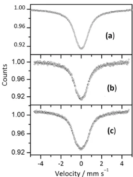
2[15]119Sn is not a magnetic atom itself. However, the transition
by gamma ray absorption occurs between the exited state of nuclear spin I = 3/2 and the ground state of I = 1/2. In
119Sn Mössbauer spectra measured under the applied
mag-netic fields, the magmag-netic sextet is expected to appear like
57Fe Mössbauer spectrum. When 119Sn Mössbauer spectra
of Sn0.8–yFe0.2SbyO2 were measured under magnetic fields of
0.3 T and 0.55 T, the peak with broaden width was observed in 119Sn Mössbauer spectra as shown in Figure 6. Thus, it is
found that even 119Sn nucleus in the bulk area is also
sensi-tive to applied magnetic fields.[15]119Sn can be used as one
of magnetic probes for dilute magnetic materials. Figure 6. 119Sn Mössbauer spectra of FexSbySn1–x–yO2–δ:
(a) no applied field; (b) 0.3 T applied field; (c) 0.55 T applied
field.[15]
DOI: 10.5562/cca2784 Croat. Chem. Acta 2015, 88(4), 579–590
Nonmagnetic Zn Doped
Fe
xSb
ySn
1–x–yO
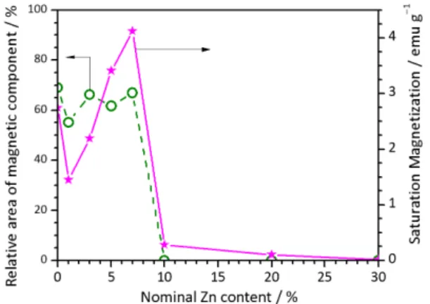
2–δ[16]XRD peaks of Zn doped FexSbySn1–x–yO2–δshow only the rutile structure of SnO2 even if doped with Zn up to 7 %,
whereas the spinel, Zn2SnO4, is additionally observed for
more than 10 % Zn doped samples. XRD peaks of Zn2SnO4
are almost similar to those of spinel ZnFe2O4. The
Mössbauer spectra are composed of two paramagnetic doublets and magnetic relaxation peaks as shown in Figure 7. A doublet with small QS was assigned to Fe3+ coordinated
with six oxygen atoms and another doublet with large QS was assigned to Fe3+ closed to oxygen vacancy. However,
both doublets are due to Fe3+, which are located close to
oxygen vacancies in different configurations as described later in section Magnetic moments and oxygen vacancies of
Fe doped SnO2 by ab initio calculation. The area intensity of
magnetic relaxation component and the saturation mag-netization are plotted for doping rate of Zn, respectively, as shown in Figure 8. It is found that the saturation magneti-zation is correlated with magnetic relaxation component of Fe3+. When more than 10 % Zn is doped, the magnetic
relaxation component disappears in the Mössbauer spec-tra, but the weak magnetization is remaining. It is con-sidered due to magnetic defects produced at the interface between the precipitated impurity and SnO2 matrix.
SnO
2Codoped With Two Transition
Metal Ions
In the case of single metal ion doped SnO2, it is known that
the saturation magnetization increases with Fe concentra-tion, but decreases with Co concentration. The codoping effect of Fe and Co doped SnO2 were studied.[17,18] The
samples were prepared by a sol-gel method and a thermal decomposition at 550 °C. The crystallite size is about 20– 40 nm in diameter. As shown in Figure 9, 1 % Fe and 0.5 %
Co codoped SnO2 enhances the saturation magnetization
(Ms). When more than 3 % Co are doped into Fe doped
SnO2, the magnetization decreases. There is a possibility
that ferrite is precipitated with increase of Co content, but the broad magnetic components as shown in Fig. 10 are absolutely different from that of CoFe2O4. It is hard to
con-sider the precipitated ferrite as the origin of ferromag-netism. It is not sure whether a double exchange interaction of Fe and Co through Sn and O occur or not, but it was confirmed from Co L absorption edge in XAFS that 1 % Co ions have the mixed valence states of 2+ and 3+. It Figure 8. Relative area intensity of magnetic components in
Mössbauer spectra and saturation magnetization of ZnxFe0.2Sb0.1Sn1–(x+0.3)O2.
Figure 9. Magnetic hysteresis of 1 % Fe and y % Co codoped
SnO2.[17]
Figure 10.57Fe Mössbauer spectra of SnO2 doped with 1 %
Croat. Chem. Acta 2015, 88(4), 579–590 DOI: 10.5562/cca2784 is considered that the local electrons existing in oxygen
deficiency and isolated Sn atoms are intermediates between ferromagnetic interactions.
The magnetization properties of Mn and Fe codoped SnO2, and Fe and Ni codoped SnO2 are also studied.[19,20]
Dilute Fe-Mn codoped SnO2 is synthesized by a
sol-gel method. The Fe-Mn codoping enhances the magnetiza-tion in comparison with the case of single metal ion doping. The Ms values are correlated with the crystallite sizes.
Mössbauer spectrometry revealed the magnetic sextet and relaxation peaks, which suggest that Fe ions doped contrib-ute to the magnetic ordering and super-paramagnetic prop-erties. X-ray absorption spectroscopy revealed that Mn3+ and
Mn2+ mixed states are dominant in Fe-Mn codoped SnO2.[19]
In the case of Fe-Ni codoped SnO2, the following
pos-sibilities for the origins of RT ferromagnetism are consid-ered. The formation of Fe and Ni complex oxides might become one of the candidates for ferromagnetic ordering, since Ni-based spinel-type ferrite is well known as a ferro-magnetic material. However, the observed doping depend-encies in Ms of Fe-Ni codoped SnO2 cannot be explained by
the simple formation of secondary phases. It is therefore deduced that the Fe-Ni codoping enhances the Msnot by
the segregation of secondary phases but by the exchange interaction.[20]
In the case of Fe-V codoped SnO2, Fe-V codoping
also enhanced the Ms, which showed the maximum Ms at
1 % V and 1 % Fe codoping. With further increase of Fe-V codoped into SnO2 host, the Ms decreased. Chemical
states of vanadium ions were deduced as V5+ states
by XAS. Mössbauer spectrometry revealed that the intensity of sextet components is related to the Ms, which
indicates that small amounts of Fe-V codoped are effective to enhance Ms.[21] It is to note that the sextet
component is like hematite, which shows weak ferromagnetism at RT although the XRD peaks of hematite are not be detected.
As compared with single metal doped SnO2, the
ferromagnetic exchange interaction is enhanced by codoping two transition metal ions. A codoping technique that employs different d electron numbers becomes essential for the stabilization of ferromagnetism with the control of both saturation magnetization (Ms) and the
coercive field (Hc) at RT.[20]
Fe ION IMPLANTED SnO
2FILM
[22]57Fe ions are implanted into SnO2 film (200 nm in thickness)
on fused quartz glass, heated at RT, 300 °C, and 500 °C, respectively. The ion implantation energy is 100 keV, and the flux is 1×10¹⁷ Fe ions/cm2. According to Monte Carlo
calculation (using the Transport of Ions in Matter: TRIM), Fe concentration reaches the maximum 10 % in the depth of
40 nm of SnO2 film. The magnetic hysteresis was a little
observed at substrate temperature of 300 °C, but disap-peared after annealing.
57
Fe Ion Implantation at
RT
[23]SnO2 film implanted with 57Fe at RT did not show Kerr effect
at all. 119Sn CEM spectra of SnO2 film implanted with
1×10¹⁷ ions/cm2 57Fe are composed of two doublets of Sn4+
(80 %) and Sn2+ (20 %). By 57Fe ion implantation, Sn4+ in
oxide film is partially reduced into Sn2+, whereas implanted 57Fe2+ is oxidized into Fe3+. With the increase of annealing
temperatures, Sn2+ ions are perfectly oxidized into Sn4+ to
form SnO2 rutile film.
57
Fe Ion Implantation of SnO
2Film at
Substrate Temperature of 500 °C
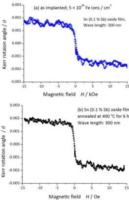
[24]SnO2 film implanted with 5×10¹⁶ Fe ions/cm2 at 500 °C
shows Kerr effect as shown in Figure 11. It is transparent Fe doped SnO2 film with high electric resistance. Two
magnetic sextets with broad peaks observed in Figure 12 (left side) are due to fine Fe3O4. The implanted ions are
segregated into a cluster at the high temperature. CEM spectra of Figure 12, (a), (b) and (c) are obtained by discriminating the energy of inner conversion electrons emitted after Mössbauer effect. The low energy electrons detected reflect the deep layer because the energy of the inner conversion electrons is lost by the upper layers, whereas the high energy electrons without the energy loss reflect the top surface layer. The two doublets of Fe3+
and Fe2+ are observed in CEM spectra. It is found from
their relative intensity that the Fe2+ species is located in
deeper layer than in top surface layers, and that magnetite is more produced in the surface layers. Even if the sample was further heated at 400 °C, the ferromagnetic behavior did not disappear. The CEM spectra of the annealed sample showed one broad sextet in addition to two doublets of Fe2+ and Fe3+. This broad
sextet is due to fine maghemite (γ-Fe2O3). Peak intensity
of paramagnetic Fe2+ in deep layers is not so affected by
annealing in air although the peak intensity of paramagnetic Fe2+ decreases and the intensity of
paramagnetic Fe3+ increases in top surface layers. The fine
magnetite in surface layers is oxidized directly into a maghemite by annealing.
CEM spectra of 3 % Sb doped SnO2 film implanted
with 57Fe are the same as the CEM spectra of 1 % Sb doped
SnO2 film implanted with 57Fe although each intensity is
different. It was found that 3 % Sb doped SnO2 film is
oxidized harder than 1 % Sb doped SnO2 film because the
relative intensity of magnetite in the former is smaller than that of the latter. The detail analysis of 3 % Sb doped SnO2
film implanted with 57Fe and further annealed at the high
DOI: 10.5562/cca2784 Croat. Chem. Acta 2015, 88(4), 579–590
Super Dilute
57Mn Ion Implanted
SnO
2Films
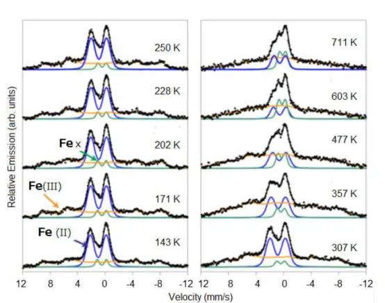
[26]57Fe emission Mössbauer spectra of radioactive 57Mn ions
implanted SnO2 film are obtained on extremely dilute
con-centrations of < 2×10–12 57Mn/cm2, or < 10–3 at % 57Mn as
shown in Figure 13. In the β-decay of 57Mn (T½ = 1.5 min),
the daughter 57Fe is obtained by average recoil energy of ER
= 40 eV, leading to the possible formation of interstitial Fe species in the case that the bond–dissociation energy is less than the recoil energy. The emission Mössbauer spectra ob-tained are composed of three components: (1) a doublet marked Fe(II) with a Gaussian distribution of quadrupole splitting (QS), assigned to high-spin Fe2+, (2) magnetic
hyperfine field distribution marked Fe(III), assigned to high-spin Fe3+ showing slow paramagnetic relaxations, 3) a
dou-blet component Fex, assigned to interstitial Fe3+, not metallic Fe. From the recoil imparted on the Fe atom in the β-decay of 57Mn, it was tempting to assign Fex to interstitial
Fe.[25] However, the sharp lined doublet (Fex) is interpreted
due to recoil produced interstitial Fe3+ from the isomer shift
values. From the temperature dependence (143 K to 711 K) as shown in Figure 14, the activation energy for dissociation of Mn-VO pairs is estimated to be 0.9(1) eV. The energy is so small that the oxygen vacancy produced easily interacts Figure 11. Magneto-optical measurement of SnO2 films
(a) as implanted; (b) after annealing at 400 °C.[24]
Croat. Chem. Acta 2015, 88(4), 579–590 DOI: 10.5562/cca2784 with 57Fe. The transformation process of implanted Fe
species can be suggested from the peak intensity ratios of the decomposed spectral components.
The paramagnetic Fe2+ species, which are located in
amorphous phases produced locally by 40 keV ion implantation, are oxidized into Fe3+ at the temperatures
higher than 330 K. The oxidized Fe3+ species, which may be
located in the crystalline phase at the higher temperatures, contribute to the paramagnetic relaxation peaks in the emission Mössbauer spectra. A part of the oxidized Fe3+
species, which are recoiled and located at the interstitial sites, contribute to the sharp doublet of Fe3+ (Fex in Figures
13 and 14). The another possible explanation is that Fe3+
atoms that are relocated from a substitutional site due to the recoil in the β−decay in an environment characterized by a high concentration of vacancy defects can more easily be incorporated into substitutional sites. Consequently, as the defects density reduces at elevated temperatures, the probability for observing interstitial Fe3+ increases.[26]
MAGNETIC MOMENTS AND OXYGEN
VACANCIES OF Fe DOPED SnO
2BY
AB INITIO
CALCULATION
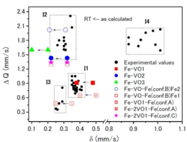
[27] The experimental Mössbauer parameters of Fe doped SnO2were summarized into four groups: Fe3+ (I1), Fe3+ (I2), Fe3+
(I3) and Fe2+ (I4) (Figure 15). Groups I1 and I2 are often
ob-served in Mössbauer spectra measured. Groups I3 and I4 are in Mössbauer spectra obtained by ion implantation and ball milling. Two main interactions (I1 and I2) observed in Mössbauer spectra can be associated to Fe-ions substituted at the Sn sites with oxygen coordination number smaller than six. That is, Fe3+ doping into SnO2 induces oxygen
va-cancy. The parameters are confirmed by ab initio calcula-tions for configuracalcula-tions of substituted Fe and oxygen vacancy (VO) as shown in Figures 16 and 17. The third inter-action (I3) is characterized by a low IS (in the order of Figure 13.57Fe emission Mössbauer spectra of dilute (ca. 5×10⁻⁴ %) 57Mn ion implanted SnO2 film in ISOLDE.[26]
Figure 14. Temperature dependence of area fraction of
components: Fex: interstitial Fe3+, Fe(II): Fe2+, and Fe(III) :
DOI: 10.5562/cca2784 Croat. Chem. Acta 2015, 88(4), 579–590 0.20 mm s–1 (0.10 mm s–1 at RT). Fe atoms replacing Sn with
oxygen vacancy are far away from the first shell of oxygen neighbours. It is not often observed in experimental data.
In the case of one Fe substituted at Sn site in SnO2
lattice and one oxygen vacancy, average Fe–ONN bond-length is in good agreement with the distances obtained from the EXAFS analysis (0.198 nm). It suggests that oxygen vacancies near the doped Fe are more stable than those near Sn in the lattice. Doping Fe ions lowers the formation energy of oxygen vacancies. The presence of an oxygen vacancy close to the Fe ions increases the split between iron 3d levels resulting in a higher magnetic moment of the Fe atoms.[27]
The cases of two irons sharing oxygen vacancies are shown to search the preferential magnetic alignment as shown in Figure 17.[27] The lowest energy state for all
configurations is the ferromagnetic one and in this case Fermi level falls onto minority 3d level. The doping with transition iron atoms reduces the formation energy of oxygen vacancy. Fe doped SnO2 systems will have more
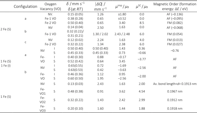
vacancies than the undoped ones. Oxygen vacancies enhance the magnetic moment of doped Fe impurities in supercell as listed in Table 1.
Oxygen vacancies prefer to be in the Fe–O–Fe path favoring the ferromagnetic coupling between Fe dopants. Small quantities of oxygen vacancies are fundamental for stabilizing local ferromagnetism in the form of bound magnetic polarons (BMPs). Two Fe ions sharing an oxygen vacancy are coupled ferromagnetically, forming a bound magnetic polaron (BMP), and that two neighbor BMPs are aligned antiparallel to each other although the formation energy is so very small as shown Figure 18.[27]
Figure 15. Isomer shift () and quadrupole splitting (Q) of
experimental data and ab initio calculation: configurations
of occupied Fe ions and oxygen vacancy (VO). The calculated
values at RT shifted by ca. –0.1 mm s–1 due to secondary
Doppler shifts (depending on the temperature) from as
calculated ones are added in the original figure.[27]
Figure 16. Configuration of one substituted Fe at Sn site and one oxygen vacancy (VO) (distance between Fe and VO: Fe-VO1 < Fe-VO2< Fe-VO3) O1 and O2 correspond to oxygen atoms in the first shell of oxygen neighbors of Fe, and O3 is
an oxygen in the second shell of Fe-oxygen neighbors.[27]
Figure 17. Configurations of Sn, Fe, O and Oxygen vacancy
(VO) in rutile structure of SnO2 doped with Fe. Part of the
2×2×2 super cell (SC) showing the different distribution of
the Fe atoms (black balls) substituted at Sn sites. Configuration a: Fe-2VO-Fe; configuration b: Fe-VO-Fe; and
Croat. Chem. Acta 2015, 88(4), 579–590 DOI: 10.5562/cca2784 In the case of 57Mn implanted SnO2 films, the
Mössbauer spectra present two doublets, one with IS = 0.48 mm s–1 and ΔQ = 0.91 mm s–1 and the other with IS =
0.89 mm s–1 and ΔQ = 2.20 mm s–1. Comparing with
calculated values for one interstitial Fe atom, there is poor agreement between experimental and calculated results, but there is good agreement with the situation where the interstitial Fe has a substitutional iron near neighbor (configuration b with one oxygen vacancy in Table 1).
Long Range Magnetic Ordering
From the calculation of the configurations as shown Figure 18, it is found that the configuration with smaller energy is
the antiferromagnetic one. Then, ferromagnetic interaction is only in short range while long range interaction is antifer-romagnetic. The energy difference between both align-ments is E = –0.05 eV; that is, the antiferromagnetic alignment is a little favored. Thus, although an increase in iron concentration would favor a neighboring situation with the increase in the number of BMP, the antiferromagnetic BMP alignment would reduce the magnetization, which is in very good agreement with the experimental results. It is to note in experimental results that the Sb5+ ions codoping
of SnO2 (Fe3+) increases the saturation magnetization. The
electron doping was simulated in two different ways: add-ing one electron to the 108-atom supercell (SC) or replacadd-ing Table 1. Calculated hyperfine parameters δ and |ΔQ|; magnetic moment in the super cell (μSC) for the different vacancy
positions studied. S: Substitutional and I: Interstitial.[27] NV, 1VO, and 2VO indicate the cases without oxygen vacancies or one
or two oxygen vacancies in the super cell (SC). In the case of Configuration b with one oxygen vacancy, the results for the δ,
the |ΔQ|, and μimp refers to each inequivalent Fe site of the structure.
Configulation Vacancy (VO)Oxygen δ/mm s–1
δ(at RT)
ΔQ /
mm s–1 μimp/μB μSC/μB
Magnetic Order (formation
energy: ΔE / eV)
2 Fe (S)
a Fe-1 VO NV. Fe-2 VO
0.15 (0.05) 0.38 (0.28) 0.50 (0.40)
1.26 0.65 0.65
±1.80 ±3.52 3.40
0.0 0.0 8.5
AF (–0.136) AF (–0.095) FM (0.082)
b NV 0.32 (0.22)/ 0.14 (0.04) 0.31 (0.21)
2.50 1.63 0.0 AF (–0.068)
Fe-1 VO 1.30 / 2.02 2.43 / 2.48 6.0 FM (0.054)
c Fe-2 VO NV 0.12 (0.02) 0.32 (0.22) 2.24 1.34 1.63 2.38 4.0 6.0 FM (0.013) FM (0.027)
1 Fe (S) 1 Fe (I)
a NV
I
S 0.50 (0.40) 0.45 (0.33) 0.50 (0.40) 0.45 (0.33) 1.43 0.73 –0.66 0.36 –0.76
Fe-VO S I 0.40 (0.30) 0.52 (0.42) 0.88 0.64 –0.17 3.45 –3.77 AF
b NV
I
S 0.65(0.55) 0.63(0.53) 0.72 0.42 –1.69 –3.63 –2.56 AF
Fe-VO S I 0.46 (0.36) 0.60 (0.50) 1.12 1.95 –2.56 0.95 –2.00 AF
1 Fe (S)
NV S 0.13 (0.03) 2.43 1.63 2.00 Av. bond length<d> 0.1913 nm
Fe-VO1 S 0.48 (0.38) 0.91 3.62 4.54 0.1967 nm
Fe-VO2 S 0.32 (0.22) 1.43 2.42 2.99 0.1912 nm
Fe-VO3 S 0.20 (0.10) 1.60 1.44 1.88 0.1918 nm
Figure 18. Part of the 3×2×3 SC showing the bound magnetic polaron model for (a) antiferromagnetic alignment and (b)
DOI: 10.5562/cca2784 Croat. Chem. Acta 2015, 88(4), 579–590 one indigenous Sn atom by a donor impurity such as Sb5+.
Both approaches suggest that the ferromagnetic coupling between BMP is the most stable configuration with E = Eanti – Eferro on the order of +0.3 eV. This result shows that
electron doping plays a fundamental role in mediating the nonlocal magnetic coupling between two BMP. From the experimental results it was considered that the overall mag-netic properties of TM-doped SnO2 are a statistical average
of different defect configurations corresponding to a real disordered system.[27]
SUMMARY
The intrinsic DMS and extrinsic DMS have been experimen-tally obtained for dilute iron doped SnO2. Concerning
intrin-sic DMS, oxygen vacancies are fundamental for the ferromagnetic response of Fe-doped SnO2. The ab initio
cal-culations show the possibility that two Fe ions sharing an oxygen vacancy are coupled ferromagnetically, forming a bound magnetic polaron (BMP), and that two neighbor BMPs are aligned antiparallel to each other. However, the formation energy for the antiferromagnetic alignment is so small that electron doping by Sb5+ can play a fundamental
role of mediating the magnetic coupling between the BMP inducing ferromagnetic alignment between the BMPs.[27]
A codoping two transition metals in SnO2 also
enhances the dilute ferromagnetism together with inducing the oxygen vacancies, and the ab initio calculation for these configurations would be studied in near future.
As the new composite material, the nano magnetic material precipitated in semiconductors, such as SnO2 film
obtained by Fe ion implantation, would be useful from the view point of practical application.
Acknowledgment. This paper presents a review article based on the author talk about oxygen defects and diluted
magnetism of SnO2 doped with Fe which was presented at
the Mediterranean Conference on the Applications of Mössbauer Effect, MECAME 2015, Zadar, Croatia. MECAME 2015 was organized in honour of Dr. Svetozar Musić on the
occasion of his 70th birthday and to celebrate his life-long
scientific contribution, particularly for his introduction of Mössbauer spectroscopy as a research technique in Croatia. Dr. Svetozar Musić has worked in the field of metal oxides chemistry for more than four decades and especially in the iron oxide chemistry. Author of this article had worked with him on the Mössbauer analysis of iron oxide nano-powders. As the result of this collaboration the common papers were
published and are listed in the references.28 These
nano-scale oxide materials are expected to be used for producing new spintronic materials by embedding them into semiconductors. Author would like to express many thanks to Dr. S. Music for giving him a chance for this research field.
Prof. Cesar Barrero from the Universidad de Antioquia have also contributed a lot to the study of DMS research since he visited the University of Tokyo, and his research group continued working in this field. Dr. H. P. Gunnlaugsson, Dr. K. Johnston, Prof. G. Langouche and the other members in ISOLDE group have engaged in emission Mössbauer spectrometry of super dilute magnetic ions doped oxide materials. This experimental research is important for development of RT- DMS materials.
One of the aims of this review paper is to show the clear relationship between magnetic moments and configurations of substituted Fe atoms and oxygen
vacancies by ab initio calculation of Fe-doped SnO2 system.
The experimental works have been done at the School of Engineering, the University of Tokyo, where author has been
working for past 35 years. The ab initio calculations were
supported by Dr. Azucena Mudarra Navarro, Prof. Claudia E. Rodriguez Torres, Prof. Leo A. Erico and so on from the University of La Plata.
There are a lot of coworkers who contributed to this research as shown in the following references. In addition to above mentioned coworkers the author would like to express special thanks to all coworkers named in the cited references, and to Prof. Akira Fujishima, Tokyo University of Science, for supporting author's study. Finally, author expresses thanks to Dr. Mira Ristić, the organizer of the MECAME 2015 for carefully reading of this manuscript.
REFERENCES
[1] T. Dietl, Semicond. Sci. Tech. 2002, 17, 377. [2] Y. Matsumoto, M. Murakami, T. Shono, T. Hasegawa,
T. Fukumura, M. Kawasaki, P. Ahmet, T. Chikyow, S. Koshihara, H. Koinuma, Science 2001, 291,854. [3] (a) T. Dietl, H. Ohno, F. Matsukura, J. Cibert, D.
Ferrand, Science 2000, 287, 1019; (b) S. J. Pearton, C. R. Abernathy, G. T. Thaler, R. Frazier, F. Ren, A. F. Hebard, Y. D. Park, D. P. Norton, W. Tang, M. Stavola, J. M. Zavada, R. G. Wilson, Physica B 2003, 340–342 39–47.
[4] S. B. Ogale, R. J. Choudhary, J. P. Buban, S. E. Lofland, S. R. Shinde, S. N. Kale, V. N. Kulkarni, J. Higgins, C. Lanci, R. Simpson, N. D. Browning, S. D. Sharma, H. D. Drew, R. L. Greene, T. Venkatesan, Phys. Rev. Lett. 2003, 91, 077205 (4p).
[5] K. Nomura, Y. Ujihira, H. Shiozawa, T. Takada, H. Reuther, E. Richter, J. Mater. Sci. Mater. Electron. 1997, 8, 301.
[6] K. Nomura, Radioisotopes 2013, 62, 857.
[7] J. M. D. Coey, M. Venkatesan, C. B. Fitzgerald, Nature
Mater. 2005, 4, 173.
Croat. Chem. Acta 2015, 88(4), 579–590 DOI: 10.5562/cca2784 [9] K. Nomura, T. Okubo, M. Nakazawa, Spectrochim.
Acta B 2004, 59, 1259.
[10] A. I. Rykov, K. Nomura, J. Sakuma, C. Barrero, Y. Yoda, T. Mitsui, Phys. Rev. B 2008, 77, 014302-1-014302-8.
[11] K. Nomura, K. Inaba, S. Iio, T. Hitosugi, T. Hasegawa, Y. Hirose, Z. Hommonay, Hyperfine Interact. 2006, 168, 1065.
[12] Z. Homonnay, K. Nomura, E. Kuzmann, A. Vértes, Y. Hirose, T. Hasegawa, Hyperfine Interact. 2008, 184, 69. [13] K. Nomura, C. A. Barrero, J. Sakuma, M. Takeda,
Phys. Rev. B 2007, 75, 184411 (13p).
[14] K. Nomura, C. A. Barrero, K. Kuwano, Y. Yamada, T. Saito, E. Kuzmann, Hyperfine Interact. 2009, 191, 25. [15] K. Nomura, E. Kuzmann, C. A. Barrero, S. Stichleutner,
Z. Homonnay, Hyperfine Interact. 2008, 184, 57. [16] K. Nomura, A. Rykov, Z. Nemeth, Y. Yoda, Hyperfine
Interact. 2012, 205,129.
[17] K. Nomura, J. Okabayashi, K. Okamura, Y. Yamada, J.
Appl. Phys. 2011, 110, 083901 (4p).
[18] S. Kono, K. Nomura, Y. Yamada, J. Okabayashi,
Hyperfine Interact. 2012, 205, 105.
[19] J. Okabayashi, K. Nomura, S. Kono, Y. Yamada, Jpn. J.
Appl. Phys. 2012, 51, 023003 (4p).
[20] J. Okayabashi, K. Nomura, S. Kono, Y. Yamada, J.
Appl. Phys. 2012, 112, 073917 (5p).
[21] J. Okabayashi, S. Kono, Y. Yamada, K. Nomura,
Hyperfine Interact. 2013, 217, 99.
[22] K. Nomura, H. Reuther, Hyperfine Interact. 2009, 191, 159.
[23] K. Nomura, Z. Németh, H. Reuther, J. Phys. Conf. Ser. 2010, 217, 012118 (4p).
[24] K. Nomura, S. Iio, Y. Hirose, H. Reuther, A. Nakanishi,
Hyperfine Interact. 2013, 217, 37.
[25] K. Nomura, S. Iio, Y. Hirose, Z. Németh, K. Yamamoto, H. Reuther, J. Nucl. Radiochem. Sci. 2010, 11, 1. [26] H. P. Gunnlaugsson, K. Nomura, T. E. Mølholt, S.
Shayestehaminzadeh, K. Johnston, R. Mantovan, H. Masenda, M. Ncube, K. Bharuth-Ram, H. Gislason, G. Langouche, D. Naidoo, S. Ólafsson, G. Weyer, the ISOLDE Collaboration, Hyperfine Interact. 2014, 226, 389.
[27] A. M. Mudarra Navarro, C. E. Rodríguez Torres, A. F. Cabrera, M. Weissmann, K. Nomura, L. A. Errico, J.
Phys. Chem. C 2015, 119, 5596.
[28] Dr. Svetozar Musić paper list, co-worked with K. Nomura: (a) A. Šarić, K. Nomura, S. Popović, N. Ljubešić, S. Musić, Mater. Chem. Phys. 1998, 52, 214; (b) A. Šarić, S. Musić, K. Nomura, S. Popović, Mater. Sci. Eng.B 1998, 56, 43; (c) A. Šarić, S. Musić. K. Nomura, S. Popović, Croat. Chem. Acta 1998, 71, 1019; (d) A. Šarić, S. Musić. K. Nomura, S. Popović, J.
Mol. Struct. 1999, 480–481, 633; (e) G. Štefanić, S.
Musić, S. Popović, K. Nomura, J. Mol. Struct. 1999,
480–481, 627; (f) S. Musić. A. Šarić, K. Nomura, S.
Popović, ACH - Models Chem. 1999, 136, 457; (g) S. Musić, A. Šarić, S. Popović, K. Nomura, T. Sawada,
Croat. Chem. Acta 2000, 73, 542; (h) G. Štefanić, B.
Gržeta, K. Nomura, R. Trojko, S. Musić, J. Alloys