THE EVOLVING WORLD OF CHRONIC KIDNEY DISEASE
MINERAL BONE DISORDER
Antonio Bellasi,
1,2Andrea Galassi,
3Mario Cozzolino,
2and Biagio Di Iorio
41. Department of Nephrology, Azienda Ospedaliera Ospedale Sant’Anna, Como, Italy 2. Department of Health Sciences, University of Milan, Italy
3. Department of Nephrology, Azienda Ospedaliera di Desio e Vimercate, Desio (MB), Italy 4. Department of Nephrology, Ospedale A. Landolfi di Solofra, Avellino, Italy
Disclosure: No potential conlict of interest. Citation: EMJ Neph. 2013;1:20-31.
ABSTRACT
Chronic kidney disease - mineral and bone disorder (CKD-MBD) is associated with a signiicant morbidity and mortality. In vitro and animal models suggest that phosphorous, calcium, parathyroid hormone, and vitamin D abnormalities, mediate the cardiovascular and bone diseases that characterise CKD-MBD and increase the risk of death. Currently, mineral abnormalities are corrected through phosphorous restriction, phosphate binders, calcimimetics and vitamin D administration. Nonetheless, data in humans that support the use of these compounds are still scarce, mainly based on observational studies. Thus, a considerable number of doubts and questions still challenge clinicians dealing with CKD patients and mineral metabolism imbalances. We herein critically review clinical evidence that support the use of diferent drugs in CKD-MBD.
Keywords: CKD-MBD, dialysis, outcome, management, needs.
INTRODUCTION
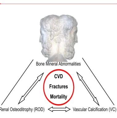
Calcium, phosphate, vitamin D and parathyroid hormone (PTH) have been repeatedly recognised as predictors of outcome in chronic kidney disease (CKD).1-4 Though the mechanisms are still poorly understood, numerous studies suggest that mineral homeostasis abnormalities are associated with bone and cardiovascular (CV) diseases that portend a poor survival.5 Hence, biochemical, CV, and bone abnormalities are now considered part of the multifaceted CKD-MBD syndrome (Figure 1). 5
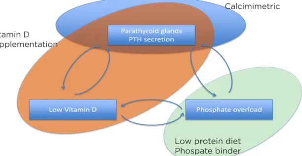
In spite of convincing preclinical data linking mineral metabolism imbalances to cardiovascular and bone diseases, clinical evidence is still far from conclusive4,6 and a considerable number of doubts and questions still challenge clinicians dealing with CKD patients and mineral metabolism imbalances. CKD-MBD is currently treated with nutritional interventions, native and active vitamin D phosphate binders, and calcimimetics administration (Figure 2). The aim of this review is to critically evaluate and summarise available evidence as well as highlight
Bone Mineral Abnormalities
Renal Osteoditrophy (ROD) Vascular Calcification (VC)
CVD
Fractures
Mortality
the numerous unanswered clinical questions on CKD-MBD management.
Diet: Facts, Promises and Expectations
Hyperphosphatemia control is perceived by nephrologists as one of the most relevant targets to achieve in CKD.4 Indeed, numerous studies have reported a close association between serum phosphorus levels and the risk of death in both subjects from the general population7,8 as well as subjects with varying degrees of renal function impairment.1-4 Furthermore, a large body of evidence suggests a direct link between phosphorous and the cardiovascular and bone systems.5 Thus, it is commonly accepted that phosphorus is a uraemic toxin, and current guidelines on mineral metabolism management recommend maintaining it within the range of normality.9-10
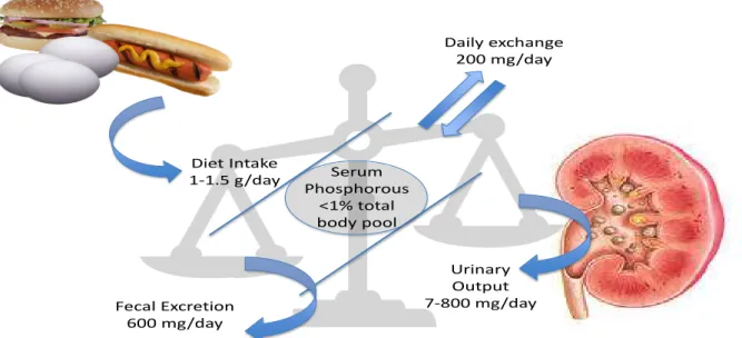
As kidney function declines, urinary phosphate excretion becomes insuicient and eventually hyperphosphataemia ensues if the phosphate daily intake remains constant.11 It is estimated that the daily phosphate intake in a standard diet in Western countries is about 1500 mg/day.11,12 Considering that faecal excretion is about 600 mg/day of which about 200 mg/day are secreted by the intestine, the amount of phosphorous absorbed by the gastrointestinal tract may approach 1100 mg/day (Figure 3).11,12 To maintain phosphorous homeostasis and keep serum
levels within the range of normality, renal excretion should match the daily intake at the expense of increasing the tubular workload of each functional nephron.13 Notably, the average phosphate level in the general population varies according to sex and menopausal status14,15 and data suggest an increased risk of unfavourable outcomes for phosphorous levels within the range of normality8,15 further corroborating the notion that serum phosphorus may not adequately relect phosphorous balance.
Two diferent strategies to lower phosphorous intake are available: low phosphate diet and phosphate binders. A low phosphorous intake can be achieved via protein restriction and quality selection.5 Indeed, Moe et al.16 showed that a vegetarian rather than a meat-based diet signiicantly reduces serum phosphorous and the phosphaturic factor ibroblast growth factor 23 (FGF23). Notably, these diferences were independent of the circadian serum and urine phosphorous changes, suggesting that phosphorous contained in the vegetarian diet is less adsorbable in the gastrointestinal tract which is possibly due to the phosphate binding to phytate.16
Cooking method and food additives are two other factors that signiicantly afect phosphorous intake.17-22 Cupisti and coworkers18 reported that 20-30 minutes boiling signiicantly reduce (20-30-50%) phosphorous burden at the expense of a minimal reduction of the protein content (9-17%).
Figure 2. CKD-MBD pathophysiology is characterised by phosphate overload, PTH hypersecretion and vitamin D depletion. Our armamentarium is composed by low protein diet and phosphate binder (light green circle) to lower phosphate overload; diferent forms of vitamin D (orange circle) to overcome vitamin D deiciency and inhibit PTH production and secretion; calcimimentics (light blue circle) to reduce PTH secretion
Parathyroid glands PTH secretion
Phosphate overload Low Vitamin D
Calcimimetric
Vitamin D
supplementation
Food additives are another source of phosphorous in prepared meals. A recent survey of best-selling processed groceries concluded that phosphorus additive-containing foods averaged 67 mg phosphorus/100 g more than matched non-additive-containing foods (about 736 mg more phosphorus per day compared with meals consisting of only additive-free foods).23 Phosphorous-based additives (phosphoric acid, tetrasodium pyrophosphate, tricalcium phosphate, disodium phosphate, monopotassium phosphate, etc.) are used to enhance taste and consistency of diferent foods such as baked goods (baking powder, cakes, frozen dough, etc.), beverages (colas, chocolate milk, buttermilk, fruit juices, sport drinks, canned milk, soy beverages), cereals, dairy, meat and egg products, fruit and vegetables, and pasta and noodles.
Inorganic phosphorous contained in food additives is highly bioavailable and adsorbed in the gastrointestinal tract to a greater extent than the organic phosphorous. It is estimated that as much as 90% versus 60% of the ingested inorganic (food additives) and organic (vegetable and meat protein) phosphorous is absorbed, respectively.21,22
Though the mechanisms are still unclear, accumulating evidence suggests the high serum levels of phosphorous are associated with increased levels of FGF23 that in turn, have been independently associated with a signiicant risk of endothelial dysfunction,24 left ventricular hypertrophy,25 CKD progression and all-cause mortality.26 In the absence of a randomised controlled clinical trial (RCT), it is
unclear whether elevated serum phosphorous or FGF23 mediates the toxicity1,26 or, alternatively, both factors contribute to the organ damage and poor survival in CKD-MBD.27
A balanced nutritional program should control both serum phosphorous and FGF23. Di Iorio et al.28 showed that a very low protein diet (0.3 g/kg of ideal body weight per day) supplemented with alpha-chetoanalogues and essential aminoacids signiicantly reduces FGF23 and phosphoremia. In 32 CKD subjects randomised to cross-over sequential treatments with either standard low protein diet (60-70 g of protein/day) or very low protein diet (25-30 g of protein/day), they reported a signiicant 33.5%, 12% and 34% reduction of FGF23, serum and urinary phosphorous levels associated with very low protein diet (VLPD), respectively.28 Of note, the two diet regimens did not difer only in the total protein intake but also in the animal/vegetal protein ratio (VLPD regimen based on vegetable protein only) and phosphorous content (350-420 mg/day versus 600-700 in VLPD and standard diet, respectively).28 Other groups have conirmed that phosphorous restriction with or without phosphate binders, is efective in controlling FGF23.29,30
Low phosphate and protein diet has also been associated with proteinuria and CKD progression reduction.19,31,32 In a seminal paper by Brunori at al.,32 it was demonstrated that life expectancy among old patients with end-stage renal disease (ESRD) was similar if patients were randomised to VLPD and conservative treatment or haemodialysis.
Urinary Output 7-800 mg/day Diet Intake
1-1.5 g/day
Fecal Excretion 600 mg/day
Daily exchange 200 mg/day
Serum Phosphorous
<1% total body pool
The most important drawback of low protein and phosphorous diet is the potential for malnutrition.33 Indeed, a balanced nutritional program should be tailored to each individual and should provide the patient with the right amount of calories and nutrients.34 In this regard, an observational study suggests that protein malnutrition maybe more detrimental than phosphorous intake and that the ideal nutritional regime should provide enough protein with minimal phosphorous burden.33
Future RCT studies should investigate the safety and the impact of low protein and VLPD on long-term survival and CKD progression, in both CKD patients not receiving and receiving dialysis. In consideration of the substantial increase of the mean age of dialysis patients, it is to be established if the recommended protein intake by current guidelines is still adequate in light of the considerable number of patients with increased levels of serum phosphorous.35 Finally, a pharmacoeconomic analysis should evaluate the cost burden connected to aproteic foods, chetoanalogue or essential aminoacid supplements.
Phosphate Binders: Facts, Promises and
Expectations
Phosphate binders are another strategy for reducing phosphate intake. These compounds share the property to bind phosphorous in the intestinal lumen, prevent its absorption and increase the faecal excretion. Various drugs are now available on the market with this indication.36,37 For ease, these compounds can be divided into two diferent groups: calcium-based phosphate binders (calcium carbonate and calcium acetate) and calcium-free phosphate binders (aluminium hydroxide, lanthanum carbonate, magnesium carbonate, sevelamer hydrochloride, and sevelamer carbonate). Alternatively, these compounds can be divided into absorbable (calcium-based binders, aluminium hydroxide, lanthanum carbonate, magnesium carbonate) and not absorbable (sevelamer hydrochloride, and sevelamer carbonate) in the gastrointestinal tract. Though all these compounds might have diferent ainity for phosphorous in the gastrointesinal tract and diferent doses have to be administered,38 clinical studies suggest that they all efectively lower serum phosphorous.36,39,40 Nonetheless, due to the diferent adsorbability in the gastrointestinal tract, the safety proile of these compounds can be profoundly diferent. Indeed, the prolonged use of aluminum-based phosphate binder is not indicated due to its accumulation and toxicity.41
The debate on containing versus calcium-free phosphate binders has characterised the last decade.36,42 Preclinical data suggest that both phosphorous and calcium can actively induce vascular calciication,43-45 a marker of vascular disease46 and a risk factor for arterial stifness47 and mortality.46,48 A seminal paper by Cozzolino and coworkers49 demonstrated that the use of sevelamer was associated with a similar phosphate control but lower extraosseous calciication than calcium-based phosphate binder. Observational data suggest that excessive calcium intake may result in a positive calcium balance that in turn has been associated with arterial stifness and vascular calciication,50,51 adynamic bone disease52,53 and, in some but not all studies, excessive mortality.35,54
A third RCT designed to test the impact of sevelamer versus calcium carbonate on hard outcomes (all-cause mortality and CKD progression) in mild to moderate CKD patients (mean creatinine clearance 30 ml/min) with hyperphosphatemia supports the notion that non-calcium-containing phosphate binders may be associated with a more favourable renal and life survival rate.57 In this study, a signiicant CAC progression attenuation was also noted.57 Although sevelamer-treated patients showed a higher CAC prevalence and burden at baseline (prevalence of CAC 62.6% versus 47.6%; P=0.02; median CAC score: 122 AU [IQR, 0–180] versus 0 AU [IQR, 0–215]; P=0.01 in the sevelamer and calcium carbonate group respectively), at study completion a signiicantly lower risk of CAC progression or de novo onset (12.8% in sevelamer-treated patients and 81.8% in calcium carbonate–treated patients) was noted.57
Other studies in ESRD patients new to48 or on maintenance dialysis61 have also investigated the diferential impact of calcium salts and calcium-free phosphate binders on vascular calciication or hard outcome.48,62 Though the majority of these trials point toward a harmful potential of calcium-containing phosphate binders, metanalyses have repeatedly failed to conirm this hypothesis.39,63,64 A recent study by Di Iorio et al.65 unfolds an almost 10-fold reduction of CV and all-cause mortality associated with sevelamer versus calcium carbonate in a large cohort (N=466) of patients new to dialysis.
Though these data suggest a diferent efect of calcium-free phosphate binders on the cardiovascular system and survival, no study has ever tested whether serum phosphorous-lowering is associated with a survival beneit. In light of the many adaptive mechanisms to hyperphosphataemia such as increased PTH and FGF23 that can modulate phosphorous toxicity and the potential calcium toxicity,66,67 future studies should address when to start in the course of CKD and to what serum phosphorus target should we aim when prescribing phosphate binders. Finally, cost-efectiveness analyses of these compounds are needed in light of the expanding epidemiology of CKD.68
Native Vitamin D: Facts, Promises and
Expectations
Native vitamin D has received growing interest in the last ten years. Every year, hundreds of manuscripts on native vitamin D associations with a variety of diseases such as osteoporosis,69 hypertension,70
cardiovascular disease,71,72 insulin resistance,73 infections,74 cancer75 and mortality76 are published. Similarly, nephrologists have traditionally linked native vitamin D deiciency to CKD progression,77 secondary hyperparathyroidism (SHPT)78 and survival79 in renal patients. The widespread association between native vitamin D and unfavourable outcomes in the general population, as well as in selected diseased sub-cohorts, together with the emerging knowledge of the extra-renal activation of native vitamin D, support the hypothesis that vitamin D deiciency is an etiologic factor rather than a mere biomarker of frailty.80
The term ‘native Vitamin D’ refers to the 25 hydroxlate vitamin D (25(OH)D) forms. Vitamin D precursors ergocalciferol (vitamin D2) and cholecalciferol (vitamin D3) are synthesised by the UV radiation in yeast and in animals starting from ergosterol and 7-dehydrocholesterol, respectively.81 In turn, vitamin D precursors are hydroxylated in the liver to form 25(OH)D2 and 25(OH)D3, respectively.81 These are the substrates that are subsequently activated to 1-25(OH)2D (calcitriol) by the renal and, to a lesser extent, by the extra-renal 1 alpha hydroxylase.82 Of note, humans do not synthesise vitamin D281 and almost 80% of vitamin D is obtained by UVB irradiation with only a minor contribution of diet intake.82
Though it is commonly prescribed as a supplement, we currently ignore what is the desirable level of 25(OH)D.69,83 It is commonly accepted that levels of 25(OH)D above 30 ng/ml, between 21 and 29 ng/ ml and below 20 ng/ml deine vitamin D suiciency, insuiciency and deiciency, respectively.82
50.000 IU of ergocalciferol are pharmacologically equivalent to 5-15.000 IU of cholecalciferol.87 However, whether or not these two forms of vitamin D may have diferent clinical implications is still unknown. Two RCTs are currently recruiting patients to compare the efect of vitamin D2 versus Vitamin D3 on mineral metabolism in CKD stage 2-5 (NCT01633853, NCT01173848) to shed light on which 25(OH)D form is better suited in CKD. Current evidence suggests a potential role for 25(OH)D as PTH lowering agent. Indeed, a recent meta-analysis by Kandula and colleagues88 concludes, based on the available observational studies, that 25(OH) D compared to placebo reduces PTH levels in CKD (about 25 pg/ml) as well as in ESRD (about 60 pg/ml) patients. However, the heterogeneity of the studies precludes speculation on what could be the best 25(OH)D regimen in CKD. Whether 25(OH)D can be used instead of VDR activator for PTH suppression in CKD is still under debate, though preliminary data suggest that paricalcitol and doxercalciferol induce a stronger PTH reduction compared to ergocalciferol and cholecalciferol in CKD 3-489,90 and ESRD patients,86 respectively. Similarly, data concerning PTH reduction by the co-administration of native and active vitamin D are still inadequate, mainly based on observational and retrospective studies.91-93 Further evidence is advocated before recommending the implementation of this combined approach.
In spite of the many pleiotropic efects described in the past decades and the substantial increase in the risk of death associated with low 25(OH) D levels,75 only a few studies have investigated the impact of native vitamin D on surrogate endpoints such as renal osetodystrophy, vascular calciication, proteinuria, LVH or survival. However, numerous RCTs are currently investigating the efect of native vitamin D on left ventricular hypertrophy (NCT01323712), insulin resistance (NCT00893451), erythropoietin dosing (NCT01395823), proteinuria (NCT01426724), immunity (NCT00892099), ateriovenous istulae maturation (NCT00912782) and physical and cognitive performance (NCT00511225, NCT01229878) to shed light on the potentials of this treatment. Finally, the NUTRIVITA study is actively randomising dialysis patients to 25(OH)D versus placebo treatment to test the efect of 25(OH)D on survival, fatal myocardial infarction, and non-fatal stroke (NCT01457001).
Due to the scarce data available, current guidelines on mineral metabolism management,10 suggest 25(OH)D deiciency replenishment as the irst step
to correct SHPT in CKD stage 3-5,10 whereas no suggestion is provided for dialysis patients. These statements are ‘not graded’ and based on expert opinion rather than on evidence.10 A considerable ongoing and future efort is needed to clarify the impact of 25(OH)D administration to CKD and dialysis patients.
Vitamin D Analogues: Facts, Promises and
Expectations
Repeated observational data described an independent association between PTH levels and unfavourable outcomes in CKD stage 3-594,95 as well as in ESRD.2,3 However, no RCTs have yet proven that an active reduction of PTH values improves such patient-centred hard outcomes as hospitalisations, cardiovascular events, CKD progression, and survival. Thus, the optimal PTH target is still uncertain in CKD as well as in ESRD subjects. KDIGO guidelines provide a low-grade suggestion to maintain PTH levels into the range of normality in CKD stage 3-5 and between two and nine-times the normal range in ESRD.10
The reduction of calcitriol levels, together with hypocalcemia and hyperphosphataemia, are the leading causes of increased PTH levels. Thus, KDIGO guidelines suggest the use of vitamin D in case of increased PTH values and its tailoring in case of PTH over-correction, hypercalcemia or hyperphosphataemia.10 The risks related to high doses of vitamin D are mainly due to phosphate and calcium overload that possibly contribute to the low achievement rate of calcium and phosphate recommended targets96 and to a poor survival in dialysis patients.3 However, selective vitamin D receptor activator (VDRA), with a stronger efect on PTH and a lesser impact on calcium and phosphate load, may improve the global achievement of serum PTH, calcium and phosphate targets reducing the vitamin D toxicity.97-99
In recent years industries have provided multiple synthetic vitamin D2 (paricalcitol and doxercalciferol) and vitamin D3 analogues (alfacalcidol, falecalcitriol and maxacalcitol). However, comparison data of diferent vitamin D analogues on mineral metabolism control, surrogate and patient-centred outcomes are currently still scarce.
and inconclusive results. Alfacalcidol was similar to calcitriol in suppressing PTH values with equal change in phosphate and calcium levels,100,101 however recent data by Hansen et al.102 did not observe signiicant diferences between alfacalcidol and paricalcitol on similar targets. Joist et al.103 observed that paricalcitol at very high doses suppressed PTH with lower elevation of phosphate and calcium levels compared to doxercalciferol. However, Fadem and coworkers104 could not detect any diference in PTH, calcium and phosphorous control when haemodialysis patients were switched from intravenous paricalcitol to doxercalciferol. No clinical data comparing doxercalciferol with alfacalcidol in humans are currently available.
More recently, a growing interest for vitamin D pleiotropic efects, related to the widespread regulation of the human genome played by VDR activation, has been observed. Albuminuria, left ventricular hypertrophy (LVH) and cardiac remodelling have all been tested as potential targets of vitamin D analogues. The activation of VDR can regulate the expression of several genes involved in glomerular and myocardial inlammation as renin,105 TGF-beta,106 antioxidant molecules,107 NFκB and RANTES.108 The VITAL study, a randomised placebo controlled trial in diabetic CKD patients, documented a dose dependent trend toward reduction of albuminuria when paricalcitol was added to RAAS inhibitors.109 Though the PRIMO study failed to demonstrate a signiicant LVH reduction,110 a post-hoc analysis documented a lower increase of brain natriuretic peptide and left atrial index in diabetic CKD patients receiving paricalcitol on top of ACE-I or ARBs compared to placebo.111 Interestingly, paricalcitol was associated with lower risk of hospitalisation in those patients with more severe LVH.110 However, no RCT has tested the efect of diferent forms of vitamin D or VDRA on hard patient-centred outcomes.
Numerous, albeit not all, observational studies suggest potential beneits beyond mineral metabolism control linked to VDRA use on hospitalisation, cardiovascular events, and mortality. Kalantar-Zadeh and coworkers3 reported a 14% reduction in all-cause hospitalisation among patients receiving paricalcitol compared to those treated with calcitriol in a large cohort of 58,058 haemodialysis patients.3 Paricalcitol112-114 and doxercalciferol114 use were both associated with lower mortality risk compared to calcitriol in other large series of patients on chronic haemodialysis. Recently
published results from the Italian FARO survey115 unexpectedly showed a better survival in dialysis patients receiving vitamin D also in the presence of PTH <150 pg/ml. However, the Dialysis Outcome and Practice Pattern Study (DOPPS) investigators failed to report on vitamin D improved survival after adjustment for confounders and diferent practice patterns.116 Hence, these encouraging observational data have to be conirmed in RCTs prior to orient stronger recommendations on vitamin analogues prescription.
Future studies should shed deinitive light on whether the use of VDRAs improve survival in CKD and ESRD as well as surrogate outcomes such as albuminuria and LVH. Finally, in consideration of the growing number of CKD patients and the high-cost burden connected to CKD management, future studies should also verify the cost-efectiveness of the use of VDRA in diferent stages of CKD.
Cinacalcet: Facts, Promises and Expectations
The existing body of evidence suggests that cinacalcet efectively lowers serum PTH, phosphorous, and calcium levels in ESRD modulating the parathyroid calcium sensing receptor ainity to serum calcium.6,117-125 Phase two and three studies show that, on average a 40-50% (250-350 pg/ml) serum PTH, a 5-8% (0.5-0.8 mg/dl) serum calcium and a 5-10% (0.2-1.0 mg/dl) serum phosphorous reduction is expected when cinacalcet is administered.6,117-125 It is conceivable that the calcium-PTH setpoint shift and the metabolic change in bone metabolism induced by this drug explain these results.126,127
design diferences it is unclear whether one of these two approaches is superior, though answering this question might be of limited clinical utility in light of the diferent pharmacological proile of calcimimetic and VDRAs.
The presence of calcium-sensing receptors in diferent tissues other than the parathyroid glands, could explain the positive impact of cinacalcet on the bones and vasculature detected in numerous preclinical data.129 In vitro and animal evidence suggest that a reduction of functional calcium-sensing receptors is associated with vascular calciication,129,130 blood pressure,131 proteinuria,132 CKD progression,132 arterial stifness and endothelial dysfunction improvement.133 Large cohort prospective studies show that calcium-sensing receptor modulation is associated with favourable clinically meaningful outcomes. Cunningham and coworkers134 showed a signiicant reduction in the risk of cardiovascular disease, bone fracture, parathyroidectomy incidence, and a parallel improvement in the general health perception among dialysis patients with secondary hyperparathyroidism. Block et al.135 documented a substantial risk reduction in all-cause and cardiovascular mortality associated with cinacalcet in a large cohort of 25,292 chronic haemodialysis patients independent of several confounders.
However, the clinical impact of cinacalcet on hard outcome is far from being established in light of the recently published results of the ADVANCE124 and EVOLVE6 trials. The ADVANCE trial was conducted to investigate whether cinacalcet in combination with low dose of vitamin D (<6 mcg paricalcitol equivalent/ week) versus lexible doses of vitamin D attenuates coronary, aorta, and cardiac valves calciication progression in a cohort of 360 prevalent haemodialysis patients. After a relatively short period of follow-up of 12 months, a trend toward CAC reduction in the cinacalcet arm (Agatston CAC scores % change: 24% (95% conidence interval: - 22%, 119%) and 31% (- 9%, 179%), in the cinacalcet and lexible vitamin D group, respectively, P=0.073) was noted. Notably the trend was consistent across all CV sites investigated for vascular calciication.124 Furthermore, the large dose of calcium-containing phosphate binders and vitamin D administered in the calcimimetic arm may contribute to explain these results.136 Finally, the EVOLVE trial was designed to test the survival beneit of cinacalcet hypothesised by observational data in haemodialysis patients. At study completion, a statistically non-signiicant trend toward reduction (relative hazard in the cinacalcet group vs. the placebo group, 0.93; 95% conidence interval, 0.85 to 1.02; P=0.11) of the composite endpoint (time until death, myocardial
Treatment evidenceType of
Head-to-head comparisons
between drugs of the
same class
Mineral metabolism
control
Tissue marker of organ damage
Survival data Pharmacoeconomic evaluation
Low phosphate diet
Observational
studies NA YES NO NO NA
RCTs NA YES NO NO NO
Phosphate binders
Observational
studies YES YES YES YES NA
RCTs YES YES YES YES NO
Native vitamin D
Observational
studies YES YES YES YES NA
RCTs NO NO NO NO NO
Activated forms of vitamin D (VDRA)
Observational
studies YES YES YES YES NA
RCTs NO YES YES NO NO
Cinacalcet
Observational
studies YES YES YES YES NA
RCTs NA YES YES YES
Inconclusive NO
infarction, hospitalisation for unstable angina, heart failure, or a peripheral vascular event) was reported.6 However, the lower than anticipated event rate, the high drop-in and out rate during follow-up (about 20%),6 signiicantly afected the statistical power (0.54)6 and the interpretability of this inconclusive RCT.
In essence, data support the notion that cinacalcet is a safe and efective drug to lower PTH in secondary hyperparathyroidism. Nonetheless, future research projects should indentify the ideal candidate that would likely increase survival and quality of life while on this treatment. Finally, though the use of cinacalcet in predialysis stages of CKD is not approved because of the risk of hypocalcemia, future studies should evaluate its eicacy and safety in CKD not dialysis dependent cases of secondary hyperparathyroidism, characterised by normal-high calcium and high phosphate in which vitamin D may further aggravate phosphorous and calcium balance.
CONCLUSION
Treatment of CKD-MBD is currently based largely on opinion rather than evidence, and many questions about CKD-MBD await answers. A tremendous efort has been performed in the attempt to clarify the natural history and pathogenic mechanisms that trigger CKD-MBD and modulate the astonishing risk connected to it. Nonetheless, a substantial degree of uncertainty on the clinical relevance and use of diferent serological and tissue biomarkers used to individualise, and titrate treatments still exists and afects patient care. Furthermore, the incompleteness (Table 1) and inconclusiveness due to various methodological laws in the few available RCTs complicate the interpretation of the available evidence and lead to a heterogeneous use of the diferent drugs we have in our armamentarium.96
Future efort is therefore needed to elucidate mechanisms and treatment of these imbalances that, at least observational data, link to a substantial risk burden2 in CKD patients.
REFERENCES
1. Bellasi A, Mandreoli M, Baldrati L, et al. Chronic kidney disease progression and outcome according to serum phosphorus in mild-to-moderate kidney dysfunction. Clin J Am Soc Nephrol. 2011;6(4):883-91. 2. Block GA, Klassen PS, Lazarus JM, et al. Mineral metabolism, mortality, and morbidity in maintenance hemodialysis. J Am Soc Nephrol. 2004;15(8):2208-18. 3. Kalantar-Zadeh K, Kuwae N, Regidor DL, et al. Survival predictability of time-varying indicators of bone disease in maintenance hemodialysis patients. Kidney Int. 2006;70(4):771-80.
4. Palmer SC, Hayen A, Macaskill P, et al. Serum levels of phosphorus, parathyroid hormone, and calcium and risks of death and cardiovascular disease in individuals with chronic kidney disease: a systematic review and meta-analysis. Jama 2011;305(11):1119-27.
5. Moe S, Drueke T, Cunningham J, et al. Deinition, evaluation, and classiication of renal osteodystrophy: a position statement from Kidney Disease: Improving Global Outcomes (KDIGO). Kidney Int. 2006;69(11):1945-53.
6. Chertow GM, Block GA, Correa-Rotter R, et al. Efect of cinacalcet on cardiovascular disease in patients undergoing dialysis. N Engl J Med. 2012;367(26):2482-94. 7. Dhingra R, Sullivan LM, Fox CS, et al. Relations of serum phosphorus and calcium levels to the incidence of
cardiovascular disease in the community. Arch Intern Med. 2007;167(9):879-85. 8. Tonelli M, Sacks F, Pfefer M, et al. Relation between serum phosphate level and cardiovascular event rate in people with coronary disease. Circulation. 2005;112(17):2627-33.
9. Gonzalez-Parra E, Tunon J, Egido J, et al. Phosphate: a stealthier killer than previously thought? Cardiovasc Pathol. 2012;21(5):372-81.
10. KDIGO clinical practice guideline for the diagnosis, evaluation, prevention, and treatment of Chronic Kidney Disease-Mineral and Bone Disorder (CKD-MBD). Kidney Int Suppl. 2009(113):S1-130. 11. Bellasi A, Cozzolino M, Adragao T, et al. Phosphate Binder in Moderate CKD: Where Are we Standing at? J Nephrol. In press.
12. Bellasi A, Kooienga L, Block GA. Phosphate binders: new products and challenges. Hemodial Int. 2006;10(3):225-34.
13. Isakova T, Wahl P, Vargas GS, et al. Fibroblast growth factor 23 is elevated before parathyroid hormone and phosphate in chronic kidney disease. Kidney Int. 2011;79(12):1370-8.
14. Cirillo M, Ciacci C, De Santo NG. Age, renal tubular phosphate reabsorption, and serum phosphate levels in adults. N Engl J Med. 2008;359(8):864-6.
15. Onufrak SJ, Bellasi A, Cardarelli F, et
al. Investigation of gender heterogeneity in the associations of serum phosphorus with incident coronary artery disease and all-cause mortality. Am J Epidemiol. 2009;169(1):67-77.
16. Moe SM, Zidehsarai MP, Chambers MA, et al. Vegetarian compared with meat dietary protein source and phosphorus homeostasis in chronic kidney disease. Clin J Am Soc Nephrol. 2011;6(2):257-64. 17. Cupisti A, Morelli E, D’Alessandro C, et al. Phosphate control in chronic uremia: don’t forget diet. J Nephrol. 2003;16(1):29-33.
18. Cupisti A, Comar F, Benini O, et al. Efect of boiling on dietary phosphate and nitrogen intake. J Ren Nutr. 2006;16(1):36-40.
19. Cianciaruso B, Bellizzi V, Brunori G, et al. Low-protein diet in Italy today: the conclusions of the Working Group from the Italian Society of Nephrology. G Ital Nefrol. 2008;25 Suppl 42:S54-7.
20. Cupisti A, D’Alessandro C. The impact of known and unknown dietary components to phosphorus intake. G Ital Nefrol. 2011;28(3):278-88.
21. Cupisti A, Benini O, Ferretti V, et al. Novel diferential measurement of natural and added phosphorus in cooked ham with or without preservatives. J Ren Nutr. 2012;22(6):533-40.
phosphorus burden in kidney disease. Semin Nephrol. 2013;33(2):180-90.
23. Leon JB, Sullivan CM, Sehgal AR. The prevalence of phosphorus-containing food additives in top-selling foods in grocery stores. J Ren Nutr. 2013;23(4):265-70
24. Yilmaz MI, Sonmez A, Saglam M, et al. Comparison of calcium acetate and sevelamer on vascular function and ibroblast growth factor 23 in CKD patients: a randomized clinical trial. Am J Kidney Dis. 2012;59(2):177-85.
25. Faul C, Amaral AP, Oskouei B, et al. FGF23 induces left ventricular hypertrophy. J Clin Invest. 2011;121(11):4393-408.
26. Isakova T, Xie H, Yang W, et al. Fibroblast growth factor 23 and risks of mortality and end-stage renal disease in patients with chronic kidney disease. Jama. 2011;305(23):2432-9.
27. Scialla JJ, Lau WL, Reilly MP, et al. Fibroblast growth factor 23 is not associated with and does not induce arterial calciication. Kidney Int. 2013 June;83(6):1159-68.
28. Di Iorio B, Di Micco L, Torraca S, et al. Acute efects of very-low-protein diet on FGF23 levels: a randomized study. Clin J Am Soc Nephrol. 2012;7(4):581-7.
29. Isakova T, Barchi-Chung A, Enield G, et al. Efects of Dietary Phosphate Restriction and Phosphate Binders on FGF23 Levels in CKD. Clin J Am Soc Nephrol. 2013;8(6):1009-1018.
30. Sigrist M, Tang M, Beaulieu M, et al. Responsiveness of FGF-23 and mineral metabolism to altered dietary phosphate intake in chronic kidney disease (CKD): results of a randomized trial. Nephrol Dial Transplant. 2013;28(1):161-9.
31. Di Iorio BR, Bellizzi V, Bellasi A, et al. Phosphate attenuates the anti-proteinuric efect of very low-protein diet in CKD patients. Nephrol Dial Transplant. 2013;28(3):632-40.
32. Brunori G, Viola BF, Parrinello G, et al. Eicacy and safety of a very-low-protein diet when postponing dialysis in the elderly: a prospective randomized multicenter controlled study. Am J Kidney Dis. 2007;49(5):569-80.
33. Noori N, Kalantar-Zadeh K, Kovesdy CP, et al. Association of dietary phosphorus intake and phosphorus to protein ratio with mortality in hemodialysis patients. Clin J Am Soc Nephrol. 2010;5(4):683-92. 34. Bellizzi V, Di Iorio BR, De Nicola L, et al. Very low protein diet supplemented with ketoanalogs improves blood pressure control in chronic kidney disease. Kidney Int. 2007;71(3):245-51.
35. Panichi V, Bigazzi R, Paoletti S, et al. Impact of calcium, phosphate, PTH abnormalities and management on mortality in hemodialysis: Results
from the RISCAVID study. J Nephrol 2010;23(5):556-562.
36. Frazao JM, Adragao T. Non-calcium-containing phosphate binders: comparing eicacy, safety, and other clinical efects. Nephron Clin Pract. 2012;120(2):108-19. 37. Tonelli M, Pannu N, Manns B. Oral phosphate binders in patients with kidney failure. N Engl J Med. 2010;362(14):1312-24.
38. Daugirdas JT, Finn WF, Emmett M, et al. The phosphate binder equivalent dose. Semin Dial. 2011;24(1):41-9.
39. Tonelli M, Wiebe N, Culleton B, et al. Systematic review of the clinical eicacy and safety of sevelamer in dialysis patients. Nephrol Dial Transplant. 2007;22(10):2856-66.
40. Goldsmith DR, Scott LJ, Cvetkovic RS, et al. Sevelamer hydrochloride: a review of its use for hyperphosphataemia in patients with end-stage renal disease on haemodialysis. Drugs. 2008;68(1):85-104. 41. K/DOQI clinical practice guidelines for bone metabolism and disease in chronic kidney disease. Am J Kidney Dis. 2003;42(4 Suppl 3):1-201.
42. Palmer SC, Craig JC, Strippoli GF. Sevelamer: a promising but unproven drug. Nephrol Dial Transplant. 2007;22(10):S2742-5.
43. Li X, Yang HY, Giachelli CM. Role of the sodium-dependent phosphate cotransporter, pit-1, in vascular smooth muscle cell calciication. Circ Res. 2006;98(7):905-12.
44. Jono S, McKee MD, Murry CE, et al. Phosphate regulation of vascular smooth muscle cell calciication. Circ Res. 2000;87(7):E10-7.
45. Giachelli CM. Vascular calciication: in vitro evidence for the role of inorganic phosphate. J Am Soc Nephrol. 2003;14(9 Suppl 4):S300-4.
46. Bellasi A, Raggi P. Vascular imaging in chronic kidney disease. Current opinion in nephrology and hypertension. 2012;21(4):382-8.
47. Raggi P, Bellasi A, Ferramosca E, et al. Association of pulse wave velocity with vascular and valvular calciication in hemodialysis patients. Kidney Int. 2007;71(8):802-7.
48. Block GA, Raggi P, Bellasi A, et al. Mortality efect of coronary calciication and phosphate binder choice in incident hemodialysis patients. Kidney Int. 2007;71(5):438-41.
49. Cozzolino M, Staniforth ME, Liapis H, et al. Sevelamer hydrochloride attenuates kidney and cardiovascular calciications in long-term experimental uremia. Kidney Int. 2003;64(5):1653-61.
50. Guerin AP, London GM, Marchais SJ, et al. Arterial stifening and vascular calciications in end-stage renal disease.
Nephrol Dial Transplant. 2000;15(7):1014-21.
51. Di Iorio B, Nargi O, Cucciniello E, et al. Coronary artery calciication progression is associated with arterial stifness and cardiac repolarization deterioration in hemodialysis patients. Kidney Blood Press Res. 2011;34(3):180-187.
52. London GM, Marty C, Marchais SJ, et al. Arterial calciications and bone histomorphometry in end-stage renal disease. J Am Soc Nephrol. 2004;15(7):1943-51.
53. Ferreira A, Frazao JM, Monier-Faugere MC, et al. Efects of sevelamer hydrochloride and calcium carbonate on renal osteodystrophy in hemodialysis patients. J Am Soc Nephrol. 2008;19(2):405-12.
54. Jean G, Lataillade D, Genet L, et al. Calcium carbonate, but not sevelamer, is associated with better outcomes in hemodialysis patients: results from the French ARNOS study. Hemodialysis international. International Symposium on Home Hemodialysis. 2011;15(4):485-92. 55. Russo D, Miranda I, Ruocco C, et al. The progression of coronary artery calciication in predialysis patients on calcium carbonate or sevelamer. Kidney Int. 2007;72(10):1255-61.
56. Block GA, Wheeler DC, Persky MS, et al. Efects of phosphate binders in moderate CKD. J Am Soc Nephrol. 2012;23(8):1407-15.
57. Di Iorio B, Bellasi A, Russo D. Mortality in kidney disease patients treated with phosphate binders: a randomized study. Clinical journal of the American Society of Nephrology : CJASN. 2012;7(3):487-93. 58. Spiegel DM, Brady K. Calcium balance in normal individuals and in patients with chronic kidney disease on low- and high-calcium diets. Kidney Int. 2012;81(11):1116-22.
59. Evenepoel P. Control of hyperphosphatemia beyond phosphate. Kidney Int. 2007;71(5):376-9.
60. Cancela AL, Oliveira RB, Graciolli FG, et al. Fibroblast growth factor 23 in hemodialysis patients: efects of phosphate binder, calcitriol and calcium concentration in the dialysate. Nephron Clin Pract. 2011;117(1):c74-82.
61. Chertow GM, Burke SK, Raggi P. Sevelamer attenuates the progression of coronary and aortic calciication in hemodialysis patients. Kidney Int. 2002;62(1):245-52.
62. Suki WN, Zabaneh R, Cangiano JL, et al. Efects of sevelamer and calcium-based phosphate binders on mortality in hemodialysis patients. Kidney Int. 2007;72(9):1130-7.
and an analysis of its potential economic impact in Canada and the United States. Kidney Int. 2004;66(3):1239-47.
64. Navaneethan SD, Palmer SC,
Craig JC, et al. Beneits and harms of phosphate binders in CKD: a systematic review of randomized controlled trials. Am J Kidney Dis. 2009;54(4):619-37. 65. Di Iorio B, et al. Sevelamer versus calcium carbonate in incident hemodialysis patients: Results of an open-label 24-month randomized clinical trial. Am J Kidney Dis. 2013; pii: S0272-6386(13)00688-4. doi: 10.1053/j. ajkd.2013.03.023. [Epub ahead of print] 66. Bolland MJ, Avenell A, Baron JA, et al. Efect of calcium supplements on risk of myocardial infarction and cardiovascular events: meta-analysis. BMJ. 2010;341:c3691.
67. Bolland MJ, Barber PA, Doughty RN, et al. Vascular events in healthy older women receiving calcium supplementation: randomised controlled trial. BMJ. 2008;336(7638):262-6.
68. Coresh J, Selvin E, Stevens LA, et al. Prevalence of chronic kidney disease in the United States. Jama. 2007;298(17):2038-47.
69. Rizzoli R, Boonen S, Brandi ML, et al. Vitamin D supplementation in elderly or postmenopausal women: a 2013 update of the 2008 recommendations from the European Society for Clinical and Economic Aspects of Osteoporosis and Osteoarthritis (ESCEO). Curr Med Res Opin. 2013;29(4):305-13.
70. Forman JP, Giovannucci E, Holmes MD, et al. Plasma 25-hydroxyvitamin D levels and risk of incident hypertension. Hypertension. 2007;49(5):1063-9.
71. Karohl C, Vaccarino V, Veledar E, et al. Vitamin D status and coronary low reserve measured by positron emission tomography: A co-twin control study. J Clin Endocrinol Metab. 2012’98(1):389-97. 72. Pilz S, Marz W, Wellnitz B, et al. Association of vitamin D deiciency with heart failure and sudden cardiac death in a large cross-sectional study of patients referred for coronary angiography. The Journal of clinical endocrinology and metabolism. 2008;93(10):3927-35.
73. Forouhi NG, Luan J, Cooper A, et al. Baseline serum 25-hydroxy vitamin d is predictive of future glycemic status and insulin resistance: the Medical Research Council Ely Prospective Study 1990-2000. Diabetes. 2008;57(10):2619-25. 74. Ginde AA, Mansbach JM, Camargo CA, Jr. Association between serum 25-hydroxyvitamin D level and upper respiratory tract infection in the Third National Health and Nutrition Examination Survey. Arch Intern Med. 2009;169(4):384-90.
75. Pilz S, Kienreich K, Tomaschitz A, et al. Vitamin D and cancer mortality: systematic review of prospective epidemiological studies. Anticancer Agents Med Chem. 2013;13(1):107-17.
76. Schottker B, Haug U, Schomburg L, et al. Strong associations of 25-hydroxyvitamin D concentrations with all-cause, cardiovascular, cancer, and respiratory disease mortality in a large cohort study. Am J Clin Nutr. 2013;97(4):782-93.
77. Ravani P, Malberti F, Tripepi G, et al. Vitamin D levels and patient outcome in chronic kidney disease. Kidney Int. 2009;75(1):88-95.
78. Urena-Torres P, Metzger M, Haymann JP, et al. Association of kidney function, vitamin D deiciency, and circulating markers of mineral and bone disorders in CKD. Am J Kidney Dis. 2011;58(4):544-53. 79. Pilz S, Iodice S, Zittermann A, et al. Vitamin D status and mortality risk in CKD: a meta-analysis of prospective studies. Am J Kidney Dis. 2011;58(3):374-82. 80. Dusso A, Gonzalez EA, Martin KJ. Vitamin D in chronic kidney disease. Best Pract Res Clin Endocrinol Metab. 2011;25(4):647-55.
81. Nair R, Maseeh A. Vitamin D: The “sunshine” vitamin. J Pharmacol Pharmacother. 2012;3(2):118-26.
82. Holick MF. Vitamin D deiciency. N Engl J Med. 2007;357(3):266-81.
83. Saliba W, Barnett O, Rennert HS, et al. The relationship between serum 25(OH) D and parathyroid hormone levels. Am J Med. 2011;124(12):1165-70.
84. Singer RF. Vitamin D in dialysis: deining deiciency and rationale for supplementation. Semin Dial. 2013;26(1):40-6.
85. Horst RL, Reinhardt TA, Ramberg CF, et al. 24-Hydroxylation of 1,25-dihydroxyergocalciferol. An unambiguous deactivation process. J Biol
Chem. 1986;261(20):9250-6.
86. Tripkovic L, Lambert H, Hart K, et al. Comparison of vitamin D2 and vitamin D3 supplementation in raising serum 25-hydroxyvitamin D status: a systematic review and meta-analysis. Am J Clin Nutr. 2012;95(6):1357-64.
87. Nigwekar SU, Bhan I, Thadhani R. Ergocalciferol and cholecalciferol in CKD. Am J Kidney Dis. 2012;60(1):139-56. 88. Kandula P, Dobre M, Schold JD, et al. Vitamin D supplementation in chronic kidney disease: a systematic review and meta-analysis of observational studies and randomized controlled trials. Clin J Am Soc Nephrol. 2011;6(1):50-62.
89. Kovesdy CP, Lu JL, Malakauskas SM, et al. Paricalcitol versus ergocalciferol for secondary hyperparathyroidism in CKD stages 3 and 4: a randomized controlled
trial. Am J Kidney Dis. 2012;59(1):58-66. 90. Moe SM, Saifullah A, LaClair RE, et al. A randomized trial of cholecalciferol versus doxercalciferol for lowering parathyroid hormone in chronic kidney disease. Clin J Am Soc Nephrol. 2010;5(2):299-306. 91. Jean G, Terrat JC, Vanel T, et al. Daily oral 25-hydroxycholecalciferol supplementation for vitamin D deiciency in haemodialysis patients: efects on mineral metabolism and bone markers. Nephrol Dial Transplant. 2008;23(11):3670-6.
92. Tokmak F, Quack I, Schieren G, et al. High-dose cholecalciferol to correct vitamin D deiciency in haemodialysis patients. Nephrol Dial Transplant. 2008;23(12):4016-20.
93. Vondracek SF, Hoody DW. Combination vitamin D therapy in stage 5 chronic kidney disease. Ann Pharmacother. 2011;45(7-8):1011-5.
94. Kovesdy CP, Ahmadzadeh S, Anderson JE, et al. Secondary hyperparathyroidism is associated with higher mortality in men with moderate to severe chronic kidney disease. Kidney Int. 2008;73(11):1296-302. 95. Bhuriya R, Li S, Chen SC, et al. Plasma parathyroid hormone level and prevalent cardiovascular disease in CKD stages 3 and 4: an analysis from the Kidney Early Evaluation Program (KEEP). Am J Kidney Dis. 2009;53(4 Suppl 4):S3-10.
96. Fernandez-Martin JL, Carrero JJ, Benedik M, et al. COSMOS: the dialysis scenario of CKD-MBD in Europe. Nephrol Dial Transplant. 2012;28(7):1922-35. 97. Llach F, Yudd M. Paricalcitol in dialysis patients with calcitriol-resistant secondary hyperparathyroidism. Am J Kidney Dis. 2001;38(5 Suppl 5):S45-50. 98. Martin KJ, Gonzalez E, Lindberg JS, et al. Paricalcitol dosing according to body weight or severity of hyperparathyroidism: a double-blind, multicenter, randomized study. Am J Kidney Dis. 2001;38(5 Suppl 5):S57-63.
99. Sprague SM, Llach F, Amdahl M, et al. Paricalcitol versus calcitriol in the treatment of secondary hyperparathyroidism. Kidney Int. 2003;63(4):1483-90.
100. el-Reshaid K, el-Reshaid W, Sugathan T, et al. Comparison of the eicacy of two injectable forms of vitamin D3 and oral one-alpha in treatment of secondary hyperparathyroidism in patients on maintenance hemodialysis. Am J Nephrol. 1997;17(6):505-10.
101. Kiattisunthorn K, Wutyam K, Indranoi A, et al. Randomized trial comparing pulse calcitriol and alfacalcidol for the treatment of secondary hyperparathyroidism in haemodialysis patients. Nephrology. 2011;16(3):277-84.
et al. No diference between alfacalcidol and paricalcitol in the treatment of secondary hyperparathyroidism in hemodialysis patients: a randomized crossover trial. Kidney Int. 2011;80(8):841-50.
103. Joist HE, Ahya SN, Giles K, et al. Diferential efects of very high doses of doxercalciferol and paricalcitol on serum phosphorus in hemodialysis patients. Clin Nephrol. 2006;65(5):335-41.
104. Fadem SZ, Al-Saghir F, Zollner G, et al. Converting hemodialysis patients from intravenous paricalcitol to intravenous doxercalciferol - a dose equivalency and titration study. Clin Nephrol. 2008;70(4):319-24.
105. Li T, Yu YT, Wang J, et al. 1,25-Dihydroxyvitamin D3 stimulates bone neovascularization by enhancing the interactions of osteoblasts-like cells and endothelial cells. J Biomed Mater Res A. 2008;86(3):583-8.
106. Tan X, Li Y, Liu Y. Paricalcitol attenuates renal interstitial ibrosis in obstructive nephropathy. J Am Soc Nephrol. 2006;17(12):3382-93.
107. Baeke F, Takiishi T, Korf H, et al. Vitamin D: modulator of the immune system. Curr Opin Pharmacol. 2010;10(4):482-96. 108. Tan X, Wen X, Liu Y. Paricalcitol inhibits renal inlammation by promoting vitamin D receptor-mediated sequestration of NF-kappaB signaling. J Am Soc Nephrol. 2008;19(9):1741-52.
109. de Zeeuw D, Agarwal R, Amdahl M, et al. Selective vitamin D receptor activation with paricalcitol for reduction of albuminuria in patients with type 2 diabetes (VITAL study): a randomised controlled trial. Lancet. 2010;376(9752):1543-51.
110. Thadhani R, Appelbaum E, Pritchett Y, et al. Vitamin D therapy and cardiac structure and function in patients with chronic kidney disease: the PRIMO randomized controlled trial. JAMA. 2012;307(7):674-84.
111. Tamez H, Zoccali C, Packham D, et al. Vitamin D reduces left atrial volume in patients with left ventricular hypertrophy and chronic kidney disease. American Heart Journal. 2012;164(6):902-9 e2. 112. Teng M, Wolf M, Lowrie E, et al. Survival of patients undergoing hemodialysis with paricalcitol or calcitriol therapy. N Engl J Med. 2003;349(5):446-56.
113. Brancaccio D, Cozzolino M, Cannella G, et al. Secondary hyperparathyroidism in chronic dialysis patients: results of the Italian FARO survey on treatment and mortality. Blood Purif. 2011;32(2):124-32. 114. Tentori F, Hunt WC, Stidley CA, et al. Mortality risk among hemodialysis patients receiving diferent vitamin D analogs. Kidney Int. 2006;70(10):1858-65.
115. Cozzolino M, Brancaccio D, Cannella G, et al. VDRA therapy is associated with improved survival in dialysis patients with serum intact PTH </= 150 pg/mL: results of the Italian FARO Survey. Nephrology, dialysis, transplantation : oicial publication of the European Dialysis and Transplant Association - European Renal Association. 2012;27(9):3588-94.
116. Tentori F, Albert JM, Young EW, et al. The survival advantage for haemodialysis patients taking vitamin D is questioned: indings from the Dialysis Outcomes and Practice Patterns Study. Nephrology, dialysis, transplantation : oicial publication of the European Dialysis and Transplant Association - European Renal Association. 2009;24(3):963-72.
117. Block GA, Martin KJ, de Francisco AL, et al. Cinacalcet for secondary hyperparathyroidism in patients receiving hemodialysis. N Engl J Med. 2004;350(15):1516-25.
118. Cooper K, Quarles D, Kubo Y, et al. Relationship between Reductions in Parathyroid Hormone and Serum Phosphorus during the Management of Secondary Hyperparathyroidism with Calcimimetics in Hemodialysis Patients. Nephron. Clinical practice. 2012;121(3-4):c124-c130.
119. Fishbane S, Shapiro WB, Corry DB, et al. Cinacalcet HCl and concurrent low-dose vitamin D improves treatment of secondary hyperparathyroidism in dialysis patients compared with vitamin D alone: the ACHIEVE study results. Clin J Am Soc Nephrol. 2008;3(6):1718-25. 120. Lindberg JS, Moe SM, Goodman WG, et al. The calcimimetic AMG 073 reduces parathyroid hormone and calcium x phosphorus in secondary hyperparathyroidism. Kidney Int. 2003;63(1):248-54.
121. Messa P, Macario F, Yaqoob M, et al. The OPTIMA study: assessing a new cinacalcet (Sensipar/Mimpara) treatment algorithm for secondary hyperparathyroidism. Clin J Am Soc Nephrol. 2008;3(1):36-45. 122. Moe SM, Chertow GM, Coburn JW, et al. Achieving NKF-K/DOQI bone metabolism and disease treatment goals with cinacalcet HCl. Kidney Int. 2005;67(2):760-71.
123. Quarles LD, Sherrard DJ, Adler S, et al. The calcimimetic AMG 073 as a potential treatment for secondary hyperparathyroidism of end-stage renal disease. J Am Soc Nephrol. 2003;14(3):575-83.
124. Raggi P, Chertow GM, Torres PU, et al. The ADVANCE study: a randomized study to evaluate the efects of cinacalcet plus low-dose vitamin D on vascular calciication in patients on hemodialysis. Nephrol Dial Transplant. 2010;26(4):1327-39.
125. Urena P, Jacobson SH, Zitt E, et al. Cinacalcet and achievement of the NKF/K-DOQI recommended target values for bone and mineral metabolism in real-world clinical practice--the ECHO observational study. Nephrol Dial Transplant. 2009;24(9):2852-9.
126. Valle C, Rodriguez M, Santamaria R, et al. Cinacalcet reduces the set point of the PTH-calcium curve. J Am Soc Nephrol. 2008;19(12):2430-6.
127. de Francisco AL, Izquierdo M, Cunningham J, et al. Calcium-mediated parathyroid hormone release changes in patients treated with the calcimimetic agent cinacalcet. Nephrol Dial Transplant. 2008;23(9):2895-901.
128. Ketteler M, Martin KJ, Wolf M, et al. Paricalcitol versus cinacalcet plus low-dose vitamin D therapy for the treatment of secondary hyperparathyroidism in patients receiving haemodialysis: results of the IMPACT SHPT study. Nephrol Dial Transplant. 2012;27(8):3270-8.
129. Alam MU, Kirton JP, Wilkinson FL, et al. Calciication is associated with loss of functional calcium-sensing receptor in vascular smooth muscle cells. Cardiovasc Res. 2009;81(2):260-8.
130. Lopez I, Mendoza FJ, Aguilera-Tejero E, et al. The efect of calcitriol, paricalcitol, and a calcimimetic on extraosseous calciications in uremic rats. Kidney Int. 2008;73(3):300-7.
131. Odenwald T, Nakagawa K, Hadtstein C, et al. Acute blood pressure efects and chronic hypotensive action of calcimimetics in uremic rats. J Am Soc Nephrol. 2006;17(3):655-62.
132. Ogata H, Ritz E, Odoni G, et al. Beneicial efects of calcimimetics on progression of renal failure and cardiovascular risk factors. J Am Soc Nephrol. 2003;14(4):959-67.
133. Ziegelstein RC, Xiong Y, He C, et al. Expression of a functional extracellular calcium-sensing receptor in human aortic endothelial cells. Biochem Biophys Res Commun. 2006;342(1):153-63.
134. Cunningham J, Danese M, Olson K, et al. Efects of the calcimimetic cinacalcet HCl on cardiovascular disease, fracture, and health-related quality of life in secondary hyperparathyroidism. Kidney Int. 2005;68(4):1793-800.
135. Block GA, Zaun D, Smits G, et al. Cinacalcet hydrochloride treatment signiicantly improves all-cause and cardiovascular survival in a large cohort of hemodialysis patients. Kidney Int. 2010;78(6):578-89.