Assembling a xylanase
–
lichenase chimera through all-atom molecular
dynamics simulations
Junio Cota
a,d,1, Leandro C. Oliveira
a,b,1, André R.L. Damásio
a, Ana P. Citadini
a, Zaira B. Hoffmam
a,
Thabata M. Alvarez
a, Carla A. Codima
a, Vitor B.P. Leite
b, Glaucia Pastore
c, Mario de Oliveira-Neto
d,
Mario T. Murakami
e, Roberto Ruller
a, Fabio M. Squina
a,⁎
aLaboratório Nacional de Ciência e Tecnologia do Bioetanol
—CTBE/CNPEM, Campinas, SP, Brazil
bDepartamento de Física, IBILCE, Universidade Estadual Paulista - UNESP, São José do Rio Preto, SP, Brazil c
Faculdade de Engenharia de Alimentos, Universidade Estadual de Campinas, Campinas, SP, Brazil
d
Departamento de Física e Biofísica, Instituto de Biociências, UNESP, Botucatu, São Paulo, Brazil
e
Laboratório Nacional de Biociências—LNBio/CNPEM, Campinas, SP, Brazil
a b s t r a c t
a r t i c l e
i n f o
Article history:
Received 8 October 2012
Received in revised form 23 January 2013 Accepted 20 February 2013
Available online 28 February 2013
Keywords:
Multifunctional enzyme Small-angle X-ray scattering Molecular dynamics Computational characterization Experimental validation
Multifunctional enzyme engineering can improve enzyme cocktails for emerging biofuel technology. Molec-ular dynamics through structure-based models (SB) is an effective tool for assessing the tridimensional arrangement of chimeric enzymes as well as for inferring the functional practicability before experimental validation. This study describes the computational design of a bifunctional xylanase–lichenase chimera (XylLich) using thexynAandbglSgenes fromBacillus subtilis. In silico analysis of the average solvent acces-sible surface area (SAS) and the root mean squarefluctuation (RMSF) predicted a fully functional chimera, with minorfluctuations and variations along the polypeptide chains. Afterwards, the chimeric enzyme was built by fusing the xynAand bglS genes. XylLich was evaluated through small-angle X-ray scattering (SAXS) experiments, resulting in scattering curves with a very accuratefit to the theoretical protein model. The chimera preserved the biochemical characteristics of the parental enzymes, with the exception of a slight variation in the temperature of operation and the catalytic efficiency (kcat/Km). The absence of substantial shifts in the catalytic mode of operation was also verified. Furthermore, the production of chimeric enzymes could be more profitable than producing a single enzyme separately, based on comparing the recombinant protein production yield and the hydrolytic activity achieved for XylLich with that of the parental enzymes. © 2013 Elsevier B.V. All rights reserved.
1. Introduction
Plant biomass saccharification and biofuel production have been de-scribed as promising renewable alternatives to petroleum and natural gas. However, the polysaccharide network in plant cell walls is one of the most complex structures in nature, which jeopardizes the produc-tion of biofuels from plant biomass. First, biomass feedstock must go through a recalcitrance-reducing step (pretreatment). Then, enzymatic cocktails are used to precisely breakdown the polysaccharides into simple sugars suitable for several bioprocesses, such as fermentation to ethanol[1].
The enzymatic cocktail for plant biomass saccharification and biofuel production must include cellulolytic hydrolases, such as cellobiohydrolases, endo-glucanases andβ-glucosidases. For
hemi-cellulose degradation, synergistic action by hydrolytic enzymes is required at the polysaccharide backbone, side chains and deco-rating units. For instance, the hydrolysis of feedstock containing arabinoxylan requires several hydrolytic enzymes, such as endo-xylanases,β-xylosidases, arabinofuranosidases, ferulic acid esterases, glucuronidases and other enzymes[2].β-glucans, polysaccharides
withβ-1,3 andβ-1,4 glucosidic linkages, are also abundant in many
plant cell walls, especially in sugarcane[3]. The enzymatic depoly-merization of 1,3-1,4-β-glucans is catalyzed by 1,4-β-D-glucan 4-glucanohydrolase (EC 3.2.1.4), 1,3-β-D-glucan 3-glucanohydrolase (EC 3.2.1.39) and 1,3-1,4-β-D-4-glucanohydrolase or lichenase (EC 3.2.1.73)[4].
The engineering of multifunctional proteins with a synergistic cat-alytic capacity has the potential to streamline biomass conversion strategies [5]. However, the unsupervised construction of enzyme ⁎ Corresponding author at: Laboratório Nacional de Ciência e Tecnologia do Bioetanol—
CTBE/CNPEM, Caixa Postal 6170, 13083-970 Campinas, São Paulo, Brazil. Tel.: +55 19 3518 3111; fax: +55 19 3518 3104.
E-mail address:[email protected](F.M. Squina).
1These authors contributed equally to this work.
1570-9639/$–see front matter © 2013 Elsevier B.V. All rights reserved.
http://dx.doi.org/10.1016/j.bbapap.2013.02.030
Contents lists available atSciVerse ScienceDirect
Biochimica et Biophysica Acta
fusions can result in nonfunctional chimeras because of misfolding and catalytic restriction[6]. Catalytic modules connected through a linker peptide are widely used because this process allows inter-domainflexibility and usually retains the original wild-type function-ality[7]. Numerous linkers have been described for protein fusion, in-cluding AAA[8], GGGG[9,10], HHHHHH[11]and (GGGGS)4[12].
An important issue regarding the construction of a protein chime-ra is how to determine the structuchime-ral organization of the domains in a simple and reliable manner. Currently, small-angle X-ray scattering (SAXS) has become a central tool in structural biology for characteriz-ing proteins in solution. Models uscharacteriz-ingflexible regions and studied with normal mode analysis[13], molecular dynamics (MD)[14–16]
or Monte Carlo simulations[17]have provided not only successful data validation but also accuratefitting of the scattering profile be-cause of the potential to explore the protein conformation in space. Motivated by low computational costs, high control of the energetic parameters and good agreement with experiments, the models based on the energy landscape theory[18] have been extensively employed in several molecular systems, including protein folding, conformational changes and dynamic molecular machines[18–24]. The combination of simulations and SAXS can also provide informa-tion about the dynamic equilibrium of proteins in soluinforma-tion[25], for in-stance, conformational changes ofholoand apoprotein states and their correlation to regulatory mechanisms[26].
The development of computational approaches for predicting chi-meric behavior, such as the stability and arrangement of the domains in solution, can contribute to the development of automated searching pipelines for optimal linkers and enzyme modules. In this paper, we de-scribed a computational approach based on energy landscape theory for designing a bifunctional enzyme containing the endo-xylanase and lichenase catalytic modules fromBacillus subtilis: XynA and BglS, respectively. The properties related to the structure of the chimera, sub-strate accessibility to the active site and dynamical behavior in solution werefirst calculated through computational tools. Then, we designed the multifunctional enzyme (XylLich), which was comprehensively evaluated and validated both biochemically and structurally (SAXS). The effectiveness of the chimera in hydrolyzing beechwood xylan and lichenan polysaccharide composites was also evaluated. Furthermore, based on the recombinant protein production yield and hydrolytic activ-ity achieved for XylLich compared with the parental enzymes, producing chimeric enzymes could be more advantageous than producing single enzymes separately.
2. Material and methods
2.1. Chimera construction for simulations
The chimera was constructed using the endo-xylanase (XynA; PDB ID: 1xxn) and endo-β-1,3-1,4-glucanase (BglS; PDB ID: 3o5s) domains connected by a 4-glycine linker. The chosen linker, which has been studied previously[9], is simple and guarantees reasonable separation between the monomers. The linker was modeled using the MOLMOL program[27], where the anglesΠ(Phi) andψ(Psi) were defined as−100° and 120°, respectively, which is an allowed region of the Ramachandran plot. The obtained structure was solvated (water TIP-3), and 5000 steps of energy minimization using the default conjugated gradient were carried out using NAMD 2.9[28]
with the CHARMM 2.2 forcefield[29]. The conformation employed for further simulations was obtained after removing the water.
2.2. Molecular dynamics simulations offlexible models
The SMOG web server[30]was used to generate the forcefield for an all-atoms model. The total energy (V) of the system for a given
conformationΓis calculated relative to the native stateΓ0by the fol-lowing equation:
VðΓ;Γ0Þ ¼∑
bondεr r−r0
ð Þ2þ∑ angleεθθ
−θ0
ð Þ2þ ∑ planar=improperεv ν
−ν0
ð Þ2
þ ∑
backboneεBB
1−cosðϕ−ϕ0Þ
½ þ12½1−cos 3ð ðϕ−ϕ0ÞÞ
þ ∑
sidechain
εSC ½1−cosðϕ−ϕ0Þ þ 1
2½1−cos 3ð ðϕ−ϕ0ÞÞ
þ ∑ contactεc
δij rij
!12 −2 δij
rij !6
" #
þ ∑
non−contactεNC δNC
rij !12
ð1Þ
The values forr0,θ0,ν0,Π0andδijwere taken from the
conforma-tion obtained in the previous step (see theChimera construction for simulationssection) and corresponded to the distance between the covalent bonds, the angle between three consecutive connected atoms, the improper/planar dihedral angles, the dihedral angles and the pair distance between the pair of atomsiandj, respectively, for all cases in the initial state.rijis the pair distance in the conformation
Γ. The energetic terms were given as a function of the contact energy εc= 1 kT, whereεr= 100εc/Å2,εθ= 20εc/rad2,ε5= 10εc/rad2and
εNC= 0.01εc. The map of interactions between the atoms was
calcu-lated using the“Shadow algorithm”[31]. The excluded volume term was defined by non-contact pairs withδNCdefined as 1.25 Å.εc,εBB
and εSC were adjusted as described by Whitford and collaborators [32]. When increasing the conformational sampling, no inter-domain interactions were included, indicating that the simulations were mostly entropically driven.
The simulation steps were integrated using the GROMACS soft-ware package 4[33]at a temperature slightly lower than the folding temperature and coupled by a thermal bath controlled by Langevin dynamics for a total of 2,000,000,000 steps using a time step = 0.0005 for a total of 100 ns.
The free energy profile was calculated using the simple histogram method[34]for the following reaction coordinates: radius of gyration (Rg) and distance from the center of mass of the XynA to the center of mass of the BglS in the chimera (CM distance Xyl-Lich), both of which were calculated using GROMACS analysis tools[33]. The conforma-tions with lower free energy values were considered the computa-tional suggestions for describing the arrangement of the domains in solution.
2.3. Assessment of the enzymatic functionality using simulations
All-atom SB were employed to simulate 5 × 10⁸steps using the energetic parameters previously defined and without modifications to the topologicalfiles generated by the SMOG web server (the con-tact map included inter-domain interactions). To evaluate possible unexpected features, the root mean squarefluctuation (RMSD) and average solvent accessible surface area (SAS) parameters per residue were calculated using GROMACS tools[33].
2.4. Assembly and protein expression of the XynA-BglS chimera (XylLich)
Genomic DNA from B. subtilis 168 was used as the template for PCR amplification of the xynA (NCBI-GI: 16078944) and bglS (NCBI-GI: 16080958) genes. The forward and reverse primers were 5′-tatatagctagcagcacagactactggcaaaa-3′ and 5′-tatataggatccccacactg ttacgttagaac-3′, respectively, for xynA, and the forward and reverse primers were 5′-tatatagctagccaaacaggtggatcgttttt-3′ and 5′-tatata ggatccttatttttttgtatagcgca-3′, respectively, for bglS. The restriction sites are underlined in the primer sequences. Two different primers were tailor designed to fuse the genes (xynA reverse 5′-accaccacc accccacactgttacgttagaac-3′andbglSforward 5′-ggtggtggtggtcaaacagg tggatcgttttt-3′) and included a four-glycine residues linker [9]
templates for the overlap extension PCR technique[35]to fuse the two genes in a unique ORF. The chimeric fragment was digested with the restriction enzymes NheI and BamHI and cloned in pET28a(+) (Novagen).
All plasmid constructions, pET-XynA, pET-BglS and pET-XylLich, were produced in BL21 DE3 cells. The protein expression was induced with afinal concentration of 0.5 mM IPTG in 0.5 L LB medium for 5 h with shaking at 200 rpm and 37 °C. The culture was harvested at the end of fermentation and resuspended in cell lysis buffer consisting of 20 mM phosphate buffer at pH 7.4 with 5 mM imidazole, 1 mM PMSF and 0.5 mg/mL lysozyme. Then, the cells were disrupted by sonica-tion. Two protein purification steps, including Ni2+-chelating af
finity and size-exclusion chromatography, were performed according to Squina et al.[36]. The purified proteins were further analyzed using SDS-PAGE, and the protein concentration was determined from the absorbance at 280 nm.
2.5. SAXS data collection and validation of the predicted conformations in solution
Small angle X-ray scattering (SAXS) data for XylLich were collected on the SAXS2 beam line at the Brazilian Synchrotron Light Laboratory at the concentrations of 1 and 2 mg/mL. The radiation wavelength was set to 1.48 Å, and a 165 mm MarCCD detector was used to record the scattering patterns. The sample-to-detector dis-tance was set to 1022.5 mm to give a range of the scattering vector qfrom 0.013 to 0.33 Å−1, whereqis the magnitude of the q-vector defined byq= 4πsinθ/λ(2θis the scattering angle). Protein samples were prepared in a buffer with 20 mM phosphate and 50 mM NaCl at pH 7.4. The SAXS patterns were integrated using Fit2D software[37], and the curves were scaled by the protein concentration. The molec-ular weight was calculated using the procedure implemented in the web tool SAXSmoW[38].
CRYSOL 2.7[39]was used to generate theoretical scattering curves and to compare the experimental and theoretical data. The parameter for goodness offitting,χ2, was defined as follows:
χ2¼ 1
Nq
Iexperimentalð Þq−Itheoreticalð Þq ςð Þq
" #2
ð2Þ
whereNqis the total number of experimental points,Iexperimental(q)
andItheoretical(q) are the experimental and theoretical intensities of
the scattering vectorq, respectively, andς(q) is the standard devia-tion of the experimental intensity values.
2.6. Assessing the enzymatic properties
All the assays were carried out with an automated pipetting system: epMotion® 5075 (Eppendorf). Reducing sugars were determined using the 3,5-dinitrosalicylic acid (DNS) method and monitored colorimetri-cally at 540 nm[40]using an Infinite® 200 PRO microplate reader (TECAN). One unit of enzyme was defined as the quantity of enzyme necessary to release reducing sugars at a rate of 1μmol per minute under standard conditions. The standard assay was conducted for 10 min in 40 mM McIlvaine's buffer with glycine at pH 6, 50 °C. The
final concentrations were 2.5 mg/mL for the substrates and 1μM for the enzymes.
The optimal pH and temperature were determined for XynA and BglS using beechwood xylan and lichenan as substrates, respectively, under standard conditions. The enzymatic activity rates of the chime-ra and independent modules were evaluated with other substchime-rates, including sugar beet, debranched arabinan, linear arabinan, rye arabinoxylan, larch arabinogalactan, galactomannan, xyloglucan, oat spelt xylan, wheat arabinoxylan and xylan beechwood. The polysac-charides were purchased from Sigma Aldrich or Megazyme.
Xylohexaose and the oligosaccharides derived from lichenan (Megazyme) were derivatized with 8-aminopyrene-1,3,6-trisulfonic acid (APTS) by reductive amination[41]. Thus, the enzymatic hydro-lysis of these labeled substrates was performed at 50 °C. Capillary zone electrophoresis (CZE) was performed in a P/ACE MQD instru-ment (Beckman Coulter) equipped with a laser-inducedfluorescence detector, as previously described[42]. A fused-silica capillary (TSP 050375, Polymicro Technologies) with an internal diameter of 50μm and total length of 31 cm was used as a separation column for the oligosaccharides. The electrophoresis conditions were 15 kV/ 70–100μA at a controlled temperature of 20 °C using sodium
phos-phate buffer (40 mM, pH 2.5).
The kinetic parameters were estimated for all enzymes from the initial rates using twelve substrate concentrations in the range of 1–30 mg/mL. The assays were carried out under standard conditions, using beechwood xylan and lichenan as substrates to assess the Vmax, Kmand kcatof the activity of xylanase and lichenase, respectively.
Far-UV circular dichroism (CD) spectra were taken on a JASCO J-810 spectropolarimeter (Jasco Inc., Tokyo, Japan.) equipped with a Peltier temperature control unit using a wavelength range of 195–
250 nm, a 0.1 cm path quartz cuvette. The solvent spectra were subtracted in all experiments to eliminate background effects. The CD spectra were the average of 8 accumulations using a scanning speed of 100 nm min−1, spectral bandwidth of 1 nm, and response time of 0.5 s. The protein concentration was 0.2 mg/mL in 50 mM so-dium phosphate buffer at pH 7.4. The thermal denaturation of the xylanase–lichenase chimera was characterized by measuring the el-lipticity changes at 218.5 nm induced by a temperature increase from 20 °C to 100 °C at a heating rate of 1 °C min−1[43].
The biotechnological potential of the chimeric enzyme was evaluat-ed under standard conditions using a polysaccharide composite pre-pared with beechwood xylan and lichenan in a 1:1 ratio (10 mg/mL). The conversion rates were assessed for XynA, BglS, XynA plus BglS mix-ture and XylLich at the same molar concentration (1μM). The amount
of reducing sugar was measured as previously described.
To compare the hydrolytic activity and recombinant protein pro-duction yield of the chimera and the parental enzymes, the enzymes were produced at the same scale as previously described. The total produced cell mass was weighed. Afterwards, the crude enzyme ex-tract was prepared through cell lysis, as previously described. The en-zymatic activities were measured under standard conditions using the crude preparations of XynA, BglS and XylLich. The total protein content was also determined for all the samples using a commercial Bradford kit from Bio-Rad®. Finally, to correlate the overall activity and the total recombinant protein production yield, a parameter dubbed activity yield (AY) was generated by dividing the total en-zyme units (U) by the total protein content (mg) and cell mass that was produced (g).
3. Results
3.1. Simulations suggested a unique ensemble of structures
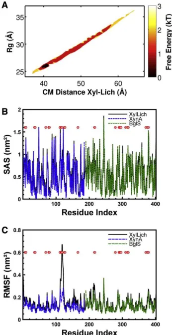
Molecular dynamics simulations were performed to characterize the most probable conformation of the chimera in solution. To assess probable arrangements of the chimera in solution, 2,000,000 confor-mations were generated through simulations using SB models and evaluated. Inter-region contacts (among XynA, BglS and the linker) were removed to allow an extensive search of the configuration space.Fig. 1A illustrates the universe of conformational possibilities derived from these simulations. The free energy profile was calculat-ed as a function of the following reaction coordinates: CM distance,Rg
a distinctive basin of low-energy conformations, which was restricted to theRgregion near 26 Å with a CM close to 42 Å, which represented
the accommodated chimera in solution. These results were obtained without any experimental support.
Conformations outside the free-energy basin revealed disagree-ments (highχ2values), which were caused by incorrect protein ar-rangements (Figs. SI-2). The compacted conformations showed deformations because of the high repulsive force between the resi-dues. The extended conformations presented local unfolding and dis-tortions near the linker. In both cases, the protein accommodation in solution was not quenched.
Simulations using XynA, BglS and the computationally suggested chimera were performed to predict possible modifications in their
functional behavior. One conformation from the basin was chosen for a new simulation (Rg= 26.7 Å,CMdistance = 42.4 Å).
Simula-tions using the chimera and the enzyme domains separately (XynA and BglS) displayed minor variations in the SAS (Fig. 1B), suggesting that the accessibility of the substrate to the pocket was not affected in the designed chimera. RMSF analysis (Fig. 1C) also showed minor variations, except for the XynA residues SER:117 and ILE:118, which were located into the“thumb”region of the protein[44,45].
3.2. Validation of the computationally predicted conformations through SAXS experiments
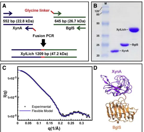
The genesxynAandbglSwere fused by PCR-mediated overlap exten-sion, and four glycine residues were included as a linker (Fig. 2A). The resulting amplicon, which was cloned into a pET28a vector, was 1209 bp long (Fig. 2A). Restriction analysis and DNA sequencing confirmed the molecular cloning. The chimera and its parental en-zymes, XynA (22.8 kDa) and BglS (26.7 kDa), were over expressed in Escherichia coliand purified through two chromatographic steps:first, nickel-affinity and then size-exclusion chromatography. The apparent molecular weight of the xylanase–lichenase chimera was 47.2 kDa (Fig. 2B).
Because the candidate conformations were obtained without pre-vious experimental information, SAXS was employed to validate the computationally predicted structures. The superimposed theoretical and experimental scattering curves of the selected conformation and the XylLich model are presented inFig. 2C. Theoretical scattering curves (I(q) versusq) within the broad basin of low-energy confor-mations were compared to the experimental scattering curve using CRYSOL. Good agreements were obtained for these conformations, i.e., lowχ2values, which were equal to 2.9 on average (some examples are presented in Figs. SI-1A and Figs. SI-1B), validating our simulation strategy. The obtained conformations displayed no obstructions or de-formations at the catalytic region, indicating that the chimera would be fully functional in vitro.
3.3. The chimera maintained the functional characteristics of the parental enzymes
The optimal temperature and pH of operation did not change drastically between the chimeric enzyme and the parental enzymes. The optimal pH for XynA and BglS was just 0.5 units lower than that of XylLich (Fig. 3A and B). There was no statistically significant shift in the xylanase temperature-dependent activity for the chimera, whereas the optimum temperature for lichenan hydrolysis was slightly greater for XylLich than for the parental enzymes (Fig. 3C and D).
The enzymes were biochemically assessed using a set of 10 natural polysaccharides. The specific activities of the chimera and the paren-tal enzymes on these polysaccharides are summarized inTable 1. XynA and XylLich hydrolyzed birchwood xylan, rye arabinoxylan and beechwood xylan more efficiently than other polysaccharides. Taking into account the standard deviation, there was no statistically significant difference between XynA and XylLich in hydrolyzing birchwood xylan and rye arabinoxylan. XylA and XylLich presented the lowest xylan-degrading activity (29–36%) on wheat arabinoxylan, which was the most complex and insoluble substrate tested. XylLich and BglS hydrolyzed lichenan andβ-glucan equally. The relative ac-tivities of XylLich and BglS on lichenan were 72 and 75% lower, re-spectively, onβ-glucan.
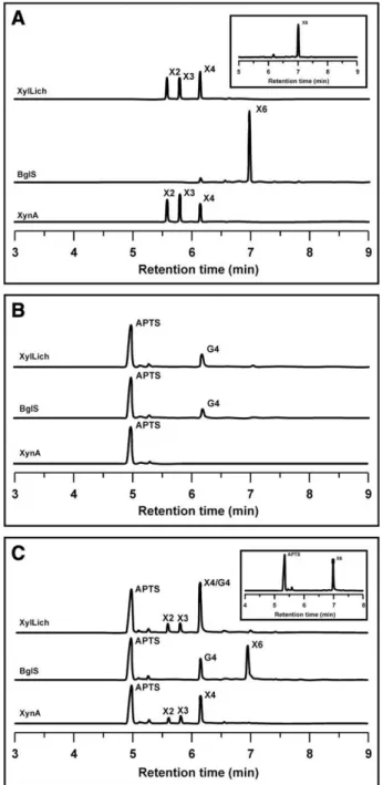
Fig. 4 describes our attempt to evaluate the mode of action of XylLich, XynA and BglS through CZE analysis of APTS-labeled oligosac-charides. Both parental and chimeric xylanase produced the same deg-radation pattern for APTS-labeled xylohexaose (X6), which included xylotetraose (X4), xylotriose (X3) and xylobiose (X2) (Fig. 4A). BglS was not able to hydrolyze X6. After hydrolysis of lichenan, XylLich and
Fig. 1.Characterization of XylLich using structure-based models (SB) through molecular dynamics (MD) simulations. (A) Thefigure on the top shows the free energy profile as a function of the radius of gyration (Rg) and the distance between the centers of mass of
Fig. 2.The chimera construction and experimental validation by SAXS. (A) Flow chart showing the process of fusion PCR for the XylLich construction. (B) SDS-PAGE of the chimera and wild-type proteins, indicating that the fused enzyme xylanase–lichenase had the predicted molecular weight. The protein molecular weight marker is shown in thefirst lane (M), and the values are displayed in kDa. (C) The small angle X-ray scattering profile for the experimental and theoretical evaluation of XylLich, which was taken from the free energy basin withχ2= 2.80,Rg= 26.0 Å andCMdistance = 40.7 Å. The scattering intensity is shown on a logarithm scale as a function of the momentum transfer (q). (D)
The XylLich model comprising XynA (purple), the linker (red) and BglS (orange). CRYSOL was employed to generate the theoretical curve and VMD[55]for the denoted cartoon.
BglS produced glucotetraose (G4), and XynA was not able to break down lichenan (Fig. 4B). As shown inFig. 4C, the enzymes were also assayed against the two substrates (X6 and lichenan) simultaneously. The results confirmed our previousfindings from using the substrates separately (Fig. 4A and B), highlighting that the mode of operation of the chimera was exactly similar to that of the parental enzymes.
Saturation assays were performed using beechwood xylan and lichenan. The kinetic constants of the wild-type and chimeric en-zymes are compared in Table 2. The maximum xylan degradation rate (Vmax) of the chimera was 30% lower than that of the parental enzyme (XynA), but for lichenan hydrolysis, the Vmaxwas 33% greater for XylLich than for BglS. Using xylan as a substrate, XylLich had a lower Km(41%) and turnover number (30%) than XynA. In contrast, the Kmof the chimeric enzyme was approximately 55% higher than that of BglS. In addition, the kcatincreased by up to 28% for XylLich using lichenan as substrate. Moreover, the catalytic efficiency (kcat/ Km) of the chimera was greater (18%) for xylan degradation and lower (18%) for lichenan hydrolysis than for the parental enzymes.
The far-UV CD spectra of XylLich showed a negative peak near 218 nm, suggesting that XylLich consists primarily ofβ-sheets (Fig.
5A). This profile was expected because the wild-type enzymes have
β-sheet predominance [46–48]. Another band was detected in the 220–230 nm region of the BglS spectrum, indicating the contribution of side-chain aromatic residues[47]. Thermal denaturation experi-ments were performed to compare the stability of the chimera and wild-type enzymes. The XylLich melting temperature (Tm) was 43.6 °C, whereas the Tm values of the parental enzymes were 53.3 °C and 42.5 °C for XynA and BglS, respectively (Fig. 5B).
3.4. Biotechnological appeal for producing the chimeric enzyme
To evaluate the biotechnological potential of using the chimeric enzyme in biomass to bio-products applications, hydrolysis assays
Table 1
Specific activities of the parental and chimeric enzymes on different types of substrates. Specific activity (U/nmol)
Substrate XynA BglS XylLich Birchwood xylan 3.73 ± 0.29 ND 2.71 ± 0.13 Beechwood xylan 3.17 ± 0.07 ND 2.87 ± 0.08 Rye arabinoxylan 3.73 ± 0.14 ND 3.03 ± 0.15 Wheat arabinoxylan 1.36 ± 0.12 ND 0.88 ± 0.07 Oat spelt xylan 3.28 ± 0.27 ND 2.15 ± 0.06 Lichenan ND 3.65 ± 0.29 3.85 ± 0.16
β-Glucan ND 5.03 ± 0.20 5.11 ± 0.07 Legend. ND means not determined. There was no activity on laminarin, xyloglucan and glucomannan (konjac) for all enzymes. Values are given by the mean ± S.D. of three independent assays.
Fig. 4.Capillary zone electrophoresis analysis of the breakdown products released by XylLich, XynA and BglS. The products after enzymatic hydrolysis of APTS-labeled xylohexaose (A), lichenan (B) and xylohexaose plus lichenan together (C) are presented. X2, X3, X4, X6 and G4 indicate the degree of polymerization of the produced xylose and glucose oligomers. The APTS-labeled xylohexaose used in the assays is indi-cated in the upper right boxes (A and C).
were performed using a polysaccharide composite comprising beechwood xylan and lichenan in a 1:1 ratio. The conversion effi cien-cies ofXynA,BglS, an equimolar mixture of the parental enzymes and the chimeric enzyme on this polysaccharide composite are described inFig. 6A. The rate of substrate conversion for XylLich was statistically equal to that of the parental enzymes combined (Fig. 6A).
The enzymatic performance was also evaluated using the crude E. colicell lysate as an enzyme source. Based on the total protein con-tent in the cell lysate and the overall mass produced byE. colicells, the amount of enzyme required to release 1μmol of reducing sugar per minute per protein milligram per gram of cell (U/mg/g) was calculated, which was defined in AY units (see methods). The chimera AY (0.35 U/mg/g) was approximately 6 times higher than XynA AY(0.06 U/mg/g) using xylan as a substrate. Likewise, the AY was 20% greater for XylLich than for BglS (Fig. 6B) using lichenan as a substrate.
4. Discussion
This study is an initial step toward the development of automated screening pipelines for optimal linkers and modules and functional chi-meras before experimental validation. XynA and BglS fromB. subtilis were chosen because these enzymes have been fully characterized and crystallographic structures are available [44–46,48–50]. Accordingly, the small and simple linker comprising four glycine residues was very well described previously[9]. However, based on the number of confor-mations available to XylLich (Figs. 1A, SI 1 and SI 2), its predicted organi-zation in solution was not obvious without computational tools or SAXS. Simulations employing SB models require significantly less com-putational time than traditional molecular dynamics using explicit water or complex forcefields. This key benefit allows the straightfor-ward investigation of several constructions and larger systems. De-spite being minimalistic approaches, SB models take into account the atomic restrictions to which real proteins are susceptible and thus corroborate experimental results[22]. The solvent properties implicitly included in these models support the calculation of an SAS, which predicts the change of the substrate accessibility to the enzyme[51]and estimates alterations in the enzymatic functionality. Simulations employing the chimeric model (χ2= 2.8) and the pa-rental enzymes (XynA or BglS) presented minor changes in the SAS. These results suggested that variations in the operation mode would not be expected because the binding site was not obstructed by deformations or steric effects between the domains. Supporting our hypothesis, the RMSF analysis (Fig. 1C) showed a similar profile for all cases. The only exception was the residue ILE:118 of the XynA thumb region located near the catalytic binding site, which could influence the enzymatic catalysis[44,45].
To validate the molecular dynamics simulations, comprehensive biochemical characterization was performed with the parental en-zymes and the chimera. Collectively, the biochemical analysis
con-firmed the simulations. The main exception was the slight variation of the XylLich temperature of operation and catalytic efficiency (kcat/Km). There were no substantial shifts in either the substrate specificities or the mode of operation. In addition, circular dichroism analysis revealed a greater similarity to the thermal denaturation properties of XylLich to BglS than those of XynA.
The minor shifts in the optimal pH and temperature of operation can be explained by an alteration in the microenvironment of the fused enzyme; one enzyme module preferentially catches protons and acidifies the local pH[52]. In addition, protein fusion can disturb the tertiary structure of a chimeric enzyme[52], which can cause dis-placements in the pH and temperature of operation, phenomena that are very well described in the literature[5,50,52].
The engineering of fused proteins has long been considered attrac-tive for industrial processes because these proteins are more efficient (based on costs and catalytic efficiency) than wild-type proteins
[5,49,50,53,54]. Along with the lack of substantial changes in the cat-alytic performance of the chimera, the parameter AY suggested an ad-vantage in producing the fused protein rather than the separate wild-type ones. Certainly more studies on XylLich properties are re-quired before use in a scaled-up process.
Table 2
Kinetic parameters of the chimeric and parental enzymes.
Substrate Beechwood xylan Lichenan Vmax
μmol/min/μmol Km
mg/mL
kcat
s−1 kmL·mgcat/Km−1·s−1 Vμmol/min/max μmol Kmg/mLm ks−cat1 mL·mgkcat/Km−1·s−1
XynA 10420 ± 497 7.87 ± 0.71 173.7 ± 8.3 22.1 ± 1.1 ND ND ND ND BglS ND ND ND ND 11430 ± 407 3.39 ± 0.26 197.2 ± 8.5 58.1 ± 2.5 XylLich 7300 ± 212 4.67 ± 0.26 121.7 ± 3.5 26.1 ± 0.8 15190 ± 1987 5.27 ± 1.13 253.2 ± 33.1 48.1 ± 6.3 Legend. ND means not determined. Values are given by the mean ± S.D. of three independent assays. The Vmaxunit was defined asμmol of reducing sugars released per minute per
μmol of enzyme.
In conclusion, this work presented a novel approach for predicting the arrangement of chimeric domains in solution before experimental validation. The computational strategy proposed herein is fast and ro-bust for characterizing a large number of enzymes in a few weeks, as well as for identifying possible binding site obstructions or large dy-namical variations differing from the single domains. The methodology can be extended to multi-domain chimeras and, by the same token, to a wide variety of biological systems. Finally, we expect that ourfindings will increase the pace offinding novel and cost-effective approaches for converting plant biomass into bio-products. One of the great challenges in biomass saccharification is decreasing the cost of enzyme production. Therefore, the use of multifunctional hydrolases, which act synergistically in plant polysaccharide degradation, is a promising venue for the improvement of enzyme cocktails for second-generation biofuels.
Acknowledgements
This research was supported by grants from FAPESP (2008/58037-9) and CNPq (475022/2011-4 and 310177/2011-1). JC received a scholar-ship from CNPq (140420/2009-6), and LCO, TMA and ARLD received scholarships from FAPESP (2011/13242-7, 2010/11499-1 and 2011/ 02169-7, respectively). X-ray scattering data were collected at the Brazilian Synchrotron Light Laboratory (LNLS) that integrates the Center of Research in Energy and Material (CNPEM). The authors would like to thank the LNLS for support. The computational analyses were supported by resources supplied by the Center for Scientific Com-puting (NCC/Grid UNESP) of São Paulo State University (UNESP) and CENAPAD-SP (Centro Nacional de Processamento de Alto Desempenho em São Paulo), project UNICAMP/FINEP-MCT.
Appendix A. Supplementary data
Supplementary data to this article can be found online athttp:// dx.doi.org/10.1016/j.bbapap.2013.02.030.
References
[1] M.E. Himmel, S.-Y. Ding, D.K. Johnson, W.S. Adney, M.R. Nimlos, J.W. Brady, T.D. Foust, Biomass recalcitrance: engineering plants and enzymes for biofuels pro-duction, Science (New York, N.Y.) 315 (2007) 804–807.
[2] T. Collins, C. Gerday, G. Feller, Xylanases, xylanase families and extremophilic xylanases, FEMS Microbiol. Rev. 29 (2005) 3–23.
[3] D.U. Lima, H.P. Santos, M.a. Tiné, F.R.D. Molle, M.S. Buckeridge, Patterns of expres-sion of cell wall related genes in sugarcane, Genet. Mol. Biol. 24 (2001) 191–198. [4] A. Planas, Bacterial 1,3-1,4-beta-glucanases: structure, function and protein
engi-neering, Biochim. Biophys. Acta 1543 (2000) 361–382.
[5] Z. Fan, K. Wagschal, C.C. Lee, Q. Kong, K.a. Shen, I.B. Maiti, L. Yuan, The construc-tion and characterizaconstruc-tion of two xylan-degrading chimeric enzymes, Biotechnol. Bioeng. 102 (2009) 684–692.
[6] S.Y. Hong, J.S. Lee, K.M. Cho, R.K. Math, Y.H. Kim, S.J. Hong, Y.U. Cho, S.J. Cho, H. Kim, H.D. Yun, Construction of the bifunctional enzyme cellulase-beta-glucosidase from the hyperthermophilic bacteriumThermotoga maritima, Biotechnol. Lett. 29 (2007) 931–936.
[7] C.J. Crasto, J.A. Feng, LINKER: a program to generate linker sequences for fusion proteins, Protein Eng. 13 (2000) 309–312.
[8] J.L. Casey, A.M. Coley, L.M. Tilley, M. Foley, Greenfluorescent antibodies: novel in vitro tools, Protein Eng. 13 (2000) 445–452.
[9] R. Arai, H. Ueda, A. Kitayama, N. Kamiya, T. Nagamune, Design of the linkers which effectively separate domains of a bifunctional fusion protein, Protein Eng. 14 (2001) 529–532.
[10] R.A. Griep, C. van Twisk, J.M. van der Wolf, A. Schots, Fluobodies: greenfl uores-cent single-chain Fv fusion proteins, J. Immunol. Methods 230 (1999) 121–130. [11] K. Morino, H. Katsumi, Y. Akahori, Y. Iba, M. Shinohara, Y. Ukai, Y. Kohara, Y.
Kurosawa, Antibody fusions withfluorescent proteins: a versatile reagent for
pro-filing protein expression, J. Immunol. Methods 257 (2001) 175–184.
[12] M. Peipp, D. Saul, K. Barbin, J. Bruenke, S.J. Zunino, M. Niederweis, G.H. Fey, Efficient eukaryotic expression offluorescent scFv fusion proteins directed against CD antigens for FACS applications, J. Immunol. Methods 285 (2004) 265–280. [13] O. Miyashita, C. Gorba, F. Tama, Structure modeling from small angle X-ray
scat-tering data with elastic network normal mode analysis, J. Struct. Biol. 173 (2011) 451–460.
[14] M. Pelikan, G.L. Hura, M. Hammel, Structure andflexibility within proteins as identified through small angle X-ray scattering, Gen. Physiol. Biophys. 28 (2009) 174–189.
[15] S. Yang, L. Blachowicz, L. Makowski, B. Roux, Multidomain assembled states of Hck tyrosine kinase in solution, Proc. Natl. Acad. Sci. U. S. A. 107 (2010) 15757–15762. [16] S. Yang, S. Park, L. Makowski, B. Roux, A rapid coarse residue-based computation-al method for X-ray solution scattering characterization of protein folds and mul-tiple conformational states of large protein complexes, Biophys. J. 96 (2009) 4449–4463.
[17] S.J. Kim, C. Dumont, M. Gruebele, Simulation-basedfitting of protein–protein in-teraction potentials to SAXS experiments, Biophys. J. 94 (2008) 4924–4931. [18] J.N. Onuchic, P.G. Wolynes, Theory of protein folding, Curr. Opin. Struct. Biol. 14
(2004) 70–75.
[19] J.D. Bryngelson, J.N. Onuchic, N.D. Socci, P.G. Wolynes, Funnels, pathways, and the energy landscape of protein folding: a synthesis, Proteins 21 (1995) 167–195. [20] L.L. Chavez, J.N. Onuchic, C. Clementi, Quantifying the roughness on the free
ener-gy landscape: entropic bottlenecks and protein folding rates, J. Am. Chem. Soc. 126 (2004) 8426–8432.
[21] C. Clementi, Coarse-grained models of protein folding: toy models or predictive tools? Curr. Opin. Struct. Biol. 18 (2008) 10–15.
[22] C. Clementi, P.A. Jennings, J.N. Onuchic, Prediction of folding mechanism for circular-permuted proteins, J. Mol. Biol. 311 (2001) 879–890.
[23] J.G. Lyubovitsky, H.B. Gray, J.R. Winkler, Mapping the cytochrome C folding land-scape, J. Am. Chem. Soc. 124 (2002) 5481–5485.
[24] L.C. Oliveira, A. Schug, J.N. Onuchic, Geometrical features of the protein folding mechanism are a robust property of the energy landscape: a detailed investiga-tion of several reduced models, J. Phys. Chem. B 112 (2008) 6131–6136. [25] M.A. Jamros, L.C. Oliveria, P.C. Whitford, J.N. Onuchic, J.A. Adams, D.K. Blumenthal,
P.A. Jennings, Proteins at work: a combined SAXS and theoretical determination of the multiple structures involved on the protein kinase functional landscape, J. Biol. Chem. 285 (2010) 36121–36128.
[26] M.A. Jamros, L.C. Oliveira, P.C. Whitford, J.N. Onuchic, J.A. Adams, et al., Substrate-specific reorganization of the conformational ensemble of CSK impli-cates novel modes of kinase function, PLoS Comput. Biol. 8 (2012) 1–8. [27] R. Koradi, M. Billeter, K. Wüthrich, MOLMOL: a program for display and analysis
of macromolecular structures, J. Mol. Graph. 14 (1996) 51–55, (29–32). [28] J.C. Phillips, R. Braun, W. Wang, J. Gumbart, E. Tajkhorshid, E. Villa, C. Chipot, R.D.
Skeel, L. Kalé, K. Schulten, Scalable molecular dynamics with NAMD, J. Comput. Chem. 26 (2005) 1781–1802.
[29] A.D. MacKerell, D. Bashford, Bellott, R.L. Dunbrack, J.D. Evanseck, M.J. Field, S. Fischer, J. Gao, H. Guo, S. Ha, D. Joseph-McCarthy, L. Kuchnir, K. Kuczera, F.T.K. Lau, C. Mattos, S. Michnick, T. Ngo, D.T. Nguyen, B. Prodhom, W.E. Reiher, B. Roux, M. Schlenkrich, J.C. Smith, R. Stote, J. Straub, M. Watanabe, J. Wiórkiewicz-Kuczera, D. Yin, M. Karplus, All-atom empirical potential for molecular modeling and dynamics studies of proteins, J. Phys. Chem. B 102 (1998) 3586–3616.
[30] J.K. Noel, P.C. Whitford, K.Y. Sanbonmatsu, J.N. Onuchic, SMOG@ctbp: simplified deployment of structure-based models in GROMACS, Nucleic Acids Res. 38 (2010) W657–W661, (Suppl.).
[31] J.K. Noel, P.C. Whitford, J.N. Onuchic, The Shadow map: a general contact defi ni-tion for capturing the dynamics of biomolecular folding and funcni-tion, J. Phys. Chem. B 116 (2012) 8692–8702.
[32] P.C. Whitford, J.K. Noel, S. Gosavi, A. Schug, K.Y. Sanbonmatsu, J.N. Onuchic, An all-atom structure-based potential for proteins: bridging minimal models with all-atom empirical forcefields, Proteins 75 (2009) 430–441.
[33] B. Hess, C. Kutzner, D.V.D. Spoel, E. Lindahl, GROMACS 4: algorithms for highly
ef-ficient, load-balanced, and scalable molecular simulation, J. Chem. Theory Comput. 4 (2008) 435–447.
[34] A.M. Ferrenberg, R.H. Swendsen, New Monte Carlo technique for studying phase transitions, Phys. Rev. Lett. 61 (1988) 2635–2638.
[35] K.L. Heckman, L.R. Pease, Gene splicing and mutagenesis by PCR-driven overlap extension, Nat. Protoc. 2 (2007) 924–932.
[36] F.M. Squina, C.R. Santos, D.a. Ribeiro, J. Cota, R.R. de Oliveira, R. Ruller, A. Mort, M.T. Murakami, R.a. Prade, Substrate cleavage pattern, biophysical characterization and low-resolution structure of a novel hyperthermostable arabinanase from
Thermotoga petrophila, Biochem. Biophys. Res. Commun. 399 (2010) 505–511. [37] A.P. Hammersley, ESRF Internal Report, ESRF97HA02T, "FIT2D: An Introduction and
Overview'', 1997. (Information obtained from: http://www.esrf.eu/computing/ scientific/FIT2D/FIT2D_REF/node268.html).
[38] H. Fischer, M. de Oliveira Neto, H.B. Napolitano, I. Polikarpov, a.F. Craievich, Determina-tion of the molecular weight of proteins in soluDetermina-tion from a single small-angle X-ray scattering measurement on a relative scale, J. Appl. Crystallogr. 43 (2009) 101–109. [39] D. Svergun, C. Barberato, M.H.J. Koch, CRYSOL—a program to evaluate X-ray
so-lution scattering of biological macromolecules from atomic coordinates D. Svergun, C. Barberato and M. H. J. Koch, J. Appl. Crystallogr. 28 (1995) 768–773. [40] G.L. Miller, Use of dinitrosalicylic acid reagent for determination of reducing
sugar, Anal. Chem. 31 (1959) 426–428.
[41] R. Naran, M.L. Pierce, A.J. Mort, Detection and identification of rhamnogalacturonan lyase activity in intercellular spaces of expanding cotton cotyledons, Plant J. 50 (2007) 95–107.
[42] C.R. Santos, F.M. Squina, A.M. Navarro, D.P. Oldiges, A.F. Leme, R. Ruller, A.J. Mort, R. Prade, M.T. Murakami, Functional and biophysical characterization of a hyperthermostable GH51 alpha-L-arabinofuranosidase fromThermotoga petrophila, Biotechnol. Lett. 33 (2011) 131–137.
-1,3-glucanase fromThermotoga petrophila, Biochem. Biophys. Res. Commun. 406 (2011) 590–594.
[44] M.T. Murakami, R.K. Arni, D.S. Vieira, L. Degrève, R. Ruller, R.J. Ward, Correlation of temperature induced conformation change with optimum catalytic activity in the recombinant G/11 xylanase A fromBacillus subtilisstrain 168 (1A1), FEBS Lett. 579 (2005) 6505–6510.
[45] D.S. Vieira, R.J. Ward, Conformation analysis of a surface loop that controls active site access in the GH11 xylanase A fromBacillus subtilis, J. Mol. Model. 18 (2012) 1473–1479.
[46] G.P. Furtado, L.F. Ribeiro, C.R. Santos, C.C. Tonoli, A.R. Souza, R.R. Oliveira, M.T. Murakami, R.J. Ward, Biochemical and structural characterization of aβ -1,3-1,4-glucanase fromBacillus subtilis168, Process Biochem. 46 (2011) 1202–1206. [47] I.B. Grishina, R.W. Woody, Contributions of tryptophan side chains to the circular
dichroism of globular proteins: exciton couplets and coupled oscillators, Faraday Discuss. 99 (1994) 245–262.
[48] R. Ruller, L. Deliberto, T.L. Ferreira, R.J. Ward, Thermostable variants of the recom-binant xylanase A fromBacillus subtilisproduced by directed evolution show re-duced heat capacity changes, Proteins 70 (2008) 1280–1293.
[49] J. Aÿ, F. Götz, R. Borriss, U. Heinemann, Structure and function of the Bacillus hy-brid enzyme GluXyn-1: native-like jellyroll fold preserved after insertion of au-tonomous globular domain, Proc. Natl. Acad. Sci. U. S. A. 95 (1998) 6613–6618. [50] L.F. Ribeiro, G.P. Furtado, M.R. Lourenzoni, A.J. Costa-Filho, C.R. Santos, S.C. Peixoto
Nogueira, J.a. Betini, M.D.L.T.M. Polizeli, M.T. Murakami, R.J. Ward, Engineering bi-functional laccase–xylanase chimerae for improved catalytic performance, J. Biol. Chem. 286 (2011) 40026–40038.
[51] M.L. Connolly, SCIENCE: solvent-accessible surfaces o f proteins and nucleic acids, Science 221 (1983) 709–713.
[52] L. Bülow, Characterization of an artificial bifunctional enzyme, beta-galactosidase/ galactokinase, prepared by gene fusion, Eur. J. Biochem. 163 (1987) 443–448. [53] L. Chang, M. Ding, L. Bao, Y. Chen, J. Zhou, H. Lu, Characterization of a bifunctional
xylanase/endoglucanase from yak rumen microorganisms, Appl. Microbiol. Biotechnol. 90 (2011) 1933–1942.
[54] R. Khandeparker, M.T. Numan, Bifunctional xylanases and their potential use in biotechnology, J. Ind. Microbiol. Biotechnol. 35 (2008) 635–644.