Deficiency of Thrombospondin-4 in Mice
Does Not Affect Skeletal Growth or Bone
Mass Acquisition, but Causes a Transient
Reduction of Articular Cartilage Thickness
Anke Jeschke1☯, Martin Bonitz1☯, Maciej Simon1,2, Stephanie Peters1, Wolfgang Baum3, Georg Schett3, Wolfgang Ruether2, Andreas Niemeier2, Thorsten Schinke1,
Michael Amling1*
1Department of Osteology and Biomechanics, University Medical Center Hamburg Eppendorf, Hamburg 20246, Germany,2Department of Orthopedics, University Medical Center Hamburg Eppendorf, Hamburg 20246, Germany,3Department of Internal Medicine 3 and Institute of Clinical Immunology, University of Erlangen-Nuremberg, Erlangen 91054, Germany
☯These authors contributed equally to this work. *[email protected]
Abstract
Although articular cartilage degeneration represents a major public health problem, the under-lying molecular mechanisms are still poorly characterized. We have previously utilized
genome-wide expression analysis to identify specific markers of porcine articular cartilage, one of them being Thrombospondin-4 (Thbs4). In the present study we analyzedThbs4expression in mice, thereby confirming its predominant expression in articular cartilage, but also identifying expression in other tissues, including bone. To study the role of Thbs4 in skeletal development and integrity we took advantage of aThbs4-deficient mouse model that was analyzed by unde-calcified bone histology. We found thatThbs4-deficient mice do not display phenotypic differ-ences towards wildtype littermates in terms of skeletal growth or bone mass acquisition. Since
Thbs4has previously been found over-expressed in bones of Phex-deficientHypmice, we additionally generatedThbs4-deficientHypmice, but failed to detect phenotypic differences towardsHyplittermates. With respect to articular cartilage we found thatThbs4-deficient mice display transient thinning of articular cartilage, suggesting a protective role of Thbs4 for joint integrity. Gene expression analysis using porcine primary cells revealed that Thbs4 is not expressed by synovial fibroblasts and that it represents the only member of the Thbs gene fam-ily with specific expression in articular, but not in growth plate chondrocytes. In an attempt to identify specific molecular effects of Thbs4 we treated porcine articular chondrocytes with human THBS4 in the absence or presence of conditioned medium from porcine synovial fibro-blasts. Here we did not observe a significant influence of THBS4 on proliferation, metabolic activity, apoptosis or gene expression, suggesting that it does not act as a signaling molecule. Taken together, our data demonstrate that Thbs4 is highly expressed in articular chondrocytes, where its presence in the extracellular matrix is required for articular cartilage integrity.
OPEN ACCESS
Citation:Jeschke A, Bonitz M, Simon M, Peters S, Baum W, Schett G, et al. (2015) Deficiency of Thrombospondin-4 in Mice Does Not Affect Skeletal Growth or Bone Mass Acquisition, but Causes a Transient Reduction of Articular Cartilage Thickness. PLoS ONE 10(12): e0144272. doi:10.1371/journal. pone.0144272
Editor:Jan Peter Tuckermann, University of Ulm, GERMANY
Received:April 28, 2015
Accepted:November 16, 2015
Published:December 2, 2015
Copyright:© 2015 Jeschke et al. This is an open access article distributed under the terms of the Creative Commons Attribution License, which permits unrestricted use, distribution, and reproduction in any medium, provided the original author and source are credited.
Data Availability Statement:All relevant data are within the paper and its Supporting Information files.
Introduction
Osteoarthritis is a highly prevalent disorder characterized by loss of articular cartilage, a unique avascular and hypocellular tissue covering the joints [1,2]. The current treatment options are limited to surgical procedures, such as microfracture, osteochondral implantation, and, in the end-stage, total joint replacement [3,4]. In addition, autologous transplantation of articular chondrocytes afterex vivoexpansion has emerged as a promising alternative therapeutic approach over the last two decades [5,6]. Since it is commonly accepted however, that any of these treatments only leads to formation of a fibrocartilagenous tissue with inferior quality compared to native articular cartilage, it is of utmost clinical importance to understand the molecular mechanisms controlling the function of articular chondrocytes. This may not only help to optimize ongoing tissue-engineering approaches, but also to identify potential molecu-lar targets for pharmacological stimulation of matrix production by articumolecu-lar chondrocytes.
One potential explanation for the paucity of knowledge regarding specific regulators of articular chondrogenesis is the shortness of articular cartilage in mice, which hinders the iden-tification of unexpected osteoarthritis phenotypes in genetically modified mouse models, unlike it was the case for other skeletal disorders [7]. For the same reason, it is extremely difficult to isolate primary articular chondrocytes from mice, which explains why many experi-ments related to arthritis have been performed with rib or epiphyseal growth plate chondro-cytes. Regardless of these limitations several studies have been performed to identify specific markers of articular cartilage in mice, and there is an increasing number of mouse models dis-playing an arthritis phenotype [8–14]. Given the potential importance of identifying molecular differences between chondrocytes from articular and non-articular cartilage, we have previ-ously performed genome-wide expression analysis with native tissues and cultured cells of por-cine origin [15]. Here it was possible to separate articular and growth plate cartilage from the bone matrix and to culture a sufficient number of chondrocytes from both sourcesex vivo. The major disadvantage of this approach was related to the use of porcine Gene Chips, which did not provide the same genetic coverage compared to the murine system.
Despite this limitation however, we made at least three important observations [15]. First, we identified common markers of cartilage, but also genes with specific expression in either growth plate or articular cartilage. Second, we found that the molecular differences between the two types of chondrocytes persisted after 10 and 20 days ofex vivoculture. Third, we defined 19 markers of articular chondrocytes, thereby raising the question, which of these are relevant regulators of articular cartilage integrity. Here we analyzed the physiological role of Thbs4, one of these previously identified markers, by studying the skeletal phenotype of a mouse deficiency model. We found thatThbs4-deficient mice do not display defects of skeletal growth or bone mass acquisition, but that articular cartilage thickness is transiently reduced.
Materials and Methods
1. Mouse models
Thbs4-deficient mice on a C57Bl/6 genetic background were purchased from the Jackson Labo-ratories (#005845). Their genotyping was performed with the primers 5´-GGG TGG GAT TAG ATA AAT GCC TGC TCT-3´, 5´-GGA GAG AGA ATA GCA AGA TCA GCT C-3´, and 5´-AAC AAG CAA TGG AAG GCA GAC CCT G-3´, giving rise to a 412 bp and a 544 bp fragment for the wildtype and mutant allele, respectively. To evaluate the impact of Thbs4 in the context of X-linked hypophosphatemic rickets,Thbs4-deficient mice were crossed with Hypmice (C57Bl/6 genetic background), which were also obtained from the Jackson Laborato-ries (#000528). To induce experimental arthritis,Thbs4-deficient mice were crossed with
transgenic mice expressing human TNFα(Tg197 on a C57Bl/6 genetic background), which
have been described elsewhere [16]. Joint swelling of the foot paws was assessed between 5 and 10 weeks of age using a clinical score graded from 0 (no swelling) to 3 (severe swelling of toes and ankle) as described previously [17]. Grip strength was analyzed on wire (diameter of 3 mm) using a score from 0 (normal grip strength) to -4 (no detectable grip strength) as described [17]. All mice were fed ad libitum and housed in a regular light/dark cycle under SPF-conditions. Animal experiments were approved by the animal facility of the University Medical Center Hamburg Eppendorf and by the“Amt für Gesundheit und Verbraucherschutz”
(Org529).
2. Cell culture
Primary murine osteoclasts were generated by differentiating bone marrow cells for 10 days with 1,25-dihydroxyvitamin D3 (10 nM) added for the whole period, and M-Csf (20 ng/ml) and Rankl (40 ng/ml) added from day 3 until day 10 [18]. Primary murine osteoblasts were iso-lated by collagenase digestion of calvariae from newborn mice and differentiated for 20 days in the presence of ascorbic acid (10 mM) and ß-glycerophosphate (50μg/ml) as described [18]. Porcine synovial tissue was obtained from the right and the left knee of 6 weeks old minipigs. Digestion of the prepared synovial membrane was performed with 1 mg/dl collagenase type 1a solution (Sigma-Aldrich, Germany) for 60–75 min at 37°C. Isolated synovial fibroblasts were seeded and cultured in Synoviocyte Basal Medium (Cell Applications, USA) supplemented with 10% heat-inactivated Synoviocyte Growth Supplement (Cell Application, USA) at normal cell culture conditions. Conditioned medium of these cells was collected for 24 hours in basal medium. To isolate chondrocytes bone-cartilage cylinders were harvested from the medial and lateral condyle of knee joints of 6 weeks old minipigs. The cylinders were separated in articular and growth plate cartilage under the dissecting microscope. Chondrocytes were released by col-lagenase type la solution (Sigma-Aldrich, Germany) as described [15]. The cells were cultured in DMEM/Hams’F12 (Biochrom, Germany) supplemented with 10% (v/v) FBS (Lonza, Ger-many) at 37°C under an atmosphere of 5% (v/v) O2and 5% (v/v) CO2. These experiments were approved by the animal facility of the University Medical Center Hamburg Eppendorf and by the“Amt für Gesundheit und Verbraucherschutz”(Org356).
3. Expression analysis
RNA from murine tissues, cultured murine osteoclasts and osteoblasts, or porcine chondro-cytes and synoviochondro-cytes was isolated using the RNeasyMini kit (Qiagen, Germany). DNase digestion was performed according to manufacturer’s instructions. Concentration and quality of RNA were measured using a NanoDrop ND-1000 system (NanoDrop Technology, USA). For RT-PCR expression analysis, 1μg of RNA was reversed transcribed using SuperScriptIII (Invitrogen, Germany) according to manufacturer’s instructions. Predesigned TaqMan gene expression assays (Applied Biosystems, Germany) were used to quantify expression of all murine genes, as well as for the porcine genesACAN,FN1,COL2A1,COL10A1,ASPN,FRZB, andSDC4. For the other porcine gene the resulting cDNA was used for a PCR reaction with gene-specific primers (PRG4: 5´-CAT CTC TCT TTG ACG GTG AGG G-3´ and 5´-GCT CCA TAG TGC AGA CTT TCT TGA-3´;THBS1: 5´-CCA GCA GCC GTT TCT ATG TTG T -3’and 5´-cct atg tga cga gga tca tgc-3´;THBS2: 5´-gac gag ttt ggg tct gtg ga-3´and 5´-cca gcg tag gtt tgg tca ta-3´;THBS3: 5´-cag gta cga ctg ctg tgg ac-3´and 5´-ggc act gtg tca ttg cat cg-3´;
THBS4: 5’-ATC CAG GCG ATC GAA ATT CTG-3’and 5’-AGG TGT CCT ATC GCT GGT
GGT CTG GAG ATG GA-3´;GAPDH: 5’-CTT CGT CAA GCT CAT TTC CTG G-3’and 5’ -AGT CAG GAG ATG CTC GGT GTG-3’) and SYBR Green Master Mix (Applied Biosystems, Germany).GAPDHexpression was used as an internal control. Relative quantification was per-formed according to theΔΔC
Tmethod, and results were expressed in linear form using the for-mula 2-ΔΔCTfor both RT-PCR assays.
4. Skeletal analysis
After sacrifice the dissected skeletons were fixed in 3.7% PBS-buffered formaldehyde for 18 hours at 4°C, before they were stored in 80% ethanol. All skeletons were analyzed by contact radiography using a Faxitron Xray cabinet (Faxitron Xray Corp., USA) to measure the length of the lumbar spine and femora. For bone histology, the lumbar vertebral bodies L3 to L6 and one tibia of each mouse were dehydrated in ascending alcohol concentrations and then embed-ded in methylmetacrylate as described previously [18]. Sections of 5μm thickness were cut in the sagittal plane on a Microtec rotation microtome (Techno-Med GmbH, Germany). For articular cartilage histology, knee joints were processed in the same way. All sections were stained by toluidine blue and von Kossa/van Gieson staining procedures as described [18]. His-tomorphometry was performed according to the ASBMR guidelines [19] using the OsteoMea-sure histomorphometry system (Osteometrics Inc., USA). Articular chondrocyte apoptosis was assessed on 5μm thin decalcified paraffin sections byin situTUNEL technology (Roche, #11684795910) according to the manufacturer´s instructions.
5. Cellular assays
To assess proliferation, metabolic activity and apoptosis, porcine articular chondrocytes were seeded into 96-well plates at a density of 1.000 cells per ml. On the next day cells were incu-bated for 24 hours with human THBS4 (R&D Systems, #2390-TH) at different concentrations in serum-free medium or in serum-free medium mixed with an equal amount of conditioned medium from porcine synovial fibroblasts. BrdU incorporation, MTT conversion and Caspase 3/7 activities were determined with commercially available systems (GE Healthcare Amer-sham, #RPM250, Sigma Aldrich, #M2158, and Promega, #G8090, respectively) according to the manufacturer´s instructions. To analyze cell adhesion 96-well-microtest plates were coated with 5μg/ml THBS4 or 5μg/ml THBS5/COMP (R&D Systems, #3134-CP) at 4°C overnight. Porcine articular chondrocytes were washed twice with HBBS/C (Hank´s Balanced Salt Solu-tion + 1 mM calcium chloride), pre-treated with tosylphenylalanyl chloromethyl ketone-treated trypsin (0.1 mg/ml) in HBBS/C, before trypsinization was stopped after incubation for 5 min at 37°C. The cells were then washed three times with HBBS/C and resuspended in HBBS/C containing 1% heat inactivated BSA. Were indicated, a monoclonal antibody to integ-rin ß1 (BD Biosciences, #552828) was added to a final concentration of 16μg/ml and incubated with the cells for 30 min at room temperature. Coated wells were washed four times with HBBS/C before addition of 3 x 10⁴cells per well. After overnight incubation at 37°C under an atmosphere of 5% (v/v) O2and 5% (v/v) CO2the number of attached cells was quantified. To assess the effects of THBS4 and/or conditioned medium from porcine synovial fibroblasts on gene expression, cells were treated for 6 hours, before RNA was isolated for qRT-PCR expres-sion analysis.
6. Serum analysis of individuals with osteoarthritis
primary OA were included. OA secondary to any known cause as well as any other arthropathy were exclusion criteria. All patients had severe, symptomatic primary OA of at least one large joint of the lower or upper extremity (knee, hip or shoulder; index joint) with radiographic joint space narrowing to less than a residual 1/3, and the request for total joint arthroplasty as the primary reason for consultation. Patients with knee or hip OA without signs and symptoms of OA in one or more additional joints were considered to have a relatively confined local dis-ease and were classified as mono-osteoarthritis (mOA). Patients who, in addition to severe OA of the index joint, displayed clinically obvious hand OA of multiple interphalangeal joints and further joints of the lower and or upper extremities were considered to be more severely affected on a systemic level and were classified as poly-osteoarthritis (pOA). As an approach to distinguish between individuals with a presumably rather small total volume of articular carti-lage affected locally by OA (mOA) versus individuals with a comparatively larger total volume of articular cartilage affected at multiple sites (pOA), we used these clinical differentiation crite-ria to build two distinct subpopulations of OA from our routine inpatient and outpatient clinic. Serum concentration of THBS4 was quantified using the antibody-based detection kit (Qayee-Bio, China). After the blood draw, serum samples were kept at room temperature for a maxi-mum of two hours before storage at -70°C until analysis without additional freeze-thaw cycles. All participants provided written informed consent. This study and consent procedure was approved by the local ethics committee (Aerztekammer Hamburg, PV4037).
7. Statistical analysis
All data presented in the manuscript were obtained from the analysis of littermates (n5) and are presented as means ± standard deviations. Statistical analysis was performed using
unpaired, two-tailed Student’s t test, and p-values below 0.05 were considered statistically significant.
Results
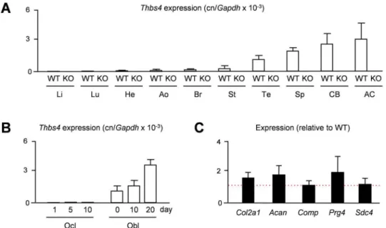
Given the previously observed predominant expression ofTHBS4in porcine articular cartilage [15], we first addressed the question, whether the same is the case in mice. We therefore iso-lated RNA from different tissues of 15 weeks old wildtype andThbs4-deficient mice to monitor Thbs4expression by qRT-PCR. Here we found, as expected only in wildtype mice, thatThbs4 is highly expressed in articular cartilage, yet there was also strong expression in other tissues, such as tendon, spleen and cortical bone (Fig 1A). We additionally analyzed, which of the two bone remodeling cell types is the primary source ofThbs4expression and performed qRT-PCR with RNA from primary osteoclasts and osteoblasts at different stages of differentiation. Here we found thatThbs4was differentially expressed in osteoblast cultures, whereasThbs4 tran-scripts were undetectable in osteoclast cultures (Fig 1B). Since articular cartilage displayed the highest expression ofThbs4in wildtype mice, we additionally analyzed ifThbs4-deficiency would affect the expression of articular chondrocyte marker genes. Here we did not observe significant differences in the expression ofCol2a1,Acan,Comp,Prg4andSdc4between wild-type andThbs4-deficient mice, suggesting that Thbs4 does not play a major role as a regulator of articular cartilage gene expression (Fig 1C). Nevertheless, given the high expression ofThbs4 in skeletal tissues, we went on to study the skeletal phenotype ofThbs4-deficient mice.
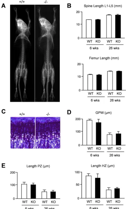
thickness did not reveal statistically significant differences between wildtype andThbs4 -defi-cient mice at 6 and 26 weeks of age (Fig 2D). Likewise, the lengths of the proliferative and hypertophic zones within the tibia growth plate were not affected byThbs4-deficiency (Fig 2E). We additionally analyzed spine sections in the same groups of mice with respect to a potential bone phenotype (Fig 3A). By quantifying trabecular bone parameters we again found no differ-ence between wildtype andThbs4-deficient littermates (Fig 3B). Collectively, these data sug-gested that Thbs4 does not function as a physiologically relevant regulator of skeletal growth, bone formation or remodeling.
Another question to be addressed was based on previously reported findings in Phex-defi-cientHypmice, a model of X-linked hypohosphatemic rickets [20,21]. These mice display defects of skeletal growth and bone matrix mineralization, which are only partially explained by hypophosphatemia [22,23]. A genome-wide expression analysis revealed thatThbs4is markedly over-expressed in cortical bone of these mice, similar toFgf23, whose increased expression inHypmice is known to cause their renal phosphate loss [24]. Since it was reason-able to speculate that a higher abundance of Thbs4 in the bone matrix could interfere with mineralization, we generatedThbs4-deficientHypmice and analyzed their skeletal phenotype. Here we found, as expected, thatHypmice displayed a severe skeletal phenotype with patho-logical enrichment of non-mineralized osteoid, yet this pathology was not affected byThbs4 -deficiency (Fig 3C). Subsequent quantification of growth plate thickness and osteoid volume confirmed that the skeletal phenotype ofHypmice was not significantly altered by additional Thbs4-deficiency (Fig 3D), thereby demonstrating that increasedThbs4expression by Phex-deficient osteoblasts is not involved in the pathogenesis of X-linked hypophosphatemic rickets. Fig 1. MurineThbs4is highly expressed in skeletal tissues.(A) qRT-PCR monitioringThbs4expression in various tissues of 15 weeks old widltype (WT) andThbs4-deficient mice (KO). Li, liver; Lu, lung; He, heart; Ao, aorta; Br, brain; St, stomach; Te, tendon; Sp, spleen; CB, cortical bone; AC, articular cartilage. Bars represent mean±SD (n = 4).Thbs4expression was undetectable in all KO tissues. (B) qRT-PCR monitoring
Thbs4expression in primary osteoclasts (Ocl) and osteoblasts (Obl) from wildtype mice at different stages of differentiation. Bars represent mean±SD (n = 3). (C) qRT-PCR monitioring expression of the indicated genes in articular cartilage of 15 weeksThbs4-deficient mice. The dotted red line indicates the expression in wildtype littermates. Bars represent mean±SD (n = 4).
To assess the articular cartilage phenotype ofThbs4-deficient mice we analyzed sections from the knee joints of 6, 26 and 52 weeks old mice and determined the thickness of the articu-lar cartilage layer in three different regions of interest (Fig 4A). Here we found that 26 weeks oldThbs4-deficient mice displayed significantly reduced articular cartilage thickness in all regions of interest, both in the femur (Fig 4B) and in the tibia (Fig 4C). This genotype-depen-dent difference was not observed at the age of 52 weeks, where the articular cartilage thickness Fig 2. Intact skeletal growth inThbs4-deficient mice.(A) Xray analysis demonstrates absence of gross skeletal abnormalities in 6 weeks oldThbs4-deficient mice. (B) Length of lumbar spine and femur in 6 and 26 weeks old wildtype (WT) andThbs4-deficient (KO) mice. (C) Toluidine blue staining of the tibia growth plates from 6 weeks old wildtype (+/+) andThbs4-deficient (-/-) mice. (D) Quantification of the tibial growth plate width (GPW) in wildtype (WT) andThbs4-deficient (KO) mice at 6 and 26 weeks of age. (E) Quantification of the lengths of the proliferative zone (PZ) and hypertrophic zone (HZ) in the same sections. All bars represent mean±SD (n = 6 per group).
was similar to 26 weeks oldThbs4-deficient mice in both genotypes. We also determined the number of chondrocytes per cartilage area in femur and tibia from the respective mice (Fig 5A). Here we did not observe statistically significant differences, and the same was the case for the percentage of apoptotic cells as assessed by TUNEL assay at the age of 26 weeks (Fig 5B). Finally, in an attempt to obtain a molecular explanation for the transient phenotype ofThbs4 -deficient mice, we monitored expression of known osteoarthritis susceptibility (OAS) genes [25–28] in articular cartilage from 26 weeks old mice. WhereasGdf5expression was not Fig 3. No impact ofThbs4-deficiency on bone mass or matrix mineralization on a wildtype orHyp
genetic background.(A) Von Kossa/van Gieson staining of spine sections from 6 and 26 weeks old wildtype (+/+) andThbs4-deficient (-/-) mice. (B) Quantification of the trabecular bone volume per tissue volume (BV/ TV) and trabecular thickness (Tb.Th.) in wildtype (WT) andThbs4-deficient (KO) mice at both ages. Bars represent mean±SD (n = 6 per group). (C) Xray analysis (top panels) and von Kossa/van Gieson staining of tibia (middle panels) or spine sections (bottom panels) from 6 weeks old Hyp mice with (+/+) or without (-/-)
Thbs4. (D) Quantification of the growth plate width (GPW) in the tibia (top) or the osteoid volume per bone volume (OV/BV) from mice of the indicated genotypes. Bars represent mean±SD (n = 5 per group). Asterisks indicate statistically significant differences towards WT controls (p<0.05).
doi:10.1371/journal.pone.0144272.g003
Fig 4. Transient thinning of articular cartilage inThbs4-deficient mice.(A) Von Kossa/van Gieson (left panels) or toluidine blue staining (right panels) of articular cartilage from the knee joints of 26 weeks old wildtype (+/+) andThbs4-deficient (-/-) mice. The three regions of interest for quantification of articular cartilage width are indicated. (B) Quantification of the articular cartilage width (ACW) in femora from wildtype (WT) andThbs4-deficient (KO) mice at 6, 26 and 52 weeks of age. (C) Quantification of the articular cartilage width (ACW) in femur and tibia sections from the same mice. All bars represent mean±SD (n = 6 per group). Asterisks indicate statistically significant differences between WT and KO (p<0.05).
detectable by qRT-PCR in samples from either genotype, we found no significant changes between wildtype andThbs4-deficient mice in terms ofFrzborMatn3expression (Fig 5C). Interestingly however,Aspn, encoding a small leucine-rich proteoglycan potentially inhibiting TGFß-dependent matrix synthesis [28,29], was expressed at lower levels in articular cartilage of 26 weeks oldThbs4-deficient mice.
To address the question, ifThbs4-deficiency would affect the severity of joint destruction in a mouse model of rheumatoid arthritis, we additionally crossedThbs4-deficient mice with mice carrying a transgene causing over-expression of human TNFα[16,17]. Here we found
that the presence of the transgene caused progressive joint swelling of the foot paws together with a decline in grip strength until the age of 12 weeks, yetThbs4-deficiency did not signifi-cantly affect these two clinical scores (S1A Fig). When we histologically analyzed the knee joints at 12 weeks of age however, we found enhanced destruction of subchondral bone specifi-cally inThbs4-deficient TNFα-transgenic mice (S1B Fig). Taken together, these findings
revealed that Thsb4 has a protective role in articular cartilage, although its deficiency does not affect the affect proliferation or apoptosis of articular chondrocytes.
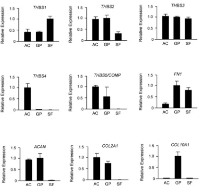
Since Thbs4 is only one member of a protein family, we next compared expression of all five thrombospondin-encoding genes in primary chondrocytes from articular and growth plate cartilage, as well as in synovial fibroblasts. To avoid any cross-contamination of these cell pop-ulations we again utilized minipigs, where the two types of cartilage, as well as synovial fibro-blasts can be undoubtedly separated. Using qRT-PCR expression analysis we found that THBS1,THBS2,THBS3andCOMP/THBS5were all expressed in both types of chondrocytes (Fig 6). With the exception ofCOMP/THBS5, we also detected their expression in synovial fibroblasts. In sharp contrast,THBS4expression was only detected in articular chondrocytes and not in any of the other cell types.
To analyze a potential impact of THBS4 on the behavior of articular chondrocytes we also used porcine cells. More specifically, we assessed cellular proliferation (Fig 7A), metabolic Fig 5. Chondrogenesis is unaffected in articular cartilage inThbs4-deficient mice.(A) Quantification of the chondrocyte number per cartilage area in femur and tibia sections from 6, 26 and 52 weeks wildtype and
Thbs4-deficient mice. Bars represent mean±SD (n = 6 per group). (B) Representative images showing TUNEL-positive cells in articular cartilage from the femora of 26 weeks old wildtype andThbs4-deficient mice. The percentage of TUNEL-positive cells is givenon the right. Values represent mean±SD (n = 6 per group). (C) qRT-PCR monitioring expression of the indicated genes in articular cartilage of 26 weeks wildtypeThbs4 -deficient mice. Bars represent mean±SD (n = 4). Asterisks indicate statistically significant differences between WT and KO (p<0.05).
activity (Fig 7B) and apoptosis (Fig 7C) over 24 hours in primary articular chondrocytes in the presence of increasing concentrations of human THBS4. As a control we performed the same assays with conditioned medium (50% final concentration) from cultured porcine synovial fibroblasts (SF-CM), and again co-administered increasing concentrations of human THBS4. Here we found that THBS4 did not cause a significant influence on any of the parameters. Interestingly however, while SF-CM did not affect proliferation or metabolic activity of the articular chondrocytes, it significantly increased the Caspase-3/7 activity, suggesting a pro-apo-ptotic influence, which was however unaffected by THBS4. Finally, since COMP/THBS5 has been shown to mediate chondrocyte attachment in an integrin-dependent manner [30], we analyzed if THBS4 would serve a similar function. To address this possibility we coated non-tissue culture plates with THBS4 or COMP, before adding porcine articular chondrocytes in the presence or absence of an antibody against ß1-integrin. After 24 hours we counted
the adherent cells and found that they attached to COMP-coated plates in a ß1-integrin-depen-dent manner (Fig 7D). In contrast, we failed to detect adherent articular chondrocytes on THBS4-coated plates.
As we observed reduced expression ofAspnin articular cartilage of 26 weeks oldThbs4 -defi-cient, we additionally treated articular chondrocytes for 6 hours with THBS4 and/or SF-CM, before isolating RNA for qRT-PCR expression analysis. When monitoring expression of the OAS genes we observed no significant influences of THBS4, either alone or in the presence of SF-CM (Fig 8A). Importantly however, expression levels ofASPN,FRZBandMATN3 (but not ofGDF5)were remarkably reduced by SF-CM, thus suggesting a direct transcriptional influ-ence by yet unidentified SF-derived molecules. Based on these findings we additionally moni-tored expression ofCOL2A1,ACAN,THBS4andSDC4, the latter gene encoding a negative Fig 6. PorcineTHBS4is specifically expressed in articular chondrocytes.Shown are the results of qRT-PCR expression analyses for all members of theTHBSfamily, as well as markers for synovial fibroblasts (FN1), chondrocytes (ACAN,COL2A1) and hypertrophic chondrocytes (COL10A1). Primary cells (AC, articular chondrocytes; GP, growth plate chondrocytes; SF, synovial fibroblasts) were derived from 6 weeks old minipigs. Shown is the relative expression (after normalization toGAPDH) towards the cell type displaying the highest expression level. All bars represent mean±SD (n = 3).
regulator of articular chondrocytes [9]. Again, we failed to detect a significant influence of THBS4, either alone or in the presence of SF-CM (Fig 8B). In contrast, whereas SF-CM caused a transcriptional repression ofACANandTHBS4,SDC4 expression was more than 5-fold induced by SF-CM, thus underscoring the suspected influence of SF-derived molecules on gene expression in articular chondrocytes. With respect to THBS4 however, our combined results suggest that it does not act as a signaling molecule directly regulating articular proliferation, differentiation or gene expression.
Discussion
The thrombospondins represent a family of secreted matricellular proteins potentially regulat-ing various processes of tissue remodelregulat-ing [31]. The five Thbs family members can be divided into two subgroups based on their domain structure and mode of multimerization. Thbs4, together with Thbs3 and Thbs5, belongs to the second subgroup considered to form a penta-meric stucture [32]. While mutations of Thbs5, better known as cartilage-oligomeric matrix protein (Comp), cause two different forms of skeletal dysplasia, the role of Thbs3 and Thbs4 in the skeleton are still poorly defined [33,34]. More specifically, while a transiently accelerated endochondral ossification has been reported for mice lacking Thbs3, the skeletal phenotype of Thbs4-deficient mice has not been analyzed previously. Interestingly however, two recent stud-ies have identified a specific function of Thbs4 in myocardial remodeling [35,36], thereby underscoring the relevance of previous findings showing thatThbs4expression is specifically induced in hypertrophic or failing hearts [37,38]. More recently,Thbs4-deficient mice were found to display an altered composition of extracellular matrices in tendons and skeletal mus-cles, which also affected the physiological functions of both tissues [39].
Fig 7. THBS4 does not affect the molecular behavior of porcine articular chondrocytes.(A) BrdU incorporation monitoring cellular proliferation of porcine articular chondrocytes in the presence of increasing concentrations of human THBS4 and/or conditioned medium from porcine synovial fibroblasts (SF-CM), as indicated. Bars represent mean±SD (n = 5). (B) MTT conversion monitoring metabolic activity of porcine articular chondrocytes under the same conditions. Bars represent mean±SD (n = 5). (C) Caspase-3/7 activity monitoring apoptosis of porcine articular chondrocytes under the same conditions. Bars represent mean±SD (n = 5). (D) Cell attachment to non-coated plates or plates coated with THBS4 or THBS5/COMP in the absence or presence of a ß1-integrin antibody. Bars represent mean±SD (n = 6). The asterisk indicates a statistically significant of the antibody (p<0.05).
Since we have previously identified Thbs4 as a marker of articular cartilage [15], our main interest was an in-depth skeletal phenotyping ofThbs4-deficient mice, thereby also addressing the question, whether its increased expression in bones fromHypmice is relevant for the path-ogenesis of X-linked hypophosphatemic rickets [24]. Through the use of undecalcified histol-ogy with subsequent histomorphometry we found thatThbs4-deficiency has no impact on skeletal growth, bone mass acquisition or skeletal remodeling, and we were able to rule out a contribution of Thbs4 to the skeletal phenotype of Phex-deficientHypmice. We did however observe a significant reduction of articular cartilage thickness in 26 weeks oldThbs4-deficient mice when compared to wildtype littermates, although there was no difference found in 6 or 52 weeks old animals. More specifically, it appeared that the age-related gain of articular cartilage Fig 8. THBS4 does not affect gene expression in porcine articular chondrocytes.(A) qRT-PCR monitoring OAS gene expression in porcine articular chondrocytes treated with human THBS4 and/or SF-CM for 6 hours. (B) qRT-PCR monitoring expression ofCOL2A1,ACAN,THBS4 and SDC4expression in the same samples. Bars represent mean±SD (n = 4). Asterisks indicate statistically significant differences towards untreated cells (p<0.05).
thickness is abolished inThbs4-deficient mice, whereas the ageing-associated articular cartilage degeneration was not accelerated [40–42]. These data indicate that Thbs4 has a protective role for articular cartilage integrity and suggest that its absence is partially compensated by other molecules, possibly Thbs family members, thereby preventing complete loss of joint surfaces in Thbs4-deficient mice. We additionally crossed theThbs4-deficiency into a TNF-transgenic background, which is commonly used to study the impact of specific molecules on the severity of rheumatoid arthritis [16,43,44]. Here we did not observe a significant impact of theThbs4 -deficiency on two clinical scores of rheumatoid arthritis, i.e. paw swelling and grip strength, yet loss of subchondral bone was apparently enhanced inThbs4-deficient TNF-transgenic mice, thereby supporting the concept that Thbs4 has a protective role in articular cartilage.
With respect to the underlying molecular mechanisms we performed experiments with por-cine articular chondrocytes, thereby avoiding the principal problem to obtain primary murine articular chondrocytes at sufficient quantity and without contaminating additional cell types. Here we administered recombinant human THBS4 to study its potential effects on different cellular parameters. Using the porcine system additionally allowed us to introduce a control, i.e. conditioned medium from synovial fibroblasts (SF-CM), since these cells appear to secrete factors modulating activities of articular chondrocytes [45]. We found, unexpectedly, that short-term treatment with SF-CM significantly increased Caspase-3/7 activity andSDC4 expression in articular chondrocyte cultures, while it reduced the expression of genes associ-ated with osteoarthritis and/or encoding components of the cartilage extracellular matrix, includingTHBS4. Albeit interesting and worth being further investigated, the most important finding related to the present study however was that THBS4 administration did not affect any of the tested parameters, and it did not protect against the negative influence of SF-CM. There-fore, although we observed a specific reduction ofAspnexpression in 26 weeks oldThbs4 -defi-cient mice, it is unlikely that this alteration is directly caused byThbs4-deficiency, since our combined analyses essentially rule out that THBS4 acts as a signaling molecule directly regulat-ing transcription in articular chondroctes. We additionally performed cell adhesion assays, thereby confirming that COMP/THBS5 mediates chondrocyte attachment in an integrin-dependent manner [30], unlike THBS4. Albeit these findings are principally consistent with the lack of differences regarding cellular density in articular cartilage between wildtype and Thbs4-deficient mice, they failed to provide a molecular explanation for the observed differ-ences. Therefore, we can only speculate about the causes of the transient reduction of articular cartilage thickness inThbs4-deficient mice. However, since Thbs4 has been shown to interact with various matrix molecules [46], this phenotype might be related to subtle differences in extracellular matrix integrity, similar to the tendons, whereThbs4-deficiency affects collagen fibrillogenesis [39].
THBS4. Although we found that the latter group displayed higher circulating levels of intact THBS4, the difference towards individuals with mono-osteoarthritis or controls was not signif-icant (S2C Fig). This implies that THBS4 is most likely not a valid biomarker of human osteo-arthritis, yet it might be useful to analyze a larger number of individuals, also including cases with other causes of articular cartilage loss (such as rheumatoid arthritis), and to analyze for the presence of THBS4 cleavage products that are potentially generated in specific pathological settings.
Supporting Information
S1 Fig. Loss of subchondral bone in TNF-transgenicThbs4-deficient mice.(A)
Quantifica-tion of foot paw swelling (left) and grip strength (right) over time in TNF-transgenic mice with (WT) or without (KO) a functionalThbs4allele. Values represent mean ± SD (n = 4 per group). (B) Von Kossa/van Gieson staining of knee joints from 12 weeks old TNF-transgenic mice with (+/+) or without (-/-) a functionalThbs4allele. The quantification of the subchon-dral bone volume is given on the right. Bars represent mean ± SD (n = 4 per group). Asterisks indicate statistically significant differences between WT and KO (p<0.05).
(TIF)
S2 Fig. THBS4 concentrations in sera from individuals with osteoarthritis.(A) Age and gender distribution of individuals with mono-osteoarthritis (n = 20). (B) Age and gender distri-bution of individuals with poly-osteoarthritis (n = 21). (C) THBS4 concentrations in the sera from patients with mono-osteoarthritis (mOA) or poly-osteoarthritis (pOA). The dotted red line indicates the mean serum concentration measured in 6 control individuals without osteo-arthritis.
(TIF)
Acknowledgments
We thank Dr. George Kolias (Fleming Institute, Vari, Greece) for kindly providing Tg197 TNFtg mice. This work was supported by grants from the Deutsche Forschungsgemeinschaft (AM103/21-1, SPP1468-IMMUNOBONE), the European Community’s Seventh Framework Programme under grant agreement n°602300 (SYBIL), and the IMI-funded project BTCure.
Author Contributions
Conceived and designed the experiments: TS GS MA AN AJ MB. Performed the experiments: AJ MB MS SP WB. Analyzed the data: AJ MB MS TS. Contributed reagents/materials/analysis tools: GS WR AN TS MA. Wrote the paper: TS.
References
1. Triche R, Mandelbaum BR. (2013) Overview of cartilage biology and new trends in cartilage stimulation. Foot Ankle Cli. 18: 1–12.
2. Conaghan PG, Kloppenburg M, Schett G, Bijlsma JW. (2014) Osteoarthritis research priorities: a report from a EULAR ad hoc expert committee. Ann Rheum Dis 73: 1442–1445. doi:
10.1136/annrheumdis-2013-204660PMID:24625626
3. van der Kraan PM. (2012) Osteoarthritis year 2012 in review: biology. Osteoarthritis Cartilage 20: 1447–1450. doi:10.1016/j.joca.2012.07.010PMID:22897882
4. Magnussen RA, Dunn WR, Carey JL, Spindler KP. (2008) Treatment of focal articular cartilage defects in the knee: a systematic review. Clin Orthop Relat Res 466: 952–962. doi:
10.1007/s11999-007-0097-zPMID:18196358
5. Tuan RS, Chen AF, Klatt BA. (2013) Cartilage regeneration. J Am Acad Orthop Surg 21: 303–311. doi:
6. Paetzold H, Goepfert C, Huber G, Hoenig E, Pörtner R, Schilling AF, et al. (2012) The development of the collagen fibre network in tissue-engineered cartilage constructs in vivo. Engineered cartilage reor-ganises fibre network. Eur Cell Mater 23: 209–221. PMID:22481225
7. Elefteriou F, Yang X. (2011) Genetic mouse models for bone studies—strengths and limitations. Bone
49: 1242–1254. doi:10.1016/j.bone.2011.08.021PMID:21907838
8. Rhee DK, Marcelino J, Baker M, Gong Y, Smits P, Lefebvre V, et al. (2005) The secreted glycoprotein lubricin protects cartilage surfaces and inhibits synovial cell overgrowth. J Clin Invest 115: 622–631.
PMID:15719068
9. Echtermeyer F, Bertrand J, Dreier R, Meinecke I, Neugebauer K, Fuerst M, et al. (2009) Syndecan-4 regulates ADAMTS-5 activation and cartilage breakdown in osteoarthritis. Nat Med 15: 1072–1076.
doi:10.1038/nm.1998PMID:19684582
10. Settle SH Jr, Rountree RB, Sinha A, Thacker A, Higgins K, Kingsley DM. (2003) Multiple joint and skel-etal patterning defects caused by single and double mutations in the mouse Gdf6 and Gdf5 genes. Dev Biol 254, 116–130. PMID:12606286
11. Raducanu A, Hunziker EB, Drosse I, Aszódi A. (2009) Beta1 integrin deficiency results in multiple abnormalities of the knee joint. J Biol Chem 284: 23780–23792. doi:10.1074/jbc.M109.039347PMID:
19586917
12. Fang H, Beier F. (2014) Mouse models of osteoarthritis: modelling risk factors and assessing out-comes. Nat Rev Rheumatol 10: 413–421. doi:10.1038/nrrheum.2014.46PMID:24662645
13. Vo N, Niedernhofer LJ, Nasto LA, Jacobs L, Robbins PD, Kang J, et al. (2013) An overview of underly-ing causes and animal models for the study of age-related degenerative disorders of the spine and synovial joints. J Orthop Res 31: 831–837. doi:10.1002/jor.22204PMID:23483579
14. Schelbergen RF, de Munter W, van den Bosch MH, Lafeber FP, Sloetjes A, Vogl T, et al. (2014) Alar-mins S100A8/S100A9 aggravate osteophyte formation in experimental osteoarthritis and predict osteo-phyte progression in early human symptomatic osteoarthritis. Ann Rheum Dis [Epub ahead of print]. 15. Hissnauer TN, Baranowsky A, Pestka JM, Streichert T, Wiegandt K, Goepfert C, et al. (2010)
Identifica-tion of molecular markers for articular cartilage. Osteoarthritis Cartilage 18: 1630–1638. doi:10.1016/j.
joca.2010.10.002PMID:20950698
16. Keffer J, Probert L, Cazlaris H, Georgopoulos S, Kaslaris E, Kioussis D, et al. (1991) Transgenic mice expressing human tumour necrosis factor: a predictive genetic model of arthritis. EMBO J 10: 4025–
4031. PMID:1721867
17. Stock M, Böhm C, Scholtysek C, Englbrecht M, Fürnrohr BG, Klinger P, et al. (2013) Wnt inhibitory fac-tor 1 deficiency uncouples cartilage and bone destruction in tumor necrosis facfac-torα-mediated experi-mental arthritis. Arthritis Rheum 65: 2310–2322. doi:10.1002/art.38054PMID:23784913
18. Albers J, Keller J, Baranowsky A, Beil FT, Catala-Lehnen P, Schulze J, et al. (2013) Canonical Wnt sig-naling inhibits osteoclastogenesis independent of osteoprotegerin. J Cell Biol 200: 537–549. doi:10.
1083/jcb.201207142PMID:23401003
19. Parfitt AM, Drezner MK, Glorieux FH, Kanis JA, Malluche H, Meunier PJ, et al. (1987) Bone histomor-phometry: standardization of nomenclature, symbols, and units. Report of the ASBMR Histomorphome-try Nomenclature Committee. J Bone Miner Res 2: 595–610. PMID:3455637
20. Strom TM, Francis F, Lorenz B, Böddrich A, Econs MJ, Lehrach H, et al. (1997) Pex gene deletions in Gy and Hyp mice provide mouse models for X-linked hypophosphatemia. Hum Mol Genet 6: 165–171.
PMID:9063736
21. Beck L, Soumounou Y, Martel J, Krishnamurthy G, Gauthier C, Goodyer CG, et al. (1997) Pex/PEX tis-sue distribution and evidence for a deletion in the 3' region of the Pex gene in X-linked hypophosphate-mic hypophosphate-mice. J Clin Invest 99: 1200–1209. PMID:9077527
22. Ecarot B, Glorieux FH, Desbarats M, Travers R, Labelle L. (1992) Defective bone formation by Hyp mouse bone cells transplanted into normal mice: evidence in favor of an intrinsic osteoblast defect. J Bone Miner Res 7: 215–220. PMID:1315116
23. Seitz S, Rendenbach C, Barvencik F, Streichert T, Jeschke A, Schulze J, et al. (2013) Retinol depriva-tion partially rescues the skeletal mineralizadepriva-tion defects of Phex-deficient Hyp mice. Bone 53: 231–
238. doi:10.1016/j.bone.2012.12.009PMID:23266491
24. Liu S, Tang W, Fang J, Ren J, Li H, Xiao Z, et al. (2009) Novel regulators of Fgf23 expression and min-eralization in Hyp bone. Mol Endocrinol 23: 1505–1518. doi:10.1210/me.2009-0085PMID:19556340
25. Miyamoto Y, Mabuchi A, Shi D, Kubo T, Takatori Y, Saito S, et al. (2007) A functional polymorphism in the 5' UTR of GDF5 is associated with susceptibility to osteoarthritis. Nat Genet 39: 529–533. PMID:
26. Stefánsson SE, Jónsson H, Ingvarsson T, Manolescu I, Jónsson HH, Olafsdóttir G, et al. (2003) Geno-mewide scan for hand osteoarthritis: a novel mutation in matrilin-3. Am J Hum Genet 72: 1448–1459.
PMID:12736871
27. Loughlin J, Dowling B, Chapman K, Marcelline L, Mustafa Z, Southam L, et al. (2004) Functional vari-ants within the secreted frizzled-related protein 3 gene are associated with hip osteoarthritis in females. Proc Natl Acad Sci U S A 101: 9757–9762. PMID:15210948
28. Kizawa H, Kou I, Iida A, Sudo A, Miyamoto Y, Fukuda A, et al. (2005) An aspartic acid repeat polymor-phism in asporin inhibits chondrogenesis and increases susceptibility to osteoarthritis. Nat Genet 37: 138–144. PMID:15640800
29. Xu L, Li Z, Liu SY, Xu SY, Ni GX. (2015) Asporin and osteoarthritis. Osteoarthritis Cartilage 23: 933–
939. doi:10.1016/j.joca.2015.02.011PMID:25689697
30. Chen FH, Thomas AO, Hecht JT, Goldring MB, Lawler J. (2005) Cartilage oligomeric matrix protein/ thrombospondin 5 supports chondrocyte attachment through interaction with integrins. J Biol Chem 280: 32655–32661. PMID:16051604
31. Stenina-Adognravi O. (2013) Thrombospondins: old players, new games. Curr Opin Lipidol 24: 401–
409. doi:10.1097/MOL.0b013e3283642912PMID:23892609
32. Stenina OI, Topol EJ, Plow EF. (2007) Thrombospondins, their polymorphisms, and cardiovascular dis-ease. Arterioscler Thromb Vasc Biol 27: 1886–1894. PMID:17569883
33. Posey KL, Yang Y, Veerisetty AC, Sharan SK, Hecht JT. (2008) Model systems for studying skeletal dysplasias caused by TSP-5/COMP mutations. Cell Mol Life Sci 65: 687–699. doi:
10.1007/s00018-007-7485-0PMID:18193163
34. Hankenson KD, Hormuzdi SG, Meganck JA, Bornstein P. (2005) Mice with a disruption of the throm-bospondin 3 gene differ in geometric and biomechanical properties of bone and have accelerated development of the femoral head. Mol Cell Biol 25: 5599–5606. PMID:15964815
35. Frolova EG, Sopko N, Blech L, Popovic ZB, Li J, Vasanji A, et al. (2012) Thrombospondin-4 regulates fibrosis and remodeling of the myocardium in response to pressure overload. FASEB J 26: 2363–
2373. doi:10.1096/fj.11-190728PMID:22362893
36. Lynch JM, Maillet M, Vanhoutte D, Schloemer A, Sargent MA, Blair NS, et al. (2012) A thrombospon-din-dependent pathway for a protective ER stress response. Cell 149: 1257–1268. doi:10.1016/j.cell.
2012.03.050PMID:22682248
37. Gabrielsen A, Lawler PR, Yongzhong W, Steinbrüchel D, Blagoja D, Paulsson-Berne G, et al. (2007) Gene expression signals involved in ischemic injury, extracellular matrix composition and fibrosis defined by global mRNA profiling of the human left ventricular myocardium. J Mol Cell Cardiol 42: 870–
883. PMID:17343875
38. Mustonen E, Aro J, Puhakka J, Ilves M, Soini Y, Leskinen H, et al. (2008) Thrombospondin-4 expres-sion is rapidly upregulated by cardiac overload. Biochem Biophys Res Commun 373: 186–191. doi:
10.1016/j.bbrc.2008.05.164PMID:18541142
39. Frolova EG, Drazba J, Krukovets I, Kostenko V, Blech L, Harry C, et al. (2014) Control of organization and function of muscle and tendon by thrombospondin-4. Matrix Biol 37: 35–48. doi:10.1016/j.matbio.
2014.02.003PMID:24589453
40. Okamura N, Hasegawa M, Nakoshi Y, Iino T, Sudo A, Imanaka-Yoshida K, et al. (2010) Deficiency of tenascin-C delays articular cartilage repair in mice. Osteoarthritis Cartilage 18: 839–848. doi:10.1016/
j.joca.2009.08.013PMID:19747998
41. Martin JA, Buckwalter JA. (2001) Roles of articular cartilage aging and chondrocyte senescence in the pathogenesis of osteoarthritis. Iowa Orthop J 21: 1–7. PMID:11813939
42. Coles JM, Zhang L, Blum JJ, Warman ML, Jay GD, Guilak F, et al. (2010) Loss of cartilage structure, stiffness, and frictional properties in mice lacking PRG4. Arthritis Rheum 62: 1666–1674. doi:10.1002/
art.27436PMID:20191580
43. Zwerina J, Redlich K, Schett G, Smolen JS. (2005) Pathogenesis of rheumatoid arthritis: targeting cyto-kines. Ann N Y Acad Sci 1051: 716–729. PMID:16127012
44. Schett G, Gravallese E. (2012) Bone erosion in rheumatoid arthritis: mechanisms, diagnosis and treat-ment. Nat Rev Rheumatol 8: 656–664. doi:10.1038/nrrheum.2012.153PMID:23007741
45. Steinhagen J, Bruns J, Niggemeyer O, Fuerst M, Rüther W, Schünke M, et al. (2010) Perfusion culture system: Synovial fibroblasts modulate articular chondrocyte matrix synthesis in vitro. Tissue Cell 42: 151–157. doi:10.1016/j.tice.2010.03.003PMID:20427066
47. Lotz M, Martel-Pelletier J, Christiansen C, Brandi ML, Bruyère O, Chapurlat R, et al. (2013) Value of bio-markers in osteoarthritis: current status and perspectives. Ann Rheum Dis 72: 1756–1763. doi:10.