www.biogeosciences.net/10/3241/2013/ doi:10.5194/bg-10-3241-2013
© Author(s) 2013. CC Attribution 3.0 License.
Biogeosciences
Geoscientific
Geoscientific
Geoscientific
Geoscientific
An overview of chemosynthetic symbioses in bivalves from the
North Atlantic and Mediterranean Sea
S. Duperron1, S. M. Gaudron1, C. F. Rodrigues1,2, M. R. Cunha2, C. Decker3, and K. Olu3
1Universit´e Pierre et Marie Curie, UMR7138 (UPMC CNRS IRD MNHN), Syst´ematique, Adaptation, Evolution, 7,
quai St. Bernard, bˆatiment A, 75005 Paris, France
2Departamento de Biologia and CESAM, Universidade de Aveiro, Campus Universit´ario de Santiago, 3810-193 Aveiro,
Portugal
3Laboratoire Environnement Profond, D´epartement Etudes des Ecosyst`emes Profonds, Centre Ifremer de Brest, BP 71,
29280 Plouzan´e, France
Correspondence to: S. Duperron (sebastien.duperron@snv.jussieu.fr)
Received: 13 November 2012 – Published in Biogeosciences Discuss.: 26 November 2012 Revised: 16 April 2013 – Accepted: 18 April 2013 – Published: 14 May 2013
Abstract. Deep-sea bivalves found at hydrothermal vents, cold seeps and organic falls are sustained by chemosynthetic bacteria that ensure part or all of their carbon nutrition. These symbioses are of prime importance for the functioning of the ecosystems. Similar symbioses occur in other bivalve species living in shallow and coastal reduced habitats worldwide. In recent years, several deep-sea species have been investigated from continental margins around Europe, West Africa, east-ern Americas, the Gulf of Mexico, and from hydrothermal vents on the Mid-Atlantic Ridge. In parallel, numerous, more easily accessible shallow marine species have been stud-ied. Herein we provide a summary of the current knowledge available on chemosymbiotic bivalves in the area ranging west-to-east from the Gulf of Mexico to the Sea of Marmara, and north-to-south from the Arctic to the Gulf of Guinea. Characteristics of symbioses in 53 species from the area are summarized for each of the five bivalve families documented to harbor chemosynthetic symbionts (Mytilidae, Vesicomyi-dae, SolemyiVesicomyi-dae, Thyasiridae and Lucinidae). Comparisons are made between the families, with special emphasis on ecology, life cycle, and connectivity. Chemosynthetic sym-bioses are a major adaptation to ecosystems and habitats exposed to reducing conditions. However, relatively little is known regarding their diversity and functioning, apart from a few “model species” on which effort has focused over the last 30 yr. In the context of increasing concern about biodiver-sity and ecosystems, and increasing anthropogenic pressure on oceans, we advocate a better assessment of the diversity
of bivalve symbioses in order to evaluate the capacities of these remarkable ecological and evolutionary units to with-stand environmental change.
1 Introduction
often related, organisms and symbioses were then identified at cold seeps and large organic falls occurring in the deep sea (Pailleret et al., 2007; Paull et al., 1984; Smith and Baco, 2003). Subsequent examples of symbioses have been found in much more readily accessible, reduced sediment habitats such as seagrass beds, mangroves, fjords, and deltas, and even in sewage sludge they have been since recorded (Du-bilier et al., 2008; Fisher, 1990), illustrating the unexpected paths emerging scientific fields can follow. As will become evident in the following text, the term “symbiosis” must be interpreted in the broadest sense here, because isolating and evaluating costs and benefits for each partner is not an easy task (Douglas, 1994).
The occurrence of chemosynthetic symbioses is one of the characteristic features of cold seeps and hydrothermal vent faunas, because they are responsible for most in situ bio-logical productivity and fuel the often high-biomass com-munities. That said, symbioses have often been overlooked in other habitats, in which symbiotic species tend not to be dominant and somewhat smaller. Nevertheless, the ability to associate with chemosynthetic bacteria is a recurring feature in the evolution of bivalves, since it has appeared indepen-dently in at least five families: the Mytilidae, Vesicomyi-dae, SolemyiVesicomyi-dae, Lucinidae and Thyasiridae. Recent discov-eries of associated bacteria in two families (Nucinellidae and Montacutidae) suggest further species and habitats should be explored (Oliver and Taylor, 2012; Oliver et al., 2013). Tight evolutionary relationships exist between metazoans and their associated symbionts in various habitats, with several stud-ies providing evidence that deep-sea vent and seep bivalve species originated from shallow-water ancestors (Craddock et al., 1995; Little and Vrijenhoek, 2003; Lorion et al., 2010; Williams et al., 2004). Examining the intra- and interspecific diversity of chemosynthetic symbioses in bivalves, within the context of functional, ecological, biogeographical and evo-lutionary trends in symbiotic systems, will provide greater insight into this adaptation to reducing habitats.
The Atlantic Ocean opened relatively recently (∼180 Mya), after fragmentation of Pangaea. Today, in the Northern Hemisphere, the Atlantic is connected with the Mediterranean Sea and the Gulf of Mexico. The Mediter-ranean only re-connected with the Atlantic following the Messinian salinity crisis (5.96–5.33 Mya), during which it essentially evaporated (Duggen et al., 2003). The Gulf of Mexico became exclusively connected to the Atlantic after the rise of the Isthmus of Panama about 3.1 Mya, restricting exchanges between the Pacific and Atlantic (Kegwin, 1978). In this context, the North Atlantic makes for an interesting area for investigating colonization, evolution and biogeog-raphy of chemosynthetic symbioses. Further, shallow-water coastal areas of the North Atlantic, Gulf of Mexico and Mediterranean have been investigated in detail for a long time, and the information regarding the diversity of bivalves is some of the most accurate available. On the continental shelf and in the deep sea, most of the sampling effort has
focused on a relatively small number of areas subject to hot and cold venting, employing manned submersibles and re-motely operated vehicles (Fig. 1). Many cold seep sites have been explored at varying depths in the Gulf of Mexico (see Cordes et al., 2010, for review), and several hydrothermal vents sites have been explored on the Mid-Atlantic Ridge (MAR) (Desbruy`eres et al., 2000; van der Heijden et al., 2012). Other areas have been less intensively investigated, such as deep hydrothermal vents in the Caribbean Sea (Piccard vent field, Mid-Cayman Ridge). Cold seeps are beginning to be explored in the Barbados prism, the east American margin (Blake Ridge), the Sea of Marmara, the eastern Mediterranean (Olimpi mud field, Anaximander mounds, Nile deep-sea fan), the Gulf of Cadiz (various mud volcanoes), the Norwegian margin (Haakon Mosby mud volcano and Storegga Slide), the Gulf of Guinea (Guiness, Regab, Kouilou), the Arctic margin (Loki’s castle), the Caribbean Sea off Colombia, and the Laurentian deep-sea fan (Connelly et al., 2012; Cosel and Olu, 2009; Van Dover et al., 2003; Gebruk et al., 2003; Gracia et al., 2012; Olu et al., 1996; Olu-LeRoy et al., 2007a; Sahling et al., 2008, Cunha et al., 2013).
north to south: Loki’s Castle (LC), Haakon Mosby
Fig. 1. Map displaying the deep-sea hydrothermal vent and cold seep sites from which chemosymbiotic bivalves were sampled in the NorthAtlantic and Mediterranean. Western Atlantic (including Gulf of Mexico) from north to south: Blake Ridge (BR), Louisiana slope (Lou), Atwater Valley (AtV), Alaminos Canyon (AlC), West Florida Escarpment (WFE), Chapopote (Cha), Mid-Cayman Ridge (MCR), Barbados prism (Bp); Mid-Atlantic Ridge (MAR) from north to south: Menez Gwen (MG), Lucky Strike (LS), Rainbow (RB), Lost City (LoC), Broken Spur (BS), Trans-Atlantic Geotraverse (TAG), Snake Pit (SP), Logatchev (Lo), 5◦S and 9◦S; eastern Atlantic from north to south: Loki’s
Castle (LC), Haakon Mosby (HMMV), Nyegga/Storegga (Nye/Sto), Skagerrak (Ska), Gulf of Cadiz (GoC), Mauritania basin (Mau), Guiness (Gui) and Regab (Reg) sites at Gulf of Guinea; Mediterranean Sea from west to east: eastern Mediterranean (EM) and Sea of Marmara (MaS). Bivalve species or sister species with a trans-Atlantic distribution are indicated by symbols, a similar shape (for example, circle) indicating same (identical color) or sister (distinct colors) species.
Here we provide an overview of the current knowledge available on chemosymbiotic bivalve species in the region ranging west-to-east from the Gulf of Mexico (GoM) to the Sea of Marmara, and north-to-south from the Arctic to the Gulf of Guinea (GoG, Fig. 1). Characteristics of symbioses are summarized for each of the five bivalve families in which chemosynthetic symbionts are documented (with a note on the recently investigated Nucinellidae), and followed by a synthesis focusing on species from the area. Although some features are shared between families, the degree of depen-dency between host and symbionts or the mode of sym-biont acquisition and transmission may vary. The final part of this review attempts to compare and contrast features in the different families to uncover general trends in bivalve
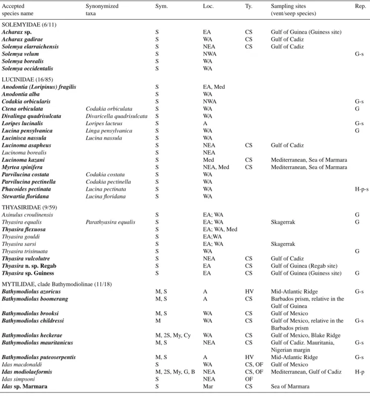
Table 1. Summary of chemosymbiotic bivalve species for which data are available regarding symbiosis in the Atlantic, Gulf of Mexico, and
Mediterranean Sea. Numbers in parentheses indicate the number of species for which symbiosis is documented using electron microscopy and molecular data, possibly including in situ hybridization of symbionts (bold). Synonymized taxa, according to the World Register of Marine Species (WORMS, www.marinespecies.org), are included as many former names were in use when symbioses were initially documented, and thus appear in the tree (Fig. 2). Species within genus “Pliocardia” were recently ascribed to undetermined genus (Audzijonyte et al., 2012). Symbiont (Sym.) types: sulfide (S)-oxidizer, methane (M)-oxidizer, (My) methylotrophs, Bacteroidetes (B), Cycloclasticus(Cy)-related and (G) for Gammaproteobacteria unrelated to other documented symbiont groups. Location (Loc.) corresponds to north (N), south (S), east (E), west (W) Atlantic (A), and Mediterranean (Med). Type (Ty.) of chemosynthesis-based ecosystem is mentioned for deep-sea species, (CS) cold seeps, (HV) hydrothermal vents, and (OF) organic falls. A non-exhaustive list of sampling sites is provided for vent and seep species (vent and seep sites illustrated in Fig. 1). Reproduction (Rep.), when documented, is either gonochoric (G) or hermaphrodite (H)(-p then means protandric), and can be seasonal (-s). For references, see text.
Accepted Synonymized Sym. Loc. Ty. Sampling sites Rep.
species name taxa (vent/seep species)
SOLEMYIDAE (6/11)
Acharax sp. S EA CS Gulf of Guinea (Guiness site)
Acharax gadirae S WA CS Gulf of Cadiz
Solemya elarraichensis S NEA CS Gulf of Cadiz
Solemya velum S NWA G-s
Solemya borealis S WA
Solemya occidentalis S WA
LUCINIDAE (16/85)
Anodontia (Loripinus) fragilis S EA, Med
Anodontia alba S WA
Codakia orbicularis S NWA G-s
Ctena orbiculata Codakia orbiculata S WA G
Divalinga quadrisulcata Divaricella quadrisulcata S WA
Loripes lucinalis Loripes lacteus S A G-s
Lucina pensylvanica Linga pensylvanica S WA G
Lucinisca nassula Lucina nassula S WA
Lucinoma asapheus S NEA CS Gulf of Cadiz
Lucinoma borealis S NEA
Lucinoma kazani S Med CS Mediterranean, Sea of Marmara
Myrtea spinifera S NEA, Med CS Mediterranean, Sea of Marmara
Parvilucina costata Codakia costata S WA
Parvilucina pectinella Codakia pectinella S WA
Phacoides pectinata Lucina pectinata S WA H-p-s
Stewartia floridana Lucina floridana S WA THYASIRIDAE (9/59)
Axinulus croulinensis S EA; WA G
Thyasira equalis Parathyasira equalis S EA; WA Skagerrak G
Thyasira flexuosa S EA; WA, Med
Thyasira gouldi S EA;WA
Thyasira sarsi S EA; WA Skagerrak
Thyasira trisinuata S WA G
Thyasira vulcolutre S NEA CS Gulf of Cadiz
Thyasira n. sp. Regab S EA CS Gulf of Guinea (Regab site)
Thyasira sp. Guiness S EA CS Gulf of Guinea (Guiness site) G MYTILIDAE, clade Bathymodiolinae (11/18)
Bathymodiolus azoricus M, S A HV Mid-Atlantic Ridge G-s
Bathymodiolus boomerang M, S A CS Barbados prism, relative in the Gulf of Guinea
Bathymodiolus brooksi M, S WA CS Gulf of Mexico
Bathymodiolus childressi M WA CS Gulf of Mexico, relative in the G-s Barbados prism
Bathymodiolus heckerae M, 2S, My, Cy WA CS Gulf of Mexico, Blake Ridge
Bathymodiolus mauritanicus M, S NEA CS Gulf of Cadiz. Mauritania, G-s Nigerian margin
Bathymodiolus puteoserpentis M, S A HV Mid-Atlantic Ridge G-s
Idas macdonaldi S WA CS, OF Gulf of Mexico
Idas modiolaeformis M, 2S, My, G, B NEA CS, OF Mediterranean, Gulf of Cadiz H-p
Idas simpsoni S NEA OF
Table 1. Continued.
Accepted Synonymized Sym. Loc. Ty. Sampling sites Rep.
species name taxa (vent/seep species)
VESICOMYIDAE (11/29)
Abyssogena southwardae S A HV, CS Barbados prism, Florida,
Mid-Atlantic Ridge, Canary Islands
Calyptogena valdiviae Vesicomya valdiviae; V. longa S EA CS Gulf of Guinea G
Christineconcha regab Calyptogena regab S EA CS Gulf of Guinea, Bay of Biscay
Elenaconcha guiness S EA CS Gulf of Guinea
Isorropodon bigoti S EA CS Gulf of Guinea G
Isorropodon megadesmus S NEA CS Gulf of Cadiz
Isorropodon perplexum S Med CS Mediterranean, Sea of Marmara
Laubiericoncha chuni Vesicomya chuni S EA CS Gulf of Guinea Laubiericoncha myriamae S NA CS Barbados prism
“Pliocardia” cordata Vesicomya cordata S WA CS Gulf of Mexico
“Pliocardia” ponderosa Calyptogena ponderosa S WA CS Gulf of Mexico; Colombia
2 Chemosymbiotic bivalves and their symbioses
2.1 Mytilidae
One clade within the family Mytilidae has successfully col-onized the deep sea, in habitats in which methane, reduced sulfur species or hydrogen is available as energy sources. These habitats include cold seeps, hydrothermal vents, or organic falls such as cetacean carcasses or sunken wood. This deep-sea mussel clade is often referred to as the sub-family Bathymodiolinae typified by the genus Bathymodio-lus Kenk and Wilson (1985), a large vent and seep mussel genus (Kenk and Wilson, 1985). In reality, several genera of smaller mussels such as Idas, Adipicola (formerly Myrina), Benthomodiolus, and Tamu, some of which were described long before the discovery of Bathymodiolus and chemosyn-thetic ecosystems, also form part of the clade. This is de-spite the fact that they have been historically included in the subfamily Modiolinae, along with Modiolus modiolus, a species often used as an out-group in deep-sea mussel phy-logenies (Distel et al., 2000; Jones et al., 2006; Lorion et al., 2010; Samadi et al., 2007). Notably, deep-sea mytilids are al-most absent from the background fauna, except for few small species within genus Dacrydium, a group in which symbio-sis has not been investigated (Duperron, 2010; Salas and Go-fas, 1997). Contrary to most other chemosymbiotic bivalves discussed in this paper, mussels are epibenthic, and occur at-tached to hard substrates including carbonates, basalts, tubes of large tube worms (Annelida: Siboglinidae) or shells of other specimens, via a byssus. Exceptions include Bathy-modiolus boomerang, which lives partly buried in the sedi-ment at the Barbados seeps (Cosel and Olu, 1998; Duperron, 2010). These mussels occur mostly in areas of diffuse fluid flow, and avoid the hottest or most anoxic niches (Fisher et al., 1988, Desbruy`eres et al, 2000). The genera themselves (e.g., Bathymodiolus, Idas, or Adipicola) remain poorly de-fined based on morphological characteristics, and are clearly non-monophyletic (Carney et al., 2006; Jones et al., 2006;
Lorion et al., 2010). Cosel (2002) re-evaluated most of the species of Bathymodiolus, based upon morphological char-acteristics of the shells and soft parts, and suggested a di-vision into four groups (Bathymodiolus thermophilus group, B. brevior group, B. heckerae/boomerang group and the B. childressi group) (Cosel, 2002). Keeping in mind the above limitations, several common features are conserved within the group, such as the overall modioliform shape, the gross anatomy, the presence of often reduced digestive tract and labial palps, and of hypertrophied gills in most species (re-viewed in Duperron, 2010). Nevertheless the most striking shared feature is the presence of bacterial symbionts associ-ated with epithelial cells in the lateral zone of gill filaments in all species. A possible exception is Idas argenteus pre-viously collected from woods in the North Atlantic, which feed on larvae of wood-boring bivalves as evidenced by the presence of remnants in their gut (Ockelmann and Dinesen, 2011). However, I. argenteus may not belong to the Bathy-modiolinae.
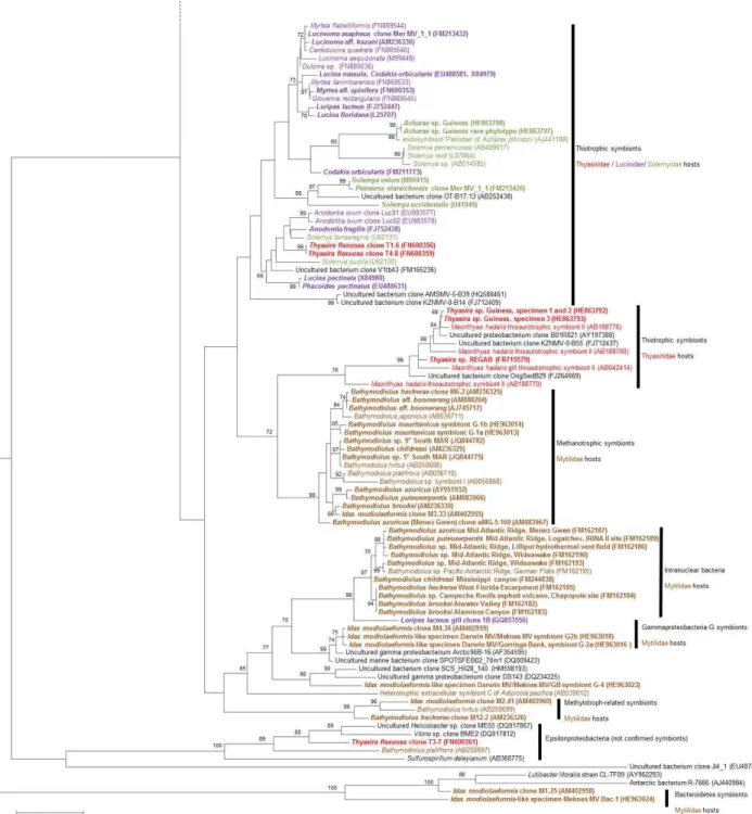
or outside the cells, within a layer of microvilli, depend-ing on the host species (Duperron et al., 2008b, 2009; Gros and Gaill, 2007; Lorion et al., 2009; Miyazaki et al., 2010). They oxidize reduced sulfur compounds (sulfide, thiosulfate) and use the energy for carbon fixation via the Calvin cy-cle, using a Type I RubisCO (Fisher et al., 1988; Nelson et al., 1995; Pimenov et al., 2002). Recently, mussel-associated thiotrophs were shown to use hydrogen as an alternative en-ergy source not only at the hydrogen-enriched ultramafic vents of Logatchev, but also at the hydrogen-poor southern MAR (5◦S and 9◦S), based on the presence of
hydrogenase-encoding genes. Pacific–Antarctic Ridge mussel symbionts also displayed the gene, but the gene was not amplified from Gulf of Mexico seep mytilids, suggesting this ability is not always present (Petersen et al., 2011). The methane-oxidizers are related to free-living Type I methanotrophs; they are intracellular and larger than thiotrophs, and dis-play Type I-typical stacked internal membranes within their cytoplasm (Cavanaugh et al., 1987). They possess methane monooxygenase, an enzyme found only in methanotrophs, and use methane as both a carbon and energy source (Cary et al., 1988; Childress et al., 1986; Duperron et al., 2007b; Pernthaler and Amann, 2004). Evidence on the occurrence of a RubisCO gene, and thus possibly an alternative au-totrophic pathway of carbon assimilation, was presented in a species from the Pacific Ocean (Elsaied et al., 2006). Re-cently, genes enabling the use of aromatic compounds were identified in a symbiotic lineage related to Cycloclasticus, present in the gills of Bathymodiolus heckerae specimens from an asphalt seep in the Gulf of Mexico (Raggi et al., 2012). The metabolism of other symbionts (methylotrophs, Gammaproteobacteria G and Bacteroidetes) has not yet been investigated, and hypotheses are based on weak phyloge-netic inferences (Duperron et al., 2007b, 2008a). For exam-ple methylotrophs in B. heckerae were hypothesized to take up methanol from nearby methane-oxidizers.
Within the area considered in the present review, large Bathymodiolus are reported from cold seeps in the Gulf of Mexico (B. childressi, B. brooksi, B. heckerae), the Barba-dos accretionary prism (B. boomerang, B. sp. B related to B. childressi), the Blake Ridge diapir (B. heckerae), the Mid-Atlantic Ridge hydrothermal vents (B. azoricus, B. puteoser-pentis, and relatives of uncertain species status at sites 5◦S
and 9◦S, displaying distinct COI haplotypes closely related
to B. azoricus). Bathymodiolus are also reported from seeps off Mauritania and the Gulf of Cadiz (B. mauritanicus), off Nigeria (B. aff. mauritanicus andB. aff. boomerang, with no detail regarding symbiosis), and at deep seeps in the Gulf of Guinea (B. aff. boomerang, Fig. 1). They are not yet doc-umented from the Mediterranean. Interestingly two groups of closely related species, B. boomerang/B. heckerae and B. mauritanicus/B. sp. B/ B. childressi, display an amphi-Atlantic distribution, which led authors to postulate a dis-persal route following the Atlantic equatorial belt (the AEB hypothesis discussed in Cordes et al., 2007; G´enio et al.,
2008; Olu et al., 2010; Olu-LeRoy et al., 2007b (Fig. 1)). Speciation could also have occurred along this belt. All but B. childressi, which has only methanotrophs, harbor multi-ple symbionts, with simultaneous occurrence of one sulfur-and one methane-oxidizer. B. heckerae specimens also have a second distinct sulfur-oxidizer, a methylotroph-related sym-biont in the northern GoM, and hydrocarbon-degrading bac-teria in the southern GoM (DeChaine et al., 2006; Distel et al., 1995; Duperron et al., 2005, 2007b; Fisher et al., 1993, Raggi et al., 2012) (Fig. 2). In fact, all Bathymodiolus species documented to harbor two or more different symbionts with distinct metabolisms are documented from seeps and vents in the Atlantic and Gulf of Mexico. Multiple symbioses are not yet documented in Bathymodiolus species from other oceans. Multiple symbiosis is a common feature of mussels within a clade that includes B. azoricus, the two mytilids from 5◦S
and 9◦S MAR, B. puteoserpentis, B. heckerae, B. boomerang
concentration in bottom waters (Duperron et al., 2011). Sta-ble isotope signatures correlated with these measurements, suggesting that specimens with more methane-oxidizers de-rived a substantially higher fraction of their carbon from methane, and a lower fraction of their sulfur from sulfide. This confirms that multiple symbioses are highly adaptable to changing habitats, both in time and space, optimizing the use of resources. In Bathymodiolus, heterotrophy plays a lim-ited role, but a fraction of carbon is derived from particulate and dissolved organic matter at shallower sites, as seen for B. azoricus at the 800 m deep Menez Gwen site (Riou et al., 2010), or during early life (Martins et al., 2008).
Symbioses in smaller mytilids from the Atlantic, Gulf of Mexico and Mediterranean have been far less documented. Tamu fisheri, which inhabits the base of Lamellibrachia luymesi (Annelida: Siboglinidae) aggregations and beds of B. childressi, and Idas simpsoni from the North Sea both harbor extracellular sulfur-oxidizing bacteria associated with their gill epithelial cells. This observation was based on electron microscopy data, but no molecular characterization is available yet (Southward, 2008). Conversely, the sulfur-oxidizing bacteria associated with Idas macdonaldi, occur-ring at 650 m on the Louisiana Slope, were characterized by 16S rRNA gene sequencing only (Won et al., 2008). Interest-ingly, this species has a well-developed digestive system in comparison with others from the group, suggesting mixotro-phy (Gustafson et al., 1998). Two Idas species were investi-gated in more detail, combining molecular and microscopic confirmative approach data. The first is an unnamed species recovered in the Sea of Marmara, tentatively ascribed to the genus Idas and labeled Idas sp. Marmara. The second is Idas modiolaeformis, a species identified in the eastern Mediter-ranean cold seeps with very close relatives recently sampled and investigated in the northeast Atlantic (Duperron et al., 2008a; Lorion et al., 2012; Ritt et al., 2012; Rodrigues et al., 2013). Despite its apparent morphological resemblance with Idas modiolaeformis, with similar habitat and depth range, Idas sp. Marmara is at best distantly related to other Idas lineages and branches quite far from other Bathymodioli-nae based on COI analysis (17 % divergence from any other mussel). Idas sp. Marmara hosts dense populations of sulfur-oxidizing bacteria in its gill epithelial cells. Idas modiolae-formis was initially described in the late 19th century, and was re-sampled only recently (Olu-LeRoy et al., 2004). It can harbor 6 distinct bacterial 16S rRNA phylotypes in its gills, including two sulfur- and one methane-oxidizers, one methylotroph, one Bacteroidetes and one belonging to the “Gammaproteobacteria G” clade (Duperron et al., 2008a) (Fig. 2). Depending on sampling years or substrates, either sulfur- or methane-oxidizers dominate in the gills, but their abundance in relation to the local geochemistry was not in-vestigated (Lorion et al., 2012). The species displays protan-dric hermaphrodism with possible gender transitions during adult life, and symbionts are absent from the gonad tissue, which support environmental acquisition later during
devel-opment or after larval settlement. The larval shell (Fig. 3f) suggests planktotrophic larvae with some dispersal capabili-ties (Gaudron et al., 2012). Idas modiolaeformis is the sister species of Idas macdonaldi known from the Gulf of Mex-ico and possibly diverged around 1.84 Mya. This amphi-Atlantic distribution led to the prediction that populations of mussels branching within the I. modiolaeformis/I. mac-donaldi clade should occur at locations in between the east-ern Mediterranean and the Gulf of Mexico. Recently, small Idas-like mussels were indeed recovered from the northeast Atlantic on organic substrates at mud volcanoes in the Gulf of Cadiz (Darwin and Mekn`es MVs) and in the Gorringe Bank (Gettysburg Seamount) (Rodrigues et al., 2013). The studied specimens display COI sequences identical (Gor-ringe) or almost identical (Mekn`es, Darwin MVs) to the specimens from the eastern Mediterranean, suggesting they belong to the same species. Possible settlement is also re-ported on wood block colonization experiments deployed in the western Mediterranean canyons at 500 m deep (personal observation, N. Le Bris, personal communication, 2012). In-terestingly, molecular and microscopic data suggest the ab-sence of methane-oxidizing symbionts. Furthermore, speci-mens from the Mekn`es and Gorringe sites do not seem to have sulfur-oxidizers either. Identified bacteria either belong to the “Gammaproteobacteria G” group, present in I. modi-olaeformis, or to a clade unrelated to any known symbiont (clone G-4), or to the Bacteroidetes. Overall, symbioses in the I. macdonaldi/I. modiolaeformis clade appear unexpect-edly variable. This could be linked with the wide range of habitats colonized, which include alfalfa and wood coloniza-tion devices, natural wood falls, carbonates close to reduced sediment, and siboglinid tubes. The plasticity of the I. mac-donaldi/I. modiolaeformis clade makes it a very good model to study at which stage of the life cycle (spawned oocytes, veliger stages, post-larvae, aposymbiotic juveniles or adult stages) and how symbionts establish, transmit and evolve over a relatively short evolutionary time scale. Other Idas species have been associated with reduced environments. In the Mediterranean, for example, I. simpsoni and I. cylindri-cus have been associated with organic falls (Pastorelli et al., 1999; Pelorce and Poutiers 2009). Another unidentified Idas was found on organic cargo in the wrecked ship Franc¸ois Vieljeux (Dando et al., 1992).
2.2 Vesicomyidae
The family Vesicomyidae consists of over 100 known species, distributed worldwide at depths from 100 to 9500 m (Baco et al., 1999; Cosel and Olu, 2009; Krylova and Sahling, 2010). As in other families of chemosymbiotic bi-valves, currently applied genera are under intense discus-sion (Krylova and Sahling, 2010, Audzijonyte et al., 2012). The subfamily Vesicomyinae, genus Vesicomya, consists of smaller species, up to 1 cm shell length, which inhabit the abyssal plain. In contrast, representatives of the Pliocardi-inae can reach up to 30 cm and colonize a variety of deep-sea reducing habitats including cold seeps, hydrothermal vents, and vertebrate carcasses (Krylova and Sahling, 2010). Most species are infaunal and possess siphons of varying length. They live with their anterior region buried into reducing sed-iment, although some vent species inhabit the cracks or small crevices on bare basalts. They thus bridge the oxic/anoxic in-terface (Cavanaugh et al., 2005). This way they can access both oxygen and dissolved carbon, as well as reduced com-pounds that seep from the subsurface or are concentrated within the underlying sediment layers that they can reach using their vermiform foot (Childress and Mickel, 1982). Characteristic features of the Pliocardiinae are a medium-to-large white shell, reduced labial palps, simple gut (Fiala-M´edioni and Le Pennec, 1987), sub-filamental tissue in the gills (Krylova and Sahling, 2010), large and thick gills, and the presence of very dense intracellular sulfur-oxidizing au-totrophic Gammaproteobacteria located within gill epithe-lial bacteriocytes. Certain species of Vesicomyidae possess hemoglobin molecules capable of transporting hydrogen sul-fide via the hemolymph, from the foot to the gill sym-bionts (Childress et al., 1993). Oxygen is acquired directly by the gill and in some species transported by circulating hemoglobin (Terwilliger et al., 1983). In some species, the gills are also organized in tubes or channels (Le Pennec et al., 1988). Several species can co-occur at a given seep or vent site, sometimes within a single clam aggregate, as observed in the Gulf of Guinea with Christineconcha re-gab and Laubiericoncha chuni (Decker et al., 2012; Krylova and Cosel, 2011). Physiological differences between some species could account for slight ecological niche differen-tiation and prevent competition (Goffredi and Barry, 2002; Decker at al., 2013).
Symbionts associated with the Vesicomyidae are related to the sulfur-oxidizing symbionts of the Mytilidae and sponges, as well as to various free-living bacteria (Fig. 2). They use reduced sulfur as an energy source and fix carbon through the Calvin cycle, but contrary to mussel symbionts, they employ a Type II instead of Type I RubisCO (Robinson and Cavanaugh, 1995). Vesicomyid symbionts form a tight clade that displays a high homogeneity among 16S rRNA se-quences; the most dissimilar sequences differ by only 8.1 % (Rodrigues et al., 2012), despite the fact that the family is quite ancient (estimates varying from 100 Mya based on the
fossil record to 22–44 Mya with molecular estimated; Lit-tle and Vrijenhoek, 2003). Symbionts are transmitted di-rectly from mother to offspring as evidenced by polymerase chain reaction (PCR) and in situ hybridization (ISH) tests us-ing symbiont-specific primers and probes (Endow and Ohta, 1990; Krueger et al., 1996b). Host and symbiont phylogenies can be superimposed, suggesting rather strict co-speciation (Peek et al., 1998a, b). However, a limited amount of lat-eral transfer does exist and may permit gene exchanges be-tween symbiont lineages (Stewart et al., 2008, 2009b; Decker et al., 2013). To date, the Vesicomyidae are the only deep-sea chemosymbiotic metazoans for which symbiont genomes are fully sequenced – namely Candidatus “Vesicomyoso-cius okutanii” and Candidatus “Ruthia magnifica”, in two species from the Pacific (Kuwahara et al., 2007; Newton et al., 2007). Genome sequencing indicates the loss of several genes and strong genome reduction compared to free-living relatives. This trait, shared with many symbionts in insect, is characteristic of maternally inherited bacterial symbionts lacking a free-living stage, and is thus congruent with pre-vious findings (Gil et al., 2004; Wernegreen et al., 2003). In Cand. “Vesicomyosocius okutanii”, the loss of the ftsZ gene involved in bacterial division suggests, for example, strong host control over the symbiont cycle (Kuwahara et al., 2007). Significant variations also exist among the metabolisms of the closely related symbionts. Cand. Ruthia magnifica does not harbor the membrane-bound nitrate reductase present in Cand. Vesicomyosocius okutanii, suggesting that only the latter is able to respire nitrate (Kleiner et al., 2012). An-other consequence of genome alteration is that the existence of active free-living forms of vesicomyid symbionts is ques-tionable. Although the issue is not settled, no environmental sequence from any active bacterium yet clusters inside the group. The only exception is the symbiont sequence from a thyasirid, Thyasira vulcolutre, which was recently shown to cluster within this group, suggesting a host shift (Fig. 2) (Ro-drigues and Duperron, 2011).
Despite the fact that approximately 29 Vesicomyidae species are documented in the Atlantic and Mediterranean Sea, symbiosis has been investigated in only 11 species (Ta-ble 1). Among these is Abyssogena southwardae, which oc-curs at seeps on the west coast of Florida and at the Bar-bados prism, with large populations on very deep mud vol-canoes. At vent sites Logatchev 5◦S and 9◦S, old shells of
suggest that species should be included in distinct genera (Decker et al., 2012). Although ultrastructural evidence ex-ists for sulfur-oxidizing symbiosis in L. myriamae, no molec-ular data have been released regarding symbionts (Olu et al., 1996). In both A. southwardae and L. myriamae, genetic data regarding symbiosis are not available for specimens from the eastern Atlantic. Another species, Pliocardia atalantae (for-merly Isorropodon atalantae), has been documented from the Gulf of Guinea to the Mid-Atlantic Ridge, but no data are available regarding symbiosis.
In the Gulf of Mexico, vesicomyids are represented by Pliocardia ponderosa (formerly Calyptogena ponderosa) and Pliocardia cordata (formerly Vesicomya cordata) (Ta-ble 1). They both display closely related sulfur-oxidizing symbionts (Fig. 2) (Brooks et al., 1987; Distel et al., 1994; Stewart et al., 2009b) and an abundance of hemoglobin (Scott and Fisher, 1995). Species from seeps in the Gulf of Guinea have recently received attention. These include Elenacon-cha guiness, Isorropodon bigoti and Calyptogena valdiviae. E. guiness was found to display a very typical vesicomyid symbiosis, as documented in many other species around the world (Duperron et al., 2012). This species displays a single symbiont 16S rRNA phylotype, with high similarity to other vesicomyid-associated symbiont sequences available in Gen-bank, and sulfur metabolism is supported by the presence of the gene encoding APS (adenylyl-sulfate) reductase and by carbon stable isotope values in the range of those reported for seep vesicomyids (Olu et al., 2009). With regards to connec-tivity, small clams of the genus Isorropodon yielded interest-ing findinterest-ings. Specimens of the three species I. perplexum, I. megadesmus and I. bigoti, collected respectively in the eastern Mediterranean, Gulf of Cadiz and Gulf of Guinea, are closely related, forming a distinct clade, based on COI phylogeny (Cosel and Salas, 2001; Rodrigues et al., 2012). Their symbionts also appear as close relatives (Fig. 2), and the association is very similar to that documented in larger clams. The genus Isorropodon consists of various species colonizing a wide range of depths (150–6800 m), and could represent a suitable case study into the barriers influencing the biogeography and evolution of vesicomyid symbioses, as demonstrated by the recent discovery of I. perplexum in the Sea of Marmara (Ritt et al., 2012). Finding closely related symbionts in closely related species is somewhat expected, although instances of non-parental acquisition are reported (Stewart et al., 2008, 2009b; Decker et al., 2013). The ho-mogeneity of symbiont populations within a single species was also questioned recently by the finding of distinct bac-terial 16S rRNA lineages in distinct specimens of Calypto-gena valdiviae (Fig. 2). Overall, new data about symbioses in the Atlantic Vesicomyidae further support the existence of a certain level of symbiont heterogeneity within species, and of environmental acquisition of non-parental symbiont strains in co-occurring host species, which can potentially lead to symbiont co-occurrence, displacement, or genetic re-combination among symbionts. These phenomena might be
of great significance for the evolution of vesicomyid sym-bioses.
2.3 Solemyidae
All documented Solemyidae live in obligate symbiosis with sulfur-oxidizing Gammaproteobacteria. They make U- or Y-shaped burrows that allow access to both the bottom sea-water, rich in oxygen, and sediment pore-waters that con-tain reduced sulfur (Fisher, 1990; Stewart and Cavanaugh, 2006). Due to their infaunal lifestyle, deep-sea Solemyidae are rarely sampled in great numbers, which limits the pos-sibility to investigate them in detail. Most data were ob-tained from coastal species. Although a weak suspension-feeding capability is retained, dependency upon symbiotic nutrition is maximal, as shown by the drastic reduction (Sole-mya velum), or even absence (Sole(Sole-mya reidi) of the diges-tive system and reduction in labial palps. Authors estimated that more than 97 % of the carbon is derived from symbionts (Conway et al., 1989; Krueger et al., 1992). Protobranch gills account for more than 35 % of total weight, with a greater surface area to volume ratio than those documented for other bivalves (Scott, 2005). Symbionts are located inside bacteri-ocytes of the gill epithelium, which alternate with symbiont-free intercalary cells. The symbiont cycle may be complex as bacterial shape is variable from coccoid- to rod-shaped, and dividing stages are rarely seen, suggesting strong host con-trol. Carbon is fixed via the Calvin cycle (Cavanaugh, 1983; Cavanaugh et al., 1988), using energy derived from the oxi-dation of reduced sulfur, most likely through the APS path-way for which they possess enzymes (Kleiner et al., 2012; Stewart and Cavanaugh, 2006). Ammonia is the nitrogen source assimilated via a host-encoded glutamine synthetase (Lee et al., 1999). Vertical transmission of the symbionts is documented in the genus Solemya based on amplification of symbiont DNA from ovaria, eggs and larvae, although con-firmation with in situ hybridizations was not obtained from oocytes and eggs (Cary, 1994; Krueger et al., 1996b). In con-trast to the Vesicomyidae, host–symbiont co-speciation is not observed.
al., 2009a). This could either be due to the limited resolu-tion of the host marker gene, or a consequence of the lat-eral acquisition of locally adapted bacteria. Enzymatic and ultrastructural data also exist for S. borealis, another small species (Conway et al., 1992). Several species have been documented at deeper depths in the area. In the Mediter-ranean, a single specimen of an undetermined Solemya is for example the only living Solemyidae species reported in the deep Mediterranean (Rodrigues et al., 2011). It was recov-ered from a soft sediment core close to a cold seep area of the Nile deep sea fan, at a depth of 1697 m. Unfortunately, no data are available regarding symbiosis, despite attempts to amplify symbiont genes. Acharax gadirae and Solemya (Petrasma) elarraichensis have been documented from deep (Yuma, Ginsburg, Jesus Baraza, Captain Arutyunov, Carlos Ribeiro, Porta MVs, 960–3902 m depth) and shallower MVs (Mercator, Mekn`es, Gemini, Kidd, Yuma, Ginsburg, Dar-win, 358–1105 m depth) respectively in the Gulf of Cadiz (Oliver et al., 2011). Another member of the genus Acharax, not yet assigned to a species, was collected at the Guiness site (580 m depth) in the Gulf of Guinea (Duperron et al., 2012). Sequences from symbionts of the two Acharax species (GoC and GoG) are almost identical to sequences of several Acharax from the Oregon, Pakistan and Indonesia margins at depths from 780 to 2940 m (Imhoff et al., 2003). S. elar-raichensis symbiont sequences are, on the other hand, closely related to those of the shallow Solemya velum (Eisen et al., 1992; Rodrigues et al., 2010). Acharax shells, but no living specimens, are documented from various locations including the Norway plateau or in the Barbados seeps (Ivanov et al., 2010; Olu et al., 1996). In addition several other species oc-cur in the area, though no information is available regarding their symbioses (Table 1).
Overall, Acharax-associated symbionts form a very tight cluster despite the geographical distance between collection sites. Meanwhile, Solemya symbionts are spread over at least three distinct clades of Gammaproteobacteria, related to Lu-cinidae and Thyasiridae symbionts (Fig. 2). This distinct clustering could reveal significant differences in the sym-bioses between the different host genera, and even among species within genus Solemya. For example, carbon fixation in symbionts of Solemya velum is carried out by a Type IA RubisCO, while a Type II RubisCO is present in Acharax sp. Guiness (Duperron et al., 2012; Robinson and Cavanaugh, 1995; Schwedock et al., 2004). Other differences could exist besides the type of RubisCO.
Additional 16S rRNA sequences were recovered besides that of gammaproteobacterial symbionts in Solemya elar-raichensis and Acharax gadirae branching within the Ep-silonproteobacteria, Betaproteobacteria, Chlamydiae, Firmi-cutes, and Actinobacteria. Their status as symbionts or pathogens was not resolved, but at least some might have been contaminants. In the Sea of Oman, a family related to Solemyidae, the Nucinellidae, was recently shown to display chemosynthetic sulfur-oxidizing bacteria in their gills based
on detailed electron microscopy approaches, but without sup-porting molecular data (Oliver and Taylor, 2012).
2.4 Thyasiridae
Around 100 Thyasiridae species are described worldwide (59 in the area reviewed herein). They occupy various habi-tats from intertidal to hadal waters, including the deepest reported chemosymbiotic species, Maorithyas hadalis (Fuji-wara et al., 2001). Thyasiridae burrow into suboxic to anoxic sediment, in particular in habitats rich in hydrocarbons or fluids (cold seeps, hydrothermal vents) and in oxygen min-imum zones. They also occur in organic-enriched habitats such as sunflower seeds in a wrecked ship off Vigo (Spinax-inus sentosus; Oliver and Holmes, 2006), colonizing artifi-cial wood substrates in the eastern Mediterranean close to seeps (Thyasira sp.; Gaudron et al., 2010), and in canyons under reducing and organic-enriched conditions (Cunha et al., 2011). As in the Vesicomyidae and Bathymodiolinae, genera are poorly defined, leading to frequent misidentifica-tions (Taylor et al., 2007). Contrary to other bivalve groups discussed here, not all members of the Thyasiridae harbor bacterial symbionts. In fact, several degrees of association, ranging from the absence of symbionts to the occurrence of very dense bacterial populations in the lateral zone of gill filaments, are documented (Dufour, 2005). This illustrates differing degrees of dependence upon symbiont-based nutri-tion (Dando and Spiro, 1993), and suggests that symbioses appeared several times during the evolution of the family. Species with symbionts harbor bacteria in large cells located in the gill epithelium. The bacteria are tightly packed into a large vacuole that is devoid of host cell cytoplasm (Fig. 3a) and separated from the outside by a net-like structure that may consist of modified microvilli (Dufour, 2005). These species burrow deeper than non-symbiotic species and use their super-extensile foot to mine for sulfide-rich pockets in the sediment, creating a network of tunnels (Dufour and Fel-beck, 2003). Some species such as Thyasira flexuosa and T. gouldi lack siphons and maintain communication with the surface seawater by a long tube made of mucus rings created by the foot (Blacknell, 1973). Symbiont chemoautotrophy is supported by the occurrence of APS reductase-encoding genes in symbionts of some species (Rodrigues and Duper-ron, 2011), and by the carbon stable isotope signatures of animal tissue that are in the range of values measured in chemosymbiotic metazoans harboring sulfur-oxidizing sym-bionts. Variability in symbiont abundances and the host nutri-tional strategy depends upon environmental conditions (pres-ence of sulfide and particles), as shown in Thyasira flexuosa, T. sarsi and Parathyasira equalis, confirming their ability to withstand fluctuating environments (Dando and Spiro, 1993; Dufour and Felbeck, 2006).
In addition, two unidentified species have been reported by Southward (1986) as having symbiotic associations, each with two distinct bacterial morphotypes, none containing sulfur vesicles. In most investigated species, only the ultra-structure was characterized. Thyasira (Parathyasira) equalis (Barents Sea) and Axinulus croulinensis (North Sea) harbor moderate amounts of extracellular symbionts, while T. tris-inuata (Florida), T. sarsi (White Sea, North Sea), T. flexu-osa (North Sea), and T. gouldi (Arctic region) harbor dense bacterial populations in their gills (Dufour, 2005). Available 16S rRNA sequences all belong to the Gammaproteobacteria and are related to free-living bacteria and sulfur-oxidizing symbionts of several metazoan groups (Fig. 2). In the area considered herein, these include T. flexuosa from Plymouth Sound (15 m depth), Thyasira aff. flexuosa from the Nile deep-sea fan cold seeps, Thyasira vulcolutre from the Car-los Ribeiro (2200 m depth) and Sagres (1562 m depth) MVs in the Gulf of Cadiz, Thyasira n. sp. from the Regab site in the Gulf of Guinea (3167 m depth), and Thyasira n. sp. Gui-ness from the GuiGui-ness site, at 580 m depth in the Gulf of Guinea (Brissac et al., 2011; Distel and Wood, 1992; Duper-ron et al., 2012; Rodrigues and DuperDuper-ron, 2011). Each of these species has a single dominant bacterial 16S rRNA phy-lotype (Fig. 2). A certain level of within-species strain het-erogeneity could exist, as shown by the co-occurrence of two highly similar yet distinct phylotypes in Thyasira n. sp. Gui-ness (Duperron et al., 2012). Interestingly, symbionts cluster in at least three clearly distinct clades within the Gammapro-teobacteria, four if we include Symbiont I of Maorithyas hadalis (Fig. 2). Sequences from T. flexuosa and Thyasira aff. flexuosa from the eastern Mediterranean cluster with various sequences from Lucinidae and Siboglinidae symbionts, and with environmental sequences. The symbiont of Thyasira sp. Guiness clusters with those of Thyasira n. sp. Regab and Maorithyas hadalis, close to environmental sequences from reducing habitats. Interestingly, the symbiont of T. vulcolutre clusters right within the clade of Vesicomyidae-associated symbionts, suggesting a recent host shift from Vesicomyi-dae to ThyasiriVesicomyi-dae (Rodrigues and Duperron, 2011). Because T. vulcolutre co-occurs with Vesicomyidae in the Gulf of Cadiz, host shift could have resulted from lateral acquisition. Symbiont phylogeny thus suggests multiple independent ori-gins of symbiotic bacteria associated with the Thyasiridae, possibly from a pool of environmental bacteria presenting features that allow them to establish interactions with meta-zoans (Imhoff et al., 2003; Rodrigues and Duperron, 2011). This observation agrees well with the hypothesis of the mul-tiple origin of symbiosis in Thyasiridae hosts. Thyasira flex-uosa, in particular, seems to be a species with a wide geo-graphical distribution (Fig. 1), occurring in the western At-lantic in Florida, in the eastern AtAt-lantic from Norway south to Galicia (Spain), and in the Mediterranean near the Iberian peninsula (Dufour, 2005; Southward, 1986). This could make T. flexuosa a good target species to investigate the relevance of the AEB hypothesis to Thyasiridae.
Occasionally, additional bacterial sequences are recov-ered, mostly Epsilonproteobacteria and Bacteroidetes, but fluorescence in situ hybridization has yet to support these as being significant symbionts (Brissac et al., 2011; Rodrigues and Duperron, 2011). Microscopy also indicates possible Spirochete-like morphotypes located extracellularly on the apex of some bacteriocytes. Interestingly, at least four stud-ies report the presence of dense virus-like inclusions within bacteria of Thyasira from four different locations: T. flexu-osa from Plymouth Sound (15 m depth) and T. gouldi from Loch Etive, Scotland (Southward and Southward, 1991), off Long Beach, Florida (Dufour, 2005), and Thyasira sp. at cold seeps in the eastern Mediterranean (central zone, site 2A, 1693 m depth) (Brissac et al., 2011). In all cases, do-decahedral inclusions occur only in some bacteriocytes, but are abundant when present. Brissac et al. (2011) indicated that these “infected” bacteriocytes displayed large lysosomal structures, possibly involved in the destruction of infected symbionts. Viral infection of symbionts is thus a recurrent feature of Thyasiridae symbioses. Another intriguing find-ing is the presence of peculiar gill cells displayfind-ing large and abundant mitochondria, unlike the situation in “normal” bi-valve gills (Southward, 1986). For example in Mendicula fer-ruginosa, which is devoid of symbionts, it has been hypoth-esized that these structures might be generating ATP by ox-idizing reduced compounds such as reduced iron, although this remains to be proven (P. R. Dando, personal communi-cation, 2013).
2.5 Lucinidae
Lucinidae were formerly classified along with Thyasiridae within the Lucinoidea, mainly because of shared morpholog-ical features. However, recent molecular evidence has shown that the families, although displaying convergent features, are not closely related (Taylor and Glover, 2000; Williams et al., 2004). Lucinidae are burrowing bivalves occurring over a wide range of depths, from coastal to at least 2500 m, and in latitudes from 60◦N to 55◦S. More than 400 (330
the functioning of lucinid symbioses originates from species in the Caribbean. In vivo experiments on Codakia orbicu-laris demonstrated symbiont absence in ovaries, testis, eggs, veliger larvae and metamorphosed juveniles reared in sterile sand (Gros et al., 1996, 1997). Symbiont-specific PCRs also failed to reveal bacterial DNA from the testis and ovaries, and TEM did not reveal bacterial shapes in Lucinoma ae-quizonata (Gros et al., 1999). Overall this supports environ-mental acquisition of symbionts after larval metamorphosis. Free-living forms of symbionts were subsequently shown to be abundant in the Thalassia testudinum seagrass envi-ronment and water (Gros et al., 2003b). The functioning of symbiosis is also intriguing. Bacteria replicate their genomic DNA but do not divide actively within hosts cells (Caro et al., 2007), and are not released by adult bivalves (Brissac et al., 2009), suggesting a strong host control. Further, symbiont reacquisition after starvation is not by within-host division of bacterial cells, but rather by capture of new bacteria courtesy of a life-long continuous ability to acquire symbionts (Gros et al., 2012). In oxygen-depleted environments, symbionts of some species can avoid competition with their host for oxy-gen resources by growing on nitrate (Duplessis et al., 2004). Carbon transfer would most likely involve the digestion of symbionts.
In the area considered, symbioses have been investigated in several coastal species, mainly from the Caribbean, near Guadeloupe and Martinique. These include Lucina pecti-nata, which lives in black reducing sediment of mangrove swamps, and Codakia orbiculata, C. pectinella, C. orbic-ularis, C. costata, and Linga pensylvanica, which live in Thalassia testudinum seagrass beds. All five seagrass bed species share a single 16S rRNA symbiont phylotype (Dis-tel et al., 1994; Durand et al., 1996). Anodontia alba (Bris-sac et al., 2011) and Divaricella quadrisulcata (Gros et al., 2003a), which live in reduced sediment near seagrass beds, also have identical symbionts to those of Codakia orbicu-laris. The symbiont from L. pectinata not only diverges from those of other lucinids, but the host tissue also displays a high concentration of haemoglobin, which suggests that the symbiosis might function in a different way (Frenkiel et al., 1996). Another species, L. nassula, occurs in seagrass beds near Florida and has a very similar symbiont sequence (Du-rand and Gros, 1996) Additionally, symbiosis was character-ized based on gill tissue ultrastructure or molecular evidence in a variety of other coastal reduced sediment or seagrass bed species including Myrtea spinifera, L. floridana, L. borealis, and Loripes lucinalis (synonymized with Loripes lacteus, for which symbiont 16S sequences are in GENBANK but un-published), with estimates that the latter could be responsi-ble for up to 16 % of the primary production observed in sea-grass bed habitats in a lagoon in upper Corsica (Dando et al., 1994, 1985, 1986; Distel et al., 1988; Johnson and Fernan-dez, 2001; Johnson et al., 2002).
Symbiosis has also been investigated in some deep-water species from the eastern Mediterranean Basin cold seeps.
Lu-cinoma aff. kazani occurs at depths between 500 and 1709 m in the Anaximander Mountains between Rhodes and Cyprus, the Olimpi area south of Greece on the Mediterranean Ridge, and the Nile deep-sea fan (North Alex MV and pockmarks in the Central area); it was also recently reported from the Sea of Marmara (Bayon et al., 2009; Olu-LeRoy et al., 2004; Ritt et al., 2010; Salas and Woodside, 2002). The second species, Myrtea sp., possibly M. amorpha described in Olu et al. (2004) and Ritt et al. (2010), is closely related to M. spinifera based on 18S and 28S gene sequences, and is la-beled Myrtea aff. spinifera in the tree (Fig. 2). It was sam-pled from the same areas as Lucinoma kazani, including the Sea of Marmara. Both L. aff. kazani and M. aff. spinifera har-bor sulfur-oxidizing bacteria in their gills (Fig. 3b), and the ultrastructure of the association, in terms of bacterial shape, presence of sulfur granules, and host tissue organization, re-sembles that of shallow water lucinids. Another deep-sea species with sulfur-oxidizing symbionts, L. asapheus, is doc-umented from Mercator MV in the Gulf of Cadiz (Rodrigues et al., 2010). In L. aff. kazani, a dominant bacterial endosym-biont was identified, and the presence of APS reductase- and RubisCO-encoding genes confirmed the chemoautotrophic potential of symbionts. Spirochete sequences were reported for L. aff. kazani and L. asapheus, but without further ev-idence they are unlikely to represent significant symbionts and were suggested as potential pathogens.
3 Comparing symbioses from North Atlantic, Gulf of Mexico and Mediterranean bivalves
3.1 Ecological trends in bivalve symbioses
Most chemosymbiotic bivalves associate with sulfur-oxidizing bacteria (Fig. 2). They usually live in habitats ex-isting within the oxic/anoxic interface, where animals can ac-quire oxygen from seawater as well as reduced sulfur com-pounds available in the underlying substrate (e.g., reduced sediment, decaying organic matter, seeping fluid, etc). In nor-mal deep-sea sediment settings, this oxic/anoxic interface does not exceed a few centimeters, or just a few millime-ters in undisturbed cold seep sediment with little-to-no mix-ing between the two layers (de Beer et al., 2006; Wenzhofer and Glud, 2002). The “feeding” behavior of chemosymbi-otic burrowing-bivalves, made possible by their morphol-ogy (long siphons, extensile foot), artificially extends this oxic layer’s depth, increasing access to reduced compounds while maintaining oxygenated conditions (Cavanaugh et al., 2005). Symbiotic Mytilidae are an exception not only be-cause they can have additional symbionts, notably methan-otrophs (Fig. 2), but also because they are epibenthic and usually do not burrow into the substrate. Thus, in order to maintain fluid supply to gill-inhabiting symbionts, they re-quire actively emitted, reduced compounds mixing with oxy-genated seawater. This might explain their success at many vents and seeps that are characterized by active venting, and organic falls that emit reduced compounds, and also their ab-sence at some other seeps and among background fauna. In contrast, other families have some representatives in reduced sediments away from active seeping sites (deep-sea or even coastal sediments).
Lucinidae and Vesicomyidae are all associated with sulfur-oxidizing symbionts, whatever their habitat or depth, al-though direct evidence is still lacking for the genus Vesi-comya sensu Krylova (Decker et al., 2012; Krylova and Sahling, 2010; Taylor and Glover, 2000). Symbiont phy-logeny indicates the absence of depth- or habitat-related clades (Fig. 2). By contrast, within Vesicomyidae hosts, re-cent phylogenies suggest recurrent events of “stepwise spe-ciation” from shallow to deep waters in different ocean basins, consistent with narrow vertical ranges for most of the vesicomyid genera (Decker et al., 2012; Krylova and Sahling, 2010). All Solemyidae also associate with sulfur-oxidizers, but the genus Acharax, which occurs at deeper sites than Solemya, is associated with a different clade of sulfur-oxidizing bacteria than members of the shallower genus (Imhoff et al., 2003). It is not known whether this difference is linked with host genus or with distinct depth ranges. In the Thyasiridae (though not all species have sym-bionts), symbiosis does exist in both shallow and deep-sea species, and in the various types of habitats colonized by this family. There are also non-symbiotic species in all habi-tats and at all depths (Dufour, 2005). Southward (1986) for
example demonstrated that individuals of Thyasira ferrug-inea found at 1500 m did not harbor symbionts, and Decker and Olu (2012) did not find evidence for a major input of symbionts in the nutrition of the host based on stable iso-tope analysis. In the Mytilidae, multiple symbioses involv-ing 2 to 6 bacterial types occur in the area considered in this study, and this might be peculiar to the area, as species from other regions have either sulfur- or methane-oxidizers only. Yet, multiple symbioses occur both at seeps and vents, and methanotrophs mostly at methane-rich sites (Duperron et al., 2009). Most mussels associated with organic falls harbor thiotrophic symbionts, but some may have methan-otrophs (identified on some Idas modiolaeformis specimens) or other symbiont types such as Gammaproteobacteria G, and may not have thiotrophs (C. Rodrigues, personal obser-vation, 2013). Bacterial densities can be rather low, suggest-ing that additional nutritional sources are probably signifi-cant. Overall depth or habitat type does not strongly influence the diversity of symbioses except in Mytilidae, in which the presence of methanotrophs is clearly linked with methane-enriched habitats, or by-products of wood degradation such as methanol.
Symbiont abundances on the other hand are clearly influ-enced by local characteristics of the habitat. This has been demonstrated in vent and seep Mytilidae, in which relative amounts of sulfur- versus methane-oxidizing bacteria reflect the availability of their respective substrates, and can also display age- or time-related variations (Le Bris and Duper-ron, 2010; Fiala-M´edioni et al., 2002; Halary et al., 2008; Riou et al., 2008). Although they only have sulfur-oxidizing bacteria, Thyasiridae can also display inter-habitat variabil-ity in symbiont densities, with higher densities when sul-fide is more abundant in the environment (Dufour and Fel-beck, 2006). Using carbon stable isotopes ratios, Dando and Spiro (1993) have shown that the contribution of chemoau-totrophic bacteria could vary inter-annually in Thyasira sarsi and T. equalis, in relation to environmental change in the habitat. In some specimens, carbon signatures of these sym-biotic thyasirids were identical to those of non-symsym-biotic bivalves in which the diet was based upon phytoplankton-derived material. This was interpreted as a consequence of mixotrophy with animals that derive distinct fractions of their diet from symbionts and from heterotrophy.
3.2 Reproduction, development and dispersal strategies of hosts
Fig. 2. Phylogeny of the Gammaproteobacteria associated with bivalve hosts based on analysis of 16S rRNA-encoding genes.
Epsilonpro-teobacteria presented include two sequences obtained from animal tissues, but not confirmed as actual symbionts, contrary to the Mytilidae-associated Bacteroidetes. Colors relate to the family to which the host belongs; sequences in bold are from species present in the area of focus for the review. For clarity, only host species name is mentioned without mention of “symbiont”. Properly identified bivalve-associated 16S rRNA sequences above 1200 bp were used; short sequences available for Bathymodiolus tangaroa, Acharax gadirae and Idas macdonaldi were not included. Tree was reconstructed using a maximum likelihood algorithm from 1129 nucleotide positions. Based on Bayesian infor-mation criterion, a Tamura 3-parameter with Gamma-distribution of rates (T92 + G) was employed. Node support values (>60 % shown) are
(Fig. 3c), indicative of lecithotrophic larval development and potentially enabling a significant larval dispersal under olig-otrophic conditions in the surrounding deep sea (Beninger and Le Pennec, 1997). Alternatively, oocytes can be smaller with a lower vitellus content (Fig. 3d), indicative of plank-totrophy where the veliger larvae will actively feed upon dissolved organic matter, phytoplankton, or microorganisms (Arellano and Young, 2009). Historically, planktotrophic lar-vae have been thought to spend an extended period in the plankton and disperse great distances, while lecithotrophic larvae live on limited reserves of vitellus, which permit lim-ited dispersal time only before metamorphosis and settlement (Thorson, 1950, Fig. 4a). This distinction might, however, be partially inaccurate in the cold deep sea, as developmen-tal and metabolic rates decrease with temperature, enabling longer dispersal time for lecithotrophic larvae, prior to com-petency and settlement, than previously thought (Le Pennec and Beninger, 2000; Tyler and Young, 1999). Much is be-lieved to depend upon the seasonality of reproduction and whether larval dispersal takes place in cold deep waters or in warmer surface waters (Fig. 4a, Tyler et al., 2007). Currently, however, such information is scarce for chemosymbiotic bi-valves.
Vesicomyidae, Solemyidae and Lucinidae bivalves gener-ally have large oocytes (from 100 µm up to 600 µm diame-ter) rich in yolk (Table 1, Fig. 3c), although a few exceptions occur (Le Pennec and Beninger, 2000). Most Vesicomyidae seem to be continuous spawners, but some also display pe-riodicity like Calyptogena pacifica and Calyptogena kilmeri from bathyal depths in Monterey Bay (Lisin et al., 1997). The large oocyte size in several species indicates that the veliger larva is probably lecithotrophic. To date, no data are avail-able regarding their dispersal capacities (Lisin et al., 1997; Parra et al., 2009). The protobranch family Solemyidae has a pericalymma larva. In Solemya velum the embryo devel-ops within a sticky and negatively buoyant gelatinous cap-sule on the seafloor and yields a juvenile stage resembling the adult and possessing gill symbionts (Gustafson and Lutz, 1992; Krueger et al., 1996b). This capsule, being adhesive, may disperse in the ocean if it has been initially attached to organic matter or sediment particles that may be resus-pended and transported by water currents. The shallow-water S. velum is widespread in the northwest Atlantic, from north-ern Florida to Nova Scotia, and larval dispersal conforms to that of a passive particle. Gelatinous egg capsules are also ob-served in the tropical Lucinidae from the Caribbean such as Codakia orbicularis (Gros et al., 1997), and in the shallow-water Thyasiridae from the North Atlantic, Thyasira gouldi (Blacknell and Ansell, 1974, Fig. 3e). Little data exists re-garding dispersal capacities in both families. Mytilidae have planktotrophic larvae, and the time spent in the water column has been inferred from the relative sizes of prodissoconch I and II (Fig. 3f). The granular prodissoconch I is produced from the energy reserves of the oocytes, while prodissoconch II develops incrementally, using energy from food during the
Fig. 3. (A) Detail of sulfur-oxidizing symbionts associated with gill
filaments of Thyasira sp. Regab (Thyasiridae) under the TEM. (B) Fluorescence in situ hybridization using a symbiont-specific probe on a cross section through 3 gill filaments of Myrtea spinifera (Lucinidae) showing the dense populations of bacteria in the lat-eral zone. Scale bar = 50 µm. (C–E) Histological sections of fe-male gonads stained with hematoxylin-eosin. Scales bars are 50 µm.
(C) Detail of an acinus (arrows delimiting) encompassing a large
lecithotrophic oocyte (fgo: fully grown oocytes of∼150 µm diam-eter) in Isorropodon bigoti (Vesicomyidae). (D) Detail of an acinus (arrows delimiting) encompassing smaller fully grown oocytes (fgo;
∼60 µm diameter) in Idas modiolaeformis (Mytilidae) that has a planktotrophic larvae. (E) Detail of a gonad (arrows delimiting) en-compassing small vitellogenic oocytes (vo)∼20 to 30 µm diameter in Thyasira sp. nov. from the Guiness site (Thyasiridae, g for gills).
(F) Early juvenile of Idas modiolaeformis, with prodissoconch II
visible in orange and the surrounding young dissoconch in white. Notice the low number of gill filaments visible by transparency. Scale bar = 100 µm.
migration of larvae up to 100 m or more above the cold seeps in the Gulf of Mexico (Arellano and Young, 2011).
Investigating dispersal patterns of bivalve hosts is prob-lematic. Early larvae and dispersal stages of chemosymbi-otic bivalves are difficult to identify in the water column, despite various methodologies based on diagnostic PCR, or in situ hybridization of larvae using specific probes having been developed (Comtet et al., 2000; Le Goff-Vitry et al., 2007; Pradillon et al., 2007). Consequently, the distributions of larvae and their positioning in the water masses are not known. The factors triggering larval settlement at a new site are poorly understood, although several authors suggest that sulfide or plume compounds may play a role (Cowen and Sponaugle, 2009; Tyler and Young, 1999; Won et al., 2003). Theoretical approaches have attempted to evaluate dispersal potential, but with severe limitations (Martins et al., 2008). For example, a recent Lagrangian-based dispersal model im-plemented for the Gulf of Mexico mussel Bathymodiolus childressi indicated that despite long larval life, larvae re-leased from relatively shallow seep habitats of the species (100 and 500 m) tended to be retained in the Gulf of Mexico, limiting the potential for trans-Atlantic dispersal (Young et al., 2012). In an earlier study, several planktotrophic gastro-pod larvae, similar in size and shape, were shown to be ca-pable of crossing and dispersing throughout the Atlantic us-ing diverse currents, indicatus-ing that the hypothesis for trans-Atlantic dispersal needs to be considered (Scheltema, 1971). Dispersal potential also depends upon the depth at which lar-vae live and, critically, whether or not they rise up the wa-ter column. Evaluating biological paramewa-ters needed to in-form dispersal, including data concerning buoyancy, swim-ming behavior, duration of larval stage, age at competency, transport mode, or feeding strategy, is critical for accurate predictions, thus emphasizing the need for more experimen-tal work. The distribution of adults, although patchy and far from being fully documented, can only provide clues, and thus alone is not sufficient.
3.3 Acquisition of symbionts
Bacteria have been detected in the ovaries and follicle cells of Vesicomyidae. In Solemya reidi, bacteria are found in ovaries, eggs, larvae and in juvenile stages. Bacteria occur in the ciliated epithelial calymma surrounding the 3 day per-icalymma larvae. Inoculation by digestion of the calymma during metamorphosis of the larvae was proposed, whereby symbionts were subsequently transported to the developing gills (Gustafson and Reid, 1988; Stewart and Cavanaugh, 2006). As already discussed, maternal inheritance in Vesi-comyidae has triggered genome reduction in symbionts, potentially limiting their ability to occur in a free-living form. This has not yet been identified in Solemya symbionts (Kleiner et al., 2012). Environmental acquisition implies the existence of free-living forms of the symbionts (Fig. 4b). Acquisition can start early in life, but the ability to acquire
Fig. 4. (A) Dispersal strategies in bivalves. Long-distance, with
planktotrophic larvae dispersing in the euphotic zone (1) or in a deeper water layer (2). Larvae can grow during dispersal and can settle very far from their birth site. Shorter-distance dispersal (3) can be achieved by lecithotrophic larvae, which live on maternal reserves and theoretically settle closer to their birth site. (B) Bacte-rial symbionts can be maternally inherited (1), acquired from free-living populations of bacteria at site of origin (2a, green bacterium) or at site of settlement (2b, yellow bacterium), or laterally acquired from another host at site of origin (3a, black bacterium) or at site of settlement (3b, red bacterium). Situations 1, 2a and 3a lead to host–symbiont co-dispersion, as emphasized by the bi-color arrows. Different transmission modes can co-exist within a given species.
bacteria seems to be retained during adult life in certain bi-valves. Indeed, symbiont-depleted Lucinidae were shown to reacquire symbionts exclusively via endocytosis throughout their life, and acquired bacteria do not divide inside the gill (Brissac et al., 2009; Caro et al., 2007). In the Mytilidae, many authors suggest that symbiont uptake occurs early in life (Fig. 4b, Salerno et al., 2005). However, repeated obser-vations of abundant endocytosis vacuoles engulfing bacteria in adult gill epithelial cells suggest that symbiont uptake may also occur in adults (Dubilier et al., 1998; Fiala-M´edioni et al., 2002). An increase in symbiont numbers after a period of starvation also occurs through within-gill bacterial division (Kadar et al., 2005). Recent results indicate that maternal and environmental modes of acquisition are not mutually ex-clusive. In Vesicomyidae, lateral acquisition of non-parental symbiont strains, probably promoted by physical closeness, has been documented in several species (Fig. 4b, Stewart et al., 2008; Decker et al., 2013).
3.4 Connectivity