Aditi Singh et al: Detection of A Neutrophil Chemotactic Factor
JPSI 1 (6), Nov – Dec 2012, 23-26
Journal of Pharmaceutical and Scientific Innovation
www.jpsionline.com
Re
searchArticle
DETECTION OF A NEUTROPHIL CHEMOTACTIC FACTOR IN JAPANESE ENCEPHALITIS PATIENTS
Aditi Singh1*, Rajesh Kulshreshtha2 & Asha Mathur3
1
Senior Lecturer, Amity Institute of Biotechnology, Amity University Uttar Pradesh, Lucknow, India
2
Research Officer, Department of Microbiology, King Georges’ Medical University, Lucknow, India
3
Professor & Head, Postgraduate Department of Pathology and Microbiology, Saraswati Dental College & Hospital, Tiwariganj, Lucknow, India
*Email: [email protected]; [email protected]
Received on: 01/08/12 Revised on: 25/10/12 Accepted on: 16/11/12
ABSTRACT:
Japanese encephalitis (JE) one of the most common cause of acute encephalitis in tropical regions, has generated much public anxiety in India. An early influx of macrophages followed by neutrophils at the site of injury in different organs in humansand mice has previously been reported. It correlated with production of a neutrophil chemotactic protein derived from macrophages. In the present study out of a total of 324 acute encephalitic patients, admitted in Gandhi memorial and associated hospitals, Lucknow, 121 patients with one or more indicators of JE virus infection were included. Significant pleocytosis (mean TLC value of 126+52 cells / mm3) in CSF and leucocytosis (>11,000 cells/mm3) in peripheral blood was observed at the time of admission. The leucocytosis
increased significantly during second week in 67% of patients. The peripheral blood mononuclear cells culture done on alternate days was tested for chemotactic activity (hMDF), which was observed to be highest in second week of illness. The direct detection of hMDF in circulation by dot blot was positive in 92% of acute serum samples, with negligible (12.5%) reactivity for convalescent sera. A correlation between the hMDF levels and severity of illness has also been observed.
Keywords: Neutrophil chemotactic factor, Japanese encephalitis, human macrophage derived factor, JEV
INTRODUCTION:
Japanese encephalitis virus (JEV, an arthropod-borne flavivirus) infection remains one of the major causes of encephalitis with significant mortality among children in India. The principal vector is Culex mosquito (C.tritaenorhynchus), which breeds extensively in rice fields. The natural cycle of JEV consists of pig mosquito pig or bird mosquito bird cycle, with pigs being the most important biological amplifiers and reservoirs1-3. JEV is transmitted to humans through the bite of infected mosquitoes. But humans are accidental, dead end hosts and human JE cases imported into non-endemic areas represent a minimal risk for subsequent transmission of the virus4. The infection with JEV ranges from nonspecific febrile illness to a severe meningoencephalomyelitis illness. After entry into the host, JEV replicates in a number of organs and generates a rapid inflammatory response in humans and in experimental animals with mononuclear and polymorphonuclear infiltration5. Neutrophils increase in number during second week of JEV infection6 and it has been reported earlier that this increase coincides with the production of a chemotactic protein (MDF) by JEV-primed splenic macrophages in mice7, with development of hypoglycemia8. MDF has been shown to play a role in the pathogenesis of JEV infection2,6. So it was thought important to investigate this aspect in human cases also and to look for any correlation with the illness.
MATERIALS AND METHODS: Materials:
Histopaque-1077 – Sigma Chemical Co., USA Hank’s balanced salt solution (HBSS) – Hi Media Laboratories Pvt. Ltd., Bombay
Superose 12 fast protein liquid chromatography column –
Pharmacia, Sweden
Milliblot system – Milipore Corporation, USA Bovine Serum Albumin – Sigma Chemical Co., USA
Horse Raddish Peroxidase Conjugated Antimouse IgG –
Sigma Chemical Co., USA
Diaminobenzidine – Sigma Chemical Co., USA
Methods Study Group
Three hundred and twenty four patients admitted with acute encephalopathic illness (acute, non-transient alteration of consciousness with or without fever, or other neurological symptoms) in Gandhi Memorial and Associated hospitals, Lucknow from both rural and urban areas were enrolled for the study. A proforma directed history was taken and examination performed on admission. Samples for virological investigations were taken in all those cases in which the cause of illness could not be otherwise established. Cerebrospinal fluid (CSF) was obtained from 82 patients. Paired sera were collected from 191 patients and single acute phase sample from 133 patients. Out of 324 patients studied, 121 patients with one or more indicators of JEV infection were included in the study.
Virus
Japanese encephalitis virus (JEV) strain 78668A isolated from human brain during an epidemic of JE at Gorakhpur in 19789 was used throughout the study.
Collection of Specimens
Aditi Singh et al: Detection of A Neutrophil Chemotactic Factor
JPSI 1 (6), Nov – Dec 2012, 23-26
JEV-specific IgM antibody by HAI test after treatment of sera with 2-mercaptoethanol (2-ME). Total and differential leukocyte counts were done in all blood samples.
Human Peripheral Blood Cell Culture
Heparinized (10U/ml) venous blood was collected from 25 JE patients at different periods of infection. The blood was allowed to stand at 370C for 1h. Buffy coat was applied to Histopaque-1077 and centrifuged at 500g for 30 min. the mononuclear cell fraction was collected, washed with Hank’s balanced salt solution (HBSS), suspended in minimum essential medium (MEM)-HEPES containing 5% foetal calf serum (FCS) and antibiotics, layered in glass Petri dishes and incubated in a humidified atmosphere of 5% CO2 at 370C at
2h. The adherent cells were washed thrice with phosphate buffered saline (PBS) and incubated with normal saline for 24h at 370C at 5000 rpm for 10 min and its chemotactic activity was assayed by skin window technique in Swiss albino mice. PBMC cultures from normal healthy individuals, of matching age and sex, served as controls. Culture supernatants were also examined for the presence of hMDF by Dot-Blot test.
Preparation of anti-hMDF antiserum
The anti-hMDF antiserum was prepared in 4-6 weeks old inbred Swiss albino mice as described by Khanna et al7. The optimum dilution of antibody which abrogated the chemotactic activity of hMDF was determined, stored at -700C and used as primary antibody when required.
Purification of human monocyte derived factor (hMDF)
The crude supernatants from cultures of JEV-stimulated human peripheral monocytes were concentrated by freeze drying in Speed Vac. The concentrated supernatant was purified on superose 12 fast protein liquid chromatography equilibrated with the elution buffer, 0.1M PBS (pH 7.4). Absorbance was monitored at 280 nm. The fractions obtained were tested for neutrophil chemotactic activity. The peak chemotactic fractions were concentrated before subjecting to SDS-PAGE for molecular weight determination.
Immunoblotting (Dot Blot)
The presence of hMDF in circulation or in culture supernatants was detected by dot-blot assay using anti-hMDF antibodies. Samples (100 μl) were blotted on nitrocellulose (NC) paper in a milliblot system. The blots were air-dried and blocked overnight at 4o C in blocking buffer, containing 3% bovine serum albumin (BSA) in 0.1M Tris HCl (pH 7.5). After three washings with PBST (0.05% Tween-20 in PBS), NC paper was incubated with primary antibody (1:500 diluted in PBST) for 1h on a rocking platform at room temperature. The NC paper was again washed three times with PBST and incubated with secondary antibody, containing anti-mouse IgG linked HRP for 1h at room temperature. After extensive washing, the blots were developed with substrate solution, containing diaminobenzidine and hydrogen peroxide. For control, normal monocyte culture supernatant and sera from normal healthy individuals were used in place of hMDF and sera from JE confirmed cases respectively.
RESULTS AND DISCUSSION Japanese encephalitis confirmed cases:
A total of 121 patients having one or more indicators of JEV infection were included in the study. These were among 324 patients admitted with acute encephalopathic illness. The diagnosis of JEV infection was made on the basis of virus isolation (10/121) from CSF or demonstration of
JEV-specific IgM antibody in CSF by MAC-ELISA (46/121) or demonstration of four fold or higher rise in HAI antibody titre in paired sera (62/121) or demonstration of JEV-specific IgM antibody in single acute phase sera after treatment with 2-mercaptoethanol (33/121). Twenty-eight normal healthy individuals were included as controls.
Effect of JEV infection on leucocytes:
Total and differential cell count was made in all CSF from patients. It was observed that JEV infection caused significant pleocytosis with mean TLC value of 126+52 cells/mm3 in JE patients compared to controls (3+0.3) cells/mm3). In differential leucocyte count (DLC), polymorphonuclear cells were present in all CSF specimens ranging from 15 to 67%.
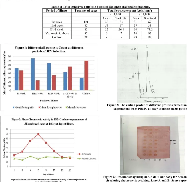
Leucopenia is a characteristic of many viral diseases10. A unique feature of JEV infection is leucocytosis with marked neutrophilia11.The total leucocyte count done in peripheral blood of 121 JE cases in the study, showed leucocytosis (>11,000 cells/mm3) in 33% patients at the time of admission. In 67% of JE patients, the leucocytes increased significantly during second week of illness. The highest count being 13,800 cells/mm3. With the duration of illness, the counts gradually decreased (Table 1).
The DLC in peripheral blood of JE confirmed patients revealed polymorphonuclear leucocytosis ranging from 47 to 65% in ~40% of patients at the time of admission. Figure 1 shows polymorphonuclear leucocytosis in peripheral blood of JE patients. There was a significant rise in percentage of neutrophils (mean count = 75 + 3%) during second week of infection as compared to control (mean count = 31+ 1%). This showed that during JEV infection leucocytosis was due to the marked increase in polymorphonuclear leucocytosis. The counts gradually declined to normal with the duration of illness.
Production of neutrophil chemotactic factor by peripheral blood mononuclear cells (PBMC) of JE patients:
Macrophages can directly influence the development of a PMN-dependent inflammatory response via the release of chemotactic factors such as IL-8, leucotriene B4 or via activating factors such as TNF-α12. An attempt was made to establish if peripheral blood monocytes in JE confirmed patients secrete such chemokine in vivo. PBMC cultures of 25 JE confirmed patients, done on alternate days showed that mononuclear cells secrete a neutrophil chemotactic factor. The PBMC culture supernatants collected on different days were assayed for chemotactic activity by skin window method. Figure 2 shows that the chemotactic activity started appearing from day 5, reaching peak on day 7, gradually declined thereafter and disappeared completely by day 15 p.i. Normal human monocyte culture supernatants did not show detectable amount of chemotactic activity. The culture supernatant from PBMC at day 7 of illness in JE patients was taken as the chemotactic factor in further study. Previous observations in mice are similar to above showing secretion of a macrophage-derived chemotactic factor during acute phase of JEV infection and disappearance of it by fourth week p.i.7
Purification of hMDF by PBMC of JE patients:
Aditi Singh et al: Detection of A Neutrophil Chemotactic Factor
JPSI 1 (6), Nov – Dec 2012, 23-26
recovered in peak 3. The chemotactic fractions were concentrated, further purified and used for molecular weight determination by sodium-dodecyl sulphate polyacrylamide gel electrophoresis (SDS-PAGE). The chemotactic factor produced by PBMC at day 7 of JE illness migrated as a single ~10 KD band, in between lysozyme and aprotinin and also parallel to hMDF prepared in vitro.
Detection of hMDF in circulation by immunoblotting:
All the acute and convalescent phase sera and culture supernatants were screened in a dot-blot test using anti-hMDF antibody (Figure 4). The test was found reactive in 92% of acute serum samples from which some representative samples are shown in lane A and B of figure 4. The day 7
PBMC culture supernatants were highly reactive against anti-hMDF antibody in immuno dot-blot (lane C). The reaction was negligible (12.5%) for convalescent sera (lane D). Lane E is showing hMDF prepared in vitro as a positive control at position 5. The normal sera shown at position 1- 4 and normal culture supernatant at position 6-8 in lane E were non reactive in the test. This is in accordance with the observations in mice that neutrophil chemotactic cytokine (MDF) was secreted during acute phase of JEV infection7. It was also observed that acute sera of all fatal cases demonstrated high levels of circulating chemotactic cytokine (figure 4, lane A) suggesting a correlation between the levels of hMDF and the severity of illness.
Table 1: Total leucocyte counts in blood of Japanese encephalitis patients. Period of illness Total no. of cases Total leucocyte count (cells/mm3)
> 11,000 < 11,000
Cases % of total Cases % of total
Ist week 121 40 33 81 67
IInd week 82 55 67 27 33
IIIrd week 82 22 26.8 60 73.2
IVth week & above 82 6 7 76 93
Control 28 - - 28 100
Figure 3: The elution profile of different proteins present in culture supernatant from PBMC at day7 of illness in JE patients.
Figure 4: Dot-blot assay using anti-hMDF antibody for demonstration of circulating chemotactic cytokine. Lane A and B: Some representative acute serum samples. Lane C: The day 7 PBMC culture supernatants
Aditi Singh et al: Detection of A Neutrophil Chemotactic Factor
JPSI 1 (6), Nov – Dec 2012, 23-26 CONCLUSION
Investigations of the molecular underpinnings of chemokine functions and their involvement in inflammatory process of all kinds are yielding information about the mechanism of pathogenesis of a number of conditions, as well as providing hope for new therapeutic insights13. The mechanism of JEV pathogenesis is still not very clear 14, and there is an immediate requirement to understand role of host factors in JEV-induced neuropathogenesis for the development of appropriate and effective therapy15. hMDF seems to be a pathogenesis related protein, further studies on its nature and structure could contribute considerably in the understanding of the pathogenesis of JEV infection in humans. The detection of hMDF in blood could be a useful prognostic marker of the disease. Observations in animal models have shown that administration of various cytokines locally, either by direct injection or by cytokine gene therapy can induce strong immunity against some inherited as well as acquired disorders such as cancer and infection by human immunodeficiency virus16. Thus owing to proinflammatory function of hMDF, the anti-hMDF antibody could be used in the preparation of vaccine against this dreaded disease, called Japanese encephalitis.
REFERENCES:
1. Johnson RT, Burke DS, Elwell M, Leake CJ, Nisalak A, Hoke CH,
Lorsomrudee W. Japanese encephalitis: immunocytochemical studies of viral antigen and inflammatory cells in fatal cases. Ann. Neurol. 1985; 18: 567-573.
2. Khanna N, Mathur A, Chaturvedi UC. Regulation of vascular
permeability by macrophage derived chemotactic factor produced in Japanese encephalitis. Immunol. Cell Biol. 1994; 72: 200-204.
3. Gubler DJ, Rochrig JT. Arboviruses (Togaviridae and Flaviviradae). In,
Collier L, Balows A, Sussman A (eds). Topley & Wilson’s
microbiology and microbial infection. 9th ed. Arnold, London; 1998;
597-600.
4. Centers for Disease Control and Prevention (CDC). Japanese
encephalitis vaccines: recommendations of the Advisory Committee on Immunization Practices (ACIP). MMWR Recomm Rep. 2010; 59(RR-1): 1-27.
5. Mathur A, Bhardwaj M, Kulshreshtha R, Rawat S, Jain A, Chaturvedi UC. Immunopathological study of spleen during JEV infection in mice. Brit. J. Exp. Path. 1988; 69: 423-432.
6. Mathur A, Khanna N, Chaturvedi UC. Breakdown of blood brain barrier
by virus induced cytokine during Japanese encephalitis virus infection. Int. J. Exp. Path. 1992; 73: 603-611.
7. Khanna N, Agnihotri M, Mathur A, Chaturvedi UC. Neutrophil
chemotactic factor produed by Japanese encephalitis virus stimulated macrophages. Clin. Exp. Immunol. 1991; 86: 299-303.
8. Tandon A, Singh A, Atrishi E, Saxena SK, Mathur A. Alteration in plasma glucose levels in Japanese encephalitis patients. Int. J. Exp. Path. 2002; 83: 39-46.
9. Mathur A, Chaturvedi UC, Tandon HO, Agarwal AK, Mathur GP, Nag
D, Prasad A, Mittal VP. Japanese encephalitis in Uttar Pradesh, India during 1978. Indian. J. Med. Res. 1982; 75: 161-169.
10. de Gruchy GC. Clinical hematology in medical practice. 3rd ed. Oxford:
ELBS and Scientific Publications. 1976; 359-403.
11. Chaturvedi UC, Mathur A, Tandon P, Natu SM, Rajvanshi A, Tandon HO. Variable effect on peripheral blood leokocytes during JE virus infection of man. Clin. Exp. Immunol. 1979; 38: 492-498.
12. Marcinkiewicz J. Neutrophil chloramines: missing links between innate
and acquired immunity. Immunol. Today. 1997, 18: 577-580.
13. Vaughan TJ, Osbourn JK, Tempest PR. Human antibodies by design.
Nat. Biotech. 1998; 16: 535-539.
14. Yang Y, Ye J, Yang X, Jiang R, Chen H, Cao S. Japanese encephalitis
virus infection induces changes of mRNA profile of mouse spleen and brain. Virol. J. 2011; 8: 80.
15. Gupta N, Bhaskar AS, Lakshmana Rao PV. Transcriptional regulation and activation of the mitogen-activated protein kinase pathway after Japanese encephalitis virus infection in neuroblastoma cells. FEMS Immunol. Med. Microbiol. 2011; 62: 110-121.
16. Gillis S, William DE. Cytokine therapy: lessons learned and future challenges. Curr. Opinion Immunol. 1998; 10: 501-503.
QUICK RESPONSE CODE
ISSN (Online) : 2277 –4572
Website
http://www.jpsionline.com
How to cite this article: