The level of circulating endothelial progenitor cells
may be associated with the occurrence and
recur-rence of chronic subdural hematoma
Yan Song,IZhitao Wang,II Li Liu,IIDong Wang,IIJianning ZhangII
ITianjin Medical University General Hospital, Tianjin Geriatrics Institute, Department of Health Care Medicine, Tianjin, China.IITianjin Medical University
General Hospital, Tianjin Neurological Institute, Department of Neurosurgery, Key Laboratory of Post-trauma Neuro-repair and Regeneration in Central Nervous System, Tianjin, China.
OBJECTIVES:The onset of chronic subdural hematoma may be associated with direct or indirect minor injuries to the head or a poorly repaired vascular injury. Endothelial progenitor cells happen to be one of the key factors involved in hemostasis and vascular repair. This study was designed to observe the levels of endothelial progenitor cells, white blood cells, platelets, and other indicators in the peripheral blood of patients diagnosed with chronic subdural hematoma to determine the possible relationship between the endothelial progenitor cells and the occurrence, development, and outcomes of chronic subdural hematoma.
METHOD: We enrolled 30 patients with diagnosed chronic subdural hematoma by computer tomography
scanning and operating procedure at Tianjin Medical University General Hospital from July 2009 to July 2011. Meanwhile, we collected 30 cases of peripheral blood samples from healthy volunteers over the age of 50. Approximately 2 ml of blood was taken from veins of the elbow to test the peripheral blood routine and coagulation function. The content of endothelial progenitor cells in peripheral blood mononuclear cells was determined by flow cytometry.
RESULTS:The level of endothelial progenitor cells in peripheral blood was significantly lower in preoperational patients with chronic subdural hematomas than in controls. There were no significant differences between the two groups regarding the blood routine and coagulation function. However, the levels of circulating endothelial progenitor cells were significantly different between the recurrent group and the non-recurrent group.
CONCLUSIONS:The level of circulating endothelial progenitor cells in chronic subdural hematoma patients was significantly lower than the level in healthy controls. Meanwhile, the level of endothelial progenitor cells in recurrent patients was significantly lower than the level in patients without recurrence. Endothelial progenitor cells may be related to the occurrence and recurrence of chronic subdural hematoma.
KEYWORDS: Chronic Subdural Hematoma; Endothelial Progenitor Cell; Injury.
Song Y, Wang Z, Liu L, Wang D, Zhang J. The level of circulating endothelial progenitor cells may be associated with the occurrence and recurrence of chronic subdural hematoma. Clinics. 2013;68(8):1084-1088.
Received for publication onJanuary 24, 2013;First review completed onFebruary 15, 2013;Accepted for publication onApril 1, 2013 E-mail: [email protected]
Tel.: 86 22 6036-3760
& INTRODUCTION
Chronic subdural hematomas (CSDHs) are common neurosurgical occurrences in intracranial hematoma, with an incidence ranging from 1-13.1%. Their mortality amounts to 1.5%,8%, and the recurrence rate is 9.2%-26.5% (1-6). The pathogenesis of CSDH remains hypothetical. There has been
significant discussion and debate regarding the complex pathogenesis of CSDH, particularly in terms of the inflammatory process (7), the head trauma etiology (8,9), the osmotic gradient theory (10-12), and recurrent hemor-rhage associated with hyperfibrinolysis (13).
The onset of CSDH may be associated with direct or indirect minor injury of the head or poor vascular repair. Accordingly, there are a large number of brittle neovascular capillaries in the outer membrane of the resulting hematoma, and continuous leaking from these structural capillary defects is the main reason for the development of CSDH. The outer membrane of the hematoma capsule generally contains numerous fragile macrocapillaries (also called sinusoidal vessels) with vascular lumina (14). When viewed with an electron microscope, these lumina are often extremely wide ($40mm in diameter), contain several blood cells and consist Copyrightß2013CLINICS– This is an Open Access article distributed under
the terms of the Creative Commons Attribution Non-Commercial License (http:// creativecommons.org/licenses/by-nc/3.0/) which permits unrestricted non-commercial use, distribution, and reproduction in any medium, provided the original work is properly cited.
No potential conflict of interest was reported.
of loose junctions between adjacent endothelial cells and a partial absence of the basement membrane and pericytes (15). Endothelial progenitor cells (EPCs) happen to be one of the key factors involved in hemostasis and vascular repair. These cells, through the secretion of related cytokines, play an extremely important role in the repair and regeneration of damaged blood vessels. Therefore, we observed the levels of EPCs, white blood cells, platelets, and other indicators in the peripheral blood of CSDH patients to determine the possible relationship between EPCs and the occurrence, development, and outcomes of CSDH.
& MATERIALS AND METHODS
Patients
We collected clinical information from 30 patients who were diagnosed with CSDH by computer tomography (CT) scanning and operating procedures at Tianjin Medical University General Hospital from July 2009 to July 2011. Sixteen patients had clear histories of minor head trauma in the last year before they were diagnosed with CSDH. All patients underwent a single burr-hole drainage surgery under local anesthesia. Four cases relapsed and recovered after reoperation. We collected peripheral blood samples 2 hours prior to the surgery and 14 days after the surgery. Meanwhile, over the same period, we collected peripheral blood samples from 30 healthy volunteers over the age of 50. Head CT scans were performed at Tianjin Medical University General Hospital in the Division of Radiology. Patients who had diabetes (16), dyslipidemia, intractable hypertension coronary heart disease, kidney disease (17), or hemorrhage disease (18) were classified as exclusions. No estrogen or anticoagulant agents were used in either group due to their potential influence on EPC levels (19,20).
Laboratory management
We took approximately 2 ml of blood from the veins of the elbow, immediately placed it in EDTA-K2 anticoagula-tion tubes, and gently mixed it upside down to prevent platelet adhesion and aggregation. Peripheral blood routine and coagulation function were tested by Tianjin Medical University General Hospital, Department of Clinical Laboratory (automatic biochemistry analyzer: SYSMEX XE-2100, Sysmex Corporation, Japan).
Because EPCs are characterized by the co-expression of the hematopoietic stem cell/progenitor markers CD34 and CD133 (21,22), we determined the content of EPCs in peripheral blood mononuclear cells (PBMNCs) by flow cytometry using dual staining with fluorescein-conjugated monoclonal antibodies against CD34/CD133 markers. PBMNCs were isolated by density centrifugation according to the manufacturers’ instructions. We incubated 106 PBMNCs with PE-labeled monoclonal mouse anti-human CD34 (BD Biosciences, USA) and FITC-labeled monoclonal mouse anti-human CD133 (BD Biosciences, USA) antibodies for 30 minutes at 4
˚
C, according to the manufacturers’ instructions. At least 100,000 events were acquired in the lymphomonocytic gate using a FACSCalibur cytometer (BD FACS Aris, USA). The number of progenitor cells was expressed as a percentage of all lymphomonocytic cells.Statistics
Chi-square tests or Student’s T tests were used to compare categorical and continuous variables among the
groups. Experimental data are presented as means¡ SD. Statistical analyses were performed using commercially available software (SPSS, ver. 18.0, SPSS Inc., Chicago, IL), and a p-value of ,0.05 was deemed to indicate statistical significance.
Ethics
The study was approved by the Tianjin Medical University General Hospital ethical standards committee on human experimentation. Written informed consent was also obtained from both patients and control participants.
& RESULTS
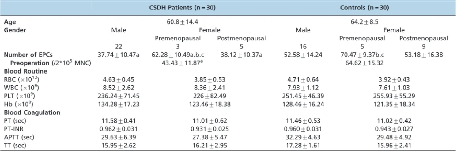
In this study, we first evaluated the EPC levels in CSDH patients treated with burr-hole drainage surgery and their association with blood routine and coagulation function. The CT scan performance of CSDH is a lunula or crescent-shaped lower-density area under the skull plate on one or both sides. High- or hybrid-density shadows occurred when the hematoma volume was large, slowly absorbed, or bleeding recurrently. The demographic characteristics of the patient and control groups are summarized (Table 1). The patients ranged in age from 45 to 82 years (average 60.8¡14.4 years, means¡SD). There were 22 men and 8 women. All of the CSDH cases underwent burr-hole surgery. All hematoma removal cases were irrigated with saline solution in the subdural space. The control group ranged in age from 52 to 75 years (average 64.2¡8.5 years, means¡SD). There were 16 men and 14 women. No significant differences in age and gender between the CSDH and control groups were identified.
The average number of circulating EPCs in preoperational CSDH patients was 43.43¡11.87 per 26105 mononuclear
cells. The average number of circulating EPCs in the health control group was 64.62¡15.32 per 26105 mononuclear cells. The EPC levels in the peripheral blood were significantly lower in preoperational CSDH patients than in controls (p= 0.013). Compared with the control group, the
numbers of EPCs in males, premenopausal females, and postmenopausal females of the CSDH group differed significantly (p,0.05). Within the CSDH or control groups, the number of EPCs in premenopausal females was higher than in the male group or in the postmenopausal female group (p,0.05). Meanwhile, there was no difference between the male group and the postmenopausal female group. There were no significant differences between the CSDH group and the control group or between males and females within the same group regarding any indices of blood routine and coagulation, such as red blood cell (RBC), white blood cell (WBC), platelet (PLT), and hemoglobin (Hb) levels; prothrombin time (PT); prothrombin time-international normalized ratio (PT-INR); activated partial thromboplastin time (APTT); and thrombin time (TT) (Table 1).
between the recurrent group and the non-recurrent group (p= 0.008) (Table 2).
& DISCUSSION
In recent years, EPCs, especially in the peripheral blood, have become the focus of research in the field of related blood vessels. EPCs are immature hematopoietic cells circulating in the peripheral blood that are capable of differentiating into mature endothelial cells, thus aiding in endothelial recovery (23). Small numbers of EPCs are circulating in the peripheral blood of every healthy adult, approximately 70-210/ml, accounting for 0.002% of PBMNCs (24). EPCs are involved not only in the formation of new blood vessels but also in the maintenance of the dynamic integrity of vascular membrane; thus, they play an important role in physiological repair. EPCs participate in these processes through mobilization, tendency, adhesion, proliferation, and differentiation. Any pathological and physiological factors resulting in reduced numbers of EPCs and fewer dysfunctions may lead to structural and functional abnormalities during neovascularization. Structural integrity and functional abnormalities during neovascularization lead to extravasation of the blood, increasing the likelihood of bleeding. Currently, it is generally accepted that the adventitial neovascularization
of hematomas and re-bleeding are key factors in the onset and development of CSDH (25). Thus, EPC reduction may be relevant to CSDH.
CSDH mainly occurs in the elderly. The incidence rate in the population over 65 years of age is 7-13/10 million, and the rate in people over 70 years of age is 17-58/10 million (26). However, the CSDH incidence in the general popula-tion is only approximately 1-3/10 million (26,27). Aging can lead to a reduction in the number of EPCs in circulation and a weakening of their function (28-30). Thus, we can infer that there are both lower numbers of EPCs and related dysfunction in the peripheral blood of CSDH patients. In this study, we measured the peripheral blood levels of EPCs in 30 CSDH patients and in 30 healthy controls. The results showed that the level of peripheral blood EPCs in CSDH patients was significantly lower compared to the control group. Meanwhile, we found that the change in the levels of EPCs in recurrent patients between postoperation and preoperation was significantly lower than the level in non-recurrent patients. These data indicate that a higher consumption of EPCs in CSDH patients and decreased EPC levels may lead to a reduced capacity to repair the endothelium, increasing the risks associated with the occurrence and recurrence of CSDH. Thus, we propose that a pharmacological means of increasing the number of circulating EPCs could help to reduce the recurrence rate of CSDH.
Through in-depth research on angiogenesis mechanisms and EPCs, we can be sure that EPCs are not only involved in the formation of new blood vessels but that they also play an important role in maintaining the dynamic integrity of the vascular intima. Any factors leading to a reduction in the number of EPCs may result in structural and func-tional abnormalities in the neovascularization process. Throughout our study, the levels of EPCs in the peripheral blood of patients with CSDH were significantly lower compared to the control group, and the levels of peripheral EPCs in the patients with postoperative recurrence were
Table 1 -Laboratory characteristics of patients and controls.
CSDH Patients (n = 30) Controls (n = 30)
Age 60.8¡14.4 64.2¡8.5
Gender Male Female Male Female
Premenopausal Postmenopausal Premenopausal Postmenopausal
22 3 5 16 5 9
Number of EPCs 37.74¡10.47a 62.28¡10.49a.b.c 38.12¡10.37a 52.58¡14.24 70.47¡9.37b.c 53.18¡16.38
Preoperation(/2*105MNC) 43.43
¡11.87a 64.62
¡15.32 Blood Routine
RBC (61012) 4.63¡0.45 3.85¡0.53 4.71¡0.64 3.92¡0.43
WBC (6109) 8.52¡2.62 8.36¡2.41 7.93¡1.12 7.61¡1.03
PLT (6109) 236.24¡71.45 226¡82.49 251.45¡46.39 255.93¡55.29
Hb (6109) 134.28¡17.23 123.46¡18.38 128.46¡16.24 121.35¡18.34
Blood Coagulation
PT (sec) 11.58¡0.41 11.01¡0.62 11.46¡0.53 11.02¡0.42
PT-INR 0.962¡0.031 0.931¡0.025 0.960¡0.031 0.943¡0.027
APTT (sec) 29.63¡6.39 27.38¡5.47 32.29¡4.63 29.48¡4.92
TT (sec) 15.95¡2.62 16.21¡2.95 17.28¡1.61 15.96¡2.41
Values are means¡SD. CSDH = chronic subdural hematoma; EPCs = endothelial progenitor cells; MNC = mononuclear cell; RBC = red blood cell; WBC = white blood cell; PLT = platelet; Hb = hemoglobin; PT = prothrombin time; PT-INR = prothrombin time-international normalized ratio; APTT = activated partial thromboplastin time; TT = thrombin time.
*T tests were used to compare continuous variables among groups if the data followed a normal distribution; otherwise, nonparametric tests were used. Chi-square tests were used to compare categorical variables among the groups. a: Compared to the control group,p,0.05; b: Compared to males in the same group,p,0.05; c: Compared to postmenopausal women in the same group,p,0.05.
Table 2 -Number of EPCs 14 days after operation.
Recurrence No recurrence p-value*
Number of CSDH Patients 6 24 NS
Number of EPCs(/2*105MNC) 8.54¡5.25a 35.52¡18.59 0.008
significantly lower than in patients who did not relapse. Therefore, we believe that the occurrence and development of CSDH may be involved in numerous aspects of the reduction in the number of EPCs in the peripheral blood. At the very least, this reduction plays an integral role in many factors. Thus, we put forward the following hypothesis. When trauma causes a split in the dural border cell layer to form a substantial subdural hematoma, cerebrospinal fluid (CSF) and/or blood accumulates as foreign matter, stimu-lating the inflammatory response and resulting in the secretion of inflammatory cytokines (31-34), vascular endothelial growth factor (VEGF) (35), and angiopoietins (36,37). Similar to granulation tissue, the purpose of new blood capillaries and the outer membrane is to wrap and absorb local foreign bodies and to promote healing.
If the number of circulating EPCs is reduced and/or bone marrow-derived EPCs cannot replenish their consumption, capillary structural defects will occur. At the same time, in the case of abnormal cerebral pulsations causing new damage to the capillaries, both the maintenance of the intimal integrity and physiological repairs cannot be completed. With these internal circles, the continuous resulting effusion from pathologically permeable capillary walls into the subdural space leads to the formation of a CSDH and its eventual expansion.
In this study, we conclude that the level of circulating EPCs in CSDH patients was significantly lower than the level in healthy controls. Additionally, the level of EPCs in recurrent patients was significantly lower than the level in patients without recurrence. Thus, we speculate that the low level of EPCs may be responsible for the occurrence of CSDH; furthermore, the continuing low level of EPCs may be associated with the recurrence of CSDH.
& ACKNOWLEDGMENTS
This work was supported by grants from the Tianjin Health Bureau Technology Foundation (No. 07KZ26) and by the National Natural Science Foundation of China (No. 81100920). No competing financial interests exist.
& AUTHOR CONTRIBUTIONS
Song Y was in charge of the experimental design and English version of the manuscript. Wang Z was responsible for the collection and testing of blood samples. Liu L was responsible for the experimental data statistics. Wang D was responsible for the collection of clinical data. Zhang J was in charge of the overall design and adjustment of the research.
& REFERENCES
1. Ramachandran R, Hegde T. Chronic subdural hematomas—causes of morbidity and mortality. Surg Neurol. 2007;67(4):367-72, http://dx.doi. org/10.1016/j.surneu.2006.07.022.
2. Kristof RA, Grimm JM, Stoffel-Wagner B. Cerebrospinal fluid leakage into the subdural space: possible influence on the pathogenesis and recurrence frequency of chronic subdural hematoma and subdural hygroma. J Neurosurg. 2008;108(2):275-80, http://dx.doi.org/10.3171/ JNS/2008/108/2/0275.
3. Katano H, Kamiya K, Mase M, Tanikawa M, Yamada K. Tissue plasminogen activator in chronic subdural hematoma as a predictor of recurrence. J Neurosurg. 2006;104(1):79-84, http://dx.doi.org/10.3171/ jns.2006.104.1.79.
4. Ko¨nig SA, Schick U, Do¨hnert J, Goldammer A, Vitzthum HE. Coagulopathy and outcome in patients with chronic subdural haema-toma. Acta Neurol Scand. 2003;107(2):110-6, http://dx.doi.org/10.1034/ j.1600-0404.2003.01340.x.
5. Hong HJ, Kim YJ, Yi HJ, Ko Y, Oh SJ, Kim JM. Role of angiogenic growth factors and inflammatory cytokine on recurrence of chronic subdural hematoma. Surg Neurol. 2009;71(2):161-5, http://dx.doi.org/10.1016/j. surneu.2008.01.023.
6. Torihashi K, Sadamasa N, Yoshida K, Narumi O, Chin M, Yamagata S. Independent predictors for recurrence of chronic subdural hematoma: a review of 343 consecutive surgical cases. Neurosurgery. 2008;63(6):1125-9, http://dx.doi.org/10.1227/01.NEU.0000335782.60059.17.
7. Virchow R. Haematoma dural matric. Verhandl. Phys Med Gasellsch. Wruzburg. 1857;7:134.
8. Trotter W. Chronic subdural haemorrhage of traumatic origin and its relation to pachymeningitis haemorrhagica interna. Br J Surg. 1914; 2(6):271-91.
9. Putnam T, Cushing H. Chronic subdural haematoma. Arch Surg. 1925; 11:328, http://dx.doi.org/10.1001/archsurg.1925.01120150002001. 10. Gardner WJB. Traumatic subdural haematoma with particular reference
to the latent interval. Arch Neurol Psychiatry. 1932;27:847-58, http://dx. doi.org/10.1001/archneurpsyc.1932.02230160088009.
11. Gitlin D. Pathogenesis of subdural collection of fluids. Pediatrics. 1955;16:345-51.
12. Labadie EL, Glover D. Chronic subdural haematoma: concepts of pathogenesis, a review. Can J Neurol Sci. 1974;1(4):222-5.
13. Labadie EL, Glover D. Local alterations of haemostatic fibrinolytic mechanisms in reforming subdural hematomas. Neurology. 1975; 25(7):669-75, http://dx.doi.org/10.1212/WNL.25.7.669.
14. Shim YS, Park CO, Hyun DK, Park HC, Yoon SH. What are the causative factors for a slow, progressive enlargement of a chronic subdural hematoma? Yonsei Med J. 2007;48(2):210-7, http://dx.doi.org/10.3349/ ymj.2007.48.2.210.
15. Nanko N, Tanikawa M, Mase M, Fujita M, Tateyama H, Miyati T, et al. Involvement of hypoxia-inducible fator-1a and vascular endothelial
growth factor in the mechanism of development of chronic subdural hematoma. Neurol Med Chir. 2009;49(9):379-85, http://dx.doi.org/10. 2176/nmc.49.379.
16. Dei Cas A, Spigoni V, Ardigo` D, Pedrazzi G, Franzini L, Derlindati E, et al. Reduced circulating endothelial progenitor cell number in healthy young adult hyperinsulinemic men. Nutr Metab Cardiovasc Dis. 2011; 21(7):512-7, http://dx.doi.org/10.1016/j.numecd.2009.11.011.
17. Lavoie JR, Stewart DJ. Genetically modified endothelial progenitor cells in the therapy of cardiovascular disease and pulmonary hypertension. Curr Vasc Pharmacol. 2012;10(3):289-99, http://dx.doi.org/10.2174/ 157016112799959413.
18. Li X, Tse HF, Yiu KH, Jia N, Chen H, Li LS, et al. Increased levels of circulating endothelial progenitor cells in subjects with moderate to severe chronic periodontitis. J Clin Periodontol. 2009;36(11):933-9, http://dx.doi.org/10.1111/j.1600-051X.2009.01481.x.
19. Bulut D, Albrecht N, Imo¨hl M, Gu¨nesdogan B, Bulut-Streich N, Bo¨rgel J, et al. Hormonal status modulates circulating endothelial progenitor cells. Clin Res Cardiol. 2007;96(5):258-63, http://dx.doi.org/10.1007/s00392-007-0494-z.
20. Martin-Yuste V, Brugaletta S, Ferreira-Gonza´lez I, Cola C, Alvarez-Contreras L, de Antonio M, et al. Endothelial progenitor cell capturing stent and short dual antiplatelet therapy in patients on chronic anti-vitamin k regimen undergoing percutaneous coronary interventions: long-term outcomes of a single centre registry. Euro Intervention. 2011;6(7):831-7.
21. Ingram DA, Caplice NM, Yoder MC. Unresolved questions, changing definitions, and novel paradigms for defining endothelial progenitor cells. Blood. 2005;106(5):1525-31, http://dx.doi.org/10.1182/blood-2005-04-1509.
22. Shin JW, Lee DW, Kim MJ, Song KS, Kim HS, Kim HO. Isolation of endothelial progenitor cells from cord blood and induction of differ-entiation by ex vivo expansion. Yonsei Med J. 2005;46(2):260-7, http:// dx.doi.org/10.3349/ymj.2005.46.2.260.
23. Asahara T, Murohara T, Sullivan A, Silver M, van der Zee R, Li T, et al. Isolation of putative progenitor endothelial cells for angiogenesis. Science. 1997;275(5302):964-7, http://dx.doi.org/10.1126/science.275. 5302.964.
24. Peichev M, Naiyer AJ, Pereira D, Zhu Z, Lane WJ, Williams M, et al. Expression of VEGFR-2 and AC133 by circulating human CD34(+) cells identifies a population of functional endothelial precursors. Blood. 2000;95(3):952-8.
25. Murakami H, Hirose Y, Sagoh M, Shimizu K, Kojima M, Gotoh K, et al. Why do chronic subdural hematomas continue to grow slowly and not coagulate? Role of thrombomodulin in the mechanism. J Neurosurg. 2002;96(5):877-84, http://dx.doi.org/10.3171/jns.2002.96.5.0877. 26. Sambasivan M. An overview of chronic subdural hematoma: experience
with 2300 cases. Surg Neurol. 1997;47(5):418-22, http://dx.doi.org/10. 1016/S0090-3019(97)00188-2.
27. Delgado-Lo´pez PD, Martı´n-Velasco V, Castilla-Dı´ez JM, Rodrı´guez-Salazar A, Galacho-Harriero AM, Ferna´ndez-Arconada O. Dexamethasone treat-ment in chronic subdural haematoma. Neurocirugia. 2009;20(4):346-59. 28. Vasa M, Fichtlscherer S, Aicher A, Adler K, Urbich C, Martin H, et al.
Number and migratory activity of circulating endothelial progenitor cells inversely correlate with risk factors for coronary artery disease. Circ Res. 2001;89(1):E1-7, http://dx.doi.org/10.1161/hh1301.093953.
patients undergoing coronary artery bypass grafting. J Am Coll Cardiol. 2003;42(12):2073-80, http://dx.doi.org/10.1016/j.jacc.2003.07.025. 30. Khakoo AY, Finkel T. Endothelial progenitor cells. Annu Rev Med.
2005;56:79-101, http://dx.doi.org/10.1146/annurev.med.56.090203.104149. 31. Fan Y, Ye J, Shen F, Zhu Y, Yeghiazarians Y, Zhu W, et al. Interleukin-6 stimulates circulating blood-derived endothelial progenitor cell angio-genesis in vitro. J Cereb Blood Flow Metab. 2008;28(1):90-8, http://dx. doi.org/10.1038/sj.jcbfm.9600509.
32. Denburg JA, van Eeden SF. Bone marrow progenitors in inflammation and repair: new vistas in respiratory biology and pathophysiology. Eur Respir. J. 2006;27(3):441-5, http://dx.doi.org/10.1183/09031936.06.00000706. 33. Suzuki M, Endo S, Inada K, Kudo A, Kitakami A, Kuroda K, et al.
Inflammatory cytokines locally elevated in chronic subdural haematoma. Acta Neurochir. 1998;140(1):51-5, http://dx.doi.org/10.1007/s007010050057. 34. Frati A, Salvati M, Mainiero F, Ippoliti F, Rocchi G, Raco A, et al. Inflammation markers and risk factors for recurrence in 35 patients with
a posttraumatic chronic subdural hematoma: a prospective study. J Neurosurg. 2004;100(1):24-32, http://dx.doi.org/10.3171/jns.2004.100. 1.0024.
35. Dome B, Dobos J, Tovari J, Paku S, Kovacs G, Ostoros G, et al. Circulating bone marrow-derived endothelial progenitor cells: characterization, mobilization, and therapeutic considerations in malignant disease. Cytometry A. 2008;73(3):186-93, http://dx.doi.org/10.1002/cyto.a.20480. 36. Hohenstein A, Erber R, Schilling L, Weigel R. Increased mRNA expression of VEGF within the hematoma and imbalance of angiopoie-tin-1 and -2 mRNA within the neomembranes of chronic subdural hematoma. J Neurotrauma. 2005;22(5):518-28, http://dx.doi.org/10. 1089/neu.2005.22.518.