Application of a Multiplex Quantitative PCR
to Assess Prevalence and Intensity Of
Intestinal Parasite Infections in a Controlled
Clinical Trial
Stacey Llewellyn1, Tawin Inpankaew2, Susana Vaz Nery3, Darren J. Gray3,4,5, Jaco J. Verweij6, Archie C. A. Clements3, Santina J. Gomes7, Rebecca Traub8, James S. McCarthy1,9*
1Clinical Tropical Medicine Laboratory, QIMR Berghofer Medical Research Institute, Herston, Queensland, Australia,2Department of Veterinary Disease Biology, Faculty of Health and Medical Science, University of Copenhagen, Copenhagen, Denmark; Department of Parasitology, Faculty of Veterinary Medicine, Kasetsart University, Bangkok, Thailand,3Research School of Population Health, College of Medicine, Biology and Environment, The Australian National University, Canberra, Australian Capital Territory, Australia,
4Molecular Parasitology Laboratory, QIMR Berghofer Medical Research Institute, Herston, Queensland, Australia,5School of Public Health, University of Queensland, Herston, Queensland, Australia,
6Laboratory for Medical Microbiology and Immunology, St Elisabeth Hospital, Tilburg, the Netherlands,
7Laboratorio Nacional da Saúde, Ministério da Saúde, Dili, Timor-Leste,8Faculty of Veterinary and Agricultural Sciences, University of Melbourne, Parkville, Victoria, Australia,9School of Medicine, University of Queensland, Queensland, Australia
Abstract
Background
Accurate quantitative assessment of infection with soil transmitted helminths and protozoa is key to the interpretation of epidemiologic studies of these parasites, as well as for moni-toring large scale treatment efficacy and effectiveness studies. As morbidity and transmis-sion of helminth infections are directly related to both the prevalence and intensity of infection, there is particular need for improved techniques for assessment of infection inten-sity for both purposes. The current study aimed to evaluate two multiplex PCR assays to determine prevalence and intensity of intestinal parasite infections, and compare them to standard microscopy.
Methodology/Principal Findings
Faecal samples were collected from a total of 680 people, originating from rural communi-ties in Timor-Leste (467 samples) and Cambodia (213 samples). DNA was extracted from stool samples and subject to two multiplex real-time PCR reactions the first targeting: Neca-tor americanus,Ancylostomaspp.,Ascarisspp., andTrichuris trichiura; and the second Entamoeba histolytica,Cryptosporidiumspp.,Giardia.duodenalis, andStrongyloides ster-coralis. Samples were also subject to sodium nitrate flotation for identification and quantifi-cation of STH eggs, and zinc sulphate centrifugal flotation for detection of protozoan OPEN ACCESS
Citation:Llewellyn S, Inpankaew T, Nery SV, Gray DJ, Verweij JJ, Clements ACA, et al. (2016) Application of a Multiplex Quantitative PCR to Assess Prevalence and Intensity Of Intestinal Parasite Infections in a Controlled Clinical Trial. PLoS Negl Trop Dis 10(1): e0004380. doi:10.1371/journal. pntd.0004380
Editor:Jeffrey Michael Bethony, George Washington University, UNITED STATES
Received:October 25, 2015
Accepted:December 18, 2015
Published:January 28, 2016
Copyright:© 2016 Llewellyn et al. This is an open access article distributed under the terms of the
Creative Commons Attribution License, which permits unrestricted use, distribution, and reproduction in any medium, provided the original author and source are credited.
Data Availability Statement:All relevant data are within the paper and its Supporting Information files.
Funding:The research was funded by a National Health and Medical Research Councilhttps://www. nhmrc.gov.au/Grant (#1013713). The funders have had no role in study design, data collection and analysis, decision to publish, or preparation of the manuscript.
parasites. Higher parasite prevalence was detected by multiplex PCR (hookworms 2.9 times higher,Ascaris1.2,Giardia1.6, along with superior polyparasitism detection with this effect magnified as the number of parasites present increased (one: 40.2% vs. 38.1%, two: 30.9% vs. 12.9%, three: 7.6% vs. 0.4%, four: 0.4% vs. 0%). Although, all STH positive sam-ples were low intensity infections by microscopy as defined by WHO guidelines the DNA-load detected by multiplex PCR suggested higher intensity infections.
Conclusions/Significance
Multiplex PCR, in addition to superior sensitivity, enabled more accurate determination of infection intensity forAscaris, hookworms andGiardiacompared to microscopy, especially in samples exhibiting polyparasitism. The superior performance of multiplex PCR to detect polyparasitism and more accurately determine infection intensity suggests that it is a more appropriate technique for use in epidemiologic studies and for monitoring large-scale inter-vention trials.
Author Summary
Gastrointestinal parasites including soil-transmitted helminths cause considerable mor-bidity worldwide, especially in resource-poor communities. Large-scale epidemiologic and treatment efficacy studies are regularly undertaken to determine the optimum ways to reduce or eliminate parasites from endemic communities, thereby reducing the burden of disease. Accurate and sensitive tests for detection of soil transmitted helminths and proto-zoa are of great importance to the success of such trials. Increasingly recognised is the importance of accurately determine the infection intensity, as morbidity and transmission pressure of helminth infections are directly related this and not just to prevalence. A vast majority of studies use standard microscopy methods which, although well accepted, may not be as accurate as more recently developed molecular techniques such as multiplex PCR. Therefore, there is need for further evaluation of multiplex PCR techniques and their ability to detect infections and provide infection intensity data. In the current study real-time PCR showed a higher sensitivity for the detection of intestinal helminths and proto-zoa especially in cases of mixed infections as well as more accurate determination of infec-tion intensity compared to microscopy.
Introduction
Gastrointestinal parasites including soil-transmitted helminths (STH) cause considerable mor-bidity worldwide, especially in resource-poor communities. The chronic effects on health are predominately attributed to the burden of disease rather than mortality [1]. The majority of the global burden is considered due to the five main STH–Ascaris lumbricoides, hookworms (Necator americanusandAncylostomaspp.),Trichuris trichiuraandStrongyloides stercoralis
control programs. This is because morbidity and transmission pressure of helminth infections are directly related to both the prevalence and intensity of infection [8,9].
Several microscopy-based techniques are available and widely used for the identification and quantification of STH eggs. The Kato-Katz (KK) thick smear technique, originally devel-oped for diagnosis of schistosomiasis [10], is currently the most widely used microscopic tech-nique, and is considered the gold standard by the World Health Organization (WHO) for assessing both prevalence and intensity of infection in helminth control programmes [11]. A major drawback of the KK is that multiple samples with multiple slides per sample are required to be examined over several days to reach high levels of sensitivity and quantitative accuracy, especially in light infections [12]. Moreover, immediate and skilled processing is required to reduce chance of false-negative results, particularly for hookworms, due to fast clearance on slides [7,13]. Alternative methods for microscopic diagnosis of STH include using concentra-tion steps, such as formalin-ether sedimentaconcentra-tion and flotaconcentra-tion techniques such as McMaster [14], simple sodium nitrate methods [15], FLOTAC [16] or mini-FLOTAC [17]. These have some advantages including increased sensitivity over KK. For example, in a recent study sodium nitrate methods proved superior for detecting low egg burdens in samples compared to quadruple KK smears and resulted in higher EPG [15]. Comparisons of KK to FLOTAC also showed equivalent [18,19] or superior [20,21] sensitivity of FLOTAC techniques. These flota-tion based techniques have drawbacks in terms of being labour intensive, having poor repro-ducibility owing to operator error, and for most tests requiring centrifugation steps [16,19]. In addition, microscopic-based techniques lack the ability to assign species-level identification of helminth eggs (e.g. those of hookworms, andAscaris). Diagnosis ofS.stercoralisis particularly challenging, as only a small number of larvae are released in stool regardless of infection inten-sity, with Baermann sedimentation or agar plate culture methods thought to provide greatest specificity and sensitivity [22,23]. Serological diagnostic methods are available for STH but their use is limited due to poor specificity in endemic areas [24].
Stained faecal smears and faecal concentration methods allow for diagnosis of protozoa. However, these too have their limitations with regards to poor sensitivity and the inability to differentiate protozoan parasite stages to a species level. For example, diagnosis ofE.histolytica
infections by microscopy misses 40% of infections [25]; and it is not possible to visually differ-entiate pathogenicE.histolyticafrom non-pathogenicEntamoeba dispar. Thus, onlyE. histoly-ticaspecific stool antigen detection tests are approved for diagnostic use by the WHO [26], although shown to have poor sensitivity compared to PCR-based methods [27,28]. More effi-cient coproantigen capture enzyme linked immunosorbent assay (ELISA) based assays can be used for diagnosis ofCryptosporidiumandGiardia, however there have been reports of false-positive and false-negative results [29].
limited data has been published to date with thorough quantitative comparisons between this method and microscopy-based techniques.
The aim of the current study was to evaluate the use of two multiplex PCRs for the detection and quantification of intestinal protozoa and helminths as tools to support large scale epidemi-ologic and treatment efficacy studies. This method was compared with results obtained with sodium nitrate flotation and zinc sulphate centrifugation for determining prevalence and intensity of intestinal parasite infections in villages in Timor-Leste and Cambodia. A specific aim was to evaluate qPCR as a method for determining infection intensity, an important parameter in both epidemiologic and anthelmintic efficacy studies.
Methods
Sample Collection
Samples were sourced from two separate parasitic surveys. Single faecal samples from 467 indi-viduals enrolled in the WASH for WORMS interventional trial in Timor-Leste (Registered with the Australian New Zealand Clinical Trials Registry; Trial registration:
ACTRN12614000680662)[43] and from a separate study of 213 individuals enrolled in a cross sectional study of intestinal parasites in northern Cambodia [44]. A single faecal sample per individual was collected within a maximum of 12 hours of defecation and divided into two ali-quots and stored separately. One sample in 5% w/v potassium dichromate solution for PCR analysis, and the other sample in 10% formalin for microscopy. Samples from Timor were transported at room temperature to Queensland Berghofer Institute of Medical Research for extraction (QIMRBerghofer) and PCR and Timor-Leste National Lab for microscopy. Cambo-dian samples were transported at room temperature to the University of Queensland (UQ) Gatton campus for microscopy and DNA extraction, and DNA transported on ice to QIMR Berghofer for PCR.
Microscopy
All faecal samples were examined microscopically and enumerated forAscaris, hookworms, andTrichuriseggs using a simple sodium nitrate flotation as previously described [45]; and for the presence of protozoa cysts and oocysts using zinc sulphate centrifugal flotation [35]. Full methods available inS1 Methods. Specific parasitologic diagnosis ofS.stercoralisinfection was not undertaken due to resource limitations that precluded agar plate culture.
DNA Extraction
Samples stored in 5% potassium dichromate were first subject to centrifugation at 2,000 g for 3 min, followed by removal of the preservative supernatant. Samples were re-suspended to 50 ml with phosphate buffered saline (1 X PBS) and the centrifugation repeated. Supernatant was again decanted off, and the sample pellet was subsequently stored at 4°C for up to a month.
DNA extraction was performed using the Powersoil DNA Isolation Kit (Mo Bio, Carlsbad, CA USA). Minor modifications were made to the manufacturer’s protocol following optimiza-tion withTrichuris vulpiseggs. Prior to extraction, samples were spiked with a known quantity of a positive control PCR target, namely a plasmid containing equine herpes virus (EHV) insert from the glycoprotein B gene [46]. Full DNA extraction protocol is available inS1 Methods.
Multiplex PCR
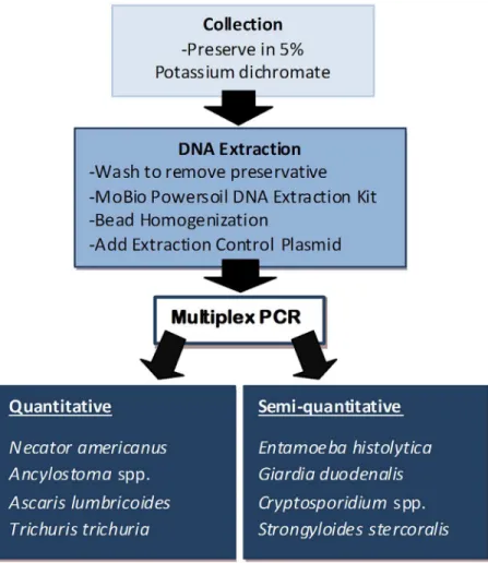
Trichiuraand EHV; the second was a semi-quantitative assay forE.histolytica, Cryptosporid-iumspp.,G.duodenalis,S.stercoralisand EHV [12,13,24,34,42,47]. Details of the primers and probes are listed in Tables1and2. However an alternateS.stercoralisforward primer was used after optimization of this assay withS.stercoralispositive DNA samples from the Northern Territory (Australia) (Deborah Holt—Personal communication). The Rotor-Gene 6000 (Qia-gen, Melbourne, VIC AUS) was used for all PCR assays, with reactions set up for both PCR reactions as previously described [30] with minor modifications. Assay protocol, optimisation and preparation of PCR controls is available inS1 Methods. A sample processing summary is displayed inFig 1.
Controls
A Ct cut-off of 31 forAscariswas established based on the limit of detection on a previously published conventional PCR, to ensure reproducibility of results [48]. The limit of detection of all other assays in terms of the maximum Ct-value considered to be positive was set at 35. All PCR assays were validated at independent laboratories (Queensland Medical Laboratory, Aus-tralia; The Task Force for Global Health, USA; St. Vincent’s Hospital, Sydney), and additional positiveE.histolytica(n = 2) andE.dispar(n = 1) control samples tested to ensure PCR speci-ficity for the pathogenic species,E.histolytica. Further confirmation ofEntamoebaspp. pres-ence in all controls was provided by a genus specific conventional PCR [49].
PCR Ct to EPG Conversion
—
Seeding Experiments
To produce a calibration curve to interpolate EPG values from PCR Ct-values a series of seed-ing experiments were conducted forAscarisspp. andN.americanusinfections.Ascaris suum
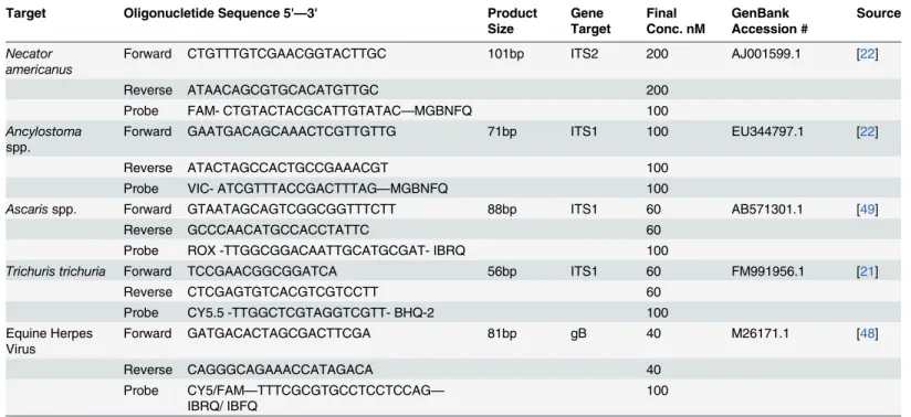
eggs were purchased from Excelsior Sentinel Inc. (Ithaca, NY), supplied in unembryonated form and stored in 5% potassium dichromate at room temperature for shipping. Hookworm Table 1. Quantitative multiplex PCR set up overview.
Target Oligonucletide Sequence 5'—3' Product
Size
Gene Target
Final Conc. nM
GenBank Accession #
Source
Necator americanus
Forward CTGTTTGTCGAACGGTACTTGC 101bp ITS2 200 AJ001599.1 [22]
Reverse ATAACAGCGTGCACATGTTGC 200
Probe FAM- CTGTACTACGCATTGTATAC—MGBNFQ 100
Ancylostoma
spp.
Forward GAATGACAGCAAACTCGTTGTTG 71bp ITS1 100 EU344797.1 [22]
Reverse ATACTAGCCACTGCCGAAACGT 100
Probe VIC- ATCGTTTACCGACTTTAG—MGBNFQ 100
Ascarisspp. Forward GTAATAGCAGTCGGCGGTTTCTT 88bp ITS1 60 AB571301.1 [49]
Reverse GCCCAACATGCCACCTATTC 60
Probe ROX -TTGGCGGACAATTGCATGCGAT- IBRQ 100
Trichuris trichuria Forward TCCGAACGGCGGATCA 56bp ITS1 60 FM991956.1 [21]
Reverse CTCGAGTGTCACGTCGTCCTT 60
Probe CY5.5 -TTGGCTCGTAGGTCGTT- BHQ-2 100
Equine Herpes Virus
Forward GATGACACTAGCGACTTCGA 81bp gB 40 M26171.1 [48]
Reverse CAGGGCAGAAACCATAGACA 40
Probe CY5/FAM—TTTCGCGTGCCTCCTCCAG—
IBRQ/ IBFQ
100
eggs were freshly isolated from threeN.americanusinfected stool samples, kindly provided by Alex Loukas (James Cook University).Ascarisand hookworm eggs were purified separately [50], and multiple 10μL aliquots counted under a microscope following staining with Lugol’s
iodine solution to determine the concentration of eggs in each sample. Purified eggs were pooled and suspended in a total of 5 ml PBS and diluted to produce a range of concentrations of eggs.Ascariseggs were prepared in triplicate in 2 ml screw top tubes in concentrations rang-ing from 200,000 EPG to 5 EPG, with an additional 200 mg negative control faecal sample added to each sample. Hookworm eggs were similarly prepared but concentrations ranged from 6,000 EPG to 250 EPG, and were only performed in duplicate due to a smaller number of available egg numbers. BothAscarisand hookworm eggs at each of the concentrations were subject to DNA extraction and multiplex PCR as stated previously. PCR Ct-values were con-verted to intensities based on assumed 100% reaction run efficiency, provided by the Rotorgene Q software (Multiplex PCR Intensity = 10−0.298Ct +9.81). This allowed the interpolation of the relation between the log transformed EPG and log transformed PCR intensity to determine a relationship between Ct-values and EPG.
Statistical Analysis
Kappa statistics were used for comparison of multiplex PCR and microscopy determined prev-alence. Analysis was performed using SPSS (IBM Corp.), Excel 2008 (Microsoft), and Graph-Pad Prism version 6.0 (GraphGraph-Pad Software Inc).
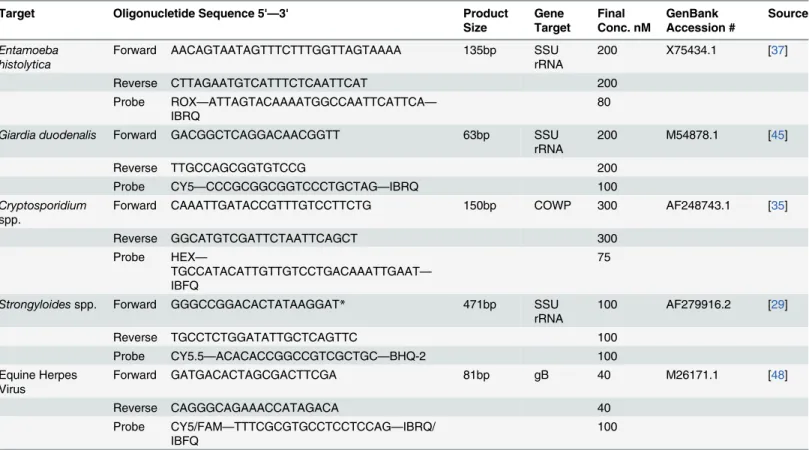
Table 2. Semi-quantitative multiplex PCR set up overview.
Target Oligonucletide Sequence 5'—3' Product
Size Gene Target Final Conc. nM GenBank Accession # Source Entamoeba histolytica
Forward AACAGTAATAGTTTCTTTGGTTAGTAAAA 135bp SSU
rRNA
200 X75434.1 [37]
Reverse CTTAGAATGTCATTTCTCAATTCAT 200
Probe ROX—ATTAGTACAAAATGGCCAATTCATTCA—
IBRQ
80
Giardia duodenalis Forward GACGGCTCAGGACAACGGTT 63bp SSU
rRNA
200 M54878.1 [45]
Reverse TTGCCAGCGGTGTCCG 200
Probe CY5—CCCGCGGCGGTCCCTGCTAG—IBRQ 100
Cryptosporidium
spp.
Forward CAAATTGATACCGTTTGTCCTTCTG 150bp COWP 300 AF248743.1 [35]
Reverse GGCATGTCGATTCTAATTCAGCT 300
Probe HEX—
TGCCATACATTGTTGTCCTGACAAATTGAAT—
IBFQ
75
Strongyloidesspp. Forward GGGCCGGACACTATAAGGAT* 471bp SSU
rRNA
100 AF279916.2 [29]
Reverse TGCCTCTGGATATTGCTCAGTTC 100
Probe CY5.5—ACACACCGGCCGTCGCTGC—BHQ-2 100
Equine Herpes Virus
Forward GATGACACTAGCGACTTCGA 81bp gB 40 M26171.1 [48]
Reverse CAGGGCAGAAACCATAGACA 40
Probe CY5/FAM—TTTCGCGTGCCTCCTCCAG—IBRQ/ IBFQ
100
*Altered from the original published primer [29]
Ethical Considerations
The WASH for WORMS interventional trial in Timor-Leste complies with the provisions con-tained in the National Statement on Ethical Conduct in Human Research and was approved by the University of Queensland Medical Research Ethics Committee (#2011000734), the ANU Human Research Ethics Committee (protocol: 2014/311), and the Timor-Leste Ethics Com-mittee of the Ministry of Health (reference 2011/51). The Cambodia sample collection study protocol [44] was approved by the National Ethics Committee for Health Research, Ministry of Health, Cambodia (NECHR, #192,) Ethics Committee of the Cantons of Stadt and Basel-land (EKBB, #18/12). Written informed consent was obtained for all participants.
Results
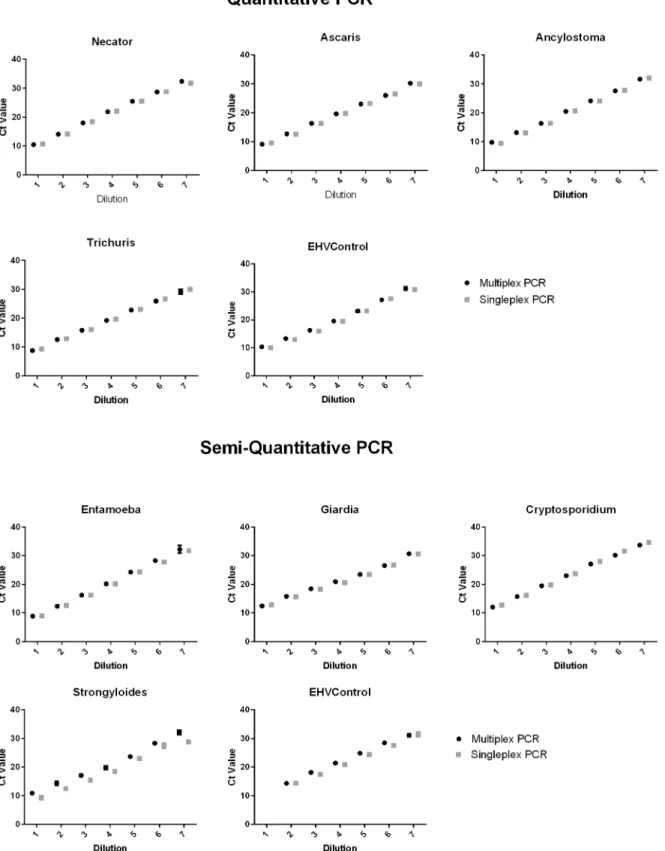
Multiplex PCR Optimisation
Optimized oligonucleotide concentrations used throughout testing are listed inTable 1. Exper-imental comparisons of plasmid control dilutions series’were undertaken between singleplex and multiplex PCR reactions (Fig 2). No or minimal effect on sensitivity and efficiency of one PCR on another was found in each multiplex PCR. Example data of standard EHV Ct-values results is shown inS1 Fig. Field samples excluded due to inhibition of the EHV control were subject to repeat analysis.
Fig 1. Flow chart of sample processing for multiplex PCR detection of intestinal parasites.
Fig 2. Multiplex to singleplex PCR Ct comparison.Assay optimization to determine effects of multiplex PCR set up on sensitivity and efficiency of PCRs compared to singleplex PCR using plasmid standard curve controls containing all PCR products.
Detection of Single Infections
—
Diagnostic Performance of PCR vs
Microscopy
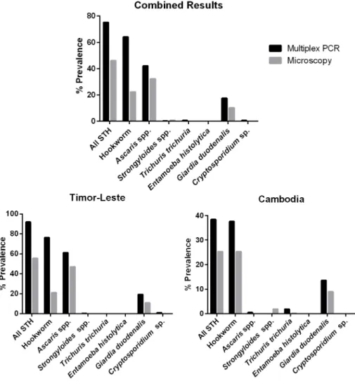
Comparison of the prevalence of parasite infection between multiplex PCR and microscopy was made individually for each data set (Fig 3;Table 2;S1 Dataset). The observed multiplex PCR prevalence was consistently higher across nearly all target organisms in both regions stud-ied. Multiplex PCR detected almost three times (2.9, 435/151) the number of hookworm infec-tions, 1.2 (260/219) times moreAscarisinfections and 1.6 (115/70) times moreGiardia
infections than microscopy at both study sites.S.stercoraliswas however detected in four of the Cambodia microscopy samples, which tested negative in multiplex PCR. The number of samples that tested negative in microscopy was twofold (2.34) higher than the number testing negative in multiplex PCR, indicating that a large proportion of infections were missed by microscopy.
Direct comparisons between diagnostic techniques on individual samples from all regions were undertaken using Kappa agreement statistics (Table 3). Results show good, moderate and fair agreement forAscaris,Giardia, and hookworms, respectively. Only target organisms with
Fig 3. Overall parasite prevalence comparison between multiplex PCR and microscopy.Data presented combined for all 680 study participants, as well as individually for Timor-Leste (467 participants) and Cambodia (213 participants), showing higher recorded percentage prevalence across all target organisms by Multiplex PCR.
over 20% prevalence were analysed. For all parasites analysed, multiplex PCR identified a large number of positive samples not detected by microscopy (Ascaris68, Hookworm 299,Giardia
69), whilst a small number of microscopy positive samples were not identified as positive by multiplex PCR (Ascaris27, Hookworm 15,Giardia24).
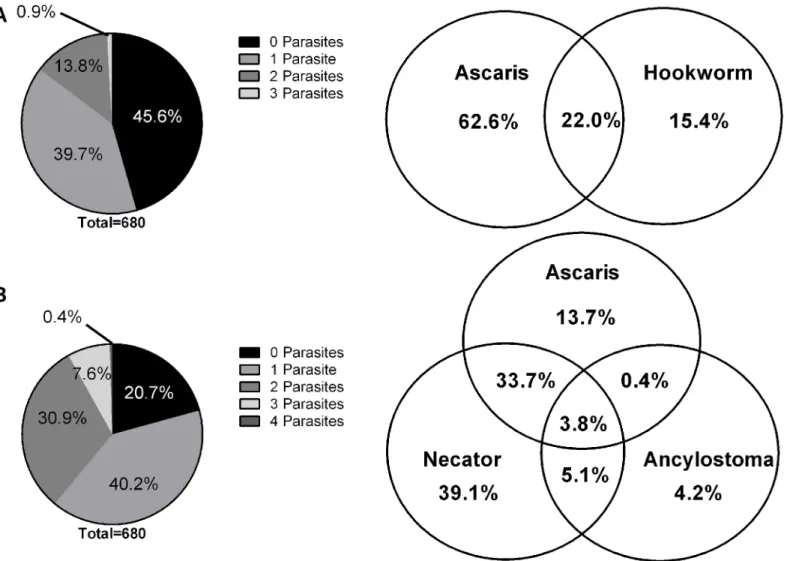
Detection of Multiple Infections
Along with a higher detection rate of all individual target organisms, the multiplex PCR approach was also superior in terms of sensitivity in detecting samples with multiple infections (Fig 4).
Increased polyparasitism was detected in multiplex PCR in comparison to microscopy, as similar levels of single parasite infections were detected but more than double (2.4 times) the number of dual parasite infections. This trend is further compounded with three parasites, with PCR detecting over 17-fold (17.3) the number of infections than microscopy. An addi-tional three samples were found to harbour four parasites (Ascaris,N.americanus, Ancylos-tomaspp., andGiardia), only detected using multiplex PCR. Coinfection with the two genera of hookworms,N.americanusandAncylostomaspp. that was detected in the multiplex PCR resulted as well in an increased prevalence of polyparasitism.
Quantitative Results
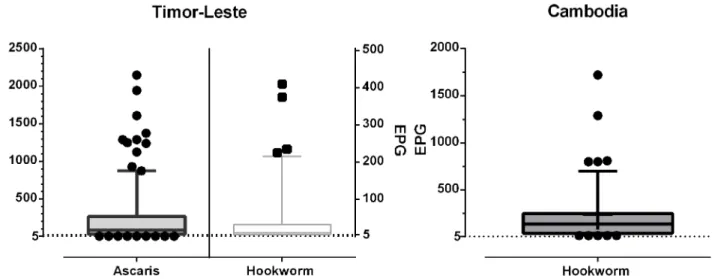
Infection intensities as determined by microscopy data for nematodes with greatest prevalence is presented inFig 5. According to WHO guidelines all infections from both study regions for both hookworm andAscariswould be classified as low intensity infections (Ascaris<5,000
EPG; hookworm<2,000 EPG). The infection intensity frequency distribution of parasites
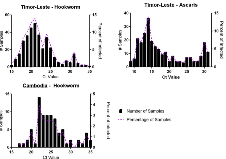
with at least 20% prevalence for Timor-Leste and Cambodia is shown inFig 6. This Multiplex PCR Ct frequency distribution graph shows the majority of infected individuals harbouring high relative infection burdens (low Ct-values).
The infection intensity ofG.duodenalewas similar in both Timor-Leste (Average Ct 24.5; range 16.2–34.3), and Cambodia (Average Ct 23.2; range 16.0–32.0). The few positive Crypto-sporidium sp. samples in Timor-Leste were all of similar infection intensities (Average Ct 30.9; range 29.0–33.0), whilst the four positiveT.trichiuraCambodian samples all were of low infec-tion intensities (Average Ct 32.7; range 31.5–33.7).
A comparison of quantitative results from both microscopy and multiplex PCR was attempted. However, data are not presented as no statistically relevant relationship was found for eitherAscarisor hookworms from both study regions. Similarly, no statistical correlation Table 3. Multiplex PCR and microscopy parasite prevalence agreement statistics.
PCR Microscopy Total Agreement (%) Kappa* SE of Kappa
POS NEG
Ascaris POS 192 68 585(86.0) 0.695 0.029
NEG 27 393
Hookworm POS 136 299 366(53.8) 0.201 0.024
NEG 15 230
Giardia POS 46 69 587 (86.3) 0.424 0.049
NEG 24 541
*Kappa Agreement Level:K<0.20Poor; 0.21–0.40Fair; 0.41–0.60Moderate; 0.61–0.80Good; 0.81–1.00Very Good
was found between PCR-determined positive samples with low infectivity levels and negative microscopy results, or vice versa.
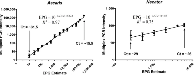
Standard curves obtained from well-defined controls presenting relationship between PCR determined Ct-values (converted to infection intensity) as a measure of EPG derived from standard microscopy practices are shown inFig 7. The interpolation of Ct-value to EPG derived from this experimental data is also presented withinFig 7, along with the Ct-value range in which this can be reliably used to estimate EPG from multiplex PCR data. A prelimi-nary example of the use of the resulting calibration curve is shown using Timor-Leste field data inS2 Fig.
Discussion
The present study further demonstrates and validates the suitability of multiplex PCR for detection and quantification ofAscaris, hookworms andGiardiain stools obtained in large scale surveys. The two multiplex PCRs inAscaris, hookworm andGiardiaendemic regions of Fig 4. Polyparasitism.Diagrams depict polyparisitism observed in the 680 combined Timor-Leste and Cambodia samples in both(A) Microscopy and(B) Multiplex PCR. Pie graph depicts total number of parasites per sample and the venn diagram details the specific division of STH coinfections for Microscopy (259Ascarisand/or hookworm positive samples) and multiplex PCR (504Ascarisand/or hookworm positive samples).*Microscopy unable to differentiate Hookworm speciesN.americanusandAncylostomaspp. -considered only as‘hookworms’for polyparasitism comparison.
Timor-Leste and Cambodia resulted in significantly higher levels of prevalence compared to microscopy alone [30] [13,36] [51]. Multiplex PCR also showed a greater ability for detection of co-infections, and provided more accurate and reliable infection intensity data; both of key importance to the assessment of disease burden due to the elevated risk of morbidity [52],
A large disparity in the prevalence rates between techniques was noted for the detection of hookworm,AscarisandGiardiawhen compared on a per sample basis. Multiplex PCR in par-ticular detected a large number of positive samples not detected in microscopy; a possible result of the failure of microscopy to detect polyparasitism, present in nearly half (49.1%) of all posi-tives samples by multiplex PCR. A small number of samples were also deemed positive by microscopy but negative in multiplex PCR. This may be due to variation in the dispersion of lar-vae, eggs and oocytes within the subsamples taken, due to the nonhomogeneous nature of the stool [53], as well as the non-uniform nature of their excretion in stool [30,54], causing variation in both techniques. Differences between techniques may also be due to errors leading to false positive results. Such errors are less likely in multiplex PCR due to rigorous controls, whilst lim-ited controls can be implemented with microscopy, which relies heavily on the technical exper-tise of the user. This is potentially the case of the fourStrongyloidespositive samples detected only by microscopy, as larvae resemble hatched hookworm larvae. Alternatively further PCR optimization may be required as there have been previous reports of PCR sensitivity issues for detection ofStrongyloides, as only low levels of larvae are present even in heavy infections [55].
Microscopy sensitivity issues of parasite detection, particularly in samples exhibiting poly-parasitism suggest further compounding issues in accurately determining infection intensity levels. Data comparing PCR Ct-values to microscopy determined EPG values have been previ-ously been reported, showing a broad range of Ct-values for each microscopy EPG value; espe-cially for microscopy negative samples [13]. This inability to link the quantitative field data using different techniques despite statistically sound prevalence agreements suggests that mul-tiplex PCR is superior for use in diagnostic testing of STH for survey work where accurate intensity data is required.
This focus on infection intensity is a major strength of the present study when considering disease burden, as STH infection prevalence alone does not provide a measure of potential morbidity, which is related directly to infection intensity [9]. Despite this, the majority of Fig 5. Intensity of infection forAscarisspp. and hookworm positive samples as determined by sodium nitrate flotation.Timor-Leste microscopy produced 219Ascarispositive samples (200 EPG average), 97 hookworm positive samples (40 EPG average). Cambodia microscopy produced 54 hookworm positive samples (60 EPG average).
studies have focused on direct prevalence comparisons between techniques, with limited reports on the quantitative abilities of the techniques [30], although some correlation data has been provided [13]. The WHO currently considers prevalence the main measure in STH con-trol programmes, with suggestion of including intensity data only if available, with treatment aims to target medium to heavy STH infections [56]. Providing this more accurate infection intensity data using the multiplex PCR technique may produce data required to establish more effective STH control programmes. The WHO infection intensity estimates may also require re-evaluation with improved intensity data to be useful in PCR-parasite intensity surveys. These estimates were established based using the KK technique, and suggest only low intensity infections when applied to the current study microscopy data (53), whilst multiplex PCR data indicated high intensity infections (low Ct-values) in the majority of individuals.
In the present study, the full relationship between Ct-values and EPG was assessed using standard curves obtained from well-defined controls, with carefully prepared measurements. The feasibility of such an approach has been shown and further indicates that PCR quantifica-tion is likely to be more accurate and that addiquantifica-tional detailed studies should be undertaken in the future. At this stage insufficient experimental data are available to be able to produce Fig 6. Multiplex PCR Ct-value frequency distribution for hookworm andAscarisspp. positive samples.Graphs show the infection intensity distribution, with lower Ct-values indicating higher infection intensities, presented for Timor-Leste hookworm (353) andAscaris(259), as well as Cambodia Hookworm (80) positive samples. As Ct-values are expressed in a continuous format, values were rounded up to the nearest integer to produce categorical data for frequency analysis.
accurate predictions across the whole range of possible Ct-values as the current interpolation is limited to within the microscopy determined EPG range in which it was tested. Additional vali-dation of theAscarisEPG interpolation is required and at higher infection intensities before it can be reliably used with field samples and significant further testing of hookworm samples is essential to produce accurate predictions of EPG. Determining this mathematical relationship between Ct-values, EPG, and by corollary adult worm burden, and assuring its accuracy repre-sents a goal in future research, vital to progress PCR methodologies as the gold standard of intestinal nematode detection. The potential shown here to convert this PCR data to the more widely understood“EPG”format may allow this technique to provide more widely accepted and reportable STH surveillance. However, attempting to enumerate egg counts by PCR with-out a truly quantitative gold standard for comparison does however, represent a limitation of this work.
Despite proving successful in parasite diagnosis and providing valuable information on infection intensity, there are limitations of multiplex PCR as a diagnostic tool. The use of PCR preservatives such as potassium dichromate and ethanol temporarily arrests further egg devel-opment, and are thought to allow accurate quantitative data after many months of storage. However extensive testing into the effect of such preservatives on the genome copy numbers for the target nematodes has not been performed but is crucial for accurate interpolation to an egg count. Further compounding the issue is the ability to relate nematode egg counts to DNA intensity, when copies of DNA increase once eggs have embryonated. Literature does indicate that once embryonated the ITS1 gene target of the PCR assays remains at a constant level [57], suggesting that accurate quantification is possible.
Further testing on preservation methods is essential in terms of the effect on the quantitative accuracy over time, both those for microscopy and multiplex PCR techniques. The maximum storage time in potassium dichromate is currently undetermined, however reports have sug-gested a one month storage period in potassium dichromate forGiardiafor optimal detection [33]. Recent reports on formalin preserved samples suggest a 15 day storage window in which microscopy can be completed to gain accurate quantitative hookworm data, with additional Fig 7. Relationship between EPG and intensity converted PCR Ct-values.Graph shows strong linear relationship (P<0.001) between sodium nitrate flotation determined EPG and Multiplex PCR Intensity upon universal log10 transformation. (95% Confidence Intervals:Ascaris- slope 1.028 to 1.089; Y-intercept 0.6405 to -0.4333; X-Y-intercept 0.4205 to 0.5895. Necator—slope 0.3151 to .7344; Y- intercept -0.6150 to 0.7130; X-intercept -2.253 to 0.8413).* PCR Intensity = 10–0.298*Ct +9.81.
decline in the ability to detect parasites after one month [52]. Microscopy on samples in this study was not reliably performed within this optimum time period; potentially resulting in the lower prevalence and intensity data for hookworms.
The main limitation of multiplex PCR in national control programmes is the capacity to implement it in parasite endemic low-resource settings, with the current need to send samples to well equipped labs for analysis. Microscopy based techniques can however, be undertaken with less resources and more affordable equipment. The material cost of processing samples and running both multiplex PCRs was estimated as AU$12.37 per sample (AU$6.05 per extrac-tion; AU$3.16 per multiplex PCR). This also represents a limitation when compared to flota-tion based microscopy, costing just AU$1 per sample (labour not included). The trade off of the additional costs and the inability for onsite analysis compared to the higher sensitivity and ability to detect multiple infections as well as the more accurate intensity data is an issue for consideration in design of epidemiologic studies and for clinical trials of effectiveness of interventions.
The use of multiplex real-time PCR for intestinal parasite diagnosis has proved to be more sen-sitive, and is more likely to detect mixed parasite infections than standard microscopy techniques. The real benefit of multiplex PCR is in its ability to more accurately determine infection intensity and the potential to report results in more understandable‘EPG’terms, which will prove to be inherently more useful in determining the success of de-worming and intervention trials.
Supporting Information
S1 Methods. Method A: Microscopy. Method B: DNA Extraction. Method C: Multiplex PCR Optimization.
(DOCX)
S1 Fig. EHV Extraction/PCR Control Example Real time PCR Results.Average EHV Ct-val-ues for each of the 37 field DNA samples compared within PCR runs to EHV-only control sample. Major outlier amplification from each run indicated in red where repeat extraction is required. Minor deviations outside 10–90thpercentile interval also considered for repeat extraction or repeat PCR. The specific source of the problem (PCR or extraction) was deter-mined through EHV data comparison between the quantitative multiplex and semi-quantita-tive multiplex.
(TIF)
S2 Fig. Intensity of Infection forAscarispositive samples as determined by PCR with
cali-bration curve.TheAscarisCt-EPG interpolation formula was used on 131 Timor-LesteAscaris
positive samples that were within the required Ct range (15.5–31.5) to allow adequate predic-tion of EPG from Ct value using calibrapredic-tion curve (EPG = 10−0.275Ct +9.622). Multiplex real-time PCR duplicates were each individually calculated with mean and SD for each sample.(A) Calibration curve example—converting PCR Ct-values to EPG(B)The infection intensity range for these 131 samples within acceptable calibration curve range.
(TIF)
S1 Dataset. (XLSX)
Acknowledgments
Timor-Leste field team and Timor-Timor-Leste National Laboratory for their efforts in collecting and cessing samples. We would also like to thank Dr. Christian Rune Stensvold for providing pro-tozoan genomic DNA for controls; Dr. Damien Stark (St Vincent’s Hospital, Sydney) for providing Entamoeba genomic DNA; Dr. Deborah Holt (Menzies School of Health Research) for providing the alternative sequence for theStrongyloidesforward primer and Dr. Thomas Nutman (NIH) for providing us withTrichurisprimer sequences. Further thanks are given to Queensland Medical Laboratories and Dr. Steven Williams (Task Force for Global Health) for their assistance in validation of results.
Author Contributions
Conceived and designed the experiments: SL TI SVN SJG DJG JJV ACAC RT JSM. Performed the experiments: SL TI SVN SJG JSM. Analyzed the data: SL TI. Contributed reagents/materi-als/analysis tools: TI SVN JJV RT JSM. Wrote the paper: SL TI SVN DJG JJV ACAC RT JSM.
References
1. WHO (2013) Soil-transmitted helminth infection: Fact Sheet No. 366.
2. Bethony J, Brooker S, Albonico M, Geiger S, Loukas A, et al. (2006) Soil-transmitted helminth infec-tions: ascariasis, trichuriasis, and hookworm. Lancet 367: 1521–1532. PMID:16679166
3. Pullan R, Smith J, Jasrasaria R, Brooker S (2014) Global numbers of infection and disease burden of soil transmitted helminth infections in 2010. Parasites & Vectors 7: 37.
4. Murray C, Vos T, Lozano R, Naghavi M, Flaxman A, et al. (2012) Disability-adjusted life years (DALYs) for 291 diseases and injuries in 21 regions, 1990–2010: a systematic analysis for the global burden of disease study 2010. Lancet 380: 2197–2223. doi:10.1016/S0140-6736(12)61689-4PMID:23245608 5. Hotez PJ, Brindley PJ, Bethony JM, King CH, Pearce EJ, et al. (2008) Helminth infections: the great
neglected tropical diseases. The Journal of Clinical Investigation 118: 1311–1321. doi:10.1172/ JCI34261PMID:18382743
6. Supali T, Verweij JJ, Wiria AE, Djuardi Y, Hamid F, et al. (2010) Polyparasitism and its impact on the immune system. Int J Parasitol 40: 1171–1176. doi:10.1016/j.ijpara.2010.05.003PMID:20580905 7. Raso G, Luginbuhl A, Adjoua CA, Tian-Bi NT, Silue KD, et al. (2004) Multiple parasite infections and their relationship to self-reported morbidity in a community of rural Cote d'Ivoire. Int J Epidemiol 33: 1092–1102. PMID:15256525
8. Anderson RM M R (1991) Infectious diseases of humans: dynamics and control. Oxford: Oxford Uni-versity Press.
9. Bundy DA, Medley GF (1992) Immuno-epidemiology of human geohelminthiasis: ecological and immu-nological determinants of worm burden. Parasitology 104 Suppl: S105–119. PMID:1589298
10. Katz N, Chaves A, Pellegrino J (1972) A simple device for quantitative stool thick-smear technique in
Schistosomiasis mansoni. Rev Inst Med Trop Sao Paulo 14: 397–400. PMID:4675644
11. Knopp S, Salim N, Schindler T, Voules DAK, Rothen J, et al. (2014) Diagnostic Accuracy of Kato-Katz, FLOTAC, Baermann, and PCR Methods for the Detection of Light-Intensity Hookworm and Strongy-loides stercoralisInfections in Tanzania. American Journal of Tropical Medicine and Hygiene 90: 535–
545. doi:10.4269/ajtmh.13-0268PMID:24445211
12. Mejia R, Vicuna Y, Broncano N, Sandoval C, Vaca M, et al. (2013) A novel, multi-parallel, real-time polymerase chain reaction approach for eight gastrointestinal parasites provides improved diagnostic capabilities to resource-limited at-risk populations. Am J Trop Med Hyg 88: 1041–1047. doi:10.4269/ ajtmh.12-0726PMID:23509117
13. Verweij J, Brienen E, Ziem J, Yelifari L, Polderman A, et al. (2007) Simultaneous detection and quantifi-cation ofAncylostoma duodenale,Necator americanus, andOesophagostomum bifurcumin fecal samples using multiplex real-time PCR. Am J Trop Med Hyg 77: 685–690. PMID:17978072
14. Levecke B, Behnke JM, Ajjampur SS, Albonico M, Ame SM, et al. (2011) A comparison of the sensitivity and fecal egg counts of the McMaster egg counting and Kato-Katz thick smear methods for soil-trans-mitted helminths. PLoS Negl Trop Dis 5: e1201. doi:10.1371/journal.pntd.0001201PMID:21695104 15. Inpankaew T, Schär F, Khieu V, Muth S, Dalsgaard A, et al. (2014) Simple Fecal Flotation Is a Superior
Alternative to Guadruple Kato Katz Smear Examination for the Detection of Hookworm Eggs in Human Stool. PLoS Neglected Tropical Diseases 8: e3313. doi:10.1371/journal.pntd.0003313PMID:
16. Cringoli G, Rinaldi L, Maurelli MP, Utzinger J (2010) FLOTAC: new multivalent techniques for qualita-tive and quantitaqualita-tive copromicroscopic diagnosis of parasites in animals and humans. Nat Protocols 5: 503–515. doi:10.1038/nprot.2009.235PMID:20203667
17. Maurelli M, Rinaldi L, Alfano S, Pepe P, Coles G, et al. (2014) Mini-FLOTAC, a new tool for copromicro-scopic diagnosis of common intestinal nematodes in dogs. Parasites & Vectors 7: 356.
18. Barda B, Zepherine H, Rinaldi L, Cringoli G, Clementi M, et al. (2013) Mini-FLOTAC and Kato-Katz: hel-minth eggs watching on the shore of Lake Victoria. Parasit Vectors 6: 220. doi: 10.1186/1756-3305-6-220PMID:23902918
19. Barda B, Cajal P, Villagran E, Cimino R, Juarez M, et al. (2014) Mini-FLOTAC, Kato-Katz and McMas-ter: three methods, one goal; highlights from north Argentina. Parasit Vectors 7: 271. doi:10.1186/ 1756-3305-7-271PMID:24929554
20. Steinmann P, Rinaldi L, Cringoli G, Du Z-W, Marti H, et al. (2015) Morphological diversity of Trichuris spp. eggs observed during an anthelminthic drug trial in Yunnan, China, and relative performance of parasitologic diagnostic tools. Acta Tropica 141, Part B: 184–189.
21. Knopp S, Rinaldi L, Khamis IS, Stothard JR, Rollinson D, et al. (2009) A single FLOTAC is more sensi-tive than triplicate Kato-Katz for the diagnosis of low-intensity soil-transmitted helminth infections. Trans R Soc Trop Med Hyg 103: 347–354. doi:10.1016/j.trstmh.2008.11.013PMID:19168197 22. Repetto SA, Alba Soto CD, Cazorla SI, Tayeldin ML, Cuello S, et al. (2013) An improved DNA isolation
technique for PCR detection ofStrongyloides stercoralisin stool samples. Acta Trop 126: 110–114. doi:10.1016/j.actatropica.2013.02.003PMID:23416126
23. Yap P, Furst T, Muller I, Kriemler S, Utzinger J, et al. (2012) Determining soil-transmitted helminth infec-tion status and physical fitness of school-aged children. J Vis Exp: e3966. doi:10.3791/3966PMID:
22951972
24. Verweij J, Canales M, Polman K, Ziem J, Brienen E, et al. (2009) Molecular diagnosis ofStrongyloides stercoralisin faecal samples using real-time PCR. Trans R Soc Trop Med Hyg 103: 342–346. doi:10. 1016/j.trstmh.2008.12.001PMID:19195671
25. Haque R, Ali IK, Akther S, Petri WA Jr. (1998) Comparison of PCR, isoenzyme analysis, and antigen detection for diagnosis ofEntamoeba histolyticainfection. J Clin Microbiol 36: 449–452. PMID:
9466756
26. Petri WA, Singh U (1999) Diagnosis and Management of Amebiasis. Clinical Infectious Diseases 29: 1117–1125. PMID:10524950
27. Roy S, Kabir M, Mondal D, Ali IKM, Petri WA, et al. (2005) Real-Time-PCR Assay for Diagnosis of Ent-amoeba histolyticaInfection. Journal of Clinical Microbiology 43: 2168–2172. PMID:15872237 28. Visser LG, Verweij JJ, Van Esbroeck M, Edeling WM, Clerinx J, et al. (2006) Diagnostic methods for
dif-ferentiation ofEntamoeba histolyticaandEntamoeba disparin carriers: performance and clinical impli-cations in a non-endemic setting. Int J Med Microbiol 296: 397–403. PMID:16753339
29. Johnston SP, Ballard MM, Beach MJ, Causer L, Wilkins PP (2003) Evaluation of Three Commercial Assays for Detection ofGiardiaandCryptosporidiumOrganisms in Fecal Specimens. Journal of Clini-cal Microbiology 41: 623–626. PMID:12574257
30. Basuni M, Muhi J, Othman N, Verweij JJ, Ahmad M, et al. (2011) A pentaplex real-time polymerase chain reaction assay for detection of four species of soil-transmitted helminths. Am J Trop Med Hyg 84: 338–343. doi:10.4269/ajtmh.2011.10-0499PMID:21292911
31. David EB, Coradi ST, Oliveira-Sequeira TCG, Ribolla PEM, Katagiri S, et al. (2011) Diagnosis of Giar-dia infections by PCR-based methods in children of an endemic area. Journal of Venomous Animals and Toxins including Tropical Diseases 17: 209–215.
32. Guy RA, Payment P, Krull UJ, Horgen PA (2003) Real-time PCR for quantification ofGiardiaand Cryp-tosporidiumin environmental water samples and sewage. Appl Environ Microbiol 69: 5178–5185. PMID:12957899
33. Kuk S, Yazar S, Cetinkaya U (2012) Stool sample storage conditions for the preservation of Giardia intestinalis DNA. Mem Inst Oswaldo Cruz 107: 965–968. PMID:23295744
34. Haque R, Roy S, Siddique A, Mondal U, Rahman SM, et al. (2007) Multiplex real-time PCR assay for detection ofEntamoeba histolytica,Giardia intestinalis, andCryptosporidiumspp. Am J Trop Med Hyg 76: 713–717. PMID:17426176
35. Traub RJ, Inpankaew T, Reid SA, Sutthikornchai C, Sukthana Y, et al. (2009) Transmission cycles of
36. ten Hove RJ, van Esbroeck M, Vervoort T, van den Ende J, van Lieshout L, et al. (2009) Molecular diag-nostics of intestinal parasites in returning travellers. Eur J Clin Microbiol Infect Dis 28: 1045–1053. doi:
10.1007/s10096-009-0745-1PMID:19415354
37. Verweij JJ, van Lieshout L (2011) Intestinal parasitic infections in an industrialized country; a new focus on children with better DNA-based diagnostics. Parasitology 138: 1492–1498. doi:10.1017/
S0031182011001211PMID:21859503
38. VERWEIJ JJ (2014) Application of PCR-based methods for diagnosis of intestinal parasitic infections in the clinical laboratory. Parasitology 141: 1863–1872. doi:10.1017/S0031182014000419PMID:
24780241
39. Verweij JJ, Stensvold CR (2014) Molecular testing for clinical diagnosis and epidemiological investiga-tions of intestinal parasitic infecinvestiga-tions. Clin Microbiol Rev 27: 371–418. doi:10.1128/CMR.00122-13
PMID:24696439
40. Gordon CA, McManus DP, Acosta LP, Olveda RM, Williams GM, et al. (2015) Multiplex real-time PCR monitoring of intestinal helminths in humans reveals widespread polyparasitism in Northern Samar, the Philippines. International Journal for Parasitology 45: 477–483. doi:10.1016/j.ijpara.2015.02.011
PMID:25858090
41. Basuni M, Mohamed Z, Ahmad M, Zakaria NZ, Noordin R (2012) Detection of selected intestinal hel-minths and protozoa at Hospital Universiti Sains Malaysia using multiplex real-time PCR. Trop Biomed 29: 434–442. PMID:23018507
42. Taniuchi M, Verweij JJ, Noor Z, Sobuz SU, Lieshout L, et al. (2011) High throughput multiplex PCR and probe-based detection with Luminex beads for seven intestinal parasites. Am J Trop Med Hyg 84: 332–337. doi:10.4269/ajtmh.2011.10-0461PMID:21292910
43. Nery SV, McCarthy JS, Traub R, Andrews RM, Black J, et al. (2015) A cluster-randomised controlled trial integrating a community-based water, sanitation and hygiene programme, with mass distribution of albendazole to reduce intestinal parasites in Timor-Leste: the WASH for WORMS research protocol. BMJ Open 5: e009293. doi:10.1136/bmjopen-2015-009293PMID:26719316
44. Inpankaew T, Schaer F, Dalsgaard A, Khieu V, Chimnoi W, et al. (2014) High Prevalence of Ancylos-toma ceylanicumHookworm Infections in Humans, Cambodia, 2012. Emerging Infectious Diseases 20: 976–982. doi:10.3201/eid2006.131770PMID:24865815
45. Inpankaew T, Traub R, Thompson R, Sukthana Y (2007) Canine parasitic zoonoses in Bangkok tem-ples. Southeast Asian J Trop Med Public Health 38: 247–255. PMID:17539273
46. Lambert SB, Whiley DM, O'Neill NT, Andrews EC, Canavan FM, et al. (2008) Comparing nose-throat swabs and nasopharyngeal aspirates collected from children with symptoms for respiratory virus identi-fication using real-time polymerase chain reaction. Pediatrics 122: e615–620. doi: 10.1542/peds.2008-0691PMID:18725388
47. Wiria A, Prasetyani M, Hamid F, Wammes L, Lell B, et al. (2010) Does treatment of intestinal helminth infections influence malaria? Background and methodology of a longitudinal study of clinical, parasito-logical and immunoparasito-logical parameters in Nangapanda, Flores, Indonesia (ImmunoSPIN Study). BMC Infectious Diseases 10: 77. doi:10.1186/1471-2334-10-77PMID:20338054
48. Traub RJ, Robertson ID, Irwin P, Mencke N, Thompson RC (2002) The role of dogs in transmission of gastrointestinal parasites in a remote tea-growing community in northeastern India. Am J Trop Med Hyg 67: 539–545. PMID:12479559
49. Verweij JJ, Polderman AM, Clark CG (2001) Genetic Variation among Human Isolates of Uninucleated Cyst-Producing Entamoeba Species. J Clin Microbiol 39: 1644–1646. PMID:11283106
50. Kotze AC, Coleman GT, Mai A, McCarthy JS (2005) Field evaluation of anthelmintic drug sensitivity using in vitro egg hatch and larval motility assays with Necator americanus recovered from human clini-cal isolates. Int J Parasitol 35: 445–453. PMID:15777920
51. Bruijnesteijn van Coppenraet LE, Wallinga JA, Ruijs GJ, Bruins MJ, Verweij JJ (2009) Parasitological diagnosis combining an internally controlled real-time PCR assay for the detection of four protozoa in stool samples with a testing algorithm for microscopy. Clin Microbiol Infect 15: 869–874. doi:10.1111/j. 1469-0691.2009.02894.xPMID:19624500
52. Barda B, Albonico M, Ianniello D, Ame SM, Keiser J, et al. (2015) How Long Can Stool Samples Be Fixed for an Accurate Diagnosis of Soil-Transmitted Helminth Infection Using Mini-FLOTAC? PLoS Negl Trop Dis 9: e0003698. doi:10.1371/journal.pntd.0003698PMID:25848772
53. Krauth SJ, Coulibaly JT, Knopp S, Traoré M, N'Goran EK, et al. (2012) An In-Depth Analysis of a Piece of Shit: Distribution ofSchistosoma mansoniand Hookworm Eggs in Human Stool. PLoS Negl Trop Dis 6: e1969. doi:10.1371/journal.pntd.0001969PMID:23285307
prevalence using three diagnostic tests in the field in the absence of a gold standard. Acta Trop 111: 125–132. doi:10.1016/j.actatropica.2009.03.006PMID:19524080
55. Verweij JJ, Canales M, Polman K, Ziem J, Brienen EA, et al. (2009) Molecular diagnosis of Strongy-loides stercoralisin faecal samples using real-time PCR. Trans R Soc Trop Med Hyg 103: 342–346. doi:10.1016/j.trstmh.2008.12.001PMID:19195671
56. Truscott J, Hollingsworth TD, Anderson R (2014) Modeling the Interruption of the Transmission of Soil-Transmitted Helminths by Repeated Mass Chemotherapy of School-Age Children. PLoS Neglected Tropical Diseases 8: e3323. doi:10.1371/journal.pntd.0003323PMID:25474477