Sensitive Simultaneous Detection of Seven Sexually
Transmitted Agents in Semen by Multiplex-PCR and of
HPV by Single PCR
Fabrı´cia Gimenes1., Fabiana Soares Medina1., Andre´ Luelsdorf Pimenta de Abreu1
, Mary Mayumi Taguti Irie1, Isis Baroni Esquic¸ati1, Nata´lia Malagutti1, Vinı´cius Rodrigo Bulla Vasconcellos1, Michele Garcia Discacciati2, Marcelo Gialluisi Bonini3, Silvya Stuchi Maria-Engler2, Marcia Edilaine
Lopes Consolaro1*
1Section of Clinical Cytology, Department of Clinical Analysis and Biomedicine, State University of Maringa´, Maringa´, Parana´, Brazil,2Clinical Chemistry and Toxicology
Department, School of Pharmaceutical Sciences, University of Sa˜o Paulo, Sa˜o Paulo, Brazil,3College of Medicine, Department of Pharmacology, University of Illinois at
Chicago, Illinois, Chicago, United States of America
Abstract
Sexually transmitted diseases (STDs) may impair sperm parameters and functions thereby promoting male infertility. To date limited molecular studies were conducted to evaluate the frequency and type of such infections in semen Thus, we aimed at conceiving and validating a multiplex PCR (M-PCR) assay for the simultaneous detection of the following STD pathogens in semen:Chlamydia trachomatis,Neisseria gonorrhoeae,Mycoplasma genitalium,Trichomonas vaginalis, Herpes virus simplex (HSV)21 and22, andTreponema pallidum; We also investigated the potential usefulness of this M-PCR assay in screening programs for semen pathogens. In addition, we aimed: to detect humanPapillomavirus(HPV) and genotypes by single PCR (sPCR) in the same semen samples; to determine the prevalence of the seven STDs, HPV and co-infections; to assess the possibility that these infections affect semen parameters and thus fertility. The overall validation parameters of M-PCR were extremely high including agreement (99.2%), sensitivity (100.00%), specificity (99.70%), positive (96.40%) and negative predictive values (100.00%) and accuracy (99.80%). The prevalence of STDs was very high (55.3%). Furthermore, associations were observed between STDs and changes in semen parameters, highlighting the importance of STD detection in semen. Thus, this M-PCR assay has great potential for application in semen screening programs for pathogens in infertility and STD clinics and in sperm banks.
Citation:Gimenes F, Medina FS, Abreu ALPd, Irie MMT, Esquic¸ati IB, et al. (2014) Sensitive Simultaneous Detection of Seven Sexually Transmitted Agents in Semen by Multiplex-PCR and of HPV by Single PCR. PLoS ONE 9(6): e98862. doi:10.1371/journal.pone.0098862
Editor:Alan Landay, Rush University, United States of America
ReceivedJanuary 15, 2014;AcceptedMay 7, 2014;PublishedJune 12, 2014
Copyright:ß2014 Gimenes et al. This is an open-access article distributed under the terms of the Creative Commons Attribution License, which permits unrestricted use, distribution, and reproduction in any medium, provided the original author and source are credited.
Funding:This work was supported by grants from Coordenac¸a˜o de Aperfeic¸oamento de Pessoal de Nı´vel superior (CAPES), AUX-PE-PRODOC 2571/2010, Brazilian Government. The funders had no role in study design, data collection and analysis, decision to publish, or preparation of the manuscript.
Competing Interests:Co-authors Marcia Lopes Edilaine Consolaro and Marcelo G Bonini are PLOS ONE Editorial Board members. This does not alter the authors’ adherence to PLOS ONE Editorial policies and criteria.
* E-mail: [email protected]
.These authors contributed equally to this work.
Introduction
It is estimated that over 340 million new cases of sexually transmitted diseases (STDs) occur annually throughout the world, with the highest incidence in developing countries. Pelvic inflammatory disease, infertility, ectopic pregnancy, chronic pelvic pain, neonatal morbidity and mortality, and genital cancer all have been associated to some degree to STDs [1]. Furthermore, an increasing risk of acquiring these infections may have a substantial impact on global susceptibility to HIV-1 transmission [2–4].
Since 1993, the World Health Organization (WHO) has established a role for genital tract infections in human infertility [5]. Most male STD pathogens seem to be involved in male reproductive tract problems, including genital injury, infections of semen, prostatitis, urethritis, epididymitis and orchitis [6]. There is possible involvement in female infertility, pregnancy complications and transmission from infected mothers to the fetus or newborn
[7]. According to recent findings 15–20% of infertile male subjects are affected by semen infection [8], and most data have been concordant with respect to the relevance of STDs to male infertility [7,8]. Several STDs in semen were associated with poor sperm quality [9] and decreased sperm concentration and motility [7]. However, there are few studies evaluating these aspects, and additional epidemiological studies in different populations and clinical scenarios are needed to determine the real impact of STD pathogens on male infertility.
screening programs for semen STD pathogens in infertility and STD clinics and in sperm banks [11,12].
Over the past three decades, diagnostics for STDs have depended nearly exclusively on traditional methods, such as culture, enzyme immunoassay, and fluorescent antibody staining, especially in developing word [13]. The need for rapid, sensitive, versatile and low cost diagnostic tools for the screening of semen samples is evident.
Here we report a validated multiplex polymerase chain reaction (M-PCR) diagnostic method to simultaneous screen forChlamydia trachomatis, Neisseria gonorrhoeae, Mycoplasma genitalium, Trichomonas vaginalis, herpes virus (HSV) types 1 and 2, andTreponema pallidum
in semen. Human Papillomavirus(HPV) and genotypes also were detected by single PCR (sPCR) in the same semen samples. We anticipate that M-PCR will potentially impact diagnostics of STD in semen and contribute to diminish male infertility in the near future.
Materials and Methods
Study population and semen specimens
The study population was selected in the Sperm Analysis Section (SAS) of Clinical and Research Laboratory of State University of Maringa´ (UEM)/Brazil between January 2012 and June 2013. Semen analysis was requested as part of a work-up for conjugal infertility investigations after failing to conceive after one year of unprotected intercourse or as a post-vasectomy sterility control. Men who had reproductive system abnormalities (e.g., varicocele), symptoms of genitourinary infections, or those that had received antibiotics within the preceding 3 months or infertility therapy within the preceding year were excluded from the study. Considering this exclusion criteria, the semen samples analyzed between January 2011 and June 2012 (prior to the study period) by SAS/UEM, the sample size calculated for our study was fixed at least 72 semen samples (confidence interval of 95% and an error estimate of 5%; EPI INFO 7.0 software).
A total of 76 men were included in the present study with a mean age of 33.4 67.2 years (range 19–51). The men signed a consent form, and this study was approved by the Committee for Ethics in Research Involving Humans at the State University of Maringa´ (UEM)/Parana´, Brazil (No. 162.209/2012).
Prior to semen collection and analysis, the men were asked to abstain from sexual intercourse or masturbation for 3–5 days. All samples for analysis were collected on site and into standard containers that had previously been shown not to have any cytotoxic effects on human spermatozoa [15]. Immediately after semen collection, the samples were placed in an incubator and liquefied at 37uC for up to 30 minutes. Semen analysis was performed according to the World Health Organization [15] criteria to determine the following variables: seminal volume, pH, sperm concentration, vitality, total progressive motility (category [a + b]), rapid progressive motility (category [a]) and morphology (normal forms) [15]. Oligospermia was defined as sperm concentration ,206106/ml, asthenospermia as sperm motility ,50% (category [a+ b]), necrospermia as sperm vitality,50% and teratospermia as normal morphology,30%.
Genomic DNA extraction
DNA was extracted using an AxyPrep Body Fluid Viral DNA/ RNA Miniprep Kit (Axygen, CA, USA) according to the manufacturer’s instructions. The quality and quantity of purified DNA were measured by spectrophotometry (NanoDrop 2000 Spectrophotometer, Thermo Scientific, Wilmington, USA).
Multiplex-PCR for 7 STD pathogens in semen
We made selected adaptations to previously designed M-PCR assays to achieve simultaneous detection of the seven selected STDs [13,14,16]. Primers were characterized by compatible melting temperatures and yielded amplicons with sizes easily separable by agarose gel electrophoresis (Table 1). The optimized protocol consisted of a reaction mixture of 25mL containing 2.5 mM of each dNTP, 0.6 mM of MgCl2, 25 mM of each primer, 5mL of extracted DNA (50 ng of total sample) and 1 U of Platinum Taq DNA polymerase (Invitrogen, CA, USA). The PCR conditions comprised thirty-five amplification cycles of denatur-ation for 10 minutes at 94uC, annealing for 1 minute at 62uC, extension for 1 minute at 72uC and final extension for 10 minutes at 72uC (Thermal cycler, Biosystem, CA, USA). The M-PCR products were electrophoresed on a 2.5% agarose gel stained with 1mg/mL ethidium bromide. In a few cases in which the bands have not been viewed in the 2.5% agarose gel, we analyzed products using an 8% polyacrylamide gel. Positive controls for all studied STDs were derived from positive clinical samples detected by reference methods, including culture and/or single-target PCR (sPCR). For validation, sPCR was also performed for the seven microorganisms in all samples studied and for positive controls using the same primers as for the M-PCR. sPCR (gold standard) is generally more sensitive than M-PCR, and cross-reactivity, which can occur during M-PCR, is avoided [17].
All clinical samples were also tested using human b -globin-specific primers GH20/PC04 as an internal control for amplifi-cation and DNA integrity under the same conditions as the M-PCR or sM-PCR reactions.
sPCR for HPV detection
This method has been in use in our laboratory for several years and consists of HPV-PCR amplification carried out using primers MY09 (5’CGTCCMAARGGAWACTGATC-39) and MY11 (59-GCMCAGGGWCATAAYAATGG-39) as described previously [18]. The reaction consisted of 2.5 mM of each dNTP, 1 U of Taq DNA polymerase (Invitrogen, Carlsbad, CA), 0.6 mM of MgCl2, 25 mM of each primer and 50 ng of extracted DNA for a final volume of 15mL. PCR products were electrophoresed on a 1.0% agarose gel, stained with 1mg/mL ethidium bromide, and photodocumented under UV light. The samples that gave a positive result by PCR were further analyzed by HPV genotyping. Co-amplification of the humanb-globin gene was performed as an internal control using primers GH20 (59-GAAGAGCCAAGGA-CAGGTAC-39) and PC04 (59 -CAACTTCATCCACGTT-CACC-39) under the same conditions as the HPV-PCR. Two types of controls were also included in each reaction series: ‘no-DNA’ (negative control) and ‘HPV-positive ‘no-DNA’ (positive control).
PCR-RFLP for HPV genotyping
18,231,233,235,239,245,251,252,253,256,258,2 59,266,268,273 and282), undetermined risk (UR- 26) and LR (26,211,230,234,240,242,243,244,254,255,261, 262,264,267,269,270,272,274,281,283,284 and2 91) (International Agency for Research on Cancer-IARC).
Statistical analysis
Statistical analysis was performed using EPI INFO 7.0 software (CDC, Atlanta, GA, USA). All the variables were expressed as absolute and relative frequencies. The Chi-square test and a crude
odds ratio (OR) with 95% confidence interval (CI) were calculated. Apvalue,0.05 was considered statistically significant.
Results
We evaluated 52 semen samples from men subjected to semen analysis due conjugal infertility (68.4%) and 24 due post-vasectomy control (31.6%). Overall, the men had a mean of 1.2 61.9 children (range 0–5), and this value was 0.560.9 (range 0– 4) for those experiencing conjugal infertility.
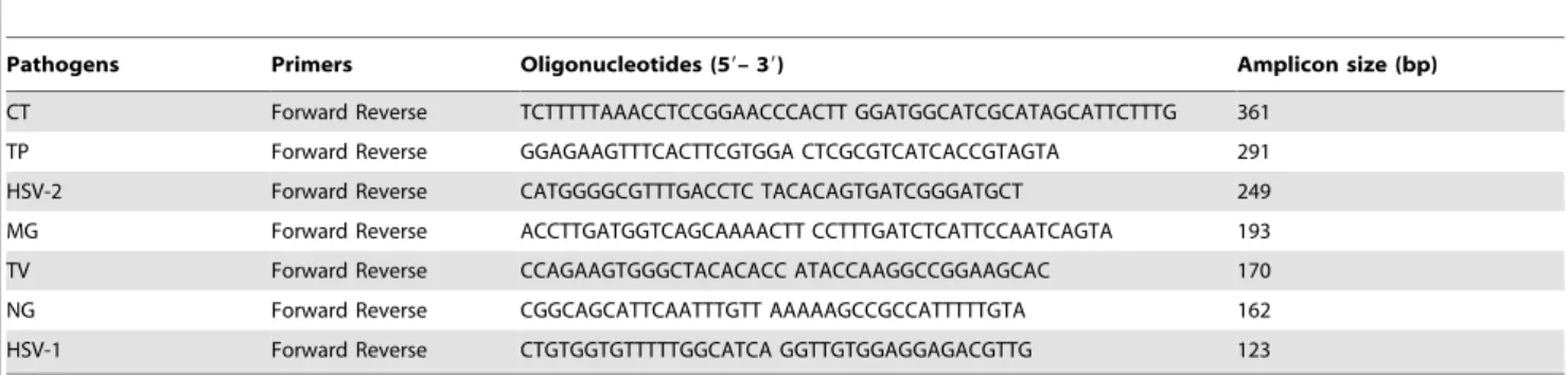
The M-PCR assay clearly distinguished and identified all seven STDs in semen samples whether alone (1 STD) or in co-infections. Table 1.Oligonucleotide primers used in the M–PCR assay.
Pathogens Primers Oligonucleotides (59– 39) Amplicon size (bp)
CT Forward Reverse TCTTTTTAAACCTCCGGAACCCACTT GGATGGCATCGCATAGCATTCTTTG 361
TP Forward Reverse GGAGAAGTTTCACTTCGTGGA CTCGCGTCATCACCGTAGTA 291
HSV-2 Forward Reverse CATGGGGCGTTTGACCTC TACACAGTGATCGGGATGCT 249
MG Forward Reverse ACCTTGATGGTCAGCAAAACTT CCTTTGATCTCATTCCAATCAGTA 193
TV Forward Reverse CCAGAAGTGGGCTACACACC ATACCAAGGCCGGAAGCAC 170
NG Forward Reverse CGGCAGCATTCAATTTGTT AAAAAGCCGCCATTTTTGTA 162
HSV-1 Forward Reverse CTGTGGTGTTTTTGGCATCA GGTTGTGGAGGAGACGTTG 123
M–PCR, multiplex–PCR; CT,C. trachomatis; TP,T. pallidum; HSV–1/–2: herpes virus simplex; MG,M. genitalium; TV,T. vaginalis; NG,N. gonorrhea; bp, base pairs.
doi:10.1371/journal.pone.0098862.t001
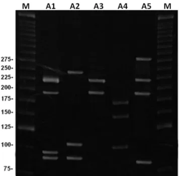
Figure 1. Electrophoretic analysis of the amplified fragments by using a multiplex polymerase chain reaction in 8% polyacrylamide gel stained with ethidium bromide.Lane C1: control ofChlamydia trachomatis(361 base pairs-bp); lane C2: control ofTreponema pallidum (291 bp); lane C3: control of HSV–2 (249 bp); lane C4: control ofMycoplasma genitalium(193 bp); lane C5: control ofTrichomonas vaginalis(170 bp); lane C6: control ofNeisseria gonorrhoeae(162 bp); lane C7: control of HSV–1(123 bp); lane A1: positive sample ofC. trachomatisand HSV–1 (361 and 123 bp); lane A2: positive sample ofT. pallidumand HSV–2 (291 and 249 bp); lane A3: positive sample ofT. vaginalisand HSV–2 (170 and 249 bp); lane A4: positive sample ofC. trachomatisandM. genitalium(361 and 193 bp); lane A5: positive sample ofT. pallidumandT. vaginalis(291 and 170 bp); lane A6: positive sample ofT. vaginalis(170 bp); lanes M1and M2, molecular weight marker (25 bp Invitrogen). Values on the left and right sides of the gel are in bp.
Final results were regarded as true positives if the sPCR was positive (gold standard). The overall agreement of M-PCR results with sPCR was 99.2%, and the validation parameters were as follows: sensitivity and negative predictive value 100%, specificity 99.7%, positive predictive value 96.4%, and accuracy 99.8%. When individually analysed the agentsC. trachomatis, M. genitalium, N. gonorrhoeae, T. pallidum, T. vaginalis and HSV-2, the M-PCR showed values of 100% for all parameters. For HSV-1, the M-PCR showed sensitivity and negative predictive values of 100%, specificity of 98.2%, positive predictive value of 75.0%, and accuracy of 98.3% (Table 2). By M-PCR, 5 semen samples (6.6%) were detected with at least 2 simultaneous STD agents. Figure 1 shows the M-PCR amplification fragments of positive semen samples for different STDs by 8% polyacrylamide gel.
Considering the 7 STDs detected by M-PCR and HPV by sPCR, 1 or more STDs were detected in 42 semen samples (55.3%). The most prevalent STD detected was HPV (n= 29), representing 38.1% of the total samples and 69.0% of the semen positive for STDs. The second most prevalent was T. vaginalis
(n= 10; 13.0% in total samples and 23.8% in semen with STD), followed byC. trachomatis(n= 6; 8.0% in total samples and 14.3% in semen with STD), HSV-1 andT. pallidum(n= 4; 5.3% in total semen samples and 9.5% of semen with STD, each),M. genitalium
andN. gonorrhoeae(n= 3; 4.0% in total samples and 7.1% in semen with STD, each) and HSV-2 (n= 2; 2.6% in total samples and 4.8% in semen with STD) (Table 3). Co-infections by 2 pathogens was in 17 out of 42 infected samples (40.5%) (Table 4). Three simultaneous STD were detected in only 1 sample (2.4% of semen with STD and 1.32% of total samples).
In the semen of men with conjugal infertility, the most prevalent STD was HPV (n= 20/52; 38.5%), followed byT. vaginalis(n = 7/ 52; 13.5%),C. trachomatisand HSV-1/-2 (n= 5/52; 9.6% each). In the semen analyzed due to post-vasectomy, the most prevalent STD was also HPV (n= 9/24; 37.5%), followed by T. vaginalis
(n= 3; 12.5%) (Table 3). Men withC. trachomatisor HSV (types 1 and 2) semen infections had a 2.5-fold greater risk of presenting conjugal infertility (OR = 2.5, 95% CI = 0.3–22.7), although the association between these STDs and conjugal infertility was not statistically significant (p= 0.4 for both).
Among HPV positive semen (n= 29), 17 (58.6%) were infected by 2 or more genotypes simultaneously representing 22.5% of the total samples and 40.5% of the semen with STDs. Figure 2 shows the sPCR amplification fragments of positive semen samples for HPV by 8% polyacrylamide gel. In regarding to carcinogenic HPV groups, 24 out of 29 (82.7%) were HR-HPV. HPV-16 (HR) was the most prevalent genotype detected (n= 13/29; 44.8%) and was found in 17.1% of the total samples and 30.9% of the semen with STDs. HPV-82 and HPV-43 (HR) were the second most prevalent (n = 5/29; 17.2% each), followed by HPV-72 (LR,
n= 4/29; 13.8%),258 (HR,n= 3/29; 10.3%);254 (LR) and266 (HR) (n= 2/29; 6.9% each). Other HPV genotypes detected in single or simultaneous infections were HPV-13,-18,-31,-44,-51,-52,-53,-59,-62,-69 and -81 (n= 1/29; 3.5% each). Table 5 shows the HPV genotypes detected in simultaneous semen HPV infections. According to Santiago et al. [19], their method with a single enzyme (HpyCH4V) application is simple enough for the detection of HPV in clinical samples but cannot be used to distinguish some HPV genotypes such as HPV 11/30, 18/68, 44/ 55, and 61/83/84, because these genotypes yield similar RFLP patterns. Chen et al. [25] used the same enzyme (HpyCH4V) for the initial RFLP and then used a second enzyme ((NlaIII) to confirm the identification. We didn’t find any of these genotypes in our analysis and the patter found by the single enzyme was very different and yielded us to well distinguish between all of them,
using the 8% polyacrylamide gel as showed in Figure 2. So, as mentioned above there was not the requirement to apply another assay with the second enzyme to confirm our results.
In regarding to semen quality parameters, the following observations were made: a decrease of seminal volume in samples with 2 or 3 simultaneous STDs orT. pallidumalone (p= 0.01 and
p= 0.02, respectively); semen with simultaneous T. vaginalis and HPV infections had a 10-fold greater risk of presenting teratospermia (OR = 10.55; 95% CI 0.8 to 13.3) (p= 0.03); a tendency toward a greater risk of semen alterations, although not statistically significant, between T. vaginalis and necrospermia (OR = 2.7; 95% CI 0.4 to 15.7) (p= 0.2), simultaneousT. vaginalis
Table 3.Frequency of STDs and reason for performing the semen analysis.
STD Totaln(%) Conjugal infertilityn(%) Post– vasectomyn(%) OR (IC 95%) P
HPV
(+) 29 (38.0) 20 (38.5) 9 (37.5) 1.1 (0.4–3.0) 0.78
(2) 47 (62.0) 32 (61.5) 15 (62.5)
HR–HPV
(+) 24 (32.0) 16 (30.8) 8 (33.3) 0.9 (0.3–2.7) 0.90
(2) 52 (68.0) 36 (69.2) 16 (66.7)
C. trachomatis
(+) 6 (8.0) 5 (9.6) 1 (4.2) 2.5 (0.3–23.6) 0.40
(2) 70 (92.0) 47 (90.4) 23 (95.8)
T. vaginalis
(+) 10 (13.0) 7 (13.5) 3 (12.5) 1.1 (0.3–4.9) 0.83
(2) 66 (87.0) 45 (86.5) 21 (87.5)
M. genitalium
(+) 3 (4.0) 2 (3.9) 1 (4.2) 0.9 (0.0–11.3) 0.90
(2) 73 (96.0) 50 (96.1) 23 (95.8)
HSV–1/–2
(+) 6 (8.0) 5 (9.6) 1 (4.2) 2.5 (0.3–23.6) 0.40
(2) 70 (92.0) 47 (90.4) 23 (95.8)
T. pallidum
(+) 4 (5.3) 4 (7.7) 0 (0) – 0.10
(2) 72 (94.7) 48 (92.3) 24 (100.0)
N. gonorrheae
(+) 3 (4.0) 2 (3.9) 1 (4.2) 0.9 (0.0–11.3) 0.90
(2) 3 (96.0) 50 (96.1) 23 (95.8)
Total 76 52 24 – –
STD, sexually transmitted diseases; HPV, humanPapillomavirus; HR–HPV, high–risk humanPapillomavirus; HSV–1/–2, herpes simplex virus; (+), positive; (2), negative.
doi:10.1371/journal.pone.0098862.t003
Table 4.Total simultaneous STDs detected by M-PCR and sPCR.
Simultaneous STDs n Total semen samplesn= 76 (%) Semen samples with STDn= 42 (%)
C. trachomatis+HSV–1 2 2.6 4.8
T. pallidum+T. vaginalis 1 1.3 2.4
HSV–2+T. vaginalis 1 1.3 2.4
HSV–2+T. pallidum 1 1.3 2.4
HPV+T. vaginalis 3 3.9 7.1
HPV+C. trachomatis 3 3.9 7.1
HPV+HSV–1 2 2.6 4.8
HPV+M. genitalium 2 2.6 4.8
HPV+N. gonorrhoe 1 1.3 2.4
HPV+T. pallidum 1 1.3 2.4
and HPV with oligospermia (OR = 2.8; 95% CI 0.2 to 3.3) (p= 0.3) and simultaneous HSV-1/-2 and HPV with teratospermia (OR = 4.6; 95% CI 0.2 to 8.2) (p= 0.2).
Discussion
To our knowledge, this is the first study to detect simultaneous bacterial and viral STD pathogens in semen using M-PCR in Brazil and Latin America. The method implemented, which represented an important advance over conventional techniques, allowed the detection of seven STDs in semen, many of which are difficult to identify using standard methods. The overall agreement of the M-PCR with sPCR was elevated (99.2%), and other validation parameters, including sensitivity, specificity, positive and negative predictive value and accuracy, were also excellent (ranging from 99.2% to 100%). Considering the agents individ-ually, the M-PCR also showed excellent values for all the parameters and detected 5 semen samples (6.6%) with co-infections.
The M-PCR assay simplifies workflow, allowing for its use in routine diagnostic laboratories with basic molecular facilities [20– 24]. Thus, this M-PCR assay has great potential to be applied in screening programs for semen pathogens in infertility and STD clinics and in sperm banks. Further investigations applying M-PCR in different populations and clinical situations may help elucidate the impact of STD pathogens on male infertility. All encompassed 7 STDs in addition to HPV were analyzed by M-PCR and sM-PCR. The overall prevalence of STDs in semen from asymptomatic men was determined to mount to 42 semen samples out of 52 (55.3%) and showed 1 or more pathogens.
HPV was the most prevalent STD pathogen in semen (n= 29; 38.1% of total semen and 69.0% of semen with STDs). Surprisingly, in 58.6% of semen samples 2 or more genotypes were detected simultaneously (22.5% of total semen and 40.5% of
Figure 2 Electrophoretic analysis of the HPV genotyping in semen using PCR-RFLP with restriction enzymeHpyCH4V in 8% polyacrylamide gel stained with ethidium bromide.Sample A1, genotypes216 (High-risk, HR) and231 (HR) in double HPV infection (216, 191, 94 and 91 base pairs-bp); A2, genotype213 (low-risk, LR) in single HPV infection (244, 103 and 91 bp); A3, genotype216 (HR) in single HPV infection (216 and 191 bp); A4, genotype218 (HR) in single HPV infection (174, 144 and 100); A5, genotypes281(LR),266 (HR) and 216 (HR) in multiple HPV infection (284, 216, 191 and 89 bp). M, molecular weight marker (25 bp).
doi:10.1371/journal.pone.0098862.g002
Table 5.HPV genotypes distribution of 17 semem samples with two or more genotypes.
Samples HPV genotypes n(%)
LR HR
1 281 16,266 1 (5.88)
2 _ 251,252 1 (5.88)
3 _ 16,282 1 (5.88)
4 _ 16,231 1 (5.88)
5 254 282 2 (11.80)
6 272 253 1 (5.88)
7 272 258 1 (5.88)
8 243 282 1 (5.88)
9 244 282 1 (5.88)
10 243 258 1 (5.88)
11 243,272 _ 1 (5.88)
12 _ 216,259 1 (5.88)
13 _ 216,258 1 (5.88)
14 _ 216,256 1 (5.88)
15 269 216 1 (5.88)
16 272 216 1 (5.88)
17 243 216 1 (5.88)
HPV, humanPapillomavirus; HR, high–risk humanPapillomavirus; LR, low-risk humanPapillomavirus.
positive semen for STDs) and HR-HPV represented 82.7% of all the HPV genotypes detected. The most prevalent HPV genotype was HPV-16 (n= 13/29, 44.8%; n= 13/76, 17.1%), followed by HPV-82 and HPV-43, both of which are HR-HPV (n= 5/29; 17.2% each). The current vaccine protects against HR HPV-16 and -18, but in our study HPV-18 genotype is not the second most frequent HR but HPV-82 and -43, which could have implications for the effectiveness of the vaccine Brazilian men.
A recent study found HPV at a much lower prevalence (10%), but the population selected for this research included only asymptomatic sexually active young men [25]. HPV infection is well-characterized in women. However, little attention has been given to the transmission of HPV through semen [26,27] because the virus is primarily transmitted through direct epithelial contact [28]. Normally, HPV infection in men is considered temporary, but little is known about the incubation time and possible viral manifestations [29].
The second most prevalent STD wasT. vaginalis(n= 10; 13.0% in total semen samples and 23.8% in semen with STDs). Knowledge of T. vaginalisin men, including in semen, is limited mainly due to difficulties associated with diagnosis including poor sensitivity of available methods [30,31]. Estimates of the preva-lence of trichomoniasis range from 6–12% of asymptomatic men to 20% of men with urethritis [32]. A study in females with trichomoniasis analyzed the urine and semen of their sexual partners and found that 72% of men were also positive for T. vaginalisdespite the fact that the majority were asymptomatic [31].
C. trachomatiswas the third most common STD (n= 6; 8.0% in total semen samples and 14.3% semen with STD). The number of men infected by this pathoges was similar to another study that showed that although up to 13.3% of young men carry genitalC. trachomatis infection, only half of these will present with any symptom and even fewer are likely to pursue treatment [33]. Thus, it has been suggested that undiagnosedC. trachomatisinfection in either partner could potentially contribute to unexplained infertility [23]. This bacterium can be located in any part of the male reproductive tract, including sexual glands, such as the prostate and seminal vesicles [34].C. trachomatisinfections of male sexual glands may cause severe complications that can threaten male fertility [23]. Urethritis is the most common clinical presentation ofC. trachomatisinfection observed in men [35], but it is normally an acute episode of chronic and silent genitourinary infection [36–38]. Evidence also suggests that upper genital tract infections in young men, including epididymitis, are attributable to
C. trachomatis [37–39]. Epididymitis is thought to be important because fertility can be affected by inflammation and obstruction, especially when both testes are affected [35].
Other STD pathogens were detected in lower abundances as follows: HSV-1 andT. pallidum(n= 4/76; 5.3% each),M. genitalium
andN. gonorrhoeae(n= 3/76; 4.0% each) and HSV-2 (n= 2/76). No previous studies were found assessing the prevalence ofT. pallidum
in semen. Although a direct toxic effect of syphilis on male fertility has not been reported in the literature, it is known that complications of syphilis can affect fertility. Syphilitic epididymitis can cause obstruction of the epididymis. Chronic obliterative endarteritis and interstitial inflammation can occur in congenital or tertiary syphilis and lead to small, fibrotic testes [40]. Gummatous lesions cause destruction of local tissue and, when occurring in the testicles, may have an impact on testicular function and fertility [41].
Among men presenting persistent or recurrent urethritis, 19% to 41% are infected withM. genitalium[42]. This bacterium is a probable cause of non-gonococcal urethritis [43,44] and is also associated with prostatitis [24,45–49]. The detection of genital mycoplasmas only in semen may indicate that these organisms are harbored in the epididymis or seminal vesicles. The influence of mycoplasmas on semenology may come from their ability to attach to spermatozoa and directly affect cellular interactions, influencing vitality, motility, morphology, cellular integrity, molecular struc-ture and the development of protective immunity to genital infection by the host or other host factors [24].
N. gonorrhoeaeinfection in men can lead to genitourinary tract inflammation (e.g., urethritis and epididymitis), obstruction and infertility [50–52]. Gonococcus is transmitted more efficiently from an infected male to a female (50% to 73% probability, independent of the number of exposures) [50,53,54]. BecauseN. gonorrhoeaeis mostly symptomatic in males, there have been very few studies regarding the relationship between this infection and male infertility. Surprisingly, we detected HSV-2 in semen less frequently than HSV-1. Most commonly, HSV-1 causes oral and genital sores, but HSV-2 is the most common cause of genital herpes. Generally, direct or indirect contact with herpetic lesions is infectious, but HSV-1 and HSV-2 have been detected in semen [22,55] and in sperm, and HSV-2 has been transmitted through donor insemination [56,57]. DNA of HSV-1 and HSV-2 has been detected in the semen from 2-50% of men with no significant difference between fertile and infertile subjects [22,56,58]. Evidence indicates that HSV infections contribute to male factor infertility either by directly invading male genital tract cells or by indirectly causing local immune responses that can negatively affect reproduction [59]. More specifically, HSV infection has been correlated to reduced sperm count, progressive motility, increased apoptosis and low sperm concentration [60,61].
Other viruses can be highly frequent in semen such as cytomegalovirus (CMV) [8,58] which is well known a STD pathogen [17]. CMV could be one of the most frequent pathogens in semen. However it was not investigated in our study, since our aim was adapt the M-PCR assay for seven agents as used by Souza et al. [14] in cervical samples to semen samples and we succeed. Thus, a new M-PCR assay allowing the detection of other STD agents would be interesting and a promising research to be standardized.
Despite being a secondary goal of our study some associations were observed between STDs and changes in semen parameters such as: decreased seminal volume in samples with 2 or 3 simultaneous STDs orT. pallidumalone; semen with simultaneous
T. vaginalis and HPV infections with a 10-fold greater risk of teratospermia. Correlations were also observed in:T. vaginaliswith necrospermia;T. vaginalisand HPV co-infections with oligosper-mia; HSV-1/-2 and HPV co-infections with teratospermia. These associations confirmed the involvement of STD pathogens with male infertility as described in few previous studies [7–9].
Author Contributions
References
1. World Health Organization (2007) Global strategy for the prevention and control of sexually transmitted infections: 2006–2015. Breaking the chain of transmission. Geneva: World Health Organization.
2. Simms I, Warburton F, Westrom L (2003) Diagnosis of pelvic inflammatory disease: time for a rethink. SexTransm Infect 79: 491–494.
3. Gore–Felton C, Vosvick M, Bendel T, Koopman C, Das B, et al. (2003) Correlates of sexually transmitted disease infection among adults living with HIV. Int J STD AIDS 14: 539–546.
4. Van B, Pol D, Kwok C, Pierre–Louis B, Rinaldi A, et al. (2008)Trichomonas vaginalisinfection and human immunodeficiency virus acquisition in African women. J Infect Dis 197: 548–554.
5. World Health Organization (1993) Manual for the standardized investigation and diagnosis of the infertile couple. UK: Cambridge University Press. 50 p. 6. Brookings C, Goldmeier D, Sadeghi–Nejad H (2013) Sexually transmitted
infections and sexual function in relation to male fertility. Korean J Urol 54: 149–156.
7. Ochsendorf FR (2008) Sexually transmitted infections: impact on male fertility. Andrologia 40: 72–75.
8. Garolla A, Pizzol D, Bertoldo A, Menegazzo M, Barzon L (2013) Sperm viral infection and male infertility: focus on HBV, HCV, HIV, HPV, HSV, HCMV, and AAV. J Reprod Immunol 100: 20–29.
9. Bezold G, Politch JA, Kiviat NB, Kuypers JM, Wolff H, et al. (2007) Prevelence of sexually transmissible pathogens in semen from asymptomatic male infertility patients with and without leukocytospermia. Fertil Steril 87: 1087–1097. 10. Rusz A, Wagenlehner F, Linn T, Diemer T, Schuppe HC, et al. (2012) Influence
of urogenital infections and inflammation on semen quality and male fertility. World J Urol 30: 23–30.
11. Olatunbosun OA, Chizen DR, Pierson RA (1998) Screening of potential semen donors for sexual transmitted diseases. West Afr J Med 17: 19–24.
12. Peeling R, Embree J (2005) Screening for sexually transmitted infection pathogens in semen samples. Can J Infect Dis Med Microbiol 16: 73–76. 13. Sankuntaw N, Sukprasert S, Engchanil C, Kaewkes W, Chantratita W, et al.
(2011) Single tube multiplex real–time PCR for the rapid detection of herpesvirus infections of the central nervous system. Mol Cell Probes 25: 114– 120.
14. Souza RP, Abreu AL, Ferreira EC, Rocha–Brischiliari SC, Carvalho MD, et al. (2013) Simultaneous Detection of Seven Sexually Transmitted Agents in Human Immunodeficiency Virus–Infected Brazilian Women by Multiplex Polymerase Chain Reaction. Am J Trop Med Hyg 30: 1199–1202.
15. World Health Organization (2010) WHO Laboratory manual for the examination and processing of human semen. Geneva: World Health Organization. 287 p.
16. Muvunyi CM, Dhont N, Verhelst R, Crucitti T, Reijans M, et al. (2011) Evaluation of a new multiplex polymerase chain reaction assay STDFinder for the simultaneous detection of 7 sexually transmitted disease pathogens. Diagn Microbiol Infect Dis 71: 29–37.
17. McIver CJ, Rismanto N, Smith C, Naing ZW, Rayner B, et al. (2009) Multiplex PCR testing detection of higher–than–expected rates of Cervical Mycoplasma, Ureaplasma, and Trichomonas and viral agent infection in sexually australian women. J Clin Microbiol 47: 1358–1363.
18. Manos MM, Waldman J, Zhang TY, Greer CE, Eichinger G, et al. (1994) Epidemiology and partial nucleotide sequence of four novel genital Human papillomaviruses. J Infect Dis 170: 1096–1099.
19. Santiago E, Camacho L, Junquera ML, Va´zquez F (2006) Full HPV typing by a single restriction enzyme. J Clin Virol 37: 38–46.
20. McKechnie ML, Hillman R, Couldwell D, Kong F, Freedman E, et al. (2009) Simultaneous identification of 14 genital microorganisms in urine by use of a multiplex PCR–based reverse line blot assay. J Clin Microbiol 47: 1871–1877. 21. Wang Y, Liang CL, Wu JQ, Xu C, Qin SX, et al. (2006) Ureaplasma urealyticum infections in the genital tract affect semen quality? Asian J Androl 8: 562–568.
22. Bezold G, Politch JA, Kiviat NB, Kuypers JM, Wolff H, et al. (2007) Prevalence of sexually transmissible pathogens in semen from asymptomatic male infertility patients with and without leukocytospermia. Fertil Steril 87: 1087–1097. 23. Cunningham KA, Beagley KW (2008) Male genital tract Chlamydial infection:
implications for pathology and infertility. Biol Reprod 79: 180–189. 24. Gdoura R, Kchaou W, Znazen A, Chakroun N, Fourati M, et al. (2008)
Screening for bacterial pathogens in semen samples from infertile men with and without leukocytospermia. Andrologia 40: 209–218.
25. Chen L, Watanabe K, Haruyama T, Kobayashi N (2013) Simple and rapid human Papillomavirus genotyping method by restriction fragment length polymorphism analysis with two restriction enzymes. J Med Virol 85: 1229– 1234.
26. Foresta C, Patassini C, Bertoldo A, Menegazzo M, Francavilla F, et al. (2011) Mechanism of human papillomavirus binding to human spermatozoa and fertilizing ability of infected spermatozoa. PLoS One 6: e15036.
27. Kaspersen MD, Larsen PB, Ingerslev HJ, Fedder J, Petersen GB, et al. (2011) Identification of multiple HPV types on spermatozoa from human sperm donors. PLoS One 6: e18095.
28. Elder K, Baker DJ, Ribes JA (2005) Infections, infertility, and assisted reproduction. Cambridge: Cambridge University Press. 412 p.
29. Foresta C, Pizzol D, Moretti A, Barzon L, Palu` G, et al. (2010) Clinical and prognostic significance of Human papillomavirus DNA in the sperm or exfoliated cells of infertile patients and subjects with risk factors. Fertil Steril 94: 1723–1727.
30. Rein M, Muller M (1990)Trichomonas vaginalisand trichomoniasis. In: Holmes KK, (eds.), sexually transmitted diseases. New York: McGraw– Hill Press. pp 481–492.
31. Hobbs MM, Lapple DM, Lawing LF, Schwebke JR, Cohen MS, et al. (2006) Methods for detection ofTrichomonas vaginalisin the male partners of infected women: implications for control of trichomoniasis. J Clin Microbiol 44: 3994– 3999.
32. Hobbs MM, Kazembe P, Reed AW, Miller WC, Nkata E, et al. (1999) Trichomonas vaginalisas a cause of urethritis in Malawian men. Sex Transm Dis 26: 381–387.
33. LaMontagne DS, Fenton KA, Randall S, Anderson S, Carter P (2004) Establishing the NationalChlamydiaScreening Program in England: results from the first full year of screening. Sex Transm Infect 80: 335–341.
34. Mackern–Oberti JP, Motrich RD, Breser ML, Sa´nchez LR, Cuffini C, et al. (2013) Chlamydia trachomatis infection of the male genital tract: An update. J Reprod Immunol 100: 37–53.
35. Eley A, Pacey AA, Galdiero M, Galdiero M, Galdiero F (2005) CanC. trachomatis directly damage your sperm? Lancet Infect Dis 5: 53–57.
36. Gonzales GF, Mun˜oz G, Sa´nchez R, Henkel R, Gallegos–Avila G, et al. (2004) Update on the impact of Chlamydia trachomatis infection on male fertility. Andrologia 36: 1–23.
37. Nadala EC, Goh BT, Magbanua JP, Barber P, Swain A, et al. (2009) Performance evaluation of a new rapid urine test for Chlamydia in men: prospective cohort study. BMJ 339: 2655.
38. Kalwij S, French S, Mugezi R, Baraitser P (2012) Using educational outreach and a financial incentive to increase general practices contribution to Chlamydia screening in South–East London 2003–2011. BMC Public Health 18: 802. 39. Furuya R, Takahashi S, Furuya S, Kunishima Y, Takeyama K, et al. (2004) Is
seminal vesiculitis a discrete disease entity? Clinical and microbiological study of seminal vesiculitis in patients with acute epididymitis. J Urol 171: 1550–1553. 40. Cheng L, Bostwick DG (2002) Essentials of anatomic pathology. Totowa:
Humana Press. 1879 p.
41. Brookings C, Goldmeier D, Sadeghi–Nejad H (2013) Sexually transmitted infections and sexual function in relation to male fertility. Korean J Urol 54: 149–156.
42. Wikstro¨m A, Jensen JS (2006) Mycoplasma genitalium: a common cause of persistent urethritis among men treated with doxycycline. Sex Transm Infect 82: 276–279.
43. Uusku¨la A, Kohl PK (2002) Genital Mycoplasmas, including Mycoplasma genitalium, as sexually transmitted agents. Int J STD AIDS 13: 79–85. 44. Jensen JS (2004)Mycoplasma genitalium: the aetiological agent of urethritis and
other sexually transmitted diseases. J Eur Acad Dermatol Venereol 18: 1–11. 45. Krieger JN, Riley DE (2002) Prostatitis: what is the role of infection.
Int J Antimicrob Agents 19: 475–479.
46. Lee JS, Kim KT, Lee HS, Yang KM, Seo JT, et al. (2013) Concordance of Ureaplasma urealyticum andMycoplasma hominis in infertile couples: impact on semen parameters. Urology 81: 1219–1224.
47. Yoshida T, Maeda S, Deguchi T, Miyazawa T, Ishiko H (2003) Rapid detection ofMycoplasma genitalium,Mycoplasma hominis,Ureaplasma parvumandUreaplasma urealyticum organisms in genitourinary samples by PCR–microtiter plate hybridization assay. J Clin Microbiol 41: 1850–1855.
48. Al–Daghistani HI, Abdel–Dayem M (2010) Clinical significance of asymptom-atic urogenitalMycoplasma hominisandUreaplasma urealyticumin relation to seminal fluid parameters among infertile Jordanian males. Middle East Fertil Soc J 15: 29–34.
49. Svenstrup HF, Fedder J, Abraham–Peskir J, Birkelund S, Christiansen G (2003) Mycoplasma genitaliumattaches to human spermatozoa. Hum Reprod 18: 2103– 2109.
50. Edwards JL, Apicella MA (2004) The molecular mechanisms used byNeisseria gonorrhoeaeto initiate infection differ between men and women. Clin Microbiol Rev 17: 965–981.
51. Pellati D, Mylonakis I, Bertoloni G, Fiore C, Andrisani A, et al. (2008) Genital tract infections and infertility. Eur J Obstet Gynecol Reprod Biol 140: 3–11. 52. Radek S, Vanda B, Miloslav S, Petra M, Eva K, et al. (2013) Bacterial infection
as a cause of infertility in humans. Epidemiol Mikrobiol Imunol 62: 26–32. 53. Bolan G, Ehrhardt AA (1999) Gender perspectives and STDs. In: Sexually
transmitted diseases. New York: McGraw–Hill Press. 117–127 p.
54. Hook EW, Handsfield HH (1999) Gonococcal infections in the adult. In: Sexually Transmitted Diseases. New York: McGraw Hill Press. 451–466 p. 55. Zuckerman RA, Lucchetti A, Whittington WL, Sa´nchez J, Coombs RW, et al.
(2009) HSV suppression reduces seminal HIV–1 levels in HIV–1/HSV–2 co– infected men who have sex with men. AIDS 23: 479–483.
56. Kotronias D, Kapranos N (1998) Detection ofHerpes simplexVirus DNA in human spermatozoa by in situ hybridization technique. In Vivo 12: 391–394. 57. Kaspersen MD, Ho¨llsberg P (2013) Seminal shedding of Human herpesviruses.
58. Neofytou E, Sourvinos G, Asmarianaki M, Spandidos DA, Makrigiannakis A (2009) Prevalence of human herpes virus types 1–7 in the semen of men attending an infertility clinic and correlation with semen parameters. Fertil Steril 91: 2487–2494.
59. Ochsendorf FR (2008) Sexually transmitted infections: impact on male fertility. Andrologia 40: 72–75.
60. Monavari SH, Vaziri MS, Khalili M, Shamsi–Shahrabadi M, Keyvani H, et al. (2013) Asymptomatic seminal infection of herpes simplex virus: impact on male infertility. J Biomed Res 27: 56–61.