© 2013 Sociedade Brasileira de Hemodinâmica e Cardiologia Intervencionista. Published by Elsevier Editora Ltda. All rights reserved.
Transcatheter Pulmonary Valve Implantation:
Systematic Literature Review
Tobias Engel Ayer Botrel
1, Otávio Augusto C. Clark
2, Marcelo C. Queiroga
3, Raul I. Rossi Filho
4,
Carlo B. Pilla
5, Raul S. Arrieta
6, Salvador Cristovão
7, Célia C. Silva
8, Cesar A. Esteves
9,
Edmundo Clarindo Oliveira
10, Luiz Carlos Simões
11, Francisco Chamié
12, Juliana Neves
13,
Roberto Max
14, Carlos A. C. Pedra
15RESUMO
Implante Transcateter de Bioprótese Valvular Pulmonar: Revisão Sistemática da Literatura
A correção cirúrgica de algumas cardiopatias congênitas com-plexas envolve a reconstrução da via de saída do ventrículo direito com a interposição de homoenxertos, biopróteses, enxertos de jugular bovina ou outros condutos valvulados entre o ventrículo direito e o tronco da artéria pulmonar. Apesar de essas cirurgias poderem ser realizadas com bai-xa mortalidade, a vida útil das válvulas ou dos condutos implantados é normalmente pequena (< 10 anos), seja por degeneração e/ou calciicação. Graus variáveis de estenose pulmonar na maioria das vezes associada à insuiciência pulmonar são consequências da degeneração dos condutos. Em 2000, Bonhoeffer et al. foram os primeiros a relatar o implante transcateter de bioprótese valvular pulmonar (ITVP) com um dispositivo que posteriormente foi denominado de válvula Melody
(Medtronic – Minneapolis, Estados Unidos). A técnica foi inicialmente desenvolvida para limitar a neces-sidade de múltiplos procedimentos cirúrgicos, substituindo, em última análise, uma nova troca cirúrgica valvular. Estudos subsequentes na Europa e Estados Unidos atestaram para a ABSTRACT
Surgical repair of some complex congenital heart diseases involves reconstruction of the right ventricular outlow tract using homografts, bioprostheses, bovine jugular grafts or other valved conduits between the right ventricle and the main pulmonary artery. Although these surgical procedures may be performed with low mortality rates, the life span of these implanted valves or conduits is usually short (< 10 years) due to either degeneration and/or calciication. Variable degrees of pulmonary stenosis, often associated with pulmonary insuf-iciency, are consequences of conduit degeneration. In 2000, Bonhoeffer et al. were the irst to report the transcatheter pul-monary valve implantation (TPVI) of a bioprosthetic pulpul-monary valve later named Melody
valve (Medtronic, Minneapolis, USA). The technique was initially developed to limit the need for multiple surgical procedures, and, ultimately, to work as a surrogate of a new surgical valve replacement. Subsequent clinical studies in Europe and the United States conirmed the safety and eficacy of this technique in a larger number of patients. Since the National Sanitary Surveillance Agency (Agência Nacional de Vigilância Sanitária – Anvisa) granted approval for clinical use of the Melody
transcatheter pulmonary biological valve in February 2103, we deemed that a judicious
Original Article
1 Oncologist. Researcher of Evidências – Credibilidade Cientíica.
Campinas, SP, Brazil.
2 Ph.D. Oncologist. Director of Evidências – Credibilidade Cientíica.
Campinas, SP, Brazil.
3 Interventional cardiologist of the Hospital da Unimed João Pessoa.
João Pessoa, PB, Brazil.
4 Master. Chief of Interventional Cardiology for Congenital Heart
Defects of the Instituto de Cardiologia da Fundação Universitária de Cardiologia. Porto Alegre, RS, Brazil.
5 Master. Physician in the Department of Pediatric Cardiology and
Interventional Cardiology of the Complexo Hospitalar Santa Casa de Porto Alegre. Porto Alegre, RS, Brazil.
6 Interventional cardiologist in the Department of Interventional
Car-diology, Heart Institute of the Hospital das Clínicas da Faculdade de Medicina da Universidade de São Paulo. São Paulo, SP, Brazil.
7 Interventional cardiologist responsible for the Sector of Interventions
in Congenital Heart Diseases of the Hospital Beneicência Portuguesa de São Paulo. São Paulo, SP, Brazil.
8 Ph.D. Chief of Pediatric Cardiology at the Escola Paulista de Medicina
da Universidade Federal de São Paulo. São Paulo, SP, Brazil.
9 Ph.D. Chief of the Medical Section of Interventions in Acquired
Val-vular Heart Diseases of the Instituto Dante Pazzanese de Cardiologia. São Paulo, SP, Brazil.
10 Ph.D. Head of the Department of Congenital Heart Defects of the
Hospital de Clinicas da Universidade Federal de Minas Gerais, Belo Horizonte, MG, Brazil.
11 Master. Chief of Cardiology at Children’s Instituto Nacional de
Car-diologia, Hospital Laranjeiras. Rio de Janeiro, RJ, Brazil.
12 Master. Chief of Interventional Cardiology and Congenital Defects of
Structural of the Hospital Federal dos Servidores do Estado, RJ, Brazil.
13 Interventional cardiologist. Chief of Hemodynamics in Congenital
Heart Diseases of the Instituto de Medicina Integral Prof. Fernando Figueira. Recife, PE, Brazil.
14 Interventional cardiologist. Head of the Department of Pediatric
Inter-ventional Cardiology of the Hospital Biocor. Belo Horizonte, MG, Brazil.
15 Ph.D. Chief of the Medical Section of Interventions in Congenital
Heart Diseases of the Instituto Dante Pazzanese de Cardiologia. São Paulo, SP, Brazil.
Correspondence to: Carlos A. C. Pedra. Av. Dr. Dante Pazzanese, 500 – 14o andar – São Paulo, SP, Brazil – CEP 04012-180
E-mail: cacpedra@uol.com.br
S
urgical correction of some complex and less frequent congenital heart diseases involves reconstruction of the right ventricular (RV) outlow tract with the interposition of homografts, bovine jugular bioprosthe-ses grafts, or other valved conduits between the right ventricle and the main pulmonary artery. Examples of diseases that may require such strategy include tetral-ogy of Fallot, pulmonary atresia with ventricular septal defect, double-outlet right ventricle with infundibular pulmonary stenosis, transposition of the great arteries with ventricular septal defect and infundibular pulmonary stenosis, and persistent truncus arteriosus. Ross procedure, used for reconstruction of the left ventricular outlow tract in case of double aortic lesion, also involves this strategy. Although these reconstructive surgeries of the right outlow tract ventricle can be performed withlow mortality,1 the duration of the implanted valves
or conduits is usually small (< ten years), due to
de-generation and/or calciication of the materials used for their manufacture. Varying degrees of pulmonary stenosis, most often associated to pulmonary failure, are the result of conduit degeneration. The earlier the interposition is performed; the lower is the durability of the conduit. Such observation results in the need
to perform several open-heartsurgeries,1 which have
great impact on patients’ health and quality of life, especially in the case of children. In addition, a new pulmonary valve replacement surgical requires the use of cardiopulmonary bypass (CPB), which can worsen
the RV function, usually already compromised.2,3
By mid-2000,4 the therapeutic alternatives to a new
surgical valve replacement were limited. The percutane-ous implantation of bare-metal stents in these stenotic conduits or bioprostheses was used both in Brazil and
abroad,5,6 aiming to increase survival and minimize the
need for repeated invasive procedures. Despite provid-ing the possibility of postponprovid-ing a new surgery, this strategy results in total pulmonary failure with currently well known deleterious effects, including arrhythmias,
ventricular dysfunction, and decreased aerobic capacity.
In 2000, Bonhoeffer et al.7 were the irst to report the
transcatheter pulmonary biological valve implantation,
later named the Melody
valve (Medtronic, Minneapolis, USA). This valve is made with bovine jugular vein tissue
and mounted on a stent.7,8 The technique was
origi-nally developed to limit the need for multiple surgical procedures, ultimately replacing a new surgical valve
exchange.1 Subsequent studies conirmed the safety and
eficacy of this technique,4,8 which has been used in
over 1,000 patients in the world, especially in Europe.9
As it occur with the less prevalent diseases, to date there have been no prospective, randomized trials with a large number of patients to deinitively guide the treatment of dysfunction in these conduits in the RV outlow tract. This is due to the dificulty in performing controlled and randomized clinical trials for a small population of patients with rare diseases. In addition, there are ethical issues that make it impossible to compare a less invasive procedure with more invasive surgical treatments. The Food and Drug Administration (FDA) has adapted to these particularities in treatment research for uncommon diseases. A review of the recent approvals demonstrates that some drugs were approved based on phase II studies and even in a historical series of cases. In the case of the transcatheter pulmonary valve
Melody
, the FDA granted humanitarian approval to the device between late 2009 and early 2010, based on a study of a large case series performed in ive ameri-cans centers of excellence. The Humanitarian Device Exemption (HDE) program was established in 1990, aiming to create an alternative path to accelerate the introduction in the market of technologies directed to the treatment of patients with rare diseases or conditions. According to FDA rules, when a device is intended to beneit patients with a disease or a condition that affects fewer than 4,000 individuals per year, there is an incentive from the U.S. government, which by a federal law offers the manufacturer exemption from the segurança e eicácia dessa técnica em um número maior de pacientes. Como a Agência Nacional de Vigilância Sanitária (Anvisa) concedeu a aprovação para o uso clínico da válvula biológica pulmonar transcateter Melody em fevereiro de 2013, consideramos necessária e oportuna a avaliação judiciosa da utilização dessa nova tecnologia antes que ela fosse aplicada em larga escala em nosso país. O objetivo deste estudo foi realizar uma revisão sistemática da literatura sobre o ITVP em pacientes com disfunções de homoenxertos, condutos valvulados e biopróteses implantados cirurgicamente na via de saída do ventrículo direito.
DESCRITORES: Valva pulmonar. Cateterismo cardíaco. Próteses valvulares cardíacas. Próteses e implantes. Revisão.
assessment of this new technology was timely and necessary before the widespread use in our country. The objective of this study was to perform a systematic literature review on the use of TPVI in patients with dysfunctional homografts, valved conduits and bioprostheses implanted surgically in the right ventricular outlow tract.
requirements of eficiency and effectiveness. However, it is necessary to demonstrate to the FDA that the device is safe for patients and that the beneits outweigh the risks. In addition, the applicant must demonstrate that there is no comparable device available for treatment or diagnosis of the same disease or condition. The goal of the humanitarian approval is to beneit a popula-tion that would not be treated if the requirements for approval of these devices for diseases and rare condi-tions were the same as for other devices. Moreover, this initiative encourages companies that manufacture medical devices to develop technologies that meet these populations’ needs.
Evidence-based medicine uses techniques and tools that assist in the search and synthesis of the best available information in the literature. Currently, these techniques are being increasingly used for creating pro-tocols and guidelines throughout the world, including Brazil. Since the Brazilian Health Surveillance Agency (Agência Nacional de Vigilância Sanitária – ANVISA)
granted approval for clinical use of the Melody
trans-catheter pulmonary biological valve in February 2103, a judicious assessment of this new technology was deemed timely and necessary before its widespread use in Brazil (Appendix).
OBJECTIVE
This study aimed to perform a systematic review the literature on the use of bioprosthesis transcatheter pulmonary valve implantation (TPVI) in patients with dysfunctional homografts, valved conduits, and surgically-implanted bioprostheses in the RV outlow tract.
METHODS
Literature review and an extensive search in the computerized databases EMBASE, LILACS, and MED-LINE were performed, using the words “transcatheter pulmonary valve” and “treatment”.
The combination of these terms provided the set of references considered for analysis. The search was restricted to articles in human subjects and of the fol-lowing types: randomized controlled trials, systematic reviews or narratives, meta-analysis, guidelines, clinical studies, and case series. The search was not restricted regarding date or language.
Articles resulting from the analysis were reviewed, as well as the references of the current guidelines. No meta-analysis of the obtained data was performed.
In the Cochrane Library, the term “transcatheter pulmonary valve” was used to search for systematic reviews.
A search was also performed in annals of con-gresses of medical specialty societies, such as the
Brazilian Society of Hemodynamics and Interventional Cardiology (Sociedade Brasileira de Hemodinâmica e Cardiologia Intervencionista – SBHCI) and the Brazil-ian Society of Cardiology (Sociedade Brasileira de Cardiologia – SBC).
The first two authors of this manuscript per-formed the literature review and initial drafting of
the manuscript; both work for Evidências company.
Subsequently, the article was critically reviewed by interventionists, specialized in congenital heart disease, who are familiar with the subject and will soon be involved in this type of procedure in Brazil. All interventionists are associated in some way to SBHCI, which supported this initiative.
Studied intervention
The intervention assessed in this review was the TPVI.
Description of the studied device for
bioprosthesis transcatheter pulmonary valve implantation
There are two types of prosthesis used in TPVI. The
oldest is called the Melody
valve, the only prosthesis approved in Brazil for use in the pulmonary position. Since almost all of the evidence found in the literature is based on the use of this device, this review will address mainly this type of prosthesis. The second prosthesis
is the SAPIEN
valve (Edwards, Irvine, United States), approved in Brazil for use in the aortic position only, although the valve itself is practically the same.
There are two components in the Melody
valve
system: the Melody
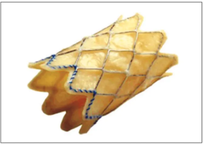
transcatheter pulmonary valve itself, model PB10 (bovine jugular valve with stent) (Figure 1)
and the Ensemble
transcatheter valve delivery system
Figure 1 – External aspect of the Melody
(Medtronic, Minneapolis, United States), NU10 model (Figures 2 and 3).
The Melody
transcatheter pulmonary valve is com-posed by a heterologous (bovine) jugular valve sutured within a platinum-iridium laser-welded stent, and the stent has gold-welded joints. The system is submitted to a inal sterilisation procedure with an adequate sterilising agent containing 1% glutaraldehyde and 20% isopropyl alcohol, in which the valve and stent are preserved and packed until they are used.
The Ensemble
transcatheter valve delivery system consists of a balloon-in-balloon catheter with a polytet-raluoroethylene retractable sheath and a distal support that is large enough to frontally carry the valve, after the valve with the stent has been compressed and adapted to the balloon. The delivery system is available in sizes of 18 mm, 20 mm, and 22 mm. The sheath of the catheter has a side port used to lush the system and a hemostatic sleeve on the sheath to minimize bleeding at insertion. The catheter has a conical distal
obturator made of polyether-b-amide (Pebax
; Arkema – Colombes, France). The delivery system is compatible with a guide wire of 0.889 mm.
Patients involved
Patients were both children and adults with objective dysfunctions (severe stenosis and/or severe insuficiencies)
in homografts, valved conduits, and bioprostheses surgi-cally implanted in the RV outlow tract.
Analysis of the results obtained in the study search
All references retrieved through the search strategies had their title and abstracts read by two researchers, the irst authors of this article. If there was any indication that a reference could meet the inclusion criteria of this study, it was included in a list of selected studies. The corresponding original article was obtained for every selected reference. Each of those articles was read by the researchers, who assessed whether they met the inclusion criteria.
All articles that met the inclusion criteria were separated for data extraction.
Data Extraction
A careful analysis and reading was performed for each article included, in order to extract data.
A speciic form for data extraction was prepared. The data from each included study were extracted independently by two reviewers. The name of the irst author and year of publication were used to identify the study. All data were extracted directly from the published articles.
Figure 2 – Delivery System (Ensemble
) developed specifically for the Melody
valve implantation. (Photos provided by Medtronic.) BIB = balloon-in-balloon.
BIB
Guidewire entry External balloon entry Internal balloon entry Hemostatic adapter
for the vascular access site
“Closed valve” indicator
Hemostatic valve sheath
Tap?
“Open valve” indicator
Catheter body
Catheter body
Balloon-size indicator
Studied variables
The following variables were assessed:
• characteristics of patients;
• characteristics of the procedure performed;
• hemodynamic results after TPVI; and
• safety data.
RESULTS
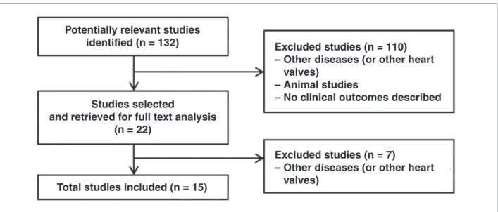
Figure 4 presents the flow chart used for identi-fication of the included studies, as recommended by the Preferred Reporting Items for Systematic reviews
and Meta-analysis (PRISMA).10 A total of 132 studies
were obtained in the first search, whose articles on other (non-pulmonary) heart valves were not used for preparation of this systematic review. Additionally, animal studies and studies without a description of clinical or laboratory outcomes were excluded. Of
this total, 15 studies met the inclusion criteria for this analysis.
Characteristics of patients and identified studies
Most studies included patients with RV outlow tract disorders (stenosis, pulmonary insuficiency, or mixed disorders), after one or more repairs for congenital heart disease with New York Heart Association (NYHA)
functional class > II.
No randomized studies or systematic reviews were retrieved on the subject. The published studies are mostly case reports or retrospective studies.
In all studies, the procedure was performed un-der general anaesthesia, after the patient had been heparinized and received prophylactic antibiotics.
The access route was reported in six studies;4,8,11-14
the femoral vein was most often chosen, followed by the jugular vein.
Age varied between studies, but most patients who underwent the implantation were young adults (Table
1). The Melody
valve was predominantly implanted in the published studies (94 %), with a success rate
> 90%. Only one study used the SAPIEN
valve.15
The mean time of procedure was 140 minutes (Table 1). Patients were discharged approximately two days
after the procedure.11,16 The main indications for the
procedure were:
• signiicant pulmonary insuficiency and/or pul -monary stenosis;
• RV dilation;
• RV dysfunction;
Figure 4 – Flow chart forstudy identiication. Figure 3 – Melody
valve mounted on the balloon (partially inlated) in the Ensemble system.
Excluded studies (n = 110)
– Other diseases (or other heart
valves)
– Animal studies
– No clinical outcomes described
Excluded studies (n = 7)
– Other diseases (or other heart
valves)
Potentially relevant studies
identified (n = 132)
Studies selected
and retrieved for full text analysis
(n = 22)
• reduction in exercise tolerance; and
• diameter of the original conduit at the RV outlow
tract > 16 mm and/or < 22 mm.
Patients with the following characteristics were excluded from the procedure:
• known heparin and acetylsalicylic acid allergies;
• pregnancy;
• active endocarditis;
• clinical or biological signs of infection; and
• obstruction of the central veins.
Description of the studies
Khambadkone, 2005 (level of evidence 4C)
The irst study retrieved was published in2005 by
Khambadkone et al.8 This study reported the data of
59 patients previously submitted to surgery at the RV outlow tract due to congenital heart disease, with signs of dysfunction of outlet right ventricle and intervention indications such as RV hypertension (more than two-thirds of systemic blood pressure) with pulmonary stenosis or pulmonary insuficiency, RV dilation or failure. Most patients (58/59) were successfully submitted to TPVI via femoral artery (Table 2). Echocardiography performed 24 hours after the TPVI confirmed the immediate hemodynamic indings, showing gradient decrease in
the RV outlow tract (63.4 ± 23.4 to 40.5 mmHg ±
18.2 mmHg; P < 0.001). The number of patients with
pulmonary insuficiency > grade II also decreased
sig-niicantly after the procedure (P < 0.001).5 There was
improvement in NYHA functional class I to II (P < 0.001).
Nuclear magnetic resonance imaging (NMRI) performed in 28 cases showed signiicant reduction in the
pulmo-nary regurgitation fraction, from 21 ± 13% to 3 ± 4%
(P < 0.001) and in RV end-diastolic volume from 94
± 28 mL to 82 ± 24 mL × beat-1 xm-2 (P < 0.001).The
rate of complications was small (Table 3).
TABLE 1
Characteristics of included studies
Study
Age, in years
(mean) Implanted valve Access route
Time of procedure
Median follow-up
Level of evidence (degree of recommendation)
Khambadkone et al. (2005)8
16 (9-43) Medtronic Melody Femoral (57)
Jugular (2)
102 min 9.8 months 4C
Coats et al. (2006)17 20 (9-51) Medtronic Melody NR 77.6 min NR 4C
Coats et al. (2007)18 21,2 (± 8,7) Medtronic Melody NR NR NR 4C
Lurz et al. (2008)4 21,2 (7-71) Medtronic Melody Femoral (148)
Jugular (7)
NR 28.4 months 4C
Momenah et al. (2009)12
14,3 (10-23) Medtronic Melody Femoral (13) 102 min 4 months 4C
Zahn et al. (2009)19 19,4 (± 7,7) Medtronic Melody NR 182 min 6 months 3 B
Nordmeyer et al. (2009)20
28 (± 5) Medtronic Melody NR 99 min 12 months 4C
Martins et al. (2010)9 19 (9-35) Medtronic Melody NR 180 min 7.8 months 4C
Asoh et al. (2010)11 15,4 (13-19) Medtronic Melody Femoral (11)
Jugular (3)
NR 12.9 months 4C
Vezmar et al. (2010)13 14,9 (10,9-19) Medtronic Melody Femoral (23)
Jugular (5)
150 min 27.6 months 4C
Kenny et al. (2011)15 30,3 (± 15,1) Edwards SAPIEN NR 144 min 6 months 4C
Eicken et al. (2011)16 21,5 (16,2-30) Medtronic Melody NR NR 12 months 4C
Biernacka et al. (2011)21 23,4 (± 5,6) Medtronic Melody NR NR 12 months 4C
Butera et al. (2013)14 24 (11-65) Medtronic Melody Femoral (59)
Jugular (4)
170 min 30 months 3B
Gillespie et al. (2013)22 NR Medtronic Melody
NR NR 12 months 4C
TABLE 2
Immediate hemodynamics results after transcatheter pulmonary valve bioprosthesis implantation
Study n
Right ventricular systolic pressure
Right ventricular outflow tract gradient
Pulmonary artery systolic pressure
Pulmonary artery diastolic pressure
Pre Post P Pre Post P Pre Post P Pre Post P
Khambadkone et al. (2005)8
59 64,4 ± 17 50,4 ± 14 <0,001 33 ± 24,6 19,5 ± 15 <0,001 NR NR NR 9,9 ± 3,7 13,5 ± 5,3 <0,001
Coats et al. (2006)17
18 72.8 ± 18 47.3 ± 9.6 <0.001 51,4 ± 21 21.7 ± 8.9 <0.001 21.4 ± 6.2 25.7 ± 8.5 0.004 10.8 ± 3.6 11.9 ± 8.6 0,16
Coats et al. (2007)18
17 51.3 ± 13 42 ± 9.7 0.003 20 ± 14 14 ± 8.5 0.042 31.3 ± 8.9 28 ± 8.1 0.253 8.9 ± 4.5 12.5 ± 5.2 0.041
Lurz et al. (2008)4
155 63 ± 18 45 ± 13 <0.001 37 ± 20 17 ± 10 <0.001 27 ± 11 29 ± 12 0.056 10 ± 4 14 ± 9 <0.001
Momenah et al. (2009)12
13 61.2 ± 14 37.6 ± 6.7 <0.05 39.6 ± 15 12.1 ± 9 <0.05 NR NR NR 8.1 ± 2.6 11.5 ± 2.8 <0.05
Zahn et al. (2009)19
30 67.7 ± 16 48.9 ± 13.7<0.001 37.2 ± 16 17.3 ± 7.3 <0.001 NR NR NR 11 ± 5.2 14.7 ± 5.1<0.001
Nordmeyer et al. (2009)20
12 NR NR NR 34 ± 6 14 ± 3 <0.01 NR NR NR NR NR NR
Martins et al. (2010)9
7 94 ± 27 44 ± 7 NR 65 ± 28 11 ± 4 NR NR NR NR 10 ± 1 14 ± 2 NR
Asoh et al. (2010)11
14 62.2 ± 21 42.4 ± 11 <0.005 36.7 ± 19 12.9 ± 7.3 <0.05 NR NR NR NR NR NR
Vezmar et al. (2010)13
28 61 ± 16 41 ± 11 <0.001 36 ± 15 12 ± 7 <0.001 26 ± 8 30 ± 9 0.02 11 ± 4 15 ± 5 0.003
Kenny et al. (2011)15
34 55.3 ± 18 42 ± 13 <0.001 26.8 ± 18 11.7 ± 8 <0.001 NR NR NR 9.3 ± 3.1 12.4 ± 5.5<0.001
Eicken et al. (2011)16
102 NR NR NR 37
(29-46) 14 (9-17)
<0.001 NR NR NR NR NR NR
Biernacka et al. (2011)21
22 NR NR NR 85 ± 39 35.6 ± 13 <0.001 NR NR NR NR NR NR
Butera et al. (2013)14
63 80 20 ± 10 <0.001 45 10 <0.001 NR NR NR NR NR NR
Gillespie et al. (2013)22
104 71.6 ± 21 46.7 ± 15 <0.001 38.7 ± 16 10.9 ± 6.7 <0.001 NR NR NR NR NR NR
NR, Not Reported or not found
Coats, 2007 (level of evidence 4C)
In 2006, Coats et al.17 reported the results of 18
patients with pure isolated pulmonary stenosis in 93 patients undergoing TPVI. The results also were favorable to the procedure. There was a signiicant improvement
in NYHA functional class I to II (P < 0.001) one month
after the procedure. The echocardiography revealed a signiicant decrease in RV systolic pressure (from 84.9
± 17.5 mmHg to 50.7 ± 14.4 mmHg; P < 0.001) and
in the RV outlow tract gradient (85.2 ± 19 mmHg to
41.1 ± 12.3 mmHg; P < 0.001). In the following year,
the same authors published data on 17 patients with
pure isolated pulmonary insuficiency undergoingTPVI.18
The results were also favorable to the procedure, with signiicant improvement in NYHA functional class from
II to I (P < 0.001). The improvement in hemodynamics
was more signiicant in patients with pure pulmonary insuficiency, when compared to those with pulmonary insuficiency, and is described in Table 2.
Lurz 2008 (level of evidence 4C)
The largest case series was published in 2008 by
Lurz et al.,4 with 155 patients, and included patients
who underwent the procedure at the initial phase of the learning curve. Most patients had NYHA functional
pulmonary stenosis, 46 had pulmonary insufficiency, and 44 had combined dysfunctions. Hemodynamic benefits are described in Table 2. The absence of reintervention in70 months was 70%. In this group, most reinterventions were performed due to valved stent fractures, which is why the preparation of the conduit with previous implantation of a conventional stent (non-valved) in conduit (pre-stenting) was later included in the technique. Another risk factor for reintervention was the finding of immediate residual
gradients > 25 mmHg, indicating the need to
elimi-nate gradients through the technique of preparing the conduit with bare-metal stents. The learning curve also played an important role in reducing the need for new procedures, with significant improvement in latest two-thirds of the treated cohort. In 2009,
three more studies12,19,20 were published, with positive
hemodynamic results for TPVI.
Zahn, 2009 (level of evidence 3B)
Zahn et al.19 reported data on 30 patients with
a success rate of the procedure in 29 Melody
valve implantations. During the study follow-up, 100% of the patients were free of new procedures, and 79% of the
24 patients with NYHA functional class ≥ II showed
functional class improvement. This study was continued, and culminated in another study that resulted in the
approval of the Melody
prosthesis by the FDA under the afore mentioned HDE provision.
Nordmeyer, 2009 (level of evidence 4C)
Nordmeyer et al.20 published the results of12 patients
undergoing TPVI, of whom 50% had predominantly pulmonary stenosis, 33% had pulmonary insuficiency, and 17% had mixed dysfunctions. The success rate of the procedure was 100 %, and no acute complications were related to the procedure.
Momenah, 2009 (level of evidence 4C)
In the study by Momenah et al.,12 the procedure was
performed in 13 patients with no acute or late procedure-related complications. This study had a follow-up of only four months. The echocardiography performed 24 hours after the TPVI procedure showed reduction in RV systolic pressure and in the RV outlow tract gradient. Three other
studies were published in 2010.9,11,13
Martins, 2010 (level of evidence 4C)
Martins et al.9 reported data on 13 patients submitted
to TPVI. The predominant dysfunction in the RV outlow tract was the mixed lesion, and all patients underwent pre-stenting of the conduit. Angiographic results showed resolution of stenosis and/or insuficiency in all patients. There were no procedure-related complications.
Vezmar, 2010 (level of evidence 4C)
In the study by Vezmar et al.,13 17 patients (61%)had
mixed lesions, nine (32%) had pure pulmonary stenosis,
TABLE 3
Rate of procedure-related complications during follow-up
Study n Stent fracture Arrhythmias Endocarditis Mortality
Khambadkone et al. (2005)8 59 12 NR 1 0
Coats et al. (2006)17 18 NR NR NR NR
Coats et al. (2007)18 17 NR NR NR NR
Lurz et al. (2008)4 155 32 2 5 4
Momenah et al. (2009)12 13 0 0 0 0
Zahn et al. (2009)19 30 1 1 NR 0
Nordmeyer et al. (2009)20 12 2 NR 1 0
Martins et al. (2010)9 7 NR NR NR 0
Asoh et al. (2010)11 14 0 NR NR 0
Vezmar et al. (2010)13 28 3 NR NR 0
Kenny et al. (2011)15 34 0 2 NR NR
Eicken et al. (2011)16 102 2 1 1 1
Biernacka et al. (2011)21 22 0 NR NR 0
Butera et al. (2013)14 63 2 1 2 3
Gillespie et al. (2013)22 104 2 NR 3 0
and two (7%) had isolated pulmonary insuficiency. The parameters of the echocardiography performed within the irst 24 hours showed decreased RV pressure and RV outlow tract gradient. One month after the TPVI, 80%of the patients had no detectable pulmonary
insuf-iciency, 68% of whom had pulmonary insuficiency ≥
grade 3 before the procedure (P < 0.001).The time free
from reintervention was 83% at 36months of follow-up.
Asoh, 2010 (level of evidence 4C)
Asoh et al.11 published a retrospective study of 14
patients with dysfunctional conduits in the RV outlow tract, of whom ten had mixed lesions, two had pulmo-nary stenosis, and two had pulmopulmo-nary insuficiency. The parameters of the echocardiography performed within the irst 24 hours also showed decrease in RV pressure
(82.2 ± 15.6 mmHg to 61 ± 10 mmHg; P < 0.01) and
in the RV outlow tract gradient (59.6 ± 26.8 mmHg
to 41 ± 19.1 mmHg; P < 0.05). The hemodynamic
im-provement of these three studies is described in Table 2.
Three studies published in 2011 were retrieved.15,16,21
Biernacka, 2011 (level of evidence 4C)
Biernacka et al.21 presented data on 22 patients
submitted to TPVI at the European Congress. There were nine patients with pure pulmonary stenosis, 11 with mixed dysfunction, and two with pure pulmonary insuficiency. The procedural success rate was 96%. They observed improvement in NYHA functional class six months after the procedure, which remained stable
at 12 months and24 months (P < 0.005). There was a
signiicant improvement in the mean pulmonary
in-suficiency fraction one month after the TPVI (15.7 ±
11.1%to 2.6 ± 2.9%; P = 0.0005).
Eicken, 2011 (level of evidence 4C)
Eicken et al.16 reported data on 102 patients, of
which 36 had pulmonary stenosis, 18 had pulmonary insuficiency and 48 had mixed lesion. Pulmonary insuficiency, assessed by nuclear magnetic resonance
(NMR), was signiicantly reduced (P < 0.001). RV-end
diastolic volume was also evaluated by NMR and de-creased from 106 mL/m² (93-133 mL/m²) to90 mL/m²
(71-108 mL/m², P < 0.001).
Kenny, 2011 (level of evidence 4C)
Kenny et al.15 published the only study using a
valve different than Melody
, the SAPIEN
Pulmonic THV (Edwards, Irvine, California, United States), which included 34 patients. The implantation success rate was 97.1%. There was improvement in hemodynamics, as
observed with the Melody
valve (Table 2). After the procedure, pulmonary insuficiency was classiied as minimal in 31 of 33 implantation procedures.
Two studies were published in 2013.14 ,22
Gillespie, 2013 (level of evidence 4C)
Gillespie et al.22 presented a series of 104 cases
from eight centers in the United States submitted to
TPVI with Melody
valve. There were hemodynamic beneits, described in Table 2, with no morbidities or deaths related to the procedure. After 12 months of follow-up, no patient had mild pulmonary insuficiency
and only four patients had a gradient > 30 mmHg in
the RV outlow tract.
Butera, 2013 (level of evidence 3B)
A prospective, multicenter study published by Butera
et al.14 reported an experience in 63 patients
undergo-ing TPVI. The success rate of the procedure was 97%. In 51 patients with pulmonary stenosis (21with pure pulmonary stenosis and 30 with associated pulmonary insuficiency), RV pressure and RV outlow tract gradient decreased signiicantly (Table 2). In the 42 patients with severe pulmonary insuficiency (12 with pure pulmo-nary insuficiency and 30 with associated pulmopulmo-nary stenosis), the degree of pulmonary insuficiency also
decreased (pre-TPVI: 42 patients with grade ≥ 2;
post-TPVI: four patients).
Immediate hemodynamic results
According to the NYHA criteria, there was statis-tically signiicant improvement in functional class in most studies (class II to I). The same occurred with hemodynamics parameters. A signiicant decrease in RV systolic pressure and reduction in the RV outlow tract gradient were observed in 100 % of the analysed studies (Table 2).
Few studies4,13,17,18 reported data on systolic
pul-monary artery pressure. For this parameter, only two
studies13,17 showed a signiicant increase of 26 mmHg
to 30 mmHg.
Regarding diastolic blood pressure in the pulmonary
artery,sevenstudies4,8,12,13,19 reported this outcome; there
was signiicant pressure increase in all of them (Table 2).
Safety
The main complications are described in Table 3. In general, the immediate mortality rate related to TPVI
was < 1%. The rate of endocarditis was small and often
associated with dental procedure without prophylaxis at
the late follow-up.4,8,16 In many studies,4,20 when stent
fracture occurred during follow-up, TPVI was performed again without complications.
Recommendations from medical specialties and regulatory entities
ANVISA approved the use of the transcatheter
pul-monary valve (Melody
• Patients with prosthetic conduits in the RV out -low tract, with pulmonary insuficiency and clinical indication for invasive or surgical intervention;
• Patients with prosthetic conduits in the RV outlow
tract with pulmonary stenosis and in whom the risk of regurgitation worsening is a relative contraindication to balloon dilation or stent implantation;
• existence of a complete conduit (circumferential)
in the RV outlow tract, whose original diameter was
≥ 16 mm and < 22 mm when implanted.
The FDA and specialty societies in cardiology in Europe (European Society of Cardiology [ESC], European Association for Cardio-Thoracic Surgery [EACTS],and National Institute for Clinical Excellence [NICE]) also approve the performance of TPVI for patients with
pul-monary insuficiency and pulpul-monary stenosis.23-26 It is
important to remember that the study that culminated
with the approval of Melody
valve by the FDA through HDE was a continuation of the irst trial published by
Zahn et al.19 In this second study with over150 patients,
recently published by McElhinney et al.,27 the risks of
procedures requiring immediate surgical intervention
were very low (<1%) and included coronary artery
compression by the valve, conduit rupture, or stent migration. Initially observed hemodynamics beneits were maintained during follow-up, with most patients in functional class I or II, non-signiicant gradients in the RV outlow tract, and adequate valve function dur-ing a three-year period. Stent fractures were observed in 25% of cases, in a mean follow-up of 12 months, and 38% of these patients needed to have a second device implanted.
The American Heart Association (AHA) recently pub-lished a document to deine guidelines for percutaneous procedures in congenital heart defects, in which they
recommend TPVI as class II A – level of evidence B.28
Recommendations for percutaneous implantation of pulmonary valve, adapted from the American Heart Association recommendations
Class IIa
Percutaneous implantation of pulmonary valve is
indicated in patients weighing > 20-30 kg in the
post-operative period of surgeries during which conduits were used to restore the continuity of the right ventricle to the pulmonary artery, and that have objective evidence of conduit dysfunction (signiicant stenosis and/or re-gurgitation) and meet the pre-established inclusion and exclusion criteria (level of evidence: B).
The SBHCI is at the inal stages of preparing its guidelines for percutaneous treatment of congenital heart diseases, which is in agreement with the posi-tion adopted by the AHA (Carlos A. C. Stone, personal communication).
FINAL CONSIDERATIONS
Dysfunctions in the RV outflow tract, especially pulmonary insufficiency when associated with pulmonary stenosis, is associated with undesirable hemodynamic effects in the long term, such as RV dilation and dys-function, tricuspid valve regurgitation, arrhythmias, and
death.1-3 The conduits used in the surgical correction
of RV outflow tract dysfunctions frequently develop regurgitation and/or progressive stenosis requiring multiple surgeries, which result in significant mobility
and mortality in these patients.1-3 The Melody
valve
prosthesis, developed by Bonhoeffer et al.7 to be
per-cutaneously implanted, brought considerable benefits to patients with dysfunctions in the RV outflow tract, as it simultaneously corrects pulmonary insufficiency and stenosis. A significant decrease in RV systolic pressure can be observed when using this valved device, due to stenosis relief in its outflow tract, also determining are duction or abolition of pulmonary reflow in those cases with predominant regurgita-tion. The hemodynamic improvement observed after TPVI and during the late follow-up of these patients was also confirmed by echocardiography assessment and magnetic resonance imaging, directly related to reduction in the RV intracavitary pressure and of the systolic gradient in its outflow tract and in the pul-monary regurgitation rate.
From a clinical standpoint, TPVI is also associated with improved functional capacity of patients. The ana-lysed studies demonstrated that, after the implantation, most of the patients developed mild symptoms or even no symptoms, little or no limitation of routine activities, and comfortat rest. This relects a signiicant improvement in quality of life of this population. TPVI also showed
immediate mortality < 1%, signiicant periprocedural
morbidity < 4%, and medium-term survival of 96.6%.
Thus, the procedure was shown to be safe, providing an increase in years of survival for the patients.
It is important to recall that TPVI is a high-complexity procedure and should be performed by surgeons acquainted with the percutaneous treatment of congenital heart disease, particularly the implant of stents in conduits and pulmonary arteries. It requires a variety of materials for its performance and possible treatment of complications (e.g. coated stent implan-tation in the conduit in case of ruptured conduit). Specific training to allow the surgeon to perform this type of procedure is necessary before its large-scale use. In this sense, SBHCI has led this initiative: the training guidelines have already been established and consolidated for percutaneous aortic valve implanta-tion in the elderly.
Finally, the aim of this article was to address more
speciically the use of the Melody
of off-label use for treatment of native outlow tracts, where there is a clear anchor point for the device (John
Cheatham, personal communication).The Melody
valve has been used in the tricuspid, mitral, and aortic positions
with optimal initial published results.29,30 Such speciic
uses must be considered in patients and institutions in an individualized and personalized manner.
CONCLUSIONS
TPVI is a safe and effective procedure in the treat-ment of dysfunctions (pulmonary stenosis, pulmonary regurgitation, or both) of homografts, bioprosthesis, and other valved conduits surgically implanted in the RV outlow. Such functional recovery is achieved without the need for CPB, and is associated with great immediate and medium-term outcomes. Although there have been no studies comparing the percutaneous and the surgical techniques, current evidence in the literature suggests that TPVI should be the irst-choice procedure or, at least, an excellent therapeutic alternative for patients with dysfunctions in the conduits of RV outlow tract.
CONFLICTS OF INTEREST
Evidências company was hired and provided services
to Medtronic to perform this systematic review. Carlos A. C. Pedra is a lecturer at Medtronic. The remaining authors declare to have no conlicts of interests.
REFERENCES
1. Hoffman JIE. Valves and conduits. In: Hoffman JIE, editor. The natural and unatural history of congenital heart disease. New York: Blackwell; 2009. p. 37-56.
2. d’Udekem Y, Rubay J, Ovaert C. Failure of right ventricular recovery of fallot patients after pulmonary valve replacement: delay of reoperation or surgical technique? J Am Coll Cardiol. 2001;37(7):2008-9.
3. Therrien J, Siu SC, McLaughlin PR, Liu PP, Williams WG, Webb GD. Pulmonary valve replacement in adults late after repair of tetralogy of fallot: are we operating too late? J Am Coll Cardiol. 2000;36(5):1670-5.
4. Lurz P, Coats L, Khambadkone S, Nordmeyer J, Boudjemline Y, Schievano S, et al. Percutaneous pulmonary valve implantation: impact of evolving technology and learning curve on clinical outcome. Circulation. 2008;117(15):1964-72.
5. Pedra CA, Justino H, Nykanen DG, Van Arsdell G, Coles JG, Williams WG, et al. Percutaneous stent implantation to ste-notic bioprosthetic valves in the pulmonary position. J Thorac Cardiovasc Surg. 2002;124(1):82-7.
6. Rossi Filho RI, Manica JLL, Borges MS, Machado PRM. Im-plante de stent não-valvado na via de saída do ventrículo direito: forma simples e efetiva de retardar nova intervenção cirúrgica. Rev Bras Cardiol Invasiva. 2009;17(1):102-9. 7. Bonhoeffer P, Boudjemline Y, Saliba Z, Hausse AO, Aggoun
Y, Bonnet D, et al. Transcatheter implantation of a bovine valve in pulmonary position: a lamb study. Circulation. 2000; 102(7):813-6.
8. Khambadkone S, Coats L, Taylor A, Boudjemline Y, Derrick G, Tsang V, et al. Percutaneous pulmonary valve implantation in humans: results in 59 consecutive patients. Circulation. 2005;112(8):1189-97.
9. Martins JD, Ewert P, Sousa L, Freitas I, Trigo C, Jalles N, et al. Percutaneous pulmonary valve implantation: initial experience. Rev Port Cardiol. 2010;29(12):1839-46.
10. Liberati A, Altman DG, Tetzlaff J, Mulrow C, Gotzsche PC, Ioannidis JP, et al. The PRISMA statement for reporting sys-tematic reviews and meta-analyses of studies that evaluate health care interventions: explanation and elaboration. Ann Intern Med. 2009;151(4):W65-94.
11. Asoh K, Walsh M, Hickey E, Nagiub M, Chaturvedi R, Lee KJ, et al. Percutaneous pulmonary valve implantation within bio-prosthetic valves. Eur Heart J. 2010;31(11):1404-9.
12. Momenah TS, El Oakley R, Al Najashi K, Khoshhal S, Al Qethamy H, Bonhoeffer P. Extended application of percutane-ous pulmonary valve implantation. J Am Coll Cardiol. 2009; 53(20):1859-63.
13. Vezmar M, Chaturvedi R, Lee KJ, Almeida C, Manlhiot C, McCrindle BW, et al. Percutaneous pulmonary valve implanta-tion in the young 2- year follo-w up. JACC Cardiovasc Interv. 2010;3(4):439-48.
ANNEX
Levels of scientific evidence according to the classification of the Oxford Centre for Evidence-Based Medicine
Grade of recommendation Level of Evidence Study Treatment
A 1A Systematic review of randomized controlled trials
1B Randomized controlled trial with narrow conidence interval
1C Therapeutic results of “all or nothing”
B 2A Systematic review of cohort studies
2B Cohort study (including low-quality randomized clinical trial)
2C Observation of therapeutic results (outcomes research); ecological study
3A Systematic review of case-control studies
3B Case-control
C 4 Case report (including cohort or case-control lower quality)
D 5 Opinion devoid of criticism or review based on raw materials
14. Butera G, Milanesi O, Spadoni I, Piazza L, Donti A, Ricci C, et al. Melody transcatheter pulmonary valve implantation. Results from the registry of the Italian society of pediatric cardiology. Catheter Cardiovasc Interv. 2013;81(2):310- 6. 15. Kenny D, Hijazi ZM, Kar S, Rhodes J, Mullen M, Makkar
R, et al. Percutaneous implantation of the Edwards SAPIEN transcatheter heart valve for conduit failure in the pulmonary position: early phase 1 results from an international multicenter clinical trial. J Am Coll Cardiol. 2011;58(21):2248-56. 16. Eicken A, Ewert P, Hager A, Peters B, Fratz S, Kuehne T, et al.
Percutaneous pulmonary valve implantation: two- centre experience with more than 100 patients. Eur Heart J. 2011; 32(10):1260 -5. 17. Coats L, Khambadkone S, Derrick G, Sridharan S, Schievano S,
Mist B, et al. Physiological and clinical consequences of relief of right ventricular outlow tract obstruction late after repair of congenital heart defects. Circulation. 2006;113(17): 2037- 44. 18. Coats L, Khambadkone S, Derrick G, Hughes M, Jones R, Mist
B, et al. Physiological consequences of percutaneous pulmo-nary valve implantation: the different behaviour of volume -and pressure overloaded ventricles. Eur Heart J. 2007;28(15):1886-93. 19. Zahn EM, Hellenbrand WE, Lock JE, McElhinney DB. Implanta-tion of the melody transcatheter pulmonary valve in patients with a dysfunctional right ventricular outlow tract conduit early results from the U.S. Clinical trial. J Am Coll Cardiol. 2009;54(18):1722-9.
20. Nordmeyer J, Lurz P, Tsang VT, Coats L, Walker F, Taylor AM, et al. Effective transcatheter valve implantation after pulmonary homograft failure: a new perspective on the Ross operation. J Thorac Cardiovasc Surg. 2009;138(1):84-8.
21. Biernacka EK, Demkow M, Spiewak M, Kowalski M, Misko J, Hoffman P, et al. Transcatheter pulmonary valve implanta-tion – results in 22 patients [Internet]. Warsaw: Institute of Cardiology; 2011 [cited 2013 May 15]. Available from: http:// spo.escardio.org/eslides/view.aspx?eevtid=48&fp=P5755 22. Gillespie MJ, Rome JJ, Levi DS, Williams RJ, Rhodes JF, Cheatham
JP, et al. Melody valve implant within failed bioprosthetic valves in the pulmonary position: a multicenter experience. Circ Cardiovasc Interv. 2012;5(6):862-70.
23. U.S. Food and Drug Administration. Medtronic Melody
Trans-catheter Pulmonary Valve (TPV) and Ensemble Transcatheter
Valve Delivery System H080002 [Internet]. Silver Spring; 2012 [cited 2012 Dec 13]. Available from: http://www. fda.gov/ MedicalDevices/ProductsandMedicalProcedures/ DeviceAp-provalsandClearances/RecentlyApprovedDevices/ucm199258. htm
24. European Association for Cardio -Thoracic Surgery (EACTS) [Internet]. Windsor; 2012 [cited 2012cDec13]. Available from: http://www.eacts.org
25. European Society of Cardiology (ESC) [Internet]. 2012 [cited 2012 Dec 13]. Available from: www.escardio.org
26. National Institute for Health and Care Excellence (NICE) [In-ternet]. London; 2012 [cited 2012 Dec 13]. Available from: www.nice.org.uk/
27. McElhinney DB, Hellenbrand WE, Zahn EM, Jones TK, Cheatham JP, Lock JE, et al. Short and medium -term outcomes after transcatheter pulmonary valve placement in the expanded multicenter US Melody valve trial. Circulation. 2010;122(5): 507- 16.
28. Feltes TF, Bacha E, Beekman RH 3rd, Cheatham JP, Feinstein JA, Gomes AS, et al. Indications for cardiac catheterization and intervention in pediatric cardiac disease: a scientiic statement from the American Heart Association. Circulation. 2011;123(22):2307-52.
29. Roberts PA, Boudjemline Y, Cheatham JP, Eicken A, Ewert P, McElhinney DB, et al. Percutaneous tricuspid valve replacement in congenital and acquired heart disease. J Am Coll Cardiol. 2011;58(2):117-22.