© 2014 Sociedade Brasileira de Hemodinâmica e Cardiologia Intervencionista. Published by Elsevier Editora Ltda. All rights reserved.
Initial Experience with Percutaneous Implantation
of the Melody™ Valve in Brazil
Marcelo Silva Ribeiro
1,2, Carlos A.C. Pedra
1,2, Rodrigo N. Costa
1,2, Raul Ivo Rossi
3,
João Luiz Manica
3, Wanda T.M. Nascimento
1,2, Luís Otávio Campanhã
1,2, Valmir F. Fontes
1,2,
Simone F. Pedra
1,2, Daniela L. Kreuzig
1,2, John P. Cheatham
4ABSTRACT
Background: Transcatheter pulmonary valve implantation is an alternative for dysfunctional conduits. We report the irst experience with the Melody™ valve implantation in Brazil. Methods: Patients with signiicant pulmonary stenosis or sig-niicant pulmonary insuficiency in conduits measuring 16 to 22 mm were enrolled. Standardized techniques were employed. The feasibility, safety and eficacy of this procedure were
assessed. Results: From December 2013, ten patients (mean
age and weight of 16.5 years and 49 kg, respectively) have undergone the procedure with a mean interval of 11.9 ± 8.6 years since the last surgery. Pulmonary insuficiency was an indication for treatment in three patients, pulmonary stenosis in two, and mixed lesion in ive. The Melody™ valve was suc-cessfully implanted in all cases. Mean right ventricular systolic pressure and right ventricle/left ventricle ratio decreased from 49.2 ± 15.9 to 35.8 ± 5.7 mmHg and from 0.55 ± 0.18 to
0.39 ± 0.08 mmHg (p < 0.01 for both). Signiicant residual
pulmonary stenosis or pulmonary insuficiency was not ob-served. One patient had a contained conduit tear requiring a covered stent and a second valve implantation. All patients were discharged within 72 hours. The valves were properly functioning in a mean follow-up of 4.1 ± 2.2 months with
no complications. Conclusions: Transcatheter Melody™ valve
implantation was feasible, safe and effective in our environment and in line with previously published data. Although more patients and a longer follow-up are required, cost-effectiveness studies should be performed for possible incorporation in the Brazilian public health system.
DESCRIPTORS: Pulmonary valve stenosis. Pulmonary valve insuficiency. Tetralogy of Fallot. Prostheses and implants.
1 Instituto Dante Pazzanese de Cardiologia, São Paulo, SP, Brazil. 2 Hospital do Coração Associação do Sanatório Sírio, São Paulo, SP, Brazil. 3 Instituto de Cardiologia, Porto Alegre, RS, Brazil.
4 Nationwide Children’s Hospital, Ohio, United States.
Correspondence to: Marcelo Silva Ribeiro. Instituto Dante Pazzanese de Cardiologia – Avenida Dr. Dante Pazzanese, 500 – Vila Mariana – CEP: 04012-180 – São Paulo, SP, Brazil
E-mail: [email protected]
Received on: 06/01/2014 • Accepted on: 08/03/2014
Original Article
RESUMO
Experiência Inicial com o Implante Percutâneo da Válvula Melody no Brasil
Introdução: O implante percutâneo da válvula pulmonar é uma alternativa para condutos com disfunção. Descrevemos aqui a
primeira experiência com o implante da válvula Melody
no
Brasil. Métodos: Foram selecionados pacientes com estenose
ou insuiciência pulmonar signiicativa em condutos de 16 a 22 mm. Foram empregadas técnicas padronizadas. Factibilida-de, segurança e eicácia desse procedimento foram avaliadas. Resultados: Desde dezembro de 2013, dez pacientes (média de idade e peso de 16,5 anos e 49 kg, respectivamente) foram submetidos ao procedimento com intervalo médio de 11,9 ± 8,6 anos desde a última cirurgia. Insuiciência pulmonar foi indicação para o tratamento em três pacientes, estenose em dois e lesão mista em cinco. A válvula Melody
foi implantada com sucesso em todos os casos. A média da pressão sistólica do ventrículo direito e a relação ventrículo direito/ventrículo esquerdo diminuíram de 49,2 ± 15,9 para 35,8 ± 5,7 mmHg
e de 0,55 ± 0,18 para 0,39 ± 0,08 mmHg (p < 0,01 para
ambos). Não observamos estenose e nem insuiciência pul-monar residual signiicativa. Um paciente teve extravasamento contido requerendo um stent coberto e um segundo implante valvular. Todos os pacientes receberam alta do hospital em 72 horas. As válvulas funcionaram adequadamente em um seguimento médio de 4,1 ± 2,2 meses, sem complicações. Conclusões: O implante percutâneo da válvula Melody
foi factível, seguro e eicaz em nosso meio, e esteve de acordo com trabalhos previamente publicados. Apesar de mais pa-cientes e um maior tempo de seguimento serem necessários, estudos de custo-efetividade devem ser realizados para sua incorporação no sistema público de saúde brasileiro.
D
ysfunctions in the right ventricular outlow tract (RVOT) are common in the late postoperative period of connection surgeries between the right ventricle (RV) and pulmonary artery (PA).1 In this context,pulmonary insuficiency (PI), especially when associated with pulmonary stenosis (PS), can result in progressive right-ventricular dilatation and dysfunction, exercise intolerance, potentially serious arrhythmias, and sudden death. Restoration of pulmonary valve function at an appropriate time can reverse this process, preserving or restoring ventricular function.2 In general, valved
conduits (for example, homograft’s, valved ducts, bovine jugular vein, and bioprostheses) have a limited life span, requiring multiple reoperations, usually in less than ten years,3-6 resulting in an accumulated incremental
morbid-ity, especially considering the low age of the patients with these cardiac diseases. Percutaneous alternatives aiming to extend the life of RV-PA conduits included stenting, which relieved the obstruction, but entailed a complete PI, with its deleterious consequences on cardiovascular dynamics.7-9
Bonhoeffer et al.10 were the first authors to
re-port the percutaneous implantation of a valve in the pulmonary position, known as the Melody™ valve (Medtronic Inc., Minneapolis, United States). Hundreds of patients were treated by this method in Europe with excellent results,11 which were later repeated in
the United States.12
Despite the extensive documentation in the literature attesting to the safety and effectiveness with the use of this device,13-15 the Melody™ valve was only approved
by the Brazilian Health Surveillance Agency (Agência Nacional de Vigilância Sanitária – ANVISA) in February 2013. In this article, the initial Brazilian experience with percutaneous implantation of the Melody™ valve is described, with a preliminary assessment of its fea-sibility, safety, and eficacy.
METHODS
Study design and eligibility
This was a multicenter, prospective, longitudinal, observational, descriptive study of a cohort of patients who underwent percutaneous implantation of the Melody™ valve in the pulmonary position, in reference centers for interventional cardiology in Brazil, after approval by the relevant research ethics committees.
Inclusion criteria are specified in the Box. Pa-tients surgically corrected with trans-annular flaps in RVOT, with dimensions > 22 mm without an appropriate anchorage point for prosthesis release, were excluded.
Demographic, clinical, and pre- and post-procedural hemodynamic data were prospectively collected.
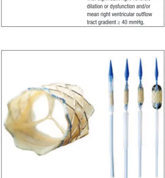
The Melody™ valve
Manufactured from a bovine jugular vein segmental patch with 18 mm diameter, the valve is sutured and mounted inside the silver-iridium Cheatham-Platinum (CP) stent, 34 mm long, and with 8 zigs of circumfer-ential arrangement (NuMED Inc., Hopkinton, United States). This device is released by an Ensemble™ delivery system speciically designed for this purpose (Medtronic Inc., Minneapolis, USA United States). This system consists of a balloon-in-balloon catheter (BIB, NuMED Inc.) measuring 18, 20, or 22 mm diameter, and mounted in a retractable jacket. The system has a 22-F proile and, at its distal end, there is a blue plastic tip, which facilitates its passage through the skin and RVOT. A retractable plastic jacket covers the valved stent and, when retracted, allows for performing a control angiogram through its side arm (Figure 1).
BOX
Indication criteria for percutaneous implantation of the Melody™ valve
Dysfunction of the right ventricle pulmonary artery conduit, since the right ventricular outlow tract has a minimum diameter of 16 mm and a maximum diameter of 22 mm in the presence of any of the indings by transthoracic echocardiography.
1. Patients with functional class II, III or IV, moderate or severe pulmonary insuficiency, and/or mean right ventricular outlow tract gradient ≥ 35 mmHg.
2. Patients with functional class I: severe pulmonary insuficiency with signiicant right ventricle dilation or dysfunction and/or mean right ventricular outlow tract gradient ≥ 40 mmHg.
Procedure
The technique employed for implanting the Melody™ valve was similar to those previously described.12 The
femoral venous approach was used in all patients. The vessel was previously prepared using two Per-close ProGlide™ (Abbott, Inc., Illinois, United States) percutaneous sutures. A high-proile DrySeal (Gore, Newark, United States) sheath (22 F, 28 cm long) was positioned to allow for multiple intravenous procedures through venous access. The arterial access was used for angiography procedures at the aortic root, or for selective coronary angiographies for deining coronary compression during a test inlation of the balloon at RVOT. Intravenous heparin was administered at a dose of 100 IU/kg and antibiotic prophylaxis was performed with cefazolin for 24 hours.
Right-chamber cardiac catheterization was fol-lowed by a RV and/or pulmonary artery angiogram in conventional projections. The length and diameter of the implant site were measured. An extra-hard guide wire (Lunderquist™; Cook Inc., Bloomington, United States) was positioned distally into PA, in order to allow for the coronary compression test, conduit preparation with stents, progression of the Ensemble™ system, and post-dilation, when necessary.
The conduit was previously dilated with progressive-diameter balloons. The initial balloon was chosen so that the smallest diameter had 80% of the conduit diameter. The balloons’ diameter was increased, so that
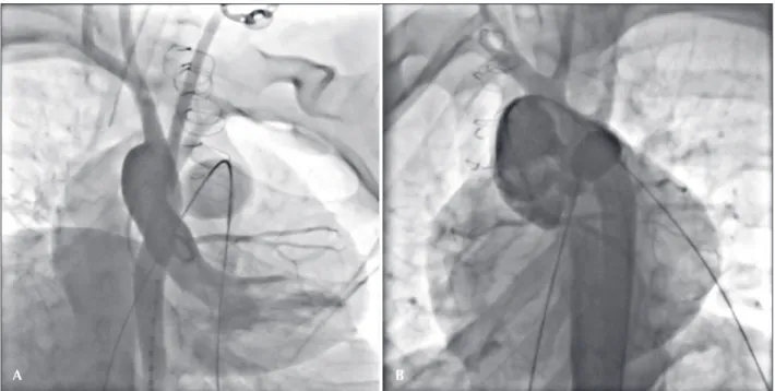
the inal balloon diameter was similar to the original diameter of the conduit, not exceeding 130-140% of this diameter. The coronary compression test was conducted by means of an angiogram of the ascending aorta, in several projections (Figure 2), while simultaneously inlating the larger-diameter balloon into the conduit. Coronary-flow reductions or a coronary proximity < 5 mm with respect to the conduit were considered as exclusion factors for the procedure.
The implant area was prepared with stent implan-tation, with dilation to a diameter close to that of the original conduit. Common stents Palmaz 4014 (Cordis Inc., Bridgewater, United States) or CP 8-zig of 34, 39, or 45 mm, mounted on BIB Max LD™ (NuMED Inc.) or Cristal (Balt, Montmorency, France) balloons were chosen. In cases where signs of dissection and/or contrast extravasation were observed after pre-dilation, a CP covered stent (NuMED Inc.) was used, instead of regular stents, as well as in cases with extensive conduit calciication. The conduit preparation was performed using long 14 F Mullins sheaths inside the DrySeal sheath. In those cases of valve implantation inside a bioprosthesis, no previous preparation was done.13 In
the case of stent elastic recoil > 2 mm, or of the pres-ence of residual waists, a high-pressure Atlas balloon (Bard, Inc., Covington, United States) was used to a full expansion of the stent within the conduit. After each step in conduit preparation (pre-dilation with balloons, stent implantation, and post-dilation), control angiograms were performed.
A B
In general, the Ensemble™ system diameter was equal or up to 2 mm larger than the original conduit diameter. The steps for mounting the valve prosthesis in its delivery system were performed as recommended by the manufacturer, with special attention given to the proper orientation of the prosthesis in the balloon and in relation to the blood low direction.
The DrySeal sheath was replaced by the Ensemble™, and the prosthesis was positioned within the previously prepared conduit, avoiding protrusion towards the pulmonary-artery bifurcation or the muscular portion of RVOT. When necessary, control angiograms were performed through the Ensemble™ side arm. After the retraction of the jacket into the Ensemble™ system, the BIB balloon was sequentially inlated, in order to release the valved stent. In cases of a short implantation zone (< 40 mm), especially if accompanied by a full PI, the conduit preparation and the Melody™ valve release were performed under a quick ventricular pacing with a RV-positioned pacemaker wire.
New pressure measurements and angiograms were obtained for evaluation of the result. According to avail-ability, one intracardiac echocardiogram was obtained after implantation.
Follow-up
Patients were observed for 72 hours in the hospital. A transthoracic echocardiogram (TTE) was performed before hospital discharge, as well as a chest X-ray and an electrocardiogram. All patients were instructed to use acetylsalicylic acid (ASA) 3 to 5 mg/kg/day (up to 100 mg/day) for 6 months. Outpatient follow-up was scheduled to return in 1, 6, and 12 months after the procedure. Patients were also instructed as to bacterial endocarditis prophylaxis for life.
Definitions and variables for evaluation of outcomes
The feasibility of the procedure was deined by a successful implantation, with stent release for conduit preparation and prosthesis release with a proper inal positioning.
The safety was assessed by the incidence of pro-cedure- or prosthesis-related complications: mortality, conduit rupture, pulmonary-vessel perforations, a need for blood products, endovascular injury, impairment of pulmonary arterial low, device embolization or fracture requiring reintervention, serious cardiac arrhythmias, and each and every complication putting the life or physical integrity of the patient at risk. The eficacy was assessed by the determination of a peak-to-peak pres-sure gradient < 25 mmHg of systemic blood prespres-sure, with RV systolic pressure < 50% of systemic blood pressure, besides PI absence, or with a trivial residual presence of PI.
Statistical analysis
Qualitative variables were expressed as absolute numbers and percentages, and quantitative variables were expressed as means and standard deviations, or medians and interquartile ranges, according to sample distribution. Student’s t-test was used to evaluate changes in quantitative variables before and after the procedure. P-values < 0.05 were considered signiicant.
RESULTS
Patients
From December 2013 to June 2014, ten patients (seven men) with a median age of 16.5 years (11-31 years) and weighing 49 kg (32-85 kg) underwent implantation of the Melody™ valve (Table 1). Most patients (60%) had an initial diagnosis of tetralogy of Fallot, or its variants. Six patients underwent two or more previous surgeries, and the mean time since the last surgery was 11.9 ± 8.6 years. One patient was in functional class III, four in functional class II, and the others in functional class I (50%). The patient in functional class III showed signiicant right ventricular dysfunction, QRS = 200 ms, and frequent episodes of non-sustained ventricular tachycardia in the 24 hours Holter monitor. The remaining patients showed good biventricular function without changes in cardiac rhythm (Table 1).
Five patients had a homograft in the pulmonary position, all ≥ 16 mm. The remaining patients had the following types of conduit: Contegra™ bovine jugular vein of 16 and 17 mm (Medtronic Inc., Minneapolis, United States) in two patients, one corrugated pericar-dium conduit of 18 mm, and a Epic™ bioprosthesis of 21 mm (St. Jude Medical, Inc., St. Paul, United States).
Implantation success and technical aspects of the procedure
All patients, except one, underwent conduit prepa-ration with stent, and Melody™ valves were properly implanted in the intended location. In one patient, two stents were necessary, due to an unequal expansion of the irst stent and to the greater length of the conduit (homograft) to be treated. In another patient, a previ-ous implantation with a covered CP stent was chosen, due to the extreme calciication associated to a severe conduit stenosis area.
artery stenosis (Figure 3), and two others only exhibited stenosis in left PA (LPA). This entire group was success-fully treated with stenting prior to valve implantation. The conduit pre-dilation was performed with balloons with a mean inal diameter of 20.8 ± 2.9 mm.
The mean diameter of the balloon used for pretreat-ment with stent was 21.6 ± 2.2 mm, and the mean of the minor diameter of the stent was 20.1 ± 1.8 mm. The stent was post-dilated with a high-pressure Atlas balloon in three patients (12, 16, and 20 atm, respectively), because of a persistent residual waist. The patient not subjected to the preparation had a calciied bioprosthesis in a semi-open position (Figure 4).
One patient, with tetralogy of Fallot and absence of right PA, was subjected to an off-label LPA implantation with the Melody™ valve. An important failure of the monocuspid device used in the RVOT transannular patch and a mild LPA stenosis (initial diameter, 16 mm) were observed, and the patient was considered as having a favorable anatomy for implantation of a conventional stent, which later served as an anchorage zone and, therefore, as a “conduit” to valve implantation.
The Melody™ prosthesis was mounted on an 18-mm Ensemble™ system in a patient implanted with a stenotic Contegra™ (17 mm); and on an 20 mm Ensemble™ in two other patients – one of them implanted with an Epic™ bioprosthesis of 21 mm (internal diameter, 18 mm). In the remaining patients (70%), 22 mm En-semble™ systems were used. The mean ratio between the diameter of the Ensemble™ system used and the conduit original diameter was 1.1 (0.95-1.35). No modiication of the delivery system or of the prosthesis assembling technique was used.
Efficacy
A signiicant reduction, in terms of post-treatment conduit obstruction, with the use of the Melody™ valve (Table 2) occurred. The PI detected by the control angiogram was not greater than trivial in any of the pa-tients, a fact proven by the intracardiac echocardiogram performed in six patients. No post-implantation change in valve shape or performance, nor the occurrence of paravalvar leakage, was detected.
Safety
A severe adverse event was observed in this cohort. After implantation, a patient with a homograft exhibiting signiicant calciication presented a contained extrava-sation of contrast in the distal portion of the conduit (Figure 5). No associated hemodynamic instability was noted. One covered CP stent was implanted, over-riding the implanted prosthesis, with immediate occlusion of the extravasation site, followed by implantation of a second Melody™ valve. This patient was transferred to the intensive care unit (ICU) after the procedure for monitoring, and was extubated within hours. The remaining patients were referred to their room after anesthetic recovery. In another patient, a Max™ LD balloon ruptured during conduit preparation. The rupture was circular, and the distal fragment of the balloon was rescued, using a loop catheter. No patient exhibited clinical or electrocardiographic changes consistent with coronary compression after treatment. There were no deaths and no need for an immediate surgical interven-tion. In two patients, during hospitalization a minor bleeding from the puncture site was observed, without formation of local hematoma and with no need for blood transfusions. All patients were discharged within 72 hours after treatment.
TABLE 1
Clinical and laboratory characteristics before valve implantation
n = 10
Male gender 7
Age, years 16.5 (11-31)
Weight, kg 49 (32-85)
Underlying diagnosis
Tetralogy of Fallot 4
Pulmonary atresia with ventricular septal communication
1
Pulmonary atresia with intact ventricular septum 2
Pulmonary stenosis 1
Double outlet right ventricle 1
Common arterial trunk 1
Type of conduit
Homograft 5
Corrugated pericardial conduit 1
Contegra™ 2
Bioprosthesis 1
Native output tract 1
Conduit diameter at the time of implantation, mm 15.5 (10-22)
Number of prior surgeries 2 (1-3)
Primary indication for valve implantation
Stenosis 2
Failure 3
Mixed 5
Mean for maximum gradient in the conduit on echocardiography, mmHg
49.2 ± 20.2
Pulmonary insuficiency on echocardiography
Absent 2
Mild 1
Moderate 1
Severe 6
MRI right ventricular ejection fraction,%
Right-ventricular end-systolic volume (indexed), mL 57 ± 5
Follow-up
All patients were followed as outpatients, with a mean of 4.1 ± 2.2 months. No patient required re-intervention. One month after treatment, one patient (previously in functional class III) reported signiicant improvement in tolerance to daily activities. All treated patients noticed some improvement. TTEs performed 24 hours and 30 days after the procedure revealed that all patients presented mean gradients < 25 mmHg through RVOT. In seven patients, no residual PI was observed;
and in two, PI was considered trivial. On chest radiog-raphy, no Melody™ valve fractures were noticed, and no clinical or echocardiographic signs of thrombosis or endocarditis were observed. There were no late complications associated with the venous access used.
DISCUSSION
This study described the irst Brazilian experience in Melody™ valve implantation. Its importance lies not only in its originality in this environment, but also in its A
C
B
D
potential application for the future assimilation of this technology by the public health system. To this end, its feasibility, safety, and eficacy must be documented. This device comes at an opportune time, since, in this environment, there is a growing population with con-genital heart disease in the late post-operative period of RV-PA conduits. In this small case series, nine of the ten treated patients had inclusion criteria similar to those classically used.14 In the only off-label case,
the anatomical substrate favored the realization of an LPA valve implantation, rather than in its commonly
used place. In the literature, there are other reports of Melody™ valve implantation in pulmonary arteries of patients with complex anatomy, resulting in similar outcomes to those observed here.16,17
Feasibility and technical aspects
The procedure was successfully completed in all patients. Percutaneous implantation of the Melody™ valve proved to be an extremely labor-intensive task, requiring high technical differentiation. Should have a A
C
B
D
TABLE 2
Immediate hemodynamic results
Basal value Basal value Post-implantation value p-value
Right-ventricular systolic pressure, mmHg 49.2 ± 15.9 49.2 ± 15.9 35.8 ± 5.7 0.008
Right-ventricular/left ventricular pressure ratio 0.55 ± 0.18 0.55 ± 0.18 0.39 ± 0.08 0.002
Right-ventricle-pulmonary artery gradient, mmHg 25.1 ± 17.9 25.1 ± 17.9 7.0 ± 6.7 0.001
A
C
B
D
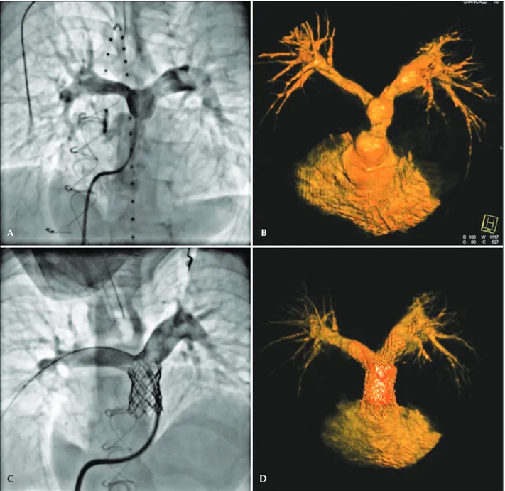
Figure 5 – Contained leakage from the conduit after implantation of a Melody™ valve, identiied by the white arrow (B). In C and D, the inal angiographic result in orthogonal projections, after the implantation of a covered stent and a second Melody™ valve.
strong background in structural and congenital heart disease; moreover, they must be familiar with a wide
the learning curve has a recognized impact on the progressive improvement of results.18
The smallest patient in our sample was 11 years old and weighed 32 kg; nevertheless, an American study has illustrated the feasibility of the procedure in patients weighing less than 30 kg, with ive patients weighing less than 18 kg.19 However, these authors observed a
higher incidence of vascular complications in this sub-group. However, such a vascular access limitation can be overcome, even with still smaller patients, through the use of a periventricular access, as already described in the literature for a patient weighing 12 kg.20
The most labor-intensive step was not the Melody™ valve implantation itself, but the conduit preparation. The preparation of the conduit with stents dissipates the vector forces and the mechanical stress imposed on the Melody™ valve, reducing the incidence of fractures in the patients’ follow-up.15 These technical aspects are
discussed below.
Efficacy
The immediate hemodynamic improvement ob-served, with signiicant reduction in the degree of stenosis within the conduit and with pulmonary valve competence restoration, was similar to that found by European and American groups.11,12,14,21 Gradients
> 30 mmHg immediately after implantation are associ-ated with lower durability of the valve and with less reintervention-free survival.13 This observation justiies
a more aggressive preparation of the conduit with the implantation of multiple stents, and the abolition of residual waists and gradients.
The follow-up is still in its early stages, but clinical and echocardiographic data obtained one month after the procedure conirmed the maintenance of valve com-petence in all treated patients, as well as the absence of residual obstruction. In the irst evaluation after a month, all patients reported some clinical improve-ment, with less fatigue when exercising. Subjectively supporting these patient reports, some studies have shown that reduction of RVSP and/or improvement of volume overload after valvular treatment are associated with improved RV and septal strain,22 culminating in
an improved global biventricular function.23-25
This preliminary study did not compare the per-cutaneous versus surgical method, and the percutane-ous treatment of cost-effectiveness was not analyzed. However, there are studies published in the literature pointing to percutaneous treatment as the best option in selected cases.26-29
Safety
A major event in one patient was observed. There was a contained conduit extravasation, without hemo-dynamic consequences or need for transfusions. The
covered CP stent implantation resulted in an occlusion of the extravasation site. Such complication underscores the need to have covered stents of various lengths available in the room.
Together with a potential coronary compression, conduit rupture is the most severe complication that can occur in this type of procedure. However, the rupture of the conduit is more unpredictable; and, to avoid this complication some measures are recom-mended, such as a progressive dilation of the conduit and a serial angiographic study after each dilation and preparation with stents. The underlying rationale is the possibility of creating a lesion of lesser magnitude in the conduit wall, due to a more conservative and progressive dilatation, with less probability of a rupture that would result in extravasation into the mediastinum and in hemodynamic instability. The ibrosis adjacent to the conduit, secondary to previous surgeries, helps to minimize the occurrence of a catastrophic rupture.
In this regard, McElhinney et al.30 studied the effects
of the use of ultra-high pressure balloons compared to standard balloons in the dilation of these conduits. It was observed that the use of ultra-high pressure balloons results in lower residual gradients and in a larger diameter of the dilated site, in spite of the higher occurrence of dissection and small leakages. Such lesions caused by the balloons can be classiied as therapeutic dissections (if < 3 mm), and contained and non-contained extravasations; the actual rupture with conduit avulsion is a very rare event.30 In the American
experience, in one patient the rupture of the conduit resulted in surgery.16 In this same study, six patients
were excluded because of the possibility of coronary compression. Although the authors have not experienced this possibility, this step of the procedure should merit special attention,7,12 mainly due to the frequent changes
in the origin and course of the coronary arteries associ-ated with the presence of tetralogy of Fallot, and the transposition of the great arteries, among others, and also variations in the spatial location of the aorta and of the conduit.31-33 Conversely, the occurrence of coronary
artery compression in patients with bioprosthesis in the pulmonary position is unlikely, due to the rigidity of its metal ring. Even in this scenario, the compression test is mandatory.
Other serious complications, such as valve em-bolization, occlusion of pulmonary arteries, and distal perforation of pulmonary branches, were described in previous studies,11,12,14,18 but are potentially preventable
with the use of an impeccable technique and the ac-cumulation of experience. In general, the percutaneous implantation of the Melody™ valve is a safe procedure, with low rates of severe associated complications or need for surgical intervention, as seen in this study.
CP stent, which surrounds the Melody™ valve, with rates of approximately 25% on a median follow-up of 13 months.16 Several factors are associated with
fractures, many of which are interconnected. Conduit blockage severity was associated with a decreased fracture-free survival, and also with a larger pres-sure gradient through the RVOT before and after valve implantation. Other predisposing factors were related to the vicinity where the prosthesis was implanted, including lower patient age, anterior chest-wall proximity, and signs of extrinsic vascular compression of the prosthesis.13 Conversely, patients
with bioprostheses are at lower risk for fractures in the metal mesh overtime.15,22 The metal ring of the
bioprosthesis probably acts as a shield against op-posing vector forces, protecting the implant.
Despite the progressive increase in the prevalence of fractures over time a inding observed in several series,13-15,19,34-37 less than half of these patients require
reintervention.15 It is estimated that about three years
after implantation, approximately 85% of patients are free of new procedures.14-16 Factors associated with an
increased risk of reintervention were the same as those identiied for an increased risk of fractures. The cases of prosthesis dysfunction can be safely and effectively treated with implantation of a new prosthesis (valve-in-valve), generally making the surgery unnecessary.13,14,35
The present study did not observe the presence of fractures, probably due to the short follow-up time.
Recently, cases of late occurrence of Melody™ valve endocarditis were described.38 Fever of unknown origin
and a quick valvular function deterioration are clinical signs suggestive of the diagnosis. About 50% of these patients respond well to a long-term systemic antibiotic therapy; however, some patients require surgery for treat-ment. Apparently, there are no well-deined risk factors, but most patients with endocarditis were male, had previous episodes of endocarditis before implantation, and were intravenous drug users. In these scenarios, the implant should probably be contraindicated. Obviously, endocarditis prophylaxis should be recommended, as in conventional situations.
CONCLUSIONS
In this preliminary study, percutaneous implantation of the Melody ™ valve for restoration of pulmonary func-tion in selected patients was feasible, safe, and effective in the short term. As there is extensive experience with this type of procedure in Europe and North America, it is speculated that the occurrence of potential adverse events may be even lower in the authors’ practice, due to the neutralization of well-established risk factors. However, a larger number of patients and a longer follow-up time are needed for a better understanding of the durability of the implant and the incidence of complications in this environment.
CONFLICTS OF INTEREST
Carlos A. C. Pedra occasionally receives medical fees from Medtronic Co. for lecturing at conferences and related events. John P. Cheatham is a consultant for Medtronic Co. and has received honoraria to work as tutor in the irst ive cases performed in this study.
FUNDING SOURCES
For ive patients in this study, the procedure was funded by the Brazilian Ministry of Health as part of a project to assess new technologies, in partnership with the Research Institute of the Hospital do Coração, Associação do Sanatório Sírio.
REFERENCES
1. Tarasoutchi F, Montera MW, Grinberg M, Barbosa MR, Piñeiro DJ, Sánchez CRM, et al. Diretriz Brasileira de Valvopatias – SBC 2011/I Diretriz Interamericana de Valvopatias – SAC 2011. Arq Bras Cardiol. 2011;97(5 Supl 1):1-67.
2. d’Udekem Y, Rubay J, Ovaert C. Failure of right ventricular recovery of fallot patients after pulmonary valve replacement: delay of reoperation or surgical technique? J Am Coll Cardiol. 2001;37(7):2008-9.
3. Askovich B, Hawkins JA, Sower CT, Minich LL, Tani LY, Stod-dard G, et al. Right ventricle-to-pulmonary artery conduit longevity: is it related to allograft size? Ann Thorac Surg. 2007;84(3):907-11.
4. Brown JW, Ruzmetov M, Rodefeld MD, Vijay P, Turrentine MW. Right ventricular outlow tract reconstruction with an allograft conduit in non-ross patients: risk factors for allograft dysfunction and failure. Ann Thorac Surg. 2005;80(2):655-63. 5. Tweddell JS, Pelech AN, Frommelt PC, Mussatto KA, Wyman
JD, Fedderly RT, et al. Factors affecting longevity of homograft valves used in right ventricular outlow tract reconstruction for congenital heart disease. Circulation. 2000;102(19 Suppl 3):III1 30-5.
6. Oosterhof T, Meijboom FJ, Vliegen HW, Hazekamp MG, Zwin-derman AH, Bouma BJ, et al. Long-term follow-up of homograft function after pulmonary valve replacement in patients with tetralogy of Fallot. Eur Heart J. 2006;27(12):1478-84. 7. Peng LF, McElhinney DB, Nugent AW, Powell AJ, Marshall
AC, Bacha EA, et al. Endovascular stenting of obstructed right ventricle-to-pulmonary artery conduits: a 15-year experience. Circulation. 2006;113(22):2598-605.
8. Fogelman R, Nykanen D, Smallhorn JF, McCrindle BW, Free-dom RM, Benson LN. Endovascular stents in the pulmonary circulation: clinical impact on management and medium-term follow-up. Circulation. 1995;92(4):881-5.
9. Powell AJ, Lock JE, Keane JF, Perry SB. Prolongation of RV-PA conduit life span by percutaneous stent implantation: intermediate-term results. Circulation. 1995;92(11):3282-8. 10. Bonhoeffer P, Boudjemline Y, Saliba Z, Merckx J, Aggoun
Y, Bonnet D, et al. Percutaneous replacement of pulmonary valvein a right-ventricle to pulmonary-artery prosthetic conduit with valve dysfunction. Lancet. 2000;356(9239):1403-5. 11. Khambadkone S, Coats L, Taylor A, Boudjemline Y, Derrick
G, Tsang V, et al. Percutaneous pulmonary valve implanta-tion in humans: results in 59 consecutive patients. Circula-tion.2005;112(8):1189-97.
with a dysfunctional right ventricular outlow tract conduit early results from the U.S. Clinical trial. J Am Coll Cardiol. 2009;54(18):1722-9.
13. McElhinney DB, Cheatham JP, Jones TK, Lock JE, Vincent JA, Zahn EM, et al. Stent fracture, valve dysfunction, and right ventricular outlow tract reintervention after transcatheter pul-monary valve implantation: patient-related and procedural risk factors in the US Melody Valve Trial. Circ Cardiovasc Interv. 2011;4(6):602-14.
14. McElhinney DB, Hellenbrand WE, Zahn EM, Jones TK, Cheatham JP, Lock JE, et al. Short- and medium-term outcomes after trans-catheter pulmonary valve placement in the expanded multicenter US melody valve trial. Circulation. 2010;122(5):507-16. 15. Vezmar M, Chaturvedi R, Lee K-J, Almeida C, Manlhiot C,
McCrindle BW, et al. Percutaneous pulmonary valve implanta-tion in the young 2-year follow-up. JACC Cardiovasc Interv. 2010;3(4):439-48.
16. Qureshi AM, Krasuski RA, Prieto LR. Percutaneous pulmo-nary valve implantation in left pulmopulmo-nary artery branch in a patient with a functional single lung. J Invasive Cardiol. 2012;24(9):E202-4.
17. Gillespie MJ, Dori Y, Harris MA, Sathanandam S, Glatz AC, Rome JJ. Bilateral branch pulmonary artery melody valve im-plantation for treatment of complex right ventricular outlow tract dysfunction in a high-risk patient. Circ Cardiovasc Interv. 2011;4(4):e21-3.
18. Lurz P, Coats L, Khambadkone S, Nordmeyer J, Boudjemline Y, Schievano S, et al. Percutaneous pulmonary valve implan-tation: impact of evolving technology and learning curve on clinical outcome. Circulation. 2008;117(15):1964-72. 19. Berman DP, McElhinney DB, Vincent JA, Hellenbrand WE,
Zahn EM. Feasibility and short-term outcomes of percutane-ous transcatheter pulmonary valve replacement in small (<30 kg) children with dysfunctional right ventricular outlow tract conduits. Circ Cardiovasc Interv. 2014;7(2):142-8.
20. Holoshitz N, Ilbawi MN, Amin Z. Perventricular Melody valve implantation in a 12 kg child. Catheter Cardiovasc Interv. 2013;82(5):824-7.
21. Lurz P, Nordmeyer J, Coats L, Taylor AM, Bonhoeffer P, Schulze-Neick I. Immediate clinical and haemodynamic beneits of restoration of pulmonary valvar competence in patients with pulmonary hypertension. Heart. 2009;95(8):646-50.
22. Moiduddin N, Asoh K, Slorach C, Benson LN, Friedberg MK. Effect of transcatheter pulmonary valve implantation on short-term right ventricular function as deshort-termined by twodimensional speckle tracking strain and strain rate imaging. Am J Cardiol. 2009;104(6):862-7.
23. Coats L, Khambadkone S, Derrick G, Sridharan S, Schievano S, Mist B, et al. Physiological and clinical consequences of relief of right ventricular outlow tract obstruction late after repair of congenital heart defects. Circulation. 2006;113(17):2037-44. 24. Coats L, Khambadkone S, Derrick G, Hughes M, Jones R, Mist
B, et al. Physiological consequences of percutaneous pulmonary
valve implantation: the different behaviour of volume- and pressure-overloaded ventricles. Eur Heart J. 2007;28(15):1886-93. 25. Lurz P, Nordmeyer J, Muthurangu V, Khambadkone S, Der-rick G, Yates R, et al. Comparison of bare metal stenting and percutaneous pulmonary valve implantation for treatment of right ventricular outlow tract obstruction: use of an x-ray/ magnetic resonance hybrid laboratory for acute physiological assessment. Circulation. 2009;119(23):2995-3001.
26. O’Byrne ML, Gillespie MJ. Will catheter interventions re-place surgery for valve abnormalities? Curr Opin Cardiol. 2014;29(1):83-90.
27. Reardon MJ. Cost-effectiveness analysis of TAVR. Methodist Debakey Cardiovasc J. 2012;8(2):26-8.
28. Gatlin SW, Kim DW, Mahle WT. Cost analysis of percutaneous pulmonary valve replacement. Am J Cardiol. 2011;108(4):572-4. 29. McElhinney DB. Recent progress in the understanding and
management of postoperative right ventricular outlow tract dysfunction in patients with congenital heart disease. Circula-tion. 2012;125(16):e595-9.
30. Hainstock MR, Marshall AC, Lock JE, McElhinney DB. Angio-plasty of obstructed homograft conduits in the right ventricular outlow tract with ultra-noncompliant balloons: assessment of therapeutic eficacy and conduit tears. Circ Cardiovasc Interv. 2013;6(6):671-9.
31. Elliott LP, Amplatz K, Edwards JE. Coronary arterial patterns in transposition complexes. Anatomic and angiocardiographic studies. Am J Cardiol. 1966;17(3):362-78.
32. Gordillo L, Faye-Petersen O, de la Cruz MV, Soto B. Coro-nary arterial patterns in double-outlet right ventricle. Am J Cardiol.1993;71(12):1108-10.
33. Meng Cc, Eckner Fa, Lev M. Coronary artery distribution in tetralogy of Fallot. Arch Surg. 1965;90:363-6.
34. Martins JDF, Ewert P, Sousa L, Freitas I, Trigo C, Jalles N, et al. Percutaneous pulmonary valve implantation: initial experi-ence. Rev Port Cardiol. 2010;29(12):1839-46.
35. Nordmeyer J, Coats L, Lurz P, Lee T-Y, Derrick G, Rees P, et al. Percutaneous pulmonary valve-in-valve implantation: a successful treatment concept for early device failure. Eur Heart J. 2008;29(6):810-5.
36. Raikou M, McGuire A, Lurz P, Bonhoeffer P, Wegmueller Y. An assessment of the cost of percutaneous pulmonary valve implantation (PPVI) versus surgical pulmonary valve replace-ment (PVR) in patients with right ventricular outlow tract dysfunction. J Med Econ. 2011;14(1):47-52.
37. Gillespie MJ, Rome JJ, Levi DS, Williams RJ, Rhodes JF, Cheatham JP, et al. Melody valve implant within failed bioprosthetic valves in the pulmonary position: a multicenter experience. Circ Cardiovasc Interv. 2012;5(6):862-70.