AIM2 Drives Joint Inflammation in a
Self-DNA Triggered Model of Chronic Polyarthritis
Christopher Jakobs1, Sven Perner2, Veit Hornung1*
1Institute of Molecular Medicine, University Hospital Bonn, University of Bonn, Bonn, Germany, 2Department of Prostate Cancer Research, Institute of Pathology, Center for Integrated Oncology Köln/ Bonn, University Hospital Bonn, Bonn, Germany
*veit.hornung@uni-bonn.de
Abstract
Mice lacking DNase II display a polyarthritis-like disease phenotype that is driven by translo-cation of self-DNA into the cytoplasm of phagocytic cells, where it is sensed by pattern rec-ognition receptors. While pro-inflammatory gene expression is non-redundantly linked to the presence of STING in these mice, the contribution of the inflammasome pathway has not been explored. To this end, we studied the role of the DNA-sensing inflammasome receptor AIM2 in this self-DNA driven disease model. Arthritis-prone mice lacking AIM2 dis-played strongly decreased signs of joint inflammation and associated histopathological find-ings. This was paralleled with a reduction of caspase-1 activation and pro-inflammatory cytokine production in diseased joints. Interestingly, systemic signs of inflammation that are associated with the lack of DNase II were not dependent on AIM2. Taken together, these data suggest a tissue-specific role for the AIM2 inflammasome as a sensor for endogenous DNA species in the course of a ligand-dependent autoinflammatory condition.
Introduction
The innate immune system has evolved a conserved set of so-called pattern recognition recep-tors (PRRs) to sense the presence of microbial pathogens. PRRs detect microbe-associated molecular patterns (MAMPs) as non-self and initiate signaling cascades geared at eliminating the microbial threat [1]. Under certain circumstances, these receptors can also respond to self-derived molecules. This, for example, includes endogenous nucleic acids that have gained access to compartments that are usually devoid of these [2]. Accidental activation of compart-mentalized PRRs by endogenous nucleic acids is additionally prevented by the presence of nucleases that degrade and thereby deplete potential self-ligands under steady state conditions. These nucleases are expressed in a compartment and cell-type specific manner, creating a non-redundant system of nuclease activity, which prevents accidental activation of nucleic acid sensing PRRs. The importance of this safeguard system is impressively documented by the fact that defects in a number of these nucleases can result in severe sterile inflammatory conditions that are triggered by nucleic acid sensing PRRs [3]. Aicardi–Goutières syndrome (AGS), for example, is a rare genetic inflammatory disorder, in which defects in nucleic acid degrading or
OPEN ACCESS
Citation:Jakobs C, Perner S, Hornung V (2015) AIM2 Drives Joint Inflammation in a Self-DNA Triggered Model of Chronic Polyarthritis. PLoS ONE 10(6): e0131702. doi:10.1371/journal.pone.0131702
Editor:Bernhard Ryffel, French National Centre for Scientific Research, FRANCE
Received:December 17, 2014
Accepted:June 5, 2015
Published:June 26, 2015
Copyright:© 2015 Jakobs et al. This is an open access article distributed under the terms of the
Creative Commons Attribution License, which permits unrestricted use, distribution, and reproduction in any medium, provided the original author and source are credited.
Data Availability Statement:All relevant data are within the paper and its Supporting Information files.
Funding:This work was supported by grants from the German Research Foundation (SFB704 and SFB670) and the European Research Council (ERC‐
2009‐StG 243046) to V.H. V.H. is member of the
excellence cluster ImmunoSensation. The funders had no role in study design, data collection and analysis, decision to publish, or preparation of the manuscript.
metabolizing enzymes can result in the spontaneous production of antiviral cytokines in cere-brospinal fluid and serum, manifesting as a sub-acute encephalopathy [4,5].
Detection of cytosolic DNA triggers at least two distinct core signaling cascades: On the one hand, cytosolic DNA leads to the activation of the nucleotidyltransferase cGAS, which upon DNA binding produces a 2’-5’linked cyclic dinucleotide second messenger molecule that in turn binds to and activates the ER-resident receptor STING. STING activation results in its translocation to a perinuclear Golgi compartment, where it achieves its signaling competent state, culminating in the activation of antiviral and pro-inflammatory gene expression [6,7]. While these two components form the non-redundant core of cytosolic DNA mediated antivi-ral gene expression, additional factors have been described to function in concert with cGAS and or STING [8]. In myeloid cells, cytosolic DNA is additionally sensed by the PYHIN protein AIM2, which forms an ASC-dependent inflammasome complex upon ligand binding. Inflam-masome activation results in the processing and activation of pro-caspase-1, which itself leads to the processing of pro-cytokines such as IL-1βand IL-18 [9–12]. At the same time, inflamma-some activation leads to the induction of a myeloid cell specific cell death, known as pyroptosis [13]. AIM2 has shown to be involved in the recognition of a number of microbial pathogens, such as DNA viruses or bacteria that release microbial DNA into the cytoplasm during their life cycle. However, a role for AIM2 in the recognition of endogenous self-DNA has not been established so far [14].
To study the possible role of AIM2 in the context of a self-DNA driven autoinflammatory disease, we made use of a DNase II-deficiency mouse model [15]. In this mouse model, absence of the lysosomal endonuclease DNase II (Dnase2) leads to a defect in disposing of DNA in lyso-somal compartments [16]. This subsequently results in the translocation of undigested DNA into the cytoplasm, which in turn results in the activation of STING leading to unabated pro-duction of type I IFNs and pro-inflammatory cytokines [17]. Spontaneous cytokine production is already seen in early fetal development ofDnase2-deficient mice, due to the fact that macro-phages play an important role in disposing of expelled nuclei from erythroid precursor cells in so-called erythroblastic islands in the fetal liver or bone marrow in the course of definitive erythropoiesis [16,18]. This macrophage-dependent type I IFN production in the context of
Dnase2-deficiency results in lethal anemia, which can be fully rescued by ablating type I IFN production or its activity (e.g.Ifnar1-/-) in these mice [19]. WhileDnase2-Ifnar1DKO mice are born healthy, macrophages in these mice are still responding to undigested DNA by the up reg-ulation of pro-inflammatory genes. In fact, 3–6 months after birthDnase2-/-Ifnar1-/-mice develop a systemic auto-inflammatory disease, most prominently affecting the joints with chronic polyarthritis mimicking rheumatoid arthritis in humans [15]. Even though accompa-nied by the production of autoantibodies, this chronic polyarthritis is independent of the adap-tive branch of the immune system, yet rather driven by the unabated production of pro-inflammatory cytokines, most prominently TNF, IL-6 and IL-1β[20]. In fact, in diseased joints ofDNase2-deficient animals these cytokines appear to regulate each other’s expression in a mutually dependent fashion. To this effect, blocking the activity of one of these cytokines blunts the expression of the others and thereby greatly ameliorates disease progression [20]. Il-18, another potential candidate cytokine that has been associated with the development and progression of arthritis, is not involved in this disease model. In fact, IL-18-/-mice still develop polyarthritis and express pro-inflammatory cytokines in the context ofDnase2-deficiency [20].
normally subjected to DNase II mediated degradation, involving a process that depends on nuclear export and subsequent autophagy-dependent delivery to lysosomal structures. As such, next to its function in degrading apoptotic material from other cells, DNase II prevents the ini-tiation of a cell autonomous innate immune response by disposing of nuclear-derived DNA.
Given the overlapping ligand spectrum of the cGAS-STING axis as well as the inflamma-some sensor AIM2, we hypothesized that AIM2 might also be activated by the presence of cyto-solic DNA in the course ofDnase2-deficiency. Moreover, we speculated that part of the disease activity in this mouse model could indeed be attributed to AIM2.
Materials and Methods
Mice
Mice deficient forDnase2andIfnar1on a C57BL/6 background were previously described [15].Aim2-/-mice originate from crossing of PGK-Cre [22] mice withAim2flox/floxmice, which were generated by Taconic Artemis on a C57BL/6 background (S1 Fig). Following the out-breeding of PGK-Cre, the genetic background ofAim2-/-mice was validated to be 99.23% C57BL/6J using SNP genotyping. Subsequently,Aim2-/-mice were backcrossed with C57BL/6J for another 6 generations. Primers used for genotyping ofAim2deficient mice:AF: 5’- TTGAA GAGATGGGACAGCAA-3’;A1: 5’-TGAACTTCCAGGACACAAAG-3’;A2: 5’-GCAAGCAG TTAACATTTTGAAGC-3’. All described strains were housed under specific pathogen-free conditions. Studies are approved by the district government of Northrhine Westphalia, here the“State Office for protection of nature, environment and consumers”(LANUV Landesamt für Natur, Umwelt und Verbraucherschutz, Leibnizstraße 10, 45659 Recklinghausen, Ger-many), which is the responsible agency. The application number is: 84.02.04.2014.A436. Ani-mal experiments and handling were also supervised by Institutional AniAni-mal Care and Use Committee (IACUC) of the medical faculty Bonn (HET, House of Experimental Therapy, Sig-mund-Freud Str. 25, 53127 Bonn, Germany). Mice did not show any signs of pain, distress or motor dysfunction. Mice were euthanized according to the FELASA guidelines by cervical dis-location at the respective time points indicated.
Clinical score
Swelling of the fore- and hind pads was analyzed in a blinded fashion, in monthly intervals and scored as follows: 0, no swelling; 1, mild swelling; 2, severe swelling. The scores were summed, and a total score (maximum 8) was assigned to each mouse [15].
Histopathology
RNA and protein isolation
Limbs of sacrificed mice were skinned and ground in liquid nitrogen and either transferred to TRIzol (Life technologies; Carlsbad, CA) for mRNA isolation or to tissue lysis buffer (T-PER, Tissue Protein Extraction Reagent, Pierce combined with Protease Inhibitor Tablets; Pierce/ Thermo Scientific; Pittsburgh, PA) for protein extraction.
Immunoblotting
Protein samples were diluted in 2x Laemmli buffer and boiled at 95°C for 5 min. Samples were separated by SDS gel electrophoresis for 2 h and further transferred to a 0.2μM Nitrocellulose membrane [23]. As indicated, blots were incubated with rabbit polyclonal antibody to anti cas-pase-1 p10 (sc-514; Santa Cruz Biotechnology; Santa Cruz, LA), rabbit monoclonal anti MMP3 (ab52915; Abcam; Cambridge, UK) and mouse monoclonal anti Viperin (MABF106; Merck Millipore; Billerica, MA). Membranes were developed using ECL Western blotting substrate (Pierce/Thermo Scientific; Pittsburgh, PA)
Quantitative real time PCR (qPCR)
Relative gene expression is shown as a ratio of the expression level of the gene of interest to that of Hypoxanthine-guanine phosphoribosyltransferase (Hprt1). The following primer sets were used:Hprt1: 5’-CCTGGTTAAGCAGTACAGCCC-3’, 5’-CAAATCCAACAAAGTCTGG CCT-3’;Il6: 5’-GTGGCTAAGGACCAAGACCA-3’, 5’- TAACGCACTAGGTTTGCCGA-3’;
Ifnb: 5’- TGGG-AGATGTCCTCAACTGC-3’, 5’- CCAGGCGTAGCTGTTGTACT-3’;Il1b: 5’-AATTGGTCATAGCCCGCACT-3’, 5’-AAGCAATGTGCTGGTGCTTC-3’;Mmp3: 5’-AT GGGCCTGGAACAGTCTTG-3’, 5’-GGTTGGTACCAGTGACATCCTC-3’;Tnf: 5’-CACAC TCACAAACCACCAAGTG-3’, 5’- ACAA-GGTACAACCCATCGGC-3’;Bst2: 5’-AGTTGG CAGGTCACAGTTGTT-3’, 5’- GAAGACCCAATCTGGAGCCC-3’;Aim25’-AGGCAGTGG GAACAAGACAG-3’, 5’- AAACTTCCTGACGCCACCC-3’
Multiplex protein analysis
Serum cytokines and cytokine expression in the joints were determined by using customized V-PLEX Proinflammatory Panel 1 (Meso Scale Discovery; Rockville, MD).
Statistical analysis
All data are presented as mean values, whereas error bars indicate SEM. Statistical tests were conducted as indicated using GraphPad Prism (GraphPad, La Jolla, CA).
Results
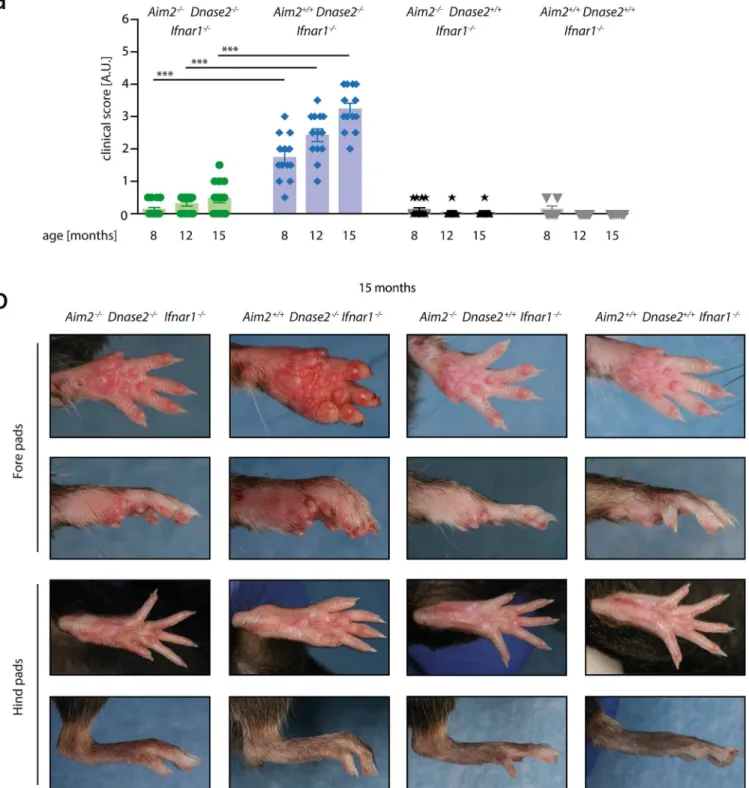
AIM2-deficiency protects from self-DNA mediated polyarthritis
To study the contribution of AIM2 toDnase2-deficiency triggered chronic polyarthritis, we crossed mice deficient inAim2with mice lackingDnase2and the common chain 1 of the type I IFN receptor (Aim2-/-,Dnase2-/-,Ifnar1-/-). As controls, mice deficient inDnase2andIfnar1
(Aim2+/+,Dnase2-/-,Ifnar1-/-), deficient inAim2andIfnar1(Aim2-/-,Dnase2+/+,Ifnar1-/-) or only deficient inIfnar1(Aim2+/+,Dnase2+/+,Ifnar1-/-) were studied. For simplicity, theIfnar1
Aim2displayed a significant mean arthritis score of 1.8 compared toDnase2-competent mice (Fig 1A). In the additional absence ofAim2,Dnase2-deficient mice (Aim2-/-x Dnase2-/-) Fig 1. Swelling of joints in the context ofDnase2-deficiency is AIM2-dependent. A:Aim2+/+Dnase2-/-Ifnar1
-/-,Aim2-/-Dnase2-/-Ifnar1
-/-,Aim2+/+
Dnase2+/+Ifnar1-/-and Aim2-/-Dnase2+/+Ifnar1-/-mice were scored for joint swelling at indicated time points in a blinded fashion. Statistical significance was assessed using a two-tailed Mann-Whitney test comparing Dnase2
-/-cohorts at 8, 12 or 15 months.B:Representative pictures of fore-and hind pads of Aim2+/+
Dnase2
-/-Ifnar1
-/-, Aim2
-/-Dnase2
-/-Ifnar1
-/-, Aim2+/+
Dnase2+/+
Ifnar1
-/-and Aim2
-/-Dnase2+/+
Ifnar1
-/-mice at the age of 15 month are shown.
showed a significant reduction in their signs of arthritis with a clinical score that was compara-ble toDnase2-competent mice. With increasing age, signs of chronic polyarthritis progressed to a score of up to 3.4 in theAim2+/+x Dnase2-/-cohort at 15 months, whereasAim2-/-x Dnase2-/-mice showed greatly reduced signs of arthritis (mean score of 0.5). Of note, in line with previous reports, arthritis was more pronounced in fore pads than in hind pads (Fig 1B). Altogether, these results indicated that AIM2 is required for the development of polyarthritis in the context ofDNase2-deficiency.
AIM2-deficiency blunts pro-inflammatory as well as antiviral gene
expression in diseased joints
As mentioned above, it has been proposed that joint inflammation in this mouse model is driven by systemic inflammation. Therefore, to elucidate the mechanism ofAim2-deficiency amelioratingDNase2-/--associated polyarthritis, we next assessed systemic signs of inflamma-tion in these mice.Dnase2-deficient mice showed markedly increased levels of TNF and IL-10 in serum, however, this was observed irrespective of theirAim2genotype (Fig 2A). Surpris-ingly, levels of the disease-relevant cytokines IL-6 and IL-1βwere not elevated in serum of
Dnase2-/-animals (Fig 2A) and cytokines associated with increased activation of the adaptive branch of the immune system (e.g. IL-2, IL-4 or IFNγ) were also not significantly increased above background level (data not shown).Dnase2-deficient mice developed marked spleno-megaly, with spleen sizes being 4–5 fold increased compared to control animals (Fig 2B). How-ever, as observed for serum levels of TNF and IL-10, the absence of AIM2 had no impact on this phenomenon. Spleen enlargement was associated with an increased expression of IFNβin splenic tissue (Fig 2C), again being independent of theAim2genotype. Moreover, anti-cyclic citrullinated peptide antibodies were also observed inDnase2-deficient mice irrespective of theirAim2genotype (Fig 2D).
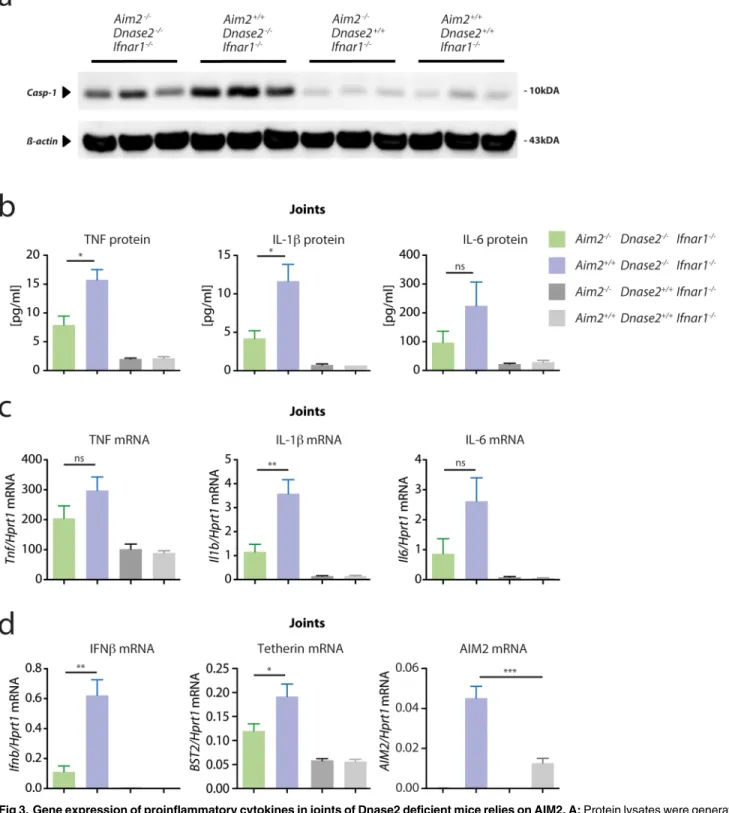
While these data could not explain the positive impact ofAim2-deficiency in this mouse model, analyzing caspase-1 activation and cytokine expression in joints ofDnase2-deficient mice revealed a different picture. Compared toDnase2-competent mice, joints ofAim2+/+x Dnase2-/-mice showed a marked increase in cleaved caspase-1, which was largely reduced in the absence of AIM2 (Fig 3A). Concomitant with this increased inflammasome activation, the expression of pro-inflammatory cytokines such as TNF, IL-6 and IL-1βexpression was strongly increased both at protein, as well as at the mRNA level in joints ofDnase2-deficient mice (Fig 3B and 3C). Of note, the expression of these pro-inflammatory cytokines was reduced in the absence ofAim2. At the same time, diseased joints also displayed a robust induction of IFNβ
and also interferon stimulated gens such asAim2,Bst2or Viperin were upregulated at the mRNA or protein level (Fig 3DandS2 Fig). This phenomenon can be explained by the fact that these genes are also directly induced upon cytosolic PRR stimulation, independently of the IFNAR loop [24]. Interestingly, the expression of these ISGs was alsoAim2-dependent, most prominently IFNβ.
AIM2 governs macrophage infiltration and local joint destruction in
Dnase2
-deficient mice
At 15 months of age, joints ofAim2+/+x Dnase2-/-mice showed severe signs of synovitis with hyperproliferation of synovial cells and massive immune cell infiltration, associated with pan-nus formation, cartilage destruction and bone erosion (Fig 4A and 4B). The immune cell infil-trate, most prominently pannus formation, was mainly dominated by the presence of
induced in joints ofAim2+/+x Dnase2-/-animals as observed by immunohistochemistry (Fig 4A and 4B) and also by qPCR and immunoblotting of joint sections (Fig 4C and 4D). In line with the arthritis scores and the pro-inflammatory cytokine profile, histological signs of Fig 2. Systemic proinflammatory status inDnase2-/-mice is independent of AIM2. A:Serum was collected fromAim2+/+Dnase2-/-Ifnar1-/-,Aim2 -/-Dnase2-/-Ifnar1
-/-,Aim2+/+Dnase2+/+Ifnar1-/-and Aim2-/-Dnase2+/+Ifnar1
-/-mice at the age of 15 month and analyzed for the depicted cytokines.B:Spleen weight and representative pictures of spleens from mice at the age of 15 month.C:Relative IFNβgene expression in spleen tissue normalized to the expression level of HPRT1 is shown.D:Anti-cyclic citrullinated peptide antibody (Anti-CCP-AB) was measured in serum samples as in (A). Data are presented as mean values + SEM, whereas statistical significance was assessed using a two-tailed, unpaired t-test comparing theDnase2-/-cohorts.
Fig 3. Gene expression of proinflammatory cytokines in joints of Dnase2 deficient mice relies on AIM2. A:Protein lysates were generated from the joints of 15 months old mice and immunoblotted for the presence of cleaved Caspase-1, whereasβ-Actin served as a loading control. Three independent protein lysates were analyzed per cohort.B:Cytokine levels of lysates as in (A) were determined.CandD:qPCR analysis of joints of the four different cohorts is shown for the indicated transcripts. The mRNA levels for the indicated cytokines are expressed relative to mRNA of HPRT1. Data are presented as mean values + SEM, whereas statistical significance was assessed using a two-tailed, unpaired t-test comparing theDnase2
-/-cohorts.
arthritis, macrophage infiltration andMmp3expression were largely decreased in the absence ofAim2.
Discussion
Altogether our data show that AIM2 plays an important role in the recognition of endogenous DNA species in the context ofDnase2-/-associated arthritis, thereby establishing AIM2 as a DAMP sensing PRR. Interestingly, the impact of AIM2 deficiency was primarily observed in diseased joints and not seen at the systemic level (e.g. splenomegaly or anti-CCP antibodies). These data imply that joint inflammation and subsequent tissue destruction are primary effects and mainly governed by local disease mechanisms and not just secondary to a systemic eleva-tion of pro-inflammatory cytokines. Moreover these results suggest that AIM2 activaeleva-tion drives disease progression in a tissue specific manner in this model. This concept is in line with a pre-vious study that has shown a predominant accumulation of self-DNA in joint tissue ofDnase2 -deficient mice, thereby arguing for an increase in ligand availability at the site of inflammation [21]. At the same time, it was also demonstrated thatDnase2-deficieny not only results in DNA accumulation resulting from undigested, phagocytized material, but also from cell-autonomous sources [21]. As such, damaged DNA expelled from nuclei can gain access to the cytosol, yet is subject to autophagy-mediated transport into DNase II containing lysosomes under normal conditions. The fact that cell autonomous and phagocytized material can both serve as ligands in the context ofDnase2-deficiency might explain why ablating AIM2 does not ameliorate all aspects of this disease model. Of note, with AIM2 being largely confined to the myeloid lineage, it is expected that AIM2 has no impact on self-DNA recognition in non-mye-loid cells.
As observed by caspase-1 immunoblotting,Dnase2-deficient mice displayed signs of inflam-masome activation in diseased joints, and consistent with its role as an inflaminflam-masome receptor,
Aim2-deficiency led to a decreased activation of caspase-1. In line with blunted inflammasome activation, IL-1βlevels were decreased in joints ofAim2-/-x Dnase2-/-mice, which could explain part of the pathomechanism being operational in this disease model [20]. Interestingly, beyond its impact on the inflammasome,Aim2deficiency also affected the expression of pro-inflammatory and antiviral genes in diseased joints, a phenomenon that had previously been ascribed to STING in this mouse model [17]. For example, expression of IFNβ, a cytokine that is completely STING-dependent in the context of cytosolic DNA recognition [25], was greatly reduced in diseased joints of AIM2-deficient mice. Whereas IFNβitself does not play a role in this disease model (globalIfnar1deficiency), these results indicate that the signaling cascade leading to IFNβproduction is indeed operational. This is also documented by the fact that IFN-stimulated genes are upregualted in joints ofDnase2-deficient mice. While these results could imply an epistatic relationship in signaling, with AIM2 functioning upstream of STING, we hypothesize that this is not the case. Most importantly,in vitroAIM2-deficient macro-phages display normal inflammatory gene expression and even increased type I IFN pro-duction upon cytosolic DNA delivery, while caspase-1 dependent processing of pro-IL-1βand pro-IL-18 and pyroptosis are abrogated [9,26,27]. These observations clearly argue against a cell-autonomous role of AIM2 in this model functioning upstream or in conjunction with intermediate grade synovitis / infiltration; 3, high grade synovitis / infiltration.C:RNAs were isolated from the joints of 15 months old mice to quantify MMP3 expression normalized to HPRT1. Data are presented as mean values + SEM, whereas statistical significance was assessed using a two-tailed, unpaired t-test comparing theDnase2-/-cohorts.D: Protein lysates were generated from the joints of 15 months old mice and immunoblotted for the presence of MMP3, whereasβ-Actin served as a loading control. Three independent protein lysates were analyzed per cohort. Data are presented as mean values + SEM, whereas statistical significance was assessed using a two-tailed Mann-Whitney test (b) or using a two-tailed, unpaired t-test (c) comparing theDnase2
-/-cohorts.
STING signaling. Moreover, the fact thatAim2deficiency differentially impacts on pro-inflam-matory cytokine expression in joints and in spleen furthermore disfavors a cell intrinsic role of AIM2 positively regulating STING-mediated signal transduction. In fact we speculate that the AIM2-dependent processing of IL-1 family cytokines and pyroptosis-dependent release of DAMPs leads to the priming of neighboring cells within joint tissue. A similar scenario has recently been proposed in the context of intramuscular DNA vaccination, whereAim2 -defi-cient animals showed greatly reduced expression of pro-inflammatory and antiviral cytokines at the sight of DNA application [28]. In this context, it is furthermore noteworthy that pro-inflammatory cytokines can dramatically enhance the responsiveness of non-myeloid cells to DNA stimulation via the STING axis [29]. Extrapolated to the in vivo situation, this would imply that AIM2 functions in a non-cell autonomous fashion to trigger inflammation, with AIM2 not being active in the cells that express pro-inflammatory cytokines. Nevertheless, it has also been reported that AIM2, when stimulated at lower ligand concentrations, can engage signaling cascades other than the canonical inflammasome pathway. To this end, it was shown that AIM2 can recruit procaspase-8 in an ASC-dependent fashion at ligand concentrations below the threshold of caspase-1 activation or in the absence of caspase-1 respectively [30,31]. While not formally established, caspase-8 recruitment by AIM2 could drive NF-KB activation and as such impact on pro-inflammatory gene expression under these conditions. Altogether, the exact mechanism of AIM2 contributing to pro-inflammatory and antiviral gene expression in this model remains to be determined, while it should be informative to dissect the contribu-tion of different cells types in thein vivosituation.
With the predominant involvement of the adaptive branch of the immune system unequiv-ocally being documented in rheumatoid arthritis, the relevance of erroneous DNA sensing by PRRs has yet to be established in this disease entity. However, theDnase2-/-model shares a number of prominent features with Systemic juvenile idiopathic arthritis (sJIA), which can be considered an autoinflammatory disease [20,32]. Having identified AIM2 as a critical pro-inflammatory driver in this disease model provides a molecular target that could be of interest for future therapeutic intervention.
Note added in proof: While this manuscript was under review, Ellen Gravallese and col-leagues have published a likewise approach of studying the role of AIM2 in the context of
Dnase2-/-xIfnar1-/-induced polyarthritis [33]. They also found a critical role for AIM2 in driv-ing disease pathology (clinical arthritis score, pro-inflammatory cytokine production in joints), yet with a slightly lower impact compared to our study. Moreover, in their analysisAim2 defi-ciency led to a considerable decrease in IL-18 levels in serum and in joint tissue, a phenomenon that we did not observe in our cohorts (data not shown). It is possible that differences in mouse strains used or housing conditions account for these discrepancies. However, given the fact that IL-18 deficiency does not protectDnase2-deficient animals from polyarthritis or pro-inflammatory gene expression [20],Aim2-dependent IL-18 production or maturation appears to be an epiphenomenon rather than a cause in this disease model. Indeed, our data suggest thatAim2deficiency dampens caspase-1 activation and associated IL-1βsecretion and that it also impacts on pro-inflammatory cytokine expression in joint tissues.
Supporting Information
S1 Fig. Generation ofAim2-deficient mice. A:The gene targeting approach that was taken to
generateAim2-/-mice is depicted.B:Representative genotyping result for wildtype,Aim2
analyzed for IL-1βproduction 6 hours after stimulation. (PDF)
S2 Fig. Viperin expression in joints ofDnase2-deficient mice.Protein lysates were generated
from the joints of 15 months old mice and immunoblotted for the presence of Viperin, whereas
β-Actin served as a loading control. Three independent protein lysates were analyzed per cohort.
(PDF)
Acknowledgments
We thank Dr. S. Nagata and Dr. Ulf Andersson for their generous support in providing us the
Dnase2a/Ifnar1-deficient mice. We kindly thank Max Rothe and Wenzel Vogel for great tech-nical assistance.
Author Contributions
Conceived and designed the experiments: VH CJ. Performed the experiments: CJ SP. Analyzed the data: CJ SP VH. Wrote the paper: VH.
References
1. Medzhitov R. Recognition of microorganisms and activation of the immune response. Nature. 2007; 449(7164):819–26. Epub 2007/10/19. doi:10.1038/nature06246PMID:17943118.
2. Barbalat R, Ewald SE, Mouchess ML, Barton GM. Nucleic acid recognition by the innate immune sys-tem. Annual review of immunology. 2011; 29:185–214. doi:10.1146/annurev-immunol-031210-101340
PMID:21219183.
3. Ablasser A, Hertrich C, Wassermann R, Hornung V. Nucleic acid driven sterile inflammation. Clin Immunol. 2013; 147(3):207–15. Epub 2013/02/20. doi:10.1016/j.clim.2013.01.003PMID:23419883.
4. Crow YJ. Aicardi-Goutieres syndrome. Handbook of clinical neurology. 2013; 113:1629–35. doi:10.
1016/B978-0-444-59565-2.00031–9PMID:23622384.
5. Lee-Kirsch MA, Wolf C, Gunther C. Aicardi-Goutieres syndrome: a model disease for systemic autoim-munity. Clinical and experimental immunology. 2014; 175(1):17–24. doi:10.1111/cei.12160PMID:
23786362; PubMed Central PMCID: PMC3898550.
6. Cai X, Chiu YH, Chen ZJ. The cGAS-cGAMP-STING pathway of cytosolic DNA sensing and signaling. Mol Cell. 2014; 54(2):289–96. Epub 2014/04/29. doi:10.1016/j.molcel.2014.03.040PMID:24766893.
7. Hornung V, Hartmann R, Ablasser A, Hopfner KP. OAS proteins and cGAS: unifying concepts in sens-ing and respondsens-ing to cytosolic nucleic acids. Nat Rev Immunol. 2014; 14(8):521–8. Epub 2014/07/19.
doi:10.1038/nri3719PMID:25033909.
8. Paludan SR, Bowie AG. Immune sensing of DNA. Immunity. 2013; 38(5):870–80. Epub 2013/05/28.
doi:10.1016/j.immuni.2013.05.004PMID:23706668; PubMed Central PMCID: PMC3683625. 9. Hornung V, Ablasser A, Charrel-Dennis M, Bauernfeind F, Horvath G, Caffrey DR, et al. AIM2
recog-nizes cytosolic dsDNA and forms a caspase-1-activating inflammasome with ASC. Nature. 2009; 458 (7237):514–8. Epub 2009/01/23. doi:10.1038/nature07725PMID:19158675; PubMed Central PMCID:
PMC2726264.
10. Fernandes-Alnemri T, Yu JW, Datta P, Wu J, Alnemri ES. AIM2 activates the inflammasome and cell death in response to cytoplasmic DNA. Nature. 2009; 458(7237):509–13. Epub 2009/01/23. doi:10.
1038/nature07710PMID:19158676; PubMed Central PMCID: PMC2862225.
11. Roberts TL, Idris A, Dunn JA, Kelly GM, Burnton CM, Hodgson S, et al. HIN-200 proteins regulate cas-pase activation in response to foreign cytoplasmic DNA. Science. 2009; 323(5917):1057–60. doi:10.
1126/science.1169841PMID:19131592.
12. Burckstummer T, Baumann C, Bluml S, Dixit E, Durnberger G, Jahn H, et al. An orthogonal proteomic-genomic screen identifies AIM2 as a cytoplasmic DNA sensor for the inflammasome. Nature immunol-ogy. 2009; 10(3):266–72. doi:10.1038/ni.1702PMID:19158679.
13. Miao EA, Rajan JV, Aderem A. Caspase-1-induced pyroptotic cell death. Immunological reviews. 2011; 243(1):206–14. doi:10.1111/j.1600-065X.2011.01044.xPMID:21884178; PubMed Central PMCID:
14. Schattgen SA, Fitzgerald KA. The PYHIN protein family as mediators of host defenses. Immunological reviews. 2011; 243(1):109–18. Epub 2011/09/03. doi:10.1111/j.1600-065X.2011.01053.xPMID:
21884171.
15. Kawane K, Ohtani M, Miwa K, Kizawa T, Kanbara Y, Yoshioka Y, et al. Chronic polyarthritis caused by mammalian DNA that escapes from degradation in macrophages. Nature. 2006; 443(7114):998–1002.
doi:10.1038/nature05245PMID:17066036.
16. Nagata S, Kawane K. Autoinflammation by endogenous DNA. Advances in immunology. 2011; 110:139–61. doi:10.1016/B978-0-12-387663-8.00004–1PMID:21762818.
17. Ahn J, Gutman D, Saijo S, Barber GN. STING manifests self DNA-dependent inflammatory disease. Proceedings of the National Academy of Sciences of the United States of America. 2012; 109 (47):19386–91. Epub 2012/11/08. doi:10.1073/pnas.1215006109PMID:23132945; PubMed Central
PMCID: PMC3511090.
18. Kawane K, Fukuyama H, Kondoh G, Takeda J, Ohsawa Y, Uchiyama Y, et al. Requirement of DNase II for definitive erythropoiesis in the mouse fetal liver. Science. 2001; 292(5521):1546–9. doi:10.1126/
science.292.5521.1546PMID:11375492.
19. Yoshida H, Okabe Y, Kawane K, Fukuyama H, Nagata S. Lethal anemia caused by interferon-beta pro-duced in mouse embryos carrying undigested DNA. Nature immunology. 2005; 6(1):49–56. doi:10.
1038/ni1146PMID:15568025.
20. Kawane K, Tanaka H, Kitahara Y, Shimaoka S, Nagata S. Cytokine-dependent but acquired immunity-independent arthritis caused by DNA escaped from degradation. Proceedings of the National Academy of Sciences of the United States of America. 2010; 107(45):19432–7. doi:10.1073/pnas.1010603107
PMID:20974942; PubMed Central PMCID: PMC2984163.
21. Lan YY, Londono D, Bouley R, Rooney MS, Hacohen N. Dnase2a Deficiency Uncovers Lysosomal Clearance of Damaged Nuclear DNA via Autophagy. Cell reports. 2014; 9(1):180–92. doi:10.1016/j.
celrep.2014.08.074PMID:25284779.
22. Lallemand Y, Luria V, Haffner-Krausz R, Lonai P. Maternally expressed PGK-Cre transgene as a tool for early and uniform activation of the Cre site-specific recombinase. Transgenic research. 1998; 7 (2):105–12. PMID:9608738.
23. Jakobs C, Bartok E, Kubarenko A, Bauernfeind F, Hornung V. Immunoblotting for active caspase-1. Methods in molecular biology. 2013; 1040:103–15. doi:10.1007/978-1-62703-523-1_9PMID:
23852600.
24. Schneider WM, Chevillotte MD, Rice CM. Interferon-stimulated genes: a complex web of host defenses. Annual review of immunology. 2014; 32:513–45. doi:
10.1146/annurev-immunol-032713-120231PMID:24555472; PubMed Central PMCID: PMC4313732.
25. Ishikawa H, Ma Z, Barber GN. STING regulates intracellular DNA-mediated, type I interferon-depen-dent innate immunity. Nature. 2009; 461(7265):788–92. doi:10.1038/nature08476PMID:19776740.
26. Rathinam VA, Jiang Z, Waggoner SN, Sharma S, Cole LE, Waggoner L, et al. The AIM2 inflammasome is essential for host defense against cytosolic bacteria and DNA viruses. Nature immunology. 2010; 11 (5):395–402. doi:10.1038/ni.1864PMID:20351692; PubMed Central PMCID: PMC2887480.
27. Panchanathan R, Duan X, Shen H, Rathinam VA, Erickson LD, Fitzgerald KA, et al. Aim2 deficiency stimulates the expression of IFN-inducible Ifi202, a lupus susceptibility murine gene within the Nba2 autoimmune susceptibility locus. Journal of immunology. 2010; 185(12):7385–93. doi:10.4049/
jimmunol.1002468PMID:21057088; PubMed Central PMCID: PMC3059233.
28. Suschak JJ, Wang S, Fitzgerald KA, Lu S. Identification of Aim2 as a sensor for DNA vaccines. Journal of immunology. 2015; 194(2):630–6. doi:10.4049/jimmunol.1402530PMID:25488991; PubMed
Cen-tral PMCID: PMC4282968.
29. Chiliveru S, Rahbek SH, Jensen SK, Jorgensen SE, Nissen SK, Christiansen SH, et al. Inflammatory cytokines break down intrinsic immunological tolerance of human primary keratinocytes to cytosolic DNA. Journal of immunology. 2014; 192(5):2395–404. doi:10.4049/jimmunol.1302120PMID:
24489095.
30. Sagulenko V, Thygesen SJ, Sester DP, Idris A, Cridland JA, Vajjhala PR, et al. AIM2 and NLRP3 inflammasomes activate both apoptotic and pyroptotic death pathways via ASC. Cell death and differ-entiation. 2013; 20(9):1149–60. doi:10.1038/cdd.2013.37PMID:23645208; PubMed Central PMCID:
PMC3741496.
31. Pierini R, Juruj C, Perret M, Jones CL, Mangeot P, Weiss DS, et al. AIM2/ASC triggers caspase-8-dependent apoptosis in Francisella-infected caspase-1-deficient macrophages. Cell death and differ-entiation. 2012; 19(10):1709–21. doi:10.1038/cdd.2012.51PMID:22555457; PubMed Central PMCID:
32. Mellins ED, Macaubas C, Grom AA. Pathogenesis of systemic juvenile idiopathic arthritis: some answers, more questions. Nature reviews Rheumatology. 2011; 7(7):416–26. doi:10.1038/nrrheum.
2011.68PMID:21647204; PubMed Central PMCID: PMC4180659.
33. Baum R, Sharma S, Carpenter S, Li QZ, Busto P, Fitzgerald KA, et al. Cutting edge: AIM2 and endoso-mal TLRs differentially regulate arthritis and autoantibody production in DNase II-deficient mice. Journal of immunology. 2015; 194(3):873–7. doi:10.4049/jimmunol.1402573PMID:25548216; PubMed