Inhibition of BACE1 Activity by a DNA
Aptamer in an Alzheimer
’
s Disease Cell
Model
Huiyu Liang1☯¤, Yusheng Shi2☯, Zhewen Kou1, Yonghua Peng1, Wenjun Chen1, Xiaowen Li1, Shuji Li1, Ying Wang3, Fang Wang1, Xingmei Zhang1*
1Key Laboratory of Psychiatric Disorders of Guangdong Province, Department of Neurobiology, School of Basic Medical Sciences, Southern Medical University, Guangzhou, China,2Department of Radiation Oncology, Nanfang Hospital, Southern Medical University, Guangzhou, China,3Cancer Research Institute, School of Basic Medical Sciences, Southern Medical University, Guangzhou, China
☯These authors contributed equally to this work.
¤ Current address: Nanmatou Community Health Service Centre, Pudong District, Shanghai, China *[email protected]
Abstract
An initial step in amyloid-β(Aβ) production includes amyloid precursor protein (APP) cleav-age viaβ-Site amyloid precursor protein cleaving enzyme 1 (BACE1). Increased levels of brain Aβhave been implicated in the pathogenesis of Alzheimer’s disease (AD). Thus,β -secretase represents a primary target for inhibitor drug development in AD. In this study, apta-mers were obtained from combinatorial oligonucleotide libraries using a technology referred to as systematic evolution of ligands by exponential enrichment (SELEX). A purified human BACE1 extracellular domain was used as a target to conduct anin vitroselection process using SELEX. Two DNA aptamers were capable of binding to BACE1 with high affinity and good specificity, withKdvalues in the nanomolar range. We subsequently confirmed that one aptamer, A1, exhibited a distinct inhibitory effect on BACE1 activity in an AD cell model. We detected the effects of M17-APPsw cells that stably expressed Swedish mutant APP after aptamer A1 treatment. Aβ40 and Aβ42 concentrations secreted by M17-APPsw cells decreased intracellularly and in culture media. Furthermore, Western blot analysis indicated that sAPPβexpression significantly decreased in the A1 treated versus control groups. These findings support the preliminary feasibility of an aptamer evolved from a SELEX strat-egy to function as a potential BACE1 inhibitor. To our knowledge, this is the first study to acquire a DNA aptamer that exhibited binding specificity to BACE1 and inhibited its activity.
Introduction
Alzheimer’s disease (AD) is a chronic degenerative disease of the central nervous system (CNS), which is primarily manifested by cognitive impairment, particularly memory deteriora-tion. The decline in daily living activities of AD patients, as well as behavioral and psychologi-cal symptoms, result in substantial emotional and financial burdens on patients, their families,
OPEN ACCESS
Citation:Liang H, Shi Y, Kou Z, Peng Y, Chen W, Li
X, et al. (2015) Inhibition of BACE1 Activity by a DNA Aptamer in an Alzheimer’s Disease Cell Model. PLoS ONE 10(10): e0140733. doi:10.1371/journal. pone.0140733
Editor:Vittorio de Franciscis, Consiglio Nazionale
delle Ricerche (CNR), ITALY
Received:May 19, 2015
Accepted:September 28, 2015
Published:October 16, 2015
Copyright:© 2015 Liang et al. This is an open
access article distributed under the terms of the
Creative Commons Attribution License, which permits unrestricted use, distribution, and reproduction in any medium, provided the original author and source are credited.
Data Availability Statement:All relevant data are
within the paper.
Funding:This work was supported by grants from
the National Science Foundation of China (No. 81471388 and No. 81272509) and the Guangdong Natural Science Foundation (No. 2014A030313351). The funders had no role in study design, data collection and analysis, decision to publish, or preparation of the manuscript.
Competing Interests:The authors have declared
and society. In recent years, the morbidity of AD has increased as a result of an aging popula-tion and increased diagnostic rates, and it has become a more serious healthcare problem [1]. The build-up of amyloid-β(Aβ) peptides in the brain has been linked to AD pathogenesis and may represent a key target for AD modification[2,3]. Aβformation occurs via sequential pro-teolytic processing of amyloid precursor protein (APP) and is catalyzed byβ- andγ-secretases.
β-site APP-cleaving enzyme 1 (BACE1) is a membrane-bound aspartic protease and the rate-limiting step in Aβgeneration, which is responsible forβ-secretase cleavage of APP [4]. Evidence indicates that BACE1 protein levels and activity are upregulated in the brains of spo-radic AD patients [5]. Furthermore, increased BACE1 levels have been reported in cerebrospi-nal fluid (CSF) of prodromal AD patients [6]. Moreover, an increased affinity of APP binding to BACE1 has been reported in patients who carry the Swedish mutation in the APP gene (APPsw), which subsequently increased Aβproduction [7]. A coding mutation in the APP gene (APPA673T) located at a site proximal to the BACE1 proteolytic site decreased BACE1 cleavage of APP and was protective against AD, which provides additional evidence that the inhibition of BACE1 cleavage of APP may protect against AD [8,9]. Previous studies have demonstrated that decreased BACE1 activity altered the amyloid burden in mice [10–14]. Thus, BACE1 represents a promising target for mechanistic-based AD treatment. To date, BACE1 inhibitor development has been very challenging, and no safe and effective BACE1 inhibitor has been used in clinical populations [15].
Aptamers are obtained from combinatorial oligonucleotide libraries using a technology referred to as systematic evolution of ligands by exponential enrichment (SELEX). These sin-gle-stranded oligonucleotides are capable of specific and high-affinity binding to target mole-cules due to their tertiary structures. Compared with conventional antibodies, aptamers have a substantial number of attractive features including low molecular weight, quick and reproduc-ible synthesisin vitro, easy modification, good stability, low toxicity, low immunogenicity and rapid tissue penetration [15,16]. Purified proteins are the most common targets for SELEX selection. Aptamers selected directly against proteins can inhibit target protein activity with high affinity and good specificity [17], and can be used to investigate the mechanisms of inter-action between proteins and nucleic acids [18]. These advantages have made aptamers excel-lent alternatives for diagnostic and therapeutic agents in CNS related pathologies, because antibody-based approaches have not demonstrated adequate sensitivity in most cases, whereas they have exhibited toxicityin vivoand the inability to cross the blood-brain barrier (BBB) effi-ciently. Although several small-molecule BACE1 inhibitors have been developed in AD research [9,10,19], there is currently no BACE1 inhibitor available on the market. Therefore, the development of a novel type of BACE1 inhibitor is very important.
This current study used a purified human BACE1 extracellular domain as a target to per-form the SELEX process, and obtained two highly efficient and specific aptamers to BACE1 (i.e. A1 and A2). The A1 aptamer decreased Aβ40and Aβ42production, as well as sAPPβ
expression, in M17-APPsw cell cultures (AD cell model). These novel findings support the ini-tial potenini-tial of A1 as a BACE1 inhibitor for the treatment of AD. To our knowledge, this is the first investigation to acquire a DNA aptamer that exhibits binding specificity to BACE1 and inhibits its activity.
Materials and Methods
Cell culture
supplemented with 10% fetal bovine serum (FBS), 100 U/ml of penicillin, 100 ug/ml of strepto-mycin (P/S), and 20 mg/ml of Geneticin in a 5% CO2/95% air atmosphere environment at 37°C.
Random library, primers and control aptamer
The synthetic single stranded DNA (ssDNA) library consists of a random sequence of 30 nt in the middle and two flanked primer hybridization sites [20]:
50-GCAATGGTACGGTACTTCC-(N30)-CAAAAGTGCACGCTACTTTGCTAA-30.
Sense strand primer P1: 50-GCAATGGTACGGTACTTCC-30.
Antisense strand primer P2: 50-TTAGCAAAGTAGCGTGCACTTTTG-30.
Structured 30antisense strand primer P3:
50-GCTAAGCGGGTGGGACTTCCTAGTCCCACCCGCTTAGCAAAGTAGCGTGCACTTTTG-30.
P1 and P2 were utilized to synthesize double-stranded DNA (dsDNA). P1 and P3 were used to synthesize ssDNA via asymmetric polymerase chain reaction (PCR). All sequences including biotin labeled ssDNA were manufactured by Invitrogen (Carlsbad, CA, USA).
SELEX procedure
This SELEX strategy was conducted as previously described with minor modifications [21]. Briefly, the purified human BACE1 extracellular domain (Sigma Aldrich, St. Louis, MO, USA) was coated onto 96-well plates at 4°C overnight, and the wells were subsequently blocked with 5% bovine serum albumin (BSA). The DNA library was heated at 95°C for five minutes and snap-cooled for five minutes on ice. Then, it was maintained at room temperature for 30 minutes in binding buffer (0.45 g of glucose, 10 mg of yeast tRNA, 5 mg of salmon sperm DNA, 0.1 g of BSA, and 0.5 ml of 1 M MgCl2in 100 ml of 1 x phosphate-buffered saline [PBS]) prior to incuba-tion with BACE1. After incubaincuba-tion, the wells were washed six times with washing buffer (0.45 g of glucose and 0.5 ml of 1 M MgCl2in 100 ml of 1 x PBS). The plate was placed on a 95°C heat block for five minutes and maintained on ice for five minutes to dissociate DNA from the target. Since the 4thround of selection by SELEX, the generated ssDNA pool was incubated with a blank well for counter selection. The unbound sequences were subsequently incubated with BACE1. BACE1 protein amount, DNA pool volume, and incubation time were gradually decreased to increase selection stringency, whereas washing time was increased during this process (Table 1).
Preparation of ssDNA via asymmetric PCR
Using P1 and P3 primers, the production of ssDNA molecules occurred via asymmetric PCR as follows [22]. One hundredμl of PCR mixture, which contained 10μl of 10 x PCR buffer, 0.2 mM
Table 1. Selection conditions in the SELEX process.
Cycle Amount of BACE1(μg) ssDNA pool (pmol) Salmon sperm DNA (μg/μl) Incubation time (minutes) washing conditions
1 7 2500 0.05 120 6×1 min
2 5 500 0.1 120 6×1 min
3 2.5 250 0.1 60 6×2 min
4a 2.5 250 0.1 40 6×2 min
5a 1 250 0.1 40 6×3 min
6a 1 250 1 30 6×3 min
7a 0.5 250 1 30 6×3 min
BACE1:β-Site amyloid precursor protein cleaving enzyme 1
aA blank control well was introduced for counter selection since the 4thround.
of dNTPs, 1μM of each primer, 20 nM of template, and 2.5 U of Taq DNA polymerase, was ther-mally cycled 30 times at 94°C for one minute, 37°C for 80 seconds, and 58°C for one minute, fol-lowed by a final extension for five minutes at 58°C. Unequal length PCR products were analyzed via electrophoresis in an 8% polyacrylamide-7M urea gel, and the lower band of interest was purified. The gel was eluted via the addition of elution buffer (0.5 M of NH4Ac, 0.2% SDS, and 1 M of EDTA [pH 8.0]). The ssDNA was collected and precipitated by the addition of 10 mM of MgCl2, 0.3 M of NaAc, and 2.5-fold ethanol by volume at -20°C overnight. Following centrifuga-tion, the pellet was rinsed with 70% ethanol and dried at room temperature.
Binding affinity assay
BACE1 (50μM per well, diluted in 1 x PBS) was coated onto 96-well plates at 4°C overnight, blocked with 5% BSA at 37°C for 60 minutes, washed with PBST (0.01% Tween 20 in 1 x PBS, pH 7.4), and incubated with different concentrations of biotinylated aptamers at 37°C for 60 minutes. Streptavidin-conjugated horseradish peroxidase (50μl, 1:1000 in PBS; Sigma Aldrich, St. Louis, MO, USA) was added to the wells and incubated for 30 minutes at 37°C. Fifty ul of TMB solution were added for an additional 20 minutes of incubation. The reaction was termi-nated with 25μl of 2 M H2SO4, and the absorbance was measured at 450 nm. Apparent equilib-rium dissociation constant (Kd) values were determined for each aptamer using nonlinear regression (SigmaPlot 12.0; Systat Software, San Jose, CA, USA) according to the following equation:Y = BmaxX/(Kd+X)[23].
Pull-down assay
RIPA buffer that contained 1% (v/v) complete protease inhibitor cocktail was added to
M17-APPsw cells. The samples were centrifuged, and the supernatant was collected. Total pro-tein levels were determined using a BCA Propro-tein Assay Reagent kit (Rockford, IL, USA). Strep-tavidin beads were incubated with biotin-labeled A1, Gp30 and U31 (250 pmol each) for 30 minutes at 37°C. Washed bead-aptamer mixtures were subsequently incubated with 600μg of cell extracts for one hour at 37°C. The mixtures were washed and heated for 10 minutes at 100°C in 50μl of 2 x sodium dodecyl sulfate-polyacrylamide gel electrophoresis (SDS-PAGE) sample buffer. The samples were subsequently analyzed via immunoblotting. The primary antibody comprised of anti-BACE1 (Sigma Aldrich, St. Louis, MO, USA) [23].
Fluorescence resonance energy transfer (FRET) assay
Cell viability assay
M17 cells were seeded in 96-well plates (density = 1×10⁴cells per well). Following 24 hours of culture, cells were treated with vehicle (DMSO) or different aptamer concentrations for 24 hours in Opti-MEM. Then, the medium that contained the vehicle or aptamers was replaced with fresh medium supplemented with MTT 0.5 mg/mL in the absence of aptamers or vehicle. Cells were subsequently cultured for an additional four hours. The medium was subsequently discarded, and 100μl of DMSO were added to the wells. Following 15 minutes of incubation with DMSO, the absorption intensities were measured at 570 nm [24].
Quantitation of A
β
40 and A
β
42
M17 cells were seeded in 6-well plates. When the cells attained 80% confluence, cultures were refreshed for 24 hours with Opti-MEM and different A1, Gp30 and U31 concentrations. The medium was obtained from the plates to determine the secreted Aβ40and Aβ42levels, and a complete protease inhibitor cocktail (Rockford, IL, USA) was added with a final concentration of 1% (v/v). The medium was subsequently centrifuged at 12,000 rpm for 20 minutes at 4°C, and the supernatant was analyzed for Aβ40and Aβ42quantitation. To quantify the intracellular Aβ40and Aβ42levels, the cells were lysed as previously described. The collected cell lysates were assayed without dilution using human Aβ40and Aβ42double-antibody sandwich ELISA kits (Invitrogen, Carlsbad, CA, USA) [24]
Western blotting
After 24 hours of treatment with A1 or vehicle, the M17 cells were harvested in 2 x SDS-PAGE sample buffer. The samples were resolved on 10% SDS-PAGE gels and transferred to PVDF membranes. Then, the membranes were blocked in 5% milk for one hour, followed by incuba-tion overnight at 4°C with the corresponding primary antibody. The primary antibody was APP, which is specific to the APP amino terminal (Abcam, Cambridge, UK), and anti-BACE1 (Sigma Aldrich, St. Louis, MO, USA). On the subsequent day, the membranes were washed three times with TBST buffer for five minutes. The membranes were then incubated in 5% milk (v/v) supplemented with anti-rabbit IgG or anti-mouse IgG for one hour at room tem-perature. Following incubation with the secondary antibody, the membranes were washed three times with TBST buffer for five minutes. The blots were then visualized via incubation with a SuperSignal West Dura chemiluminescence kit (Pierce Biotechnology, Rockford, IL, USA). Images were obtained via an AlphaImager HP System (Alpha Innotech, San Leandro, CA, USA).β-actin was used as a reference control for protein loading. The bands were quanti-fied using Fluorchem Q SA software (Alpha Innotech, Santa Clara, CA, USA).
Statistical analysis
Statistical analysis included one-way analysis of variance (ANOVA) for more than two groups or independent-samplet-tests for two groups (SPSS 13.0; SPSS Inc., Chicago, IL, USA).Kd val-ues for each aptamer were calculated using nonlinear regression (SigmaPlot 12.0; Systat Soft-ware, San Jose, CA, USA).P<0.05 andP<0.01 compared with the control group were considered statistically significant.
Results
Aptamer selection
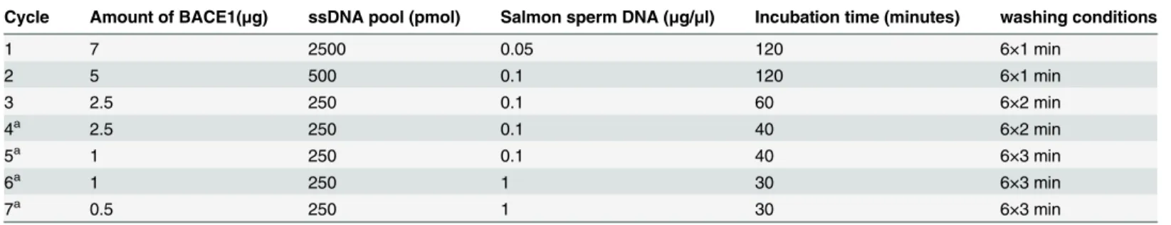
identified using 7M urea 8% denatured polyacrylamide gel electrophoresis (Fig 1A). Selected pool enrichment resulted in the evolution of potential aptamer candidates, which were investi-gated using indirect ELISA. First, ssDNAs from each round were labeled with biotin; then, using streptavidin-conjugated horseradish peroxidase, the biotinylated aptamers reacted with BACE1 via ELISA. Initial library Gp30 and ssDNA obtained from the 1st, 3rd, 5thand 7th rounds were applied to perform the assay. Absorbance was increased as the selection process proceeded, which suggested enrichment in the 7thround (Fig 1B). The ssDNA obtained from the final round was amplified, cloned, and sequenced. Two sequences were identified based on their primary sequence similarity and were regarded as potential aptamer candidates (A1 and A4) (Table 2). Both sequences had a primarily stem-loop structure as indicated by the second-ary structure analysis (Fig 1C and 1D).
Aptamers that specifically bind to BACE1 with high affinity
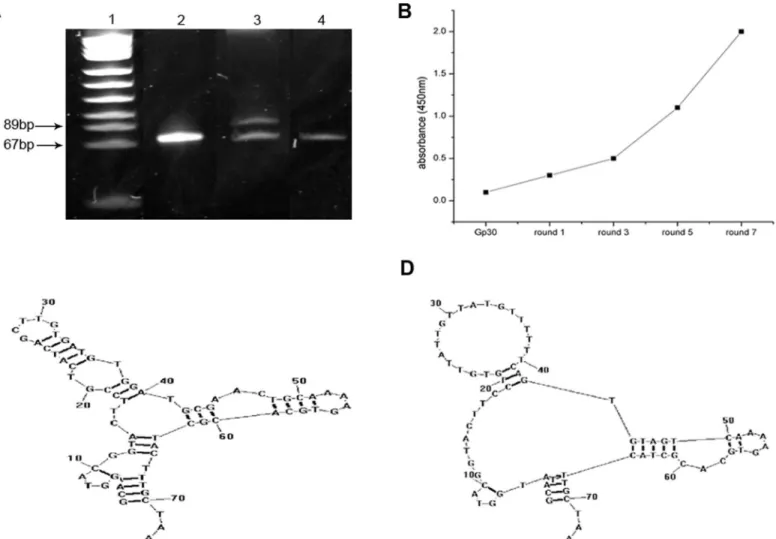
Indirect ELISA was performed to determine the binding affinity of the selected aptamers to BACE1. Anti-BACE1 antibody and an unrelated aptamer, U31, were used as the control. Similar
Fig 1. Aptamer generation using the SELEX process.(A) Seven M urea 8% denatured polyacrylamide gel electrophoresis was used to confirm ssDNA, which was separated by asymmetric PCR after each round. Lane 1: pUC18 DNA/Msp l marker. Lane 2: initial library Gp30. Lane 3: unequal length PCR products. Lane 4: target ssDNA. (B) Selected pool enrichment was monitored via indirect ELISA. (C-D) Secondary structures of aptamers A1 (C) and A4 (D) predicted by RNAstructure 5.7 software (Mathews lab,http://rna.urmc.rochester.edu/RNAstructure.html).
to anti-BACE1, the binding curves of A1 and A4 to BACE1 fit well (Fig 2A–2D) withKdvalues
in the nanomolar range (Table 2). These findings indicate that A1 and A4 can specifically bind to BACE1 with high affinity. A1 had the highest repetition frequency and was subsequently selected for the pull-down assay, which illustrated that A1 specifically interacted with BACE1 protein in M17-APPsw cells, whereas Gp30 and U31 did not exhibit binding to BACE1 proteins (Fig 2E).
Aptamer A1 specifically inhibits BACE1 activity in vitro
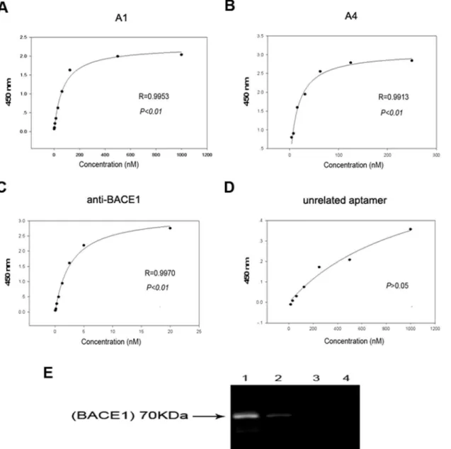
Next, a fluorescence resonance energy transfer assay was conducted to confirm the inhibitory effect of A1 on BACE1 activityin vitro. The BACE1 substrate is linked to a fluorescent donor on one end and a quenching acceptor group on the other end. As a result of intramolecular energy transfer to the quenching acceptor, fluorescence from the donor can be quenched by the acceptor. Following substrate cleavage via BACE1 enzymes, the energy transfer is disturbed, which results in fluorescent signal enhancement. The fluorescence of the substrate is signifi-cantly reduced when the substrate cleavage is blocked by an inhibitor. Based on this principle, various concentrations of A1 were added into a reaction system that contained BACE1 and its fluorescent substrate; the changes in the fluorescence intensity were subsequently detected. A BACE1 specific inhibitor ([Asn670, Sta671, Val672]-Amyloidβ/A4 Precursor Protein 770 Fragment 662–675), Gp30, and U31 served as controls. Following increases in the concentra-tion, the fluorescence intensity in the standard inhibitor group gradually decreased, with a sig-nificant decrease at a concentration of 500 nM compared with the positive control (P<0.01). The fluorescence intensity in the A1 group significantly decreased at a concentration of 250 nM (P<0.01). No significant decrease in the fluorescence intensities occurred in the Gp30 or U31 groups (Fig 3A and 3B). The data of BACE1 activity were converted from percent inhibi-tion to probit values, which were drawed versus the log of the BACE1 specific inhibitor or apta-mer A1 concentration. A1 inhibited BACE1 activity with an IC50value of 139.81 nM. Under the same experimental conditions, standard inhibitor inhibited the activity of BACE1 in a con-centration-dependent manner (IC50= 242.50 nM;Fig 3C and 3D). These findings demonstrate that A1 can act as a potent inhibitor of BACE1 activityin vitro.
Aptamer A1 specifically inhibits BACE1 activity in an AD cell model
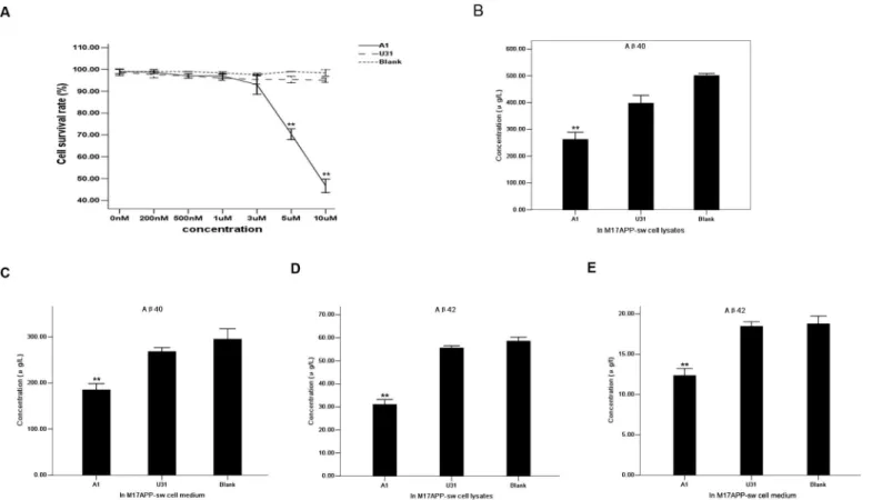
MTT was used to evaluate the effect of different A1 concentrations on cell survival.
M17-APPsw cell viability was not affected at a final concentration of 3μM (Fig 4A). Thus, a 3μM A1 concentration was applied for cell incubation. The resulting cell lysates and media were resolved on a double-antibody sandwich ELISA. A 3μM U31 treated group and a blank control (without treatment) group served as the controls. Aβ40and Aβ42concentrations secreted by M17-APPsw cells decreased intracellularly and in culture media compared with the
Table 2. Sequences of selected aptamers andKdvalues of A1, A4 and anti-BACE1.
Name Sequence Kd(nM)a
A1 GCAATGGTACGGTACTTCCGTCATCAGCTTGTGATGTGGATGCGAACTGCAAAAGTGCACGCTACTTTGCTAAb 68.5655±8.1237
A4 GCAATGGTACGGTACTTCCTGTGTTATTGTTATGTTTTTTCAGTGTAGTCAAAAGTGCACGCTACTTTGCTAAb 15.3497±2.0262
Anti-BACE1 2.7498±0.2785
BACE1:β-Site amyloid precursor protein cleaving enzyme 1
a
Michaelis-Menten binding curves used to evaluateKd(nM) were performed as described in the Materials and Methods section. Standard deviation
values were determined from three independent experiments.
b
Bold underlined letters represent random sequences of 30 nt in length.
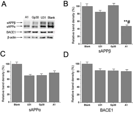
control group (Fig 4B–4E). Western blot analysis indicated that sAPPβexpression was signifi-cantly decreased in the A1 treated group compared with the control group, whereas no sub-stantial differences in sAPPαand BACE1 expression levels were identified among the groups (Fig 5). These results suggest that A1 can inhibit BACE1 activity in an AD cell model.
Discussion
This study employed the SELEX process on a microwell plate, in which a purified extracellular region of human BACE1 protein was used as the target, and two DNA aptamers against
Fig 2. Selected aptamers to BACE1 exhibit good specificity and high affinity.(A-D) Binding curves of A1, A4, anti-BACE1 and an unrelated aptamer, U31. Mean absorbances for each aptamer concentration from three independent experiments were used to create the binding curve. (E) The specific interaction of aptamer A1 with BACE1 protein was identified via affinity purification on streptavidin beads of M17-APPsw cell lysate treated with biotin-labeled A1, followed by immunoblotting with anti-BACE1 antibody. A band at 70 kDa corresponded to the molecular weight of BACE1 and indicated that BACE1 was the specific target of the aptamer. Lane 1: A1 with M17-APPsw cell lysate. Lane 2: GP30 with M17-APPsw cell lysate. Lane 3: U31 with M17-APPsw cell lysate. Lane 4: blank control (no aptamer) group. Three independent experiments were performed.
BACE1 were identified. Since the 4thround, a blank well was introduced for counter selection to exclude the enrichment of nonspecific sequences. Although many modified SELEX methods have been developed such as capillary electrophoresis SELEX and whole-cell SELEX, tradi-tional SELEX technology possesses the superiority of rapid and simple operation, as well as excellent specificity of selected aptamers to target proteins. The fitted curves and lowKdvalues of these aptamers, which were determined by indirect ELISA, indicate high affinity and speci-ficity to BACE1.
Aptamers to proteins often bind in functionally important parts of a molecule, such as a substrate binding pocket or allosteric site, to modulate physiological function [25]. Aptamers can execute their functions by directly binding to the target protein without affecting its gene expression, in contrast to other small noncoding RNAs that inhibit gene expression such as microRNAs, siRNAs and antisense [26]. Aptamers exhibit direct effects for treating diseases both within and outside the CNS [27,28]. The FRET assay demonstrated the strong inhibitory effect of A1 on BACE1 activityin vitro, in which the IC50value of A1 was smaller than a stan-dard inhibitor. We speculate that there may be sequence specificity between A1 and the
Fig 3. Specific inhibitory effect of aptamer A1 on BACE1 activityin vitro. (A-B)Effects of different concentrations of a standard inhibitor (A), A1 (straight line in B), Gp30 (dotted line in B), and U31 (dashed line in B) on fluorescence intensity determined by FRET assay are shown. (C-D) Inhibition profile of BACE1 activity by a standard inhibitor (C) and aptamer A1 (D) is shown. The data are represented as the mean±SD of three independent experiments. *P<0.05 and**P<0.01 compared with the positive control group.
catalytic site of BACE1, which thus enables A1 to bind and interfere with BACE1. It may be possible to truncate part of the aptamer’s sequence to determine the functional regions of A1. Although RNA aptamers specific to the cytoplasmic domain of BACE1 have been reported [29,
30], these aptamers only provide a useful tool to elucidate the functions of the short cyto-plasmic tail of BACE1. These aptamers are not suitable for AD drug development, because the enzymatic activity of BACE1 resides in the extracellular domain and the cytoplasmic domain of BACE1 is difficult to bind to aptamers without the facilitation of other carriers.
In addition, the uncompromising nature of the protease active site makes it difficult to manipulate inhibitor structures critical for improved drug properties [30]. Major obstacles in BACE1 inhibitor development were low oral bioavailability and restricted brain exposure caused by affinity for drug transporter proteins, which result in low drug concentrations at the target site [31,32]. Intrinsic characteristics of aptamers make them easy to incorporate into biocarriers to penetrate the BBB and improve target delivery [33,34]. Furthermore, aptamers have unique properties; suggesting that they are promising tools for the diagnosis and treat-ment of various diseases. Aptamers can discriminate between different conformations of the same target protein, and exhibit their capability to quantify the expression level of different protein forms in diagnostic applications. They can also exhibit a potential interference with the target product in therapeutic applications [26]. Decreased Aβ40and Aβ42production and sAPPβlevels following A1 treatment in M17-APPsw cell cultures illustrate the impressive
Fig 4. Specific inhibitory effect of aptamer A1 on BACE1 activity in an AD cell model.(A) Effects of different concentrations of A1 on M17-APPsw cell survival rate using MTT assay. A1 group: straight line. U31 group: dashed line. Blank control group: dotted line. The data are represented as the mean±SD (n= 3).**P<0.01 compared with the 0 nM group. (B-C) Comparison of Aβ40concentrations secreted within M17-APPsw cells (B) and media (C) among the
A1, U31 and blank control groups. (D-E) Comparison of Aβ42concentrations secreted within M17-APPsw cells (D) and media (E) among these groups. In
inhibitory effects of A1 on BACE1 activity, which supports the preliminary possibility that A1 could represent a potential BACE1 inhibitor. This finding requires further confirmation viain vivoexperiments in an AD animal model, and the potential side effects of A1 must also be determined. Western blot analysis indicated the sAPPαlevels were not affected by A1. This may be due to the impact on theα-pathway after partialβ-pathway inhibition, which was too subtle to alter sAPPαlevels; and majority of APPs were cleaved byα-secretase.
Several factors should be considered in applying aptamersin vivosuch as the degradation of unmodified nucleic acids, short shelf-life, and ability to penetrate the BBB. There are several ways to improve chemical stability and extend the half lives of aptamers by modificationin vivo(such as base modification, backbone modification, and PEG modification). Some apta-mers that could penetrate the BBB were screened viain vivoSELEX. Aptamers can also be delivered across the BBB by liposomes, nanoparticles and protein transduction domains [34]. In future experiments, we plan to determine the effects of these aptamers in AD models and explore the method and mechanism of aptamer delivery across the BBB, as well as their effects in AD animal models. We will express a series of proteins according the catalytic site of the BACE1 protein, including point mutant or domain-negative peptides or proteins, EMSA or a pull down assay will subsequently be applied to determine the aptamer A1 interactions with expressed and purified peptides and to identify the important amino acids or peptides in the catalytic site and molecular simulation using software.
Fig 5. Specific inhibitory effect of aptamer A1 on BACE1 activity in an AD cell model.(A) Western blot analysis of sAPPα, sAPPβand BACE1 expressions in the A1, Gp30, U31 and blank control groups. (B-D) Quantitative analysis of the sAPPβ(B), sAPPα(C) and BACE1 (D) bands among groups. The data are represented as the mean±SD (n= 3).**P<0.01 compared with the Gp30 and blank groups.#P<0.05
compared with the U31 group.
In conclusion, this preliminary study regarding aptamer identification using a SELEX strat-egy provides novel evidence that aptamers have the ability to inhibit BACE1 activity in an AD cell model and supports its potential in the development of a novel and specific BACE1 inhibitor.
Author Contributions
Conceived and designed the experiments: XMZ. Performed the experiments: HYL YSS ZWK YHP. Analyzed the data: HYL YSS. Contributed reagents/materials/analysis tools: WJC XWL SJL YW FW. Wrote the paper: HYL YSS.
References
1. Prince M, Bryce R, Albanese E, Wimo A, Ribeiro W, Ferri CP. The global prevalence of dementia: a systematic review and metaanalysis. Alzheimer's & dementia: the journal of the Alzheimer's Associa-tion. 2013; 9(1):63–75 e2. Epub 2013/01/12. doi:10.1016/j.jalz.2012.11.007PMID:23305823. 2. Selkoe DJ, Schenk D. Alzheimer's disease: molecular understanding predicts amyloid-based
therapeu-tics. Annual review of pharmacology and toxicology. 2003; 43:545–84. Epub 2002/11/05. doi:10.1146/ annurev.pharmtox.43.100901.140248PMID:12415125.
3. Huang Y, Mucke L. Alzheimer mechanisms and therapeutic strategies. Cell. 2012; 148(6):1204–22. Epub 2012/03/20. doi:10.1016/j.cell.2012.02.040PMID:22424230; PubMed Central PMCID: PMC3319071.
4. Cole SL, Vassar R. BACE1 structure and function in health and Alzheimer's disease. Current Alzheimer research. 2008; 5(2):100–20. Epub 2008/04/09. PMID:18393796.
5. Stockley JH, O'Neill C. The proteins BACE1 and BACE2 and beta-secretase activity in normal and Alz-heimer's disease brain. Biochemical Society transactions. 2007; 35(Pt 3):574–6. Epub 2007/05/22. doi: 10.1042/BST0350574PMID:17511655.
6. Zhong Z, Ewers M, Teipel S, Burger K, Wallin A, Blennow K, et al. Levels of beta-secretase (BACE1) in cerebrospinal fluid as a predictor of risk in mild cognitive impairment. Archives of general psychiatry. 2007; 64(6):718–26. Epub 2007/06/06. doi:10.1001/archpsyc.64.6.718PMID:17548753.
7. Vassar R, Bennett BD, Babu-Khan S, Kahn S, Mendiaz EA, Denis P, et al. Beta-secretase cleavage of Alzheimer's amyloid precursor protein by the transmembrane aspartic protease BACE. Science. 1999; 286(5440):735–41. Epub 1999/10/26. PMID:10531052.
8. Jonsson T, Atwal JK, Steinberg S, Snaedal J, Jonsson PV, Bjornsson S, et al. A mutation in APP pro-tects against Alzheimer's disease and age-related cognitive decline. Nature. 2012; 488(7409):96–9. Epub 2012/07/18. doi:10.1038/nature11283PMID:22801501.
9. Jeppsson F, Eketjall S, Janson J, Karlstrom S, Gustavsson S, Olsson LL, et al. Discovery of AZD3839, a potent and selective BACE1 inhibitor clinical candidate for the treatment of Alzheimer disease. The Journal of biological chemistry. 2012; 287(49):41245–57. Epub 2012/10/11. doi:10.1074/jbc.M112. 409110PMID:23048024; PubMed Central PMCID: PMC3510823.
10. Fukumoto H, Takahashi H, Tarui N, Matsui J, Tomita T, Hirode M, et al. A noncompetitive BACE1 inhib-itor TAK-070 ameliorates Abeta pathology and behavioral deficits in a mouse model of Alzheimer's dis-ease. The Journal of neuroscience: the official journal of the Society for Neuroscience. 2010; 30 (33):11157–66. Epub 2010/08/20. doi:10.1523/JNEUROSCI.2884-10.2010PMID:20720123. 11. Kimura R, Devi L, Ohno M. Partial reduction of BACE1 improves synaptic plasticity, recent and remote
memories in Alzheimer's disease transgenic mice. Journal of neurochemistry. 2010; 113(1):248–61. Epub 2010/01/22. doi:10.1111/j.1471-4159.2010.06608.xPMID:20089133; PubMed Central PMCID: PMC2921968.
12. Takahashi H, Fukumoto H, Maeda R, Terauchi J, Kato K, Miyamoto M. Ameliorative effects of a non-competitive BACE1 inhibitor TAK-070 on Abeta peptide levels and impaired learning behavior in aged rats. Brain research. 2010; 1361:146–56. Epub 2010/09/21. doi:10.1016/j.brainres.2010.09.032PMID: 20849831.
13. Giusti-Rodriguez P, Gao J, Graff J, Rei D, Soda T, Tsai LH. Synaptic deficits are rescued in the p25/ Cdk5 model of neurodegeneration by the reduction of beta-secretase (BACE1). The Journal of neuro-science: the official journal of the Society for Neuroscience. 2011; 31(44):15751–6. Epub 2011/11/04. doi:10.1523/JNEUROSCI.3588-11.2011PMID:22049418; PubMed Central PMCID: PMC3248793. 14. Evin G, Barakat A, Masters CL. BACE: Therapeutic target and potential biomarker for Alzheimer's
15. Probst G, Xu YZ. Small-molecule BACE1 inhibitors: a patent literature review (2006–2011). Expert opinion on therapeutic patents. 2012; 22(5):511–40. Epub 2012/04/20. doi:10.1517/13543776.2012. 681302PMID:22512789.
16. Keefe AD, Pai S, Ellington A. Aptamers as therapeutics. Nature reviews Drug discovery. 2010; 9 (7):537–50. Epub 2010/07/02. doi:10.1038/nrd3141PMID:20592747.
17. Burgstaller P, Jenne A, Blind M. Aptamers and aptazymes: accelerating small molecule drug discovery. Current opinion in drug discovery & development. 2002; 5(5):690–700. Epub 2003/03/13. PMID: 12630289.
18. Kettenberger H, Eisenfuhr A, Brueckner F, Theis M, Famulok M, Cramer P. Structure of an RNA poly-merase II-RNA inhibitor complex elucidates transcription regulation by noncoding RNAs. Nature struc-tural & molecular biology. 2006; 13(1):44–8. Epub 2005/12/13. doi:10.1038/nsmb1032PMID: 16341226.
19. Yu YJ, Atwal JK, Zhang Y, Tong RK, Wildsmith KR, Tan C, et al. Therapeutic bispecific antibodies cross the blood-brain barrier in nonhuman primates. Sci Transl Med. 2014; 6(261):261ra154. Epub 2014/11/08. doi:10.1126/scitranslmed.30098356/261/261ra154[pii]. PMID:25378646.
20. Li S, Xu H, Ding H, Huang Y, Cao X, Yang G, et al. Identification of an aptamer targeting hnRNP A1 by tissue slide-based SELEX. The Journal of pathology. 2009; 218(3):327–36. Epub 2009/03/18. doi:10. 1002/path.2543PMID:19291713.
21. Liu X, Zhang D, Cao G, Yang G, Ding H, Liu G, et al. RNA aptamers specific for bovine thrombin. Jour-nal of molecular recognition: JMR. 2003; 16(1):23–7. Epub 2003/01/31. doi:10.1002/jmr.604PMID: 12557236.
22. Wu X, Liang H, Tan Y, Yuan C, Li S, Li X, et al. Cell-SELEX aptamer for highly specific radionuclide molecular imaging of glioblastoma in vivo. PloS one. 2014; 9(6):e90752. Epub 2014/03/08. doi:10. 1371/journal.pone.0090752PMID:24603483; PubMed Central PMCID: PMC3948368.
23. Cao X, Li S, Chen L, Ding H, Xu H, Huang Y, et al. Combining use of a panel of ssDNA aptamers in the detection of Staphylococcus aureus. Nucleic acids research. 2009; 37(14):4621–8. Epub 2009/06/06. doi:10.1093/nar/gkp489PMID:19498077; PubMed Central PMCID: PMC2724295.
24. Lu Q, Chen WY, Zhu ZY, Chen J, Xu YC, Kaewpet M, et al. L655,240, acting as a competitive BACE1 inhibitor, efficiently decreases beta-amyloid peptide production in HEK293-APPswe cells. Acta phar-macologica Sinica. 2012; 33(12):1459–68. Epub 2012/07/31. doi:10.1038/aps.2012.74PMID: 22842730; PubMed Central PMCID: PMC4001842.
25. Song KM, Lee S, Ban C. Aptamers and their biological applications. Sensors (Basel). 2012; 12(1):612–
31. Epub 2012/03/01. doi:10.3390/s120100612PMID:22368488; PubMed Central PMCID: PMC3279232.
26. de Franciscis V, Esposito CL, Catuogno S, Cellai L, Cerchia L. Aptamers as innovative diagnostic and therapeutic agents in the central nervous system. CNS & neurological disorders drug targets. 2009; 8 (5):393–401. Epub 2009/08/26. PMID:19702567.
27. Cerchia L, Esposito CL, Jacobs AH, Tavitian B, de Franciscis V. Differential SELEX in human glioma cell lines. PloS one. 2009; 4(11):e7971. Epub 2009/12/04. doi:10.1371/journal.pone.0007971PMID: 19956692; PubMed Central PMCID: PMC2776989.
28. Schiller GJ, O'Brien SM, Pigneux A, Deangelo DJ, Vey N, Kell J, et al. Single-agent laromustine, a novel alkylating agent, has significant activity in older patients with previously untreated poor-risk acute myeloid leukemia. Journal of clinical oncology: official journal of the American Society of Clinical Oncol-ogy. 2010; 28(5):815–21. Epub 2009/12/23. doi:10.1200/JCO.2009.24.2008PMID:20026800. 29. Rentmeister A, Bill A, Wahle T, Walter J, Famulok M. RNA aptamers selectively modulate protein
recruitment to the cytoplasmic domain of beta-secretase BACE1 in vitro. RNA. 2006; 12(9):1650–60. Epub 2006/08/05. doi:10.1261/rna.126306PMID:16888322; PubMed Central PMCID: PMC1557694. 30. Xu Y, Li MJ, Greenblatt H, Chen W, Paz A, Dym O, et al. Flexibility of the flap in the active site of
BACE1 as revealed by crystal structures and molecular dynamics simulations. Acta crystallographica Section D, Biological crystallography. 2012; 68(Pt 1):13–25. Epub 2011/12/24. doi:10.1107/ S0907444911047251PMID:22194329.
31. Sankaranarayanan S, Holahan MA, Colussi D, Crouthamel MC, Devanarayan V, Ellis J, et al. First demonstration of cerebrospinal fluid and plasma A beta lowering with oral administration of a beta-site amyloid precursor protein-cleaving enzyme 1 inhibitor in nonhuman primates. The Journal of pharma-cology and experimental therapeutics. 2009; 328(1):131–40. Epub 2008/10/16. doi:10.1124/jpet.108. 143628PMID:18854490.
33. Cheng Y, Morshed RA, Auffinger B, Tobias AL, Lesniak MS. Multifunctional nanoparticles for brain tumor imaging and therapy. Advanced drug delivery reviews. 2014; 66:42–57. Epub 2013/09/26. doi: 10.1016/j.addr.2013.09.006PMID:24060923; PubMed Central PMCID: PMC3948347.