Vol. 51, Special Number: pp. 77-82, December 2008
ISSN 1516-8913 Printed in Brazil BRAZILIAN ARCHIVES OF
BIOLOGY AND TECHNOLOGY
A N I N T E R N A T I O N A L J O U R N A L
Aptamer-Based
Radiopharmaceuticals
for
Diagnostic
Imaging and Targeted Radiotherapy of Epithelial Tumors
Sotiris Missailidis1*, Alan Perkins2, Sebastião David Santos-Filho3, Adenilson de Souza da Fonseca3,4 and Mario Bernardo-Filho3,5
1Department of Chemistry and Analytical Sciences; The Open University; Walton Hall; Milton Keynes; MK6 7AA; s.missailidis@open.ac.uk; United Kingdom. 2Department of Medical Physics; University of Nottingham; Nottingham - UK. 3Departamento de Biofísica e Biometria; Instituto de Biologia Roberto Alcantara Gomes; Universidade do Estado do Rio de Janeiro; Av. 28 de Setembro, 87; 20551030; Rio de Janeiro - RJ - Brasil. 4Centro Universitário Serra dos Órgãos;Centro de Ciências da Saúde; Av. Alberto Torres, 111; 25964004; Teresópolis - RJ - Brasil. 5Instituto Nacional do Câncer; Coordenadoria de Pesquisa Básica; 20230130; Rio de Janeiro - RJ - Brasil
ABSTRACT
In the continuous search for earlier diagnosis and improved therapeutic modalities against cancer, based on our constantly increasing knowledge of cancer biology, aptamers hold the promise to expand on current antibody success, but overcoming some of the problems faced with antibodies as therapeutic or delivery agents in cancer. However, as the first aptamer reached the market as an inhibitor against angiogenesis for the treatment of macular degeneration, aptamers have found only limited applications or interest in oncology, and even less as radiopharmaceuticals for diagnostic imaging and targeted radiotherapy of tumours. Yet, the chemistry for the labelling of aptamers and the options to alter their pharmacokinetic properties, to make them suitable for use as radiopharmaceuticals is now available and recent advances in their development can demonstrate that these molecules would make them ideal delivery vehicles for the development of targeted radiopharmaceuticals that could deliver their radiation load with accuracy to the tumour site, offering improved therapeutic properties and reduced side effects.
Key words: Aptamer, radiopharmaceutical, diagnostic imaging, radiotherapy, epithelial tumor
INTRODUCTION
Our knowledge on cancer has changed
dramatically over the last few years and, with it, our approach to diagnosis and therapy. A lot has become known about the causes, onset and spread of the disease, the different features of individual cancers and their origin. Scientific knowledge and
technological development are constantly
improving the outlook on cancer. Yet, even though prevention methods, through changes in lifestyle, vaccinations etc. can significantly reduce the cancer incidence, there are still going to be cancers, caused by various factors, even if these
are spontaneous mutations. Thus, there is clearly a need for improved therapeutic approaches to make these cancers treatable conditions, instead of life-threatening or debilitating diseases. There is, thus, a continuous effort to develop novel therapeutic approaches, based on surgical improvements and
novel chemotherapeutic or radiotherapeutic
When chemotherapy first became established, there were predictions that, within a few years, radiotherapy would be a thing of the past. Yet, radiotherapy remains a very successful treatment, both radical and palliative, and is indicated in over 50% of all cancer cases. Improvements to existing techniques, and development of new ones, are happening at a rapid rate. One of the progress
areas in radiotherapy is the use of
radiopharmaceuticals and molecular targeted radiotherapy approaches to deliver radiation specifically to the cancer site, improving clinical outcome and reducing side effects.
Radiopharmaceuticals have traditionally only been used when there is a high uptake by a particular part of the body, such as Iodine-131 by the thyroid. Otherwise, the use of a radiopharma-ceutical would be dangerous, often causing more damaging than therapeutic effect. So, the majority of radiotherapy approaches are focused on external beam therapy, which is now quite accurate. However, research on new ways of targeting cancers with radiopharmaceuticals is likely to result in the wider use of unsealed source radiotherapy, with the possibility that treatment may be individually tailored to the patient’s cancer. Such molecular targeted radiotherapy approaches are evolving into using targeted therapeutics to deliver radiotherapy specifically to the tumour site, where a seed can not be introduced. With the use of appropriate targeting agents, such as antibodies and aptamers, a new generation of targeted radiopharmaceuticals has emerged, with conjugated antibodies already in the market, such as Zevalin, and others in clinical development. Aptamers used as delivery agents for radiotherapy are currently at preclinical stage (Borbas et al., 2007). The development of coupling techniques to such delivery agents led to the use of new chelators and different metals that could emit alpha or beta particles for cancer radiotherapy and can now be directed specifically at the tumour site.
It is not surprising that the above developments in molecular radiotherapy came at a time when the biggest boom in anticancer therapeutics has been in the area of biological medicines. Antibodies and nucleic acid therapeutics have been obtaining FDA approval faster than ever before, and dozens of these reagents are now in clinical trials. Antibodies are the most well-established biological agents, with more than 40 monoclonal antibodies currently in clinical trials against various forms of
definition in gamma-camera or MRI imaging, by
carrying radionuclides or contrast agents
respectively, specifically at the disease site.
Aptamers in the Literature
Aptamers as therapeutic or diagnostic agents in cancer have been recently reviewed (Khan and Missailidis, 2008; Makwana et al., 2008) as has the potential of aptamers as radiopharmaceuticals
in the diagnostic imaging and targeted
radiotherapy (Perkins and Missailidis, 2007; Missailidis and Perkins, 2007; Ferreira and Missailidis, 2007). Yet, references to aptamers in radiotherapy or as radiopharmaceuticals have been sporadic at best and the field is clearly lacking in development. To evaluate the current state of research in the field of aptamers, alone and in relationship to cancer, therapy, diagnosis, imaging, radiotherapy and radiopharmaceuticals over the past 18 years, since the first aptamer paper appeared, we have used two of the largest scientific databases, PubMed and Web of Science, with some interesting results.
The searches were performed in PubMed (http://www.ncbi.nlm.nih.gov/entrez/query.fcgi) and the Web of Science in the period 1990 to 2008 using the words: (i) aptamer*, (ii) aptamer* and therapy, (iii) aptamer* and diagnosis, (iv) aptamer* and cancer, (v) aptamer* and imaging, (vi) aptamer* and
radiopharmaceuticals, (vii) aptamer* and radiotherapy. The data were obtained on August 14th, 2008.
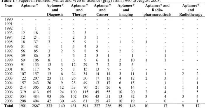
Our analysis of the scientific literature on aptamers since 1990, has been very interesting (see Table 1). First of all, we note that, although the first
papers on aptamers appeared almost
simultaneously in 1990 by two independent groups, led by Gold and Ellington respectively (Tuerk and Gold, 1990; Ellington and Szostak, 1990), there is no reference on aptamers appearing on either database till 1992. This, however, may be related to the term ‘aptamer’ not being coined and used widely until later on. Furthermore, although PubMed appears to be more widely accessible, Web of Science presented consistently a larger number of hits on aptamers and additional keywords we searched for, with exception to diagnosis.
Another interesting point identified was that, although cancer is currently one of the most researched scientific areas, only 12% (according to PubMed) and ~8.3% (according to the Web of Science) of publications on aptamers are related to cancer. This clearly indicates that aptamers have found wider applications in other areas of research, with only limited attention to their great potential in cancer. Furthermore, few papers are related to aptamers and radiopharmaceuticals or aptamers and radiotherapy.
Table 1 - Papers in PubMed (white) and Web of Science (gray) from 1990 to August 2008.
Year Aptamer* Aptamer*
and Diagnosis
Aptamer* and Therapy
Aptamer* and Cancer
Aptamer* and imaging
Aptamer* and Radio-pharmaceuticals
Aptamer* and Radiotherapy
1990 - - - -
1991 - - - -
1992 1 1 1 - - - -
1993 12 18 1 - 2 3 - - - -
1994 12 24 1 - 2 3 1 - - - -
1995 18 37 3 - 5 9 1 - 1 2 1 - - -
1996 31 48 - 1 5 4 5 - - - -
1997 56 85 3 2 6 8 9 - 2 2 - 1 - -
1998 59 86 3 - 6 2 3 1 - 1 - 1 - -
1999 59 105 8 1 6 9 6 1 2 10 1 1 - -
2000 91 133 13 3 12 29 7 2 2 5 - - - -
2001 81 117 9 5 8 15 13 9 - 3 - - - -
2002 107 157 13 6 24 34 14 14 3 11 1 1 2 2
2003 122 207 23 11 26 50 17 13 4 12 2 3 1 3
2004 157 243 20 9 28 49 13 17 6 15 - 1 1 1
2005 214 305 35 12 53 70 21 26 6 14 - 1 1 1
2006 319 413 65 24 100 115 45 55 10 20 2 4 1 5
2007 354 484 93 36 102 130 43 51 13 32 3 4 3 4
2008 208 404 42 30 46 61 35 47 10 19 - 0 - 1
Total 1901 2867 333 140 431 591 227 236 59 146 10 17 9 17
These are spread throughout the years, with one or two papers each year, with the exception of 2007
research on this field, with only one research paper this year. Given the success of aptamers in disease
diagnosis and therapy, their radiolabelling
potential remains an unexplored field.
Aptamer Design
High affinity and specificity aptamers are, primarily, generated via an in vitro selection process referred to as SELEX. However, recently, alternative selection processes have also been developed (Drabovich et al., 2005; Berezovski et al., 2005). The basic SELEX protocol is an evolutionary, iterant stringent process involving a combinatorial library of randomized nucleic acid sequences, with structural variations of more than 1015 different molecules, flanked by primers that allow PCR amplification. This is subjected to the selected molecular target for a series of events of binding, partitioning of unbound aptamers from the bound, followed by elution and amplification of the bound aptamers, which are subsequently further subjected to the target. This process is repeated for several rounds, typically from 8 to 12, to obtain, through competitive binding, one or few aptamer sequences with high specificity and affinity to the chosen target. Selected aptamers are cloned and sequenced, to reveal the binding sequences. Many alterations can be made to the SELEX protocol, such as on the approach employed to present the target or the manner in
which the aptamer-target complexes are
partitioned. Alterations in the presentation of the target can entail a method of counter selection or ToggleSELEX, whereas variations in partitioning
include photocrosslinking and capillary
electrophoresis (CE) reviewed by Hamula et al. (2006).
Chemistry and Labelling
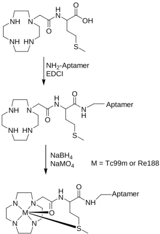
Oligonucleotides can now be purchased with a number of modifications from various providers of customised oligonucleotides. Such modifications can facilitate the interaction of aptamers with moieties of interest in labelling and to improve their pharmacokinetic properties. Thus, an amino modification at the aptamer terminus (either 3’ or 5’) can facilitate the interaction of the aptamer with a chelator. Ideally, however, this interaction would be post-labelling of the chelator with a
metal (Fig. 1), to avoid losing radioactivity, which, for a metal like technetium-99m that has a 6hr half life, is a particularly important trait.
Improving Pharmacokinetic Properties
Two major disadvantages have plagued aptamers since their initial development stage. The first major obstacle has been the degradation of oligonucleotides by nucleases. However, this issue has been resolved by a number of techniques. First of all, DNA aptamers have been shown to be far more resistant to nuclease degradation than their RNA counterparts. Second, modifications at the 3’ and/or 5’ end of the aptamer can secure nuclease resistance and increase the half-life of the molecule from minutes to hours (Khan and Missailidis, 2008; Makwana et al., 2008). One such easy modification we have used on the design of an aptamer radiopharmaceutical has been an inverted base on the unlabelled end of the aptamer (Borbas et al., 2007).
The second issue is aptamers’ pharmacokinetic
properties. Aptamers are reasonably small
(~10kDa), hydrophilic molecules that are easily cleared by the kidneys. Whilst this is interesting for diagnostic applications, where the aptamer radiopharmaceutical is cleared from the system rapidly, decreasing any background signal, it is devastating for therapy, where 90% of the aptamer can wash out in the first 15min. Thus, pegylation of the aptamer on one end with radiolabelling on the other can quickly and confidently increase the half life of the molecule in serum, whilst conferring nuclease resistance. PEG molecules of various molecular weights have been used with aptamers, but only one with a radiolabelled aptamer (Hicke et al., 2006).
Aptamer Radiopharmaceuticals Concluding Remarks
NH NH
HN N
H N
S N H O
O NH
NH
HN N
H N
S OH O
O
N N
N N
H N
S NH O
O M
NH2-Aptamer
EDCI
NaBH4
NaMO4 M = Tc99m or Re188
Aptamer
Aptamer
Figure 1 - Post-labelling of an aptamer with a chelator of interest.
Furthermore, through labelling and PEGylation has the possibility to act as diagnostic, imaging or therapeutic agents. A few years ago, an agreement between a major aptamer company and the NCI had been reached to develop and test aptamers against all major known tumour markers. Yet, to this date, only about 10% of papers on aptamers are related to cancer and only a handful on
radiopharmaceuticals. What is particularly
interesting, however, is that although there appear to be 2000-3000 papers, the World Intellectual Property Organisation indicates 138 patents in PCT with the world aptamer on the front page, but 3606 patents when searched for aptamer in ´all fields´ and 7671 hits on aptamer are found in Free
Patents Online
(http://www.freepatentsonline.com/). This is
consistent with the great commercial interest of aptamers. Research publications on aptamers as radiopharmaceuticals remain limited and, resulting mostly from research of our group and collaborators worldwide.
ACKNOWLEDGEMENTS
The authors would like to acknowledge the support of CNPq, FAPERJ, UERJ, The Open University and the Breast Cancer Campaign.
RESUMO
para tumores. A química para a marcação de aptâmeros e as opções para alterar suas propriedades radiofarmacocinéticas, para torná-los mais adequados para uso como radiofármacos, é agora disponível e os avanços recentes no seu desenvolvimento podem demonstrar que essas moléculas poderiam ser ideais como veículos para o desenvolvimento de radiofármacos sítio-dirigidos que poderiam levar radiação com precisão para o tumor, oferecendo melhores propriedades terapêuticas e reduzidos efeitos indesejados.
REFERENCES
Bacher, J. M.; Ellington, A. D. (1998), Nucleic acid selection as a tool for drug discovery, Drug Discov Today, 3, 265-273.
Belimezi, M., “Cancer Immunotherapy”. In-Anticancer Therapeutics, Missailidis S (editor). Wiley and Sons Ltd, United Kingdom.
Berezovski, M.; Drabovich, A.; Krylova, S. M.; Musheev, M.; Okhonin, V.; Petrov, A.; Krylov, S. N. (2005), Nonequilibrium capillary electrophoresis of equilibrium mixtures: a universal tool for development of aptamers. J Am Chem Soc., 127, 3165–71.
Borbas, K. E.; Ferreira, C. S. M.; Perkins, A.; Bruce, J. I.; Missailidis, S. (2007), “Design and synthesis of mono- and multimeric targeted radiopharma-ceuticals based on novel cyclen ligands coupled to anti-MUC1 aptamers for the diagnostic imaging and targeted radiotherapy of cancer”, J Bioconj Chem., 18, 1205-1212.
D’Amico, D. J.; Masonson, H. N.; Patel, M.; Adamis, A. P.; Cunningham, E. T. Jr.; Guyer, D. R.; Katz, B. (2006), Pegaptanib sodium for neovascular age-related macular degeneration: two-year safety results of the two prospective, multicenter, controlled clinical trials. Ophthalmology, 113, 992–1001. Drabovich, A.; Berezovski, M.; Krylov, S. N. (2005),
Selection of smart aptamers by equilibrium capillary electrophoresis of equilibrium mixtures (ECEEM). J Am Chem Soc., 127, 11224–11225.
Ellington, A. D.; Szostak, J. W. (1990), In vitro selection of RNA molecules that bind specific ligands. Nature, 346(6287), 818–822.
Ferreira, C. S. M.; Missailidis, S. (2007), Aptamer-based therapeutics and their potential in radiopharmaceutical design. Brazilian Arch Biol Technol., 50, 63-76.
Ferreira, C. S. M.; Papamichael, K.; Guilbault, G.; Schwarzacher, T.; Gariepy, J.; Missailidis, S. (2008), DNA aptamers against MUC1: Design of aptamer-antibody sandwich ELISA for early tumour diagnosis, Anal Bioanal Chem., 390, 1039-1050. Hamaguchi, N.; Ellington, A.; Stanton, M. (2001),
Aptamer Beacons for the Direct Detection of Proteins Anal Biochem., 294, 126–131.
Hamula, C. L. A.; Guthrie, J. W.; Zhang, H.; Li, X. F.; Le, X. C. (2006), Selection and analytical applications of aptamers. Trac-Trends Anal Chem., 25, 681–691.
Hesselberth, J.; Robertson, M. P.; Jhaveri, S.; Ellington, A. (2000), In vitro selection of nucleic acids for diagnostic applications Rev Mol Biotechnol., 74, 15-25.
Hicke, B. J.; Stephens, A. W.; Gould, T. (2006), Tumour targeting by an aptamer. J Nucl Med., 47, 668–678.
Jayasena, S. D. (1999), Aptamers: An emerging class of molecules that rival antibodies in diagnostics Clin Chem., 45, 1628-1650.
Jhaveri, S.; Rajendran, M.; Ellington, A. D. (2000), In vitro selection of signaling aptamers Nature Biotechnol., 18, 1293-1297.
Khan, H.; Missailidis, S. (2008), “Aptamers in oncology: A diagnostic perspective”, Gene Ther Mol Biol., 12, 111-128.
Makwana, V.; Simmons, S.; Missailidis, S. “Aptamers as Anticancer Agents”. In-Anticancer Therapeutics, Missailidis S (editor). Wiley and Sons Ltd, United Kingdom.
Missailidis, S., “Future Trends in Cancer Therapeutics”. In-Anticancer Therapeutics, Missailidis S (editor). Wiley and Sons Ltd, UK.
Missailidis, S.; Perkins A. (2007), “Aptamers as Novel Radiopharmaceuticals: Their applications and Future Prospects in diagnosis and therapy”, Cancer Biother Radiopharmaceuticals, 22, 453-468.
Morris, K. N.; Jensen, K. B.; Julin, C. M.; Weil, M.; (1998), High affinity ligands from in vitro selection: Complex targets. Proc Natl Acad Sci USA, 95, 2902– 2907.
Perkins, A.; Missailidis, S. (2007), “Radiolabelled aptamers for tumour imaging and therapy“, Quarterly J Nucl Med Mol. Imag., 51, 292-296.
Potyrailo, R. A.; Conrad, R. C.; Ellington, A. D.; Hieftje, G. M. (1998), Adapting selected nucleic acid ligands (aptamers) to biosensors Anal Chem., 70, 3419–3425.
Tuerk, C.; Gold, L. (1990), Systematic evolution of ligands by exponential enrichment – Rna ligands to bacteriophage-T4 DNA-polymerase. Science, 249(4968), 505–510.