ISJ 7: 165-180, 2010
ISSN 1824-307X
REVIEW
Role of α
2-macroglobulin in the immune responses of invertebrates
PB Armstrong
Marine Biological Laboratory, 7 MBL Street, Woods Hole, MA 02543, and Department of Molecular and Cellular Biology, University of California, Davis, CA 95616, USA
Accepted June 30, 2010
Abstract
Although proteases play essential roles in the lives of all organisms, they are also important
agents of disease and pathogenesis in metazoans. Most notably, proteases are essential virulence
factors for a broad array of prokaryote and eukaryote parasites. One strategy used by the immune
system of metazoans to defend against parasitic attack is to neutralize the toxins and essential
virulence factors that allow the parasite to gain entry to the host and to survive and proliferate in the
internal environment of the metazoan host. The particular defense system of interest to the present
review is the system of endogenous protease inhibitors that operate to inactivate the secreted
proteases utilized by invading parasites during the infection cycle within the host. Protease inhibitors
are of two broad classes, active-site inhibitors that bind to and inactivate the active sites of target
proteases and the
α
2-macroglobulin class of inhibitors that operate as opsonins to bind and mark
proteases in a manner that allows the subsequent endocytosis and intracellular proteolytic degradation
of the
α
2-macroglobulin-protease complex. Members of the
α
2-macroglobulin class of inhibitors interact
with target proteases by the novel process of enfolding the protease into a pocket within the interior of
the
α
2-macroglobulin molecule, which is followed by binding of the complex to the
α
2-macroglobulin
receptor at the surfaces of macrophages and other endocytotic cells and its endocytosis and
degradation. In contrast to the active-site protease inhibitors, each of which is specialized to interact
with a small subset of all endopeptidases, the
α
2-macroglobulin inhibitors are remarkably promiscuous,
binding proteases of all enzymatic classes and origins. This characteristic allows
α
2-macroglobulin to
play an important role in immune defense because this one protein is capable of binding and
neutralizing the diverse array of proteases that function as virulence factors of the diverse array of
parasites out there in the environment of metazoa.
Key Words: α2-macroglobulin; thiol ester protein family; protease inhibitor; innate immunity
Introduction
An important barrier to the successful reproduction of metazoans is the disease and premature death that attends the attack by parasites. Barriers to parasitic attack are the integuments, which limit the range of parasites that can gain entry to the internal milieu, and the immune system, which defends against parasites and their toxic products both at the surface and in the internal spaces. Parasites may be unicellular or multicellular, prokaryote, eukaryote, or virus. In coelomate animals, the most important organ of the
___________________________________________________________________________
Corresponding author: Peter B Armstrong
Department of Molecular and Cellular Biology University of California, Davis, CA 95616-8535, USA E-mail: [email protected]
immune system is the blood, presumably because the blood has ready access to all parts of the body and is best prepared to concentrate defense effectors at sites of pathogen invasion. This review will be concerned with one of the important immune effector proteins found in the plasma of all major taxa of metazoans, α2-macroglobulin.
remodeling of the extracellular matrix (Lopez-Otin and Bond, 2008). However, in addition, proteases, whether of endogenous or exogenous origin, are capable of considerable mischief when free in the blood and tissue spaces. They are important agents in a variety of connective tissue diseases (Perlmutter and Pierce, 1989), contribute to neoplastic invasion and metastasis (Deryugina and Quigley, 2006; Kessenbrock et al., 2010), and are important virulence factors contributing to the pathogenicity of prokaryotic and eucaryotic parasites (Armstrong, 2006; McKerrow et al., 2006). In response to protease challenge, animals have evolved a diverse array of protease inhibitors (Travis and Salvesen, 1983). In mammals, approximately 3-5 % of the plasma proteins are protease inhibitors (Laskowski and Kato, 1980) and in the American horseshoe crab, Limulus polyphemus, the hemolymph protease inhibitor,
α2-macroglobulin, is the third most abundant protein in the hemolymph (Enghild et al., 1990). α2-Macroglobulin is one of the most abundant proteins of human plasma, at a concentration of 2-4 mg/ml (Sottrup-Jensen, 1989) and is the second-most abundant protein of the hemolymph of the cephalopod, Sepia (Vanhoorelbeke et al., 1993).
α2-Macroglobulin, the subject of this review, is present in representatives of several metazoan phyla, including coelenterates (Fujito et al., 2010) and representative species of the Deuterostomia (Echinodermata, Tunicata, Vertebrata), Ecdysozoa (Arthropoda, Nematoda), and Lophotrochozoa (Mollusca) [for reviews see (Armstrong and Quigley, 1999; Armstrong, 2006)]. A diverse variety of Gram-negative eubacteria have acquired a gene encoding α2-macroglobulin from eukaryote associates (Budd
et al., 2004; Doan and Gettins, 2008).
Mechanisms for attack on proteases
The protease inhibitors are of two fundamental classes, the active-site inhibitors, which bind to and inactivate the activate site of the targeted endopeptidase (Huntington et al., 2000), and the
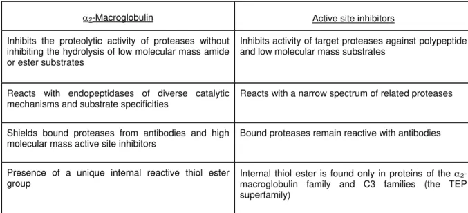
α2-macroglobulins, which react by a unique mechanism that involves the physical entrapment of the target protease within the folds of a molecule of α2-macroglobulin (Table 1). By binding to the active site of the protease, the active-site inhibitors destroy both its proteolytic activity against proteins and its ability to cleave the ester and amide bonds of low molecular mass artificial substrates. Dedication for a particular active site means that the active-site inhibitors are restricted in their activity to one particular class, or even sub-class, of protease. By contrast, α2-macroglobulin shows a unique mechanism for interaction with target proteases which is initiated by its proteolytic cleavage at a defined motif that is constructed as an exposed and highly flexible stretch of 30 - 40 amino acids that presents a suite of peptide bonds that are inviting targets for most extant proteases (Sottrup-Jensen et al., 1989). This means that, whatever the protease, it will likely find a target for proteolytic attack at this “bait” region of the α2-macroglobulin molecule.
This is the basis for the broadly reactive ability of α2-macroglobulin to bind proteases of diverse enzymatic reactivity and source. The forms of α2-macroglobulin found in the tick (Saravanan et al., 2003; Buresova et al., 2009), horseshoe crab (Husted et al., 2002), and carp (Mutsuro et al., 2000) show multiple forms of the protein that differ by bait region sequence. Presumably this functions to expand the list of proteases recognized by the α2-macroglobulins of these species. Proteolytic cleavage of the bait region is followed by the rapid re-folding of the α2-macroglobulin molecule so as to produce an enclosed interior pocket that now contains the target protease, entrapped within the folds of the α2-macroglobulin protein (Starkey and Barrett, 1973). Protease entrapment disables the enzyme’s ability to proteolyze macromolecular substrates too large to penetrate the α2-macroglobulin cage, but leaves intact the ability of the entrapped enzyme to hydrolyze low molecular mass substrates small enough to enter the α2-macroglobulin cage and interact with the active site of the protease (Starkey and Barrett, 1973). One diagnostic feature to show that α2-macroglobulin is responsible for a protease inhibitory activity of an uncharacterized sample is the demonstration that the sample inhibits proteolytic but not amidolytic activity of an exogenously added protease. I am not aware of any other enzyme inhibitor that operates by this unique “trap” mechanism.
The conformational change in the protease-activated α2-macroglobulin molecule exposes a domain at its carboxy terminus, the receptor-binding domain, that now targets the molecule, with its entrapped cargo of protease, for receptor-mediated endocytosis and proteolytic degradation by phagocytes and other cells that display the α2-macroglobulin receptor at the cell surface (Van Leuven, 1984). In this fashion, α2-macroglobulin operates as an opsonin that delivers proteases of every enzymatic class to endocytotic cells of the innate immune system for internalization and destruction.
Several strands of evidence support this unusual scheme for the removal of potentially destructive proteolytic enzymes. Active site protease inhibitors block both the proteolytic activity and the amidase and esterase activities against low molecular mass substrates. In contrast, proteases bound to α2-macroglobulin remain active against low molecular mass substrates while losing the ability to cleave peptide bonds of proteins (Barrett and Starkey, 1973; Quigley and Armstrong, 1983). This was interpreted to mean that the active site of α2-macroglobulin-bound proteases remained intact but was sterically blocked from reacting with macromolecular substrates. Protein sequencing of α2-macroglobulins from mammals (Sottrup-Jensen
Table 1 Comparison of α2-macroglobulin with active site protease inhibitors
α2-Macroglobulin Active site inhibitors
Inhibits the proteolytic activity of proteases without inhibiting the hydrolysis of low molecular mass amide or ester substrates
Inhibits activity of target proteases against polypeptide and low molecular mass substrates
Reacts with endopeptidases of diverse catalytic mechanisms and substrate specificities
Reacts with a narrow spectrum of related proteases
Shields bound proteases from antibodies and high molecular mass active site inhibitors
Bound proteases remain reactive with antibodies
Presence of a unique internal reactive thiol ester group
Internal thiol ester is found only in proteins of the α2-macroglobulin family and C3 families (the TEP superfamily)
sequencing has facilitated the molecular characterization of the receptor-recognition domain that is exposed by proteolytic cleavage (Sottrup-Jensen et al., 1986; Van Leuven et al., 1986; Enghild et al., 1989b; Holtet et al., 1994). The molecular compaction responsible for the physical entrapment of a protease molecule was initially shown by the retarded elution of the protease-reacted form during gel filtration chromatography and by the increased electrophoretic mobility of the reacted form during non-denaturing polyacrylamide gel electrophoresis. This latter observation generated the term for the reacted and more compact form of the molecule as the “fast form” (Barrett et al., 1979). The molecular compaction has also been demonstrated by direct electron microscopic comparison of α2-macroglobulin molecules before and after reaction with a protease (Armstrong et al., 1991).
The spectrum of proteases that interact with α2-macroglobulin is unusually broad and includes proteases of all major enzymatic classes. Although active-site protease inhibitors have been shown to function in immune defense (Kanost, 1999), the promiscuous reactivity of α2-macroglobulin has adapted this single molecule to function as a particularly effective scavenger of the novel proteases introduced by parasites and pathogens. Indeed, the α2-macroglobulins of vertebrates and invertebrates have been shown to react against the exoproteases of important parasites (Freedman, 1991; Araujo-Jorge et al., 1992; Fryer et al., 1996; Srimal and Armstrong, 1996; Scharfstein, 2006). The broad spectrum of reactivity of α2-macroglobulin contrasts with the much more restricted reactivity of the naturally-occurring active-site protease inhibitors and is another feature that can be used to demonstrate that an uncharacterized protease inhibitor is, indeed, α2-macroglobulin.
Interestingly, some microbial proteases are immune to certain versions of α2-macroglobulin. In some instances, the protease is too large to fit into the interior hydrophilic pocket of the reacted form of α2-macroglobulin [the collagenase of Chlostridium perfringens has a molecular mass greater than 100 kDa (Abe et al., 1989)] but other non-reactive enzymes cleave peptide bonds not found in the bait region of certain versions of α2-macroglobulin. Human α2-macroglobulin lacks the lysine residues in the bait region targeted by the lysyl endopeptidase of Acromobacter lyticus and fails to react with that enzyme (Ikai et al., 1999). A related example of this restriction is the relative insensitivity of the proteases of the Limulus clotting system to
Limulus α2-macroglobulin, but with a sensitivity of
those same proteases to human α2-macroglobulin (Armstrong et al., 1984; Iwaki et al., 1994). The
Limulus clotting protease specifically cleaves the clottable protein, coagulogen, at the carboxyl sides of two Arg residues with the sequences Leu-Gly-Arg and Ser-Gly-Arg, which are conserved in coagulogens isolated from the four extant species of horseshoe crabs (Iwanaga et al., 1992). Limulus
found in the hemolymph of that organism (Hergenhain et al., 1987).
Entrapment in the α2-macroglobulin cage both prevents the entrapped enzyme from accessing external proteins as substrates for proteolytic attack and protects the entrapped enzyme molecule from inactivation by macromolecular active site protease inhibitors. In other words, the protease molecule cannot get out of the α2-macroglobulin cage, but neither can macromolecular protein inhibitors get in. This has allowed the development of a specific and semi-quantitative assay for α2-macroglobulin, in which an uncharacterized sample is reacted first with trypsin, next with excess soybean trypsin inhibitor (STI, Mr 21.5 kDa), and then the residual trypsin activity is assayed with the low molecular mass amide substrate, BApNA (Na-benzoyl-DL-arginine p-nitroanilide) (Ganrot, 1966; Armstrong et al., 1985). Any trypsin not sequestered by
α2-macroglobulin is inactivated by STI and the only enzyme still able to hydrolyze BApNA is that which is protected within the α2-macroglobulin cage. In the absence of α2-macroglobulin in the sample, degradation of BApNA is zero. In the presence of increasing amounts of α2-macroglobulin, the hydrolysis of BApNA is increased in a dose-dependent fashion, indicative of increasing amounts of α2-macroglobulin-sequestered, and thus protected, trypsin. A positive reaction in a properly controlled STI-protection assay is a sure indicator for the presence of α2-macroglobulin in the sample, but the assay is subject to false negative responses that can occur if the sample also contains low molecular mass trypsin inhibitors small enough that they can enter the α2-macroglobulin cage and inactivate bound enzyme or if the α2-macroglobulin in the sample contacts proteases during sample collection and storage. Because α2-macroglobulin is large, low molecular mass trypsin inhibitors can be removed by dialysis and premature reaction of α2-macroglobulin can often be prevented by the inclusion of appropriate cocktails of low molecular mass protease inhibitors that can subsequently be removed by dialysis prior to assay with the STI-protection assay.
We have seen now three diagnostic features of the α2-macroglobulin family of protease inhibitors: a broad reactive capacity against proteases of all classes, a unique molecular trap mechanism for interaction with target proteases, and a failure to inactivate the active site of the entrapped protease. A fourth diagnostic feature of the members of the α2-macroglobulin family of proteins is the presence of a reactive internal thiol ester bond linking cysteinyl and glutamyl residues,
⋅⋅⋅⋅G-C-G-E-Q-N-M⋅⋅⋅⋅
* S____ C=O
which in Limulus α2-macroglobulin are Cys-999 and
Glx-1002 (Iwaki et al., 1996). The thiol ester bond is cleaved when α2-macroglobulin experiences proteolytic attack on the bait domain, thereby exposing the cysteinyl thiol and the carbonyl of
glutamic acid. This was seen by the exposure of a new titratable thiol group, the thiol of Cys-999 in
Limulus α2-macroglobulin (Armstrong and Quigley,
1987). The newly-exposed carbonyl group of the glutamyl residue is highly reactive and can engage in isopeptide bonding with ε-amino groups of lysines of protein targets in the reaction environment (Fig. 1). In human α2-macroglobulin, crosslinking of the thiol esterified glutamic acid is preferentially with the entrapped protease (Sottrup-Jensen et al., 1990b), whereas with α2-macroglobulin from the American horseshoe crab, Limulus, the isopeptide bonds crosslink chains of α2-macroglobulin itself (Dolmer
et al., 1996). Although the internal thiol ester of
α2-macroglobulin is stable in the intact protein, it will react with small primary amines such as ammonium and methylamine in the absence of proteolysis (Fig. 1). An important diagnostic feature of α2-macroglobulin is its sensitivity to methylamine. The contribution of α2-macroglobulin to the protease inhibitory activities of a biological sample is inactivated following methylamine treatment.
Protease clearance
Although α2-macroglobulin is usually thought of as a protease inhibitor, it might better be considered a protease-binding molecule whose principal function is to deliver its protease cargo to an endocytotic protease clearance pathway. In this context, unreacted α2-macroglobulin serves the recognition function and the protease-reacted fast-form α2-macroglobulin serves the delivery function. The role of α2-macroglobulin in protease clearance is especially well illustrated in Limulus because
Fig. 1 Activation and cleavage of the internal thiol ester of α2-macroglobulin exposes a new thiol group on the
cysteine and a reactive (-carbonyl on the glutamyl residue, which in Limulus α2-macroglobulin are Cys999 and Glx1002 . The reactive internal thiol ester of members of the α2-macroglobulin protein family is cleaved following proteolysis at the distantly-located protease bait region of the protein. Thiol ester cleavage generates an activated (-carbonyl at the glutamyl residue and a free thiol at the cystenyl residue (top line of the diagram). The reactive glutamyl can form amide linkages with proteins (right arm of the diagram). The thiol ester can also react slowly with small primary amines, such as methylamine (left arm of diagram), even in the absence of proteolytic cleavage at the bait region. Methylamine treatment eliminates many of the functional activities of α2-macroglobulin in parallel with its inactivation of the thiol ester. In general, sensitivity of a molecular function such as protease inhibition to treatment with methylamine is a useful test for the possibility that that function is dependent on the activity of a protein of the thiol ester protein family.
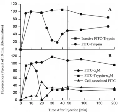
fluoresceinated trypsin and fast-form α2-macroglobulin associate with the blood cells at the very stages that these proteins are being cleared from the hemolymph (Fig. 2B).
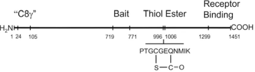
The conformational change of protease-reacted α2-macroglobulin serves both to entrap the protease and to expose a previously cryptic domain at the carboxyl terminus, the receptor-binding domain (Fig. 3) (Sottrup-Jensen et al., 1986; Van Leuven et al., 1986; Enghild et al., 1989b; Holtet et al., 1994), that is recognized by cell surface receptors, leading to the binding and endocytosis of the protease-α2-macroglobulin complex (Van Leuven, 1984). This removes the protease from the circulation, thereby neutralizing its potentially damaging actions. The three major steps in protease recognition, capture, and delivery are mediated in part by three identifiable domains of the α2-macroglobulin molecule, the bait region, the thiol ester domain, and the receptor-recognition domain (Fig. 3). Presumably, there are additional domains that are under molecular strain in the unreacted form of
α2-macroglobulin and that are most actively involved in the active change in molecular shape following protease reaction and hydrolysis of the thiol ester, putative “hinge-domains”. In the absence of high resolution information of the structure of the native and fast-forms of the molecule, these have not been characterized.
Fig. 2 Clearance of proteases from the hemolymph of Limulus is mediated by α2-macroglobulin. Fluoresceinated
proteins were injected into the lumen of the heart and their concentration in the peripheral hemolymph drawn from the leg joints was measured with a fluorometer. Fluoresceinated protein reached the periphery by 5 - 10 min after injection. Enzymatically active trypsin, but not trypsin inactivated by treatment with PMSF, was substantially cleared from the hemolymph by 30 min (Fig. 1A). The fluorescence that appeared at later times was of low molecular mass, and presumably consisted of peptide digest products of the injected protein. Fast-form, but not slow-form Limulus α2-macroglobulin showed a similar pattern of clearance from the hemolymph. During the period
of clearance of fast-form α2-macroglobulin from the hemolymph, fluorescence accumulated in the blood cells. This is interpreted to indicate that clearance of the introduced protease is mediated by its binding to α2-macroglobulin and that the blood cells participate in the uptake and clearance of the α2-macroglobulin-protease complex.
May et al., 2007). Of practical interest is its ability to bind two or more mols/mol of a 39 kDa endogenous ligand, LRP receptor-associated protein (RAP) (Ashcom et al., 1990; Herz et al., 1991). RAP functions as a dedicated chaperone and regulator of members of the LDL receptor family of proteins (Bu, 1998) and its high affinity binding makes RAP-conjugated Sepharose an appropriate affinity matrix for purification of these proteins.
To date, a receptor for protease-reacted α2-macroglobulin has not been identified in an invertebrate. The family of proteins that includes vertebrate CD91 is, like α2-macroglobulin itself, an ancient family that was present prior to the great evolutionary radiation that established the divergent deuterostome and protostome invertebrate superphyla. Representatives of the CD91 family have been found in modern representatives of lineages that diverged at the time of the Precambrian radiation, notably in vertebrates, the nematode, Caenorhabditis elegans (Yochem and Greenwald, 1993), and the arthropod, Drosophila melanogaster (Locus tag Dmel CG33087). Whether invertebrate orthologs of vertebrate CD91 operate as α2-macroglobulin receptors has not been
decided. The Limulus blood cell does contain a protein of very high molecular mass that, like CD91, binds RAP (Aimes et al., 1995) and human CD91 binds to protease-reacted Limulus α2-macroglobulin
(Iwaki et al., 1996). Although these observations hint at a possibility that the clearance pathway for protease-reacted Limulus α2-macroglobulin involves
an ortholog of CD91, they fall well short of any convincing demonstration of that prospect.
Phylogenetic distribution of the α2
-macroglobulins
Fig. 3 Domain structure of Limulus α2-macroglobulin. Based on the amino acid sequence of Limulus
α2-macroglobulin (Iwaki et al., 1996), it is possible to identify important functional domains by comparison with the already established domain structure of human α2-macroglobulin. The amino terminal end of Limulus
α2-macroglobulin has a unique stretch identified as the C8 (domain, based on modest sequence identity with the human defense protein C8 (of the mammalian complement pathway. The bait region contains a sequence of amino acids that present target sites for nearly all proteases. Proteolysis of the bait region initiates the compaction of α2-macroglobulin that is responsible for entrapment of the molecule of reacting protease. The thiol ester domain contains the unique thiol ester bond linking cysteine-999 and glutamic acid-1002. The receptor-binding domain at the carboxyl terminus of the polypeptide chain is exposed following molecular compaction and makes the protease-reacted form of α2-macroglobulin a target for binding to the cell surface α2-macroglobulin receptor.
suppressed the action of serine, thiol, and metalloproteases against [14C]-casein, a protein substrate, but that failed to suppress hydrolysis of the amide substate BApNA by trypsin. The antiprotease activity of hemolymph was eliminated if
Limulus hemolymph was treated with methylamine. This suggested the presence of α2-macroglobulin in the hemolymph of this invertebrate (Quigley et al., 1982). Our early analysis was facilitated by the absence of other trypsin inhibitors in Limulus
hemolymph (Quigley and Armstrong, 1983). Subsequently, we showed that Limulus hemolymph protected trypsin from inactivation by the macromolecular active-site inhibitor, soybean trypsin inhibitor (Armstrong et al., 1985), that purified
Limulus α2-macroglobulin did indeed contain an
internal thiol ester domain (Armstrong and Quigley, 1987), and that the protein from Limulus shows significant sequence identity in key functional domains with human α2-macroglobulin (Sottrup-Jensen et al., 1990a; Iwaki et al., 1996). Although overall sequence identity between human and
Limulus α2-macroglobulin is relatively low, the thiol
ester domain shows significant sequence conservation, the receptor binding domains shows conservation of key basic amino acids, and the bait regions shows no sequence similarity.
The strict conservation of the thiol ester domain of the various α2-macroglobulins has facilitated the molecular cloning of the genes for a variety of invertebrates from c-DNA libraries. With the advent of complete genomic sequences for an ever-increasing variety of species, data mining has revealed additional examples of genes encoding α2-macroglobulin from these species. For example, the presence of genes for α2-macroglobulin in Gram-negative bacteria was first demonstrated using the data-mining strategy (Budd et al., 2004). These methods have identified both representatives of α2-macroglobulin that, upon further investigation, have shown the unique suites of functional and structural
characteristics of canonical α2-macroglobulin, and have also revealed novel related proteins, several of which await functional characterization.
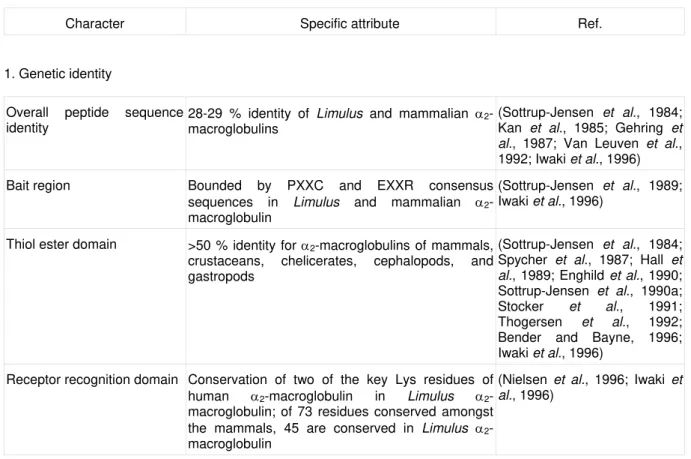
The α2-macroglobulin family includes the canonical α2-macroglobulins, a variety of similar protease-binding proteins, including pregnancy-zone protein of humans, α1I3 of the rat, ovomacroglobulin of avian and reptile eggs, and proteins to be discussed below that were initially identified from the vertebrate complement system (Sottrup-Jensen, 1987). Additionally, humans have two newly identified and incompletely characterized members of the family, CD109, which is linked to the cell surface by GPI-bonding (Lin et al., 2002), and CPAMD8, an acute-phase protein expressed by kidney, brain, and testis (Li et al., 2004), and the settlement-inducing protein of the barnacle Balanus amphitrite is an α2-macroglobulin (Dreanno et al., 2006). The different α2-macroglobulins show differing orders of multimerization. Human (Jones et al., 1972; Swenson and Howard, 1979) and snail (Bender and Bayne, 1996) α2-macroglobulins are homotetramers, whereas many forms of the α2-macroglobulins from a variety of lower vertebrates and arthropods are homodimers (Starkey and Barrett, 1982; Feldman and Pizzo, 1984; Sand et al., 1985; Feldman and Pizzo, 1986; Spycher et al., 1987; Enghild et al., 1990; Armstrong et al., 1991), and a few monomeric forms of α2-macroglobulin have been identified (Enghild et al., 1989a). As indicated above, the molecular homologues of the canonical α2-macroglobulins have a wide phylogenetic distribution and share a defined suite of structural and functional characters (Table 2).
Thiol ester protein family
Table 2 Molecular and functional conservation of the α2-macroglobulins
Character Specific attribute Ref.
1. Genetic identity
Overall peptide sequence identity
28-29 % identity of Limulus and mammalian
α2-macroglobulins
(Sottrup-Jensen et al., 1984; Kan et al., 1985; Gehring et al., 1987; Van Leuven et al., 1992; Iwaki et al., 1996)
Bait region Bounded by PXXC and EXXR consensus sequences in Limulus and mammalian
α2-macroglobulin
(Sottrup-Jensen et al., 1989; Iwaki et al., 1996)
Thiol ester domain >50 % identity for α2-macroglobulins of mammals, crustaceans, chelicerates, cephalopods, and gastropods
(Sottrup-Jensen et al., 1984; Spycher et al., 1987; Hall et al., 1989; Enghild et al., 1990; Sottrup-Jensen et al., 1990a; Stocker et al., 1991; Thogersen et al., 1992; Bender and Bayne, 1996; Iwaki et al., 1996)
Receptor recognition domain Conservation of two of the key Lys residues of human α2-macroglobulin in Limulus
α2-macroglobulin; of 73 residues conserved amongst the mammals, 45 are conserved in Limulus
α2-macroglobulin
(Nielsen et al., 1996; Iwaki et al., 1996)
2. Biochemical homology
Susceptibility of bait region to proteolysis by proteases of all catalytic mechanisms
Reactivity of purified α2-macroglobulins of mammals, arthropods and molluscs to serine, cysteine, and metalloproteases
(Starkey and Barrett, 1977; Quigley and Armstrong, 1985; Hergenhahn and Soderhall, 1985; Enghild et al., 1990; Thogersen et al., 1992; Bender and Bayne, 1996)
Molecular compaction; non-covalent trapping of proteases
Molecular compaction of protease-reacted α2-macroglobulin of mammals and Limulus
(Barrett and Starkey, 1973; Barrett et al., 1979; Quigley et al., 1991; Armstrong et al., 1991)
Covalent isopeptide bonding of thiol esterified glutamyl residue
Covalent bonding to trapped protease in mammalian α2-macroglobulin; covalent cross-linking to Lys-254 of partner chain of α2-macroglobulin dimer in Limulus
(Sottrup-Jensen et al., 1980; Sottrup-Jensen et al., 1990b; Jacobsen and Sottrup-Jensen, 1993; Dolmer et al., 1996)
Protease treatment exposes the recognition stretch for receptor-binding
Thiol ester-reacted Limulus α2-macroglobulin binds
the human α2-macroglobulin receptor
(Sottrup-Jensen et al., 1986; Van Leuven et al., 1986; Enghild et al., 1989b; Iwaki et al., 1996)
3. Functional similarity
Protease clearance Mammals and Limulus utilize α2-macroglobulin for
the clearance of proteases from the internal milieu
architecture (Janssen et al., 2005; Doan and Gettins, 2007), sequence similarity in key functional domains, and most, but not all, contain a stable internal thiol ester bond linking the cysteinyl and glutamyl residues of the thiol ester motif. Members of this family that lack the internal thiol ester include complement component C5 (Tack, 1983) and ovostatin from the albumin of the chicken egg (Nagase et al., 1983) and Drosophila Mcr (NCBI accession number Y11116). In all three proteins, the Cys has has been substituted with another residue (Nielsen and Sottrup-Jensen, 1993). But these proteins retain the other signature features that validate their assignment to the thiol ester protein family and ovostatin retains the ability to capture proteases by the molecular trap mechanism (Nagase and Harris, 1983).
The taxonomy of the TEP family is still a work in progress. One classification identifies two subfamilies, C3 and A2M (Sottrup-Jensen et al., 1985; Fujito et al., 2010) named for their founding members, human complement component C3 and human α2-macroglobulin, respectively. A second scheme posits three distinct subfamilies, C3, A2M, and insect Tep. Members of the insect Teps show sequence relatedness intermediate between the C3 and A2M subfamilies and functional similarity to members of the C3 subfamily. They also, like C3, the canonical member of the C3 subfamily, have a Histidine residue (His 951 in Drosophila Tep1r; His 1,106 in human C3) in position to convert the reactive Glx of the activated thiol ester to an intermediate that favors its subsequent covalent linkage to hydroxyl groups rather than showing the default reactivity with amines (Dodds et al. 1966; Law and Dodds 1997). This residue is Asp, Asn, or Ala in thiol ester proteins where the covalent reactivity of the thiol ester is with amines. Subfamily membership is based on amino acid sequence and functional domain relatedness and is reflective of important functional differences between the members of the subfamilies. The best-characterized function of the A2M family members is protease binding, whereas the best-characterized function of the members of the C3 and insect Tep proteins is covalent binding to the surfaces of foreign cells to mark them for immune destruction (Dodds and Sim, 1997; Blandin et al., 2008). As will be described below, some members of the A2M subfamily, with well established protease-binding characters also, like members of the C3 and insect Tep subfamilies, bind to the surfaces of foreign cells and facilitate their phagocytotic uptake. I am unaware of any reports of members of the C3 or insect Tep proteins showing the unique protease capture abilities of the members of the a2M subfamily. The common ancestor of the TEP family is presumably ancient because representatives of both the C3 and A2M subfamilies are found in cnidarians, which are basal metazoans. The sea anemone, Haliplanella has two members of the C3 subfamily and two of the A2M subfamily (Fujito et al., 2010). Representatives of both families have been identified in vertebrates (Sottrup-Jensen et al., 1985), non-vertebrate chordates (Nonaka et al., 1999), echinoderms (Al-Sharif et al., 1998), and representatives of the
Ecdysozoans, the horseshoe crab (Zhu et al., 2005; Ariki et al., 2008), and the Lophotrochozoans, a squid (Castillo et al., 2009) and a clam (Prado-Alvarez et al., 2009). The insect Tep subfamily has been reported only from insects and possibly nematodes (Blandin and Levashina, 2004), but not from other arthropods. It is important to note that the subdivision of the thiol ester proteins into just two or just three major subfamilies is not yet definitive. There are several TEP proteins that may not fit within a two- or three-subfamily classification schemes (Lin et al., 2002; Li et al., 2004; Dreanno
et al., 2006).
Although the best-characterized function of the proteins of the A2M subfamily, both from vertebrates and invertebrates, is protease binding, biochemical pathways other than protease clearance have been identified. The α2-macroglobulins also modulate cell proliferation and cell survival pathways (LaMarre et al., 1991; Shi et al., 2008), protease-independent pathways of the innate immune system (Swarnakar et al., 2000; Arnold et al., 2006; Craig-Barnes et al., 2010), antigen delivery in the operation of the adaptive immune system (Bowers et al., 2009), and can operate as molecular chaparones (Yerbury et al., 2009). As mentioned above, CD91, the canonical cell-surface receptor for protease-reacted
α2-macroglobulin recognizes and binds a diverse suite of ligands and is an essential agent for a diverse array of important biological processes.
The best-characterized function for members of the C3 subfamily is their covalent binding to the surfaces of foreign cells to target them for immune destruction, a function shared with members of the insect Tep subfamily. An appealing example of this is the protein TEP1, the mediator of an important pathway for the immune defense of the mosquito vector against the protozoan parasite, Plasmodium falciparum, the agent of human malaria (reviewed in Blandin et al., 2008). TEP1 is a member of the thiol ester protein family from the Anopheles mosquito. TEP1 binds to bacteria and Plasmodium cells and targets the cells for phagocytosis. The experimental elevation of the concentration of TEP1 in the hemolymph of the mosquito reduces infection rates and experimental reduction increases susceptibility of the mosquito to infection (reviewed in Volohonsky
et al., 2010). Different members of the insect Teps show specialization of their targeting to the surfaces of different classes of microbes, for example, with
Drosophila Mcr targeting Candida albicans cells,
Drosophila TepIII targeting S. aureus, and
Drosophila TepII targeting E. coli (Stroschein-Stevenson et al., 2006).
Prospects for future research
review of this topic. A full characterization of the function(s) of the α2-macroglobulins in a diverse array of species will involve both the functional characterization of the molecule in vitro, which has been the principal topic of this review, and the elucidation of its function in vivo. What functions in the animal require α2-macroglobulin? The gene knock-out mouse has been available for a decade and a half and shows a surprisingly mild phenotype (Umans et al., 1995; Umans et al., 1999). Interpretation is complicated by the artificially sanitary conditions of the life of the laboratory mouse. This mouse model will be particularly interesting when its sensitivity to an extended suite of protease-wielding pathogens is investigated (Coutinho et al., 1999). In appropriate model invertebrates, RNAi knock-down trials are possible and reduction of expression of various TEPs have been shown to significantly diminish host resistance to infection by some, but not all pathogens (Buresova et al., 2009; Volohonsky et al., 2010). One imagines refinements on this strategy where modified versions of α2-macroglobulin are used to reconstitute function in α2-macroglobulin-depleted animals. In this context, the modified α2-macroglobulin constructs might feature bait regions with enhanced or restricted sensitivity to the various proteases of an appropriate pathogen (Ikai et al., 1999). These could provide information on the roles of different proteases of the pathogen in the infection cycle and on the precise roles for α2-macroglobulin in immune defense. For example, might forms of α2-macroglobulin modified to include the appropriate novel cleavage sites for previously unrecognized microbial proteases now offer protection to the host from those bacteria?
A related challenge to the functional characterization of the diverse members of the thiol ester protein family is the high resolution structural characterization of representative thiol ester protein family members for a detailed understanding of the relation of protein structure to the several functions of different members of this protein family. The goal of establishing a detailed understanding of the relations between protein structure and function is best exemplified by the characterization of human C3 (Janssen et al., 2005) and an insect Tep (Baxter
et al., 2007). The establishment of a high resolution structural characterization of α2-macroglobulin has proven elusive (Jenner et al.,1998) but the insights provided by the characterization of the domain structure of C3 have been used to develop a model for human α2-macroglobulin (Doan and Gettins, 2007). One additional bonus of this line of research will be the provision of information for the refinement of the still-unsettled molecular taxonomy of the thiol ester protein superfamily.
It will be interesting and illuminating to identify and characterize the clearance pathways for protease-reacted α2-macroglobulin in taxa other than mammals. To date, the sole experimental study is the investigation of the protease clearance pathway for the horseshoe crab (Melchior et al., 1995) and this has implicated a cell-based clearance pathway that is selective for fast-form α2-macroglobulin. Initial evidence described above
hints at a role for a role for a cell surface receptor with properties similar to mammalian CD91 in this pathway. Although CD91 is the best-characterized cell surface receptor for mammalian α2-macroglobulin, a second cell surface protein, GRP78, has been shown to bind protease-reacted α2-macroglobulin with nM affinity (Quinones et al., 2008; Gonzalez-Gronow et al., 2009). GRP78 is a protein found in diverse eukaryotes and it will be interesting to discover if this or other undiscovered receptors operate to bind α2-macroglobulin in different metazoans.
In mammals, both α2-macroglobulin and its receptor, CD91, have been shown to contribute to the operation of a number of physiological processes that are independent of protease clearance. It will be interesting to identify and characterize the possible diversity of functions of α2-macroglobulin in invertebrates. A scattering of examples have already been identified. In invertebrates, α2-macroglobulin also regulates the activities of other immune effector proteins. In the shrimp, Penaeusmonodon, α2-macroblobulin binds syntenin (Tonganunt et al., 2005). Syntenin is an acute-phase protein that is dramatically up-regulated in shrimp infected with the white spot syndrome virus. In the horseshoe crab, Limulus, protease-activated, but not native α2-macroglobulin binds and inactivates the cytolytic actions of limulin (Armstrong et al., 1998; Swarnakar et al., 2000). Cytolytic destruction of foreign cells is a ubiquitous strategy of metazoan immune systems (Canicatti, 1990; Gabay, 1994). In mammals, cytolytic destruction of foreign cells is mediated in part by the multi-protein complement system (Law and Reid, 1995), whereas the hemolymph cytolytic pathway of
Limulus is mediated by the single protein limulin, a sialic acid-binding member of the pentraxin gene family (Armstrong et al., 1996; Harrington et al., 2008). The adaptive significance of the α2-macroglobulin-limulin binding reaction has not been identified, but the pentraxins of Limulus are multi-functional mediators of immunity, with potentially important roles in the inactivation of bacteral lipopolysaccharide (Ng et al., 2004) and in the formation of hydrophilic pores across the lipopolysaccharide-rich outer membrane of Gram-negative bacteria (Harrington et al., 2009). It will be interesting to discover if the binding to protease-reacted α2-macroglobulin affects other functions of the Limulus pentraxins or the pentraxins of other invertebrates. Perhaps the reduced cytolytic activity of limulin complexed with α2-macroglobulin is correlated with augmentation of other immune functions of limulin.
subsequent immune destruction. It will be interesting to determine if this functional characterization is universal amongst metazoans or whether there is functional diversity for members of the C3 family from different taxa. For example are there instances where proteins of the C3 or insect Tep sub-families function in a manner similar to the A2Ms as protease binding proteins in addition to their canonical cell-binding functions or are there members of the A2M family that, in addition to the display of an ability to bind proteases, also function as opsonins that promote the phagocytosis of foreign cells that have become decorated with surface-associated α2-macroglobulin? For example, murine α2-macroglobulin binds to the surfaces of the pathogen, Trypanosome cruzi (Coutinho et al., 1997), and a form of α2-macroglobulin from a hard tick binds target bacteria and promotes their ingestion by phagocytes (Buresova et al., 2009).
Conclusion
It is the solemn duty of every animal to live to adulthood and to reproduce the species. Survival requires efficient means to thwart the myriad invading pathogens that would compromise that survival. Since many invertebrates regularly live to a considerable age, at least 20 years for Limulus, 80 years for lobster, and 375 years for the ocean quahog, a mollusc (Finch, 1990; Philipp and Abeke, 2010), while inhabiting highly septic environments, they have to possess efficient immune processes. These are largely of the innate class of immune systems, because invertebrates lack lymphocytes and the traditional RAG-mediated adaptive immune system, which are restricted to the vertebrate lineage (Marchalonis and Schluter, 1990; Agrawal et al., 1998). As we and others have shown, certain of these innate immune systems arose early in evolution and have been faithfully preserved in species of diverse lineage, and inhabiting very different environments and displaying very different life styles.
For example both vertebrates and arthropods utilize the pentraxin (a.k.a., the C-reactive protein) and the TEP families of proteins to protect from invading parasites. Interest in immunity in invertebrates is driven by a basic curiosity in how species other than our own survives pathogenic attack and may have application to the development
___________________________________________________________________________
List of abbreviations:
A2M, the α2-macroglobulin family of the TEP protein superfamily; BApNA, Na-benzoyl-DL-arginine p-nitroanilide; C3, the complement component 3 family of the TEP protein superfamily; CD91, the canonical cell-surface receptor for fast-form α2-macroglobulin and also known as LRP1, LDL-receptor-related protein/α2-macroglobulin-receptor; MA, methylamine, a small primary amine that reacts with the internal thiol ester of the TEPs; PMSF, phenylmethylsulfonylfluoride; RAP, CD91-associated protein, a natural ligand of CD91; STI, soybean trypsin inhibitor; TEP, the superfamily of proteins characterized by the presence of a stable
internal thiol ester motif and other conserved motifs. of rational veterinary care for aquacultured invertebrates of commercial importance and the development of improvements in the application of disease organisms for the biological control of invertebrates that are agricultural pests or disease vectors.
Acknowledgments
Supported by grant 0344360 from the National Science Foundation.
References
Abe K, Yamamoto K, Sinohara H. Proteinase inhibitory spectrum of mouse murinoglobulin and α-macroglobulin. J. Biochem. 106: 564-568, 1989.
Agrawal A, Eastman QM, Schatz DG. Transposition mediated by RAG1 and RAG2 and its implications for the evolution of the immune system. Nature 394: 744-751, 1998.
Aimes RT, Quigley JP, Swarnakar S, Strickland D, Armstrong PB. Preliminary investigations on the scavenger receptors of the amebocyte of the American horseshoe crab, Limuluspolyphemus. Biol. Bull. (Woods Hole) 189: 225-226, 1995. Al-Sharif WZ, Sunyer JO, Lambris JD, Smith LC.
Sea urchin coelomocytes spedifically express a homologue of the complement component C3. J. Immunol. 160: 2983-2997, 1998.
Araujo-Jorge TC, Lage MJF, Rivera MT, Carlier Z, Van Leuven F. Trypanozoma cruzi-enhanced α-macroglobulin levels correlate with the resistance of BALB/cj mice to acute infection. Parasitol. Res. 78: 215-221,1992.
Ariki S, Takahara S, Shibata T, Fukuoka T, Ozaki A, Endo Y, et al. Factor C acts as a lipopolysaccharide-responsive C3 convertase in horseshoe crab complement activation. J. Immunol. 181: 7994-8001, 2008.
Armstrong PB. Proteases and protease inhibitors: a balance of activities in host-pathogen interaction.Immunobiology 211: 263-281, 2006. Armstrong PB, Levin J, Quigley JP. Role of
endogenous proteinase inhibitors in the regulation of blood clotting system of the horseshoe crab, Limulus polyphemus.
Thrombos. Haemost. 52: 2631-2634, 1984. Armstrong PB, Mangel WF, Wall JS, Hainfield JF,
Van Holde KE, Ikai A, et al. Structure of
α2-macroglobulin from the arthropod Limulus polyphemus. J. Biol. Chem. 266: 2526-2530, 1991.
Armstrong PB, Melchior R, Swarnakar S, Quigley JP. α2-Macroglobulin does not function as a C3 homologue in the hemolymph hemolytic system of the American horseshoe crab, Limulus.Mol. Immunol. 35: 47-53, 1998.
Armstrong PB, Quigley JP. Limulus
α2-macroglobulin. First evidence in an invertebrate for a protein containing an internal thiol ester bond.Biochem. J. 248: 703-707, 1987.
375-390, 1999.
Armstrong PB, Rossner MT, Quigley JP. An α2-macroglobulin-like activity in the blood of chelicerate and mandibulate arthropods.J. Exp. Zool. 236: 1-9, 1985.
Armstrong PB, Swarnakar S, Srimal S, Misquith S, Hahn EA, Aimes RT, et al. A cytolytic function for a sialic acid-binding lectin that is a member of the pentraxin family of proteins. J. Biol. Chem. 271: 14717-14721, 1996.
Arnold JN, Wallis R, Willis AC, Harvey DJ, Royle L, Dwek RA, et al. Interaction of mannan binding lectin with α2-macroglobulin via exposed oligomannose glycans: a conserved feature of the thiol ester protein family? J. Biol. Chem. 281: 6955-6963, 2006.
Ashcom JD, Tiller SE, Dickerson K, Cravens JL, Argraves WS, Strickland DK. The human α2-macroglobulin receptor: identification of a 420- kD cell surface glycoprotein specific for the activated conformation of α2-macroglobulin. J. Cell Biol. 110: 1041-1048, 1990.
Barrett AJ, Brown MA, Sayers CA. The electrophoretically 'slow' and 'fast' forms of the α2-macroglobulin molecule. Biochem. J. 181: 401-418, 1979.
Barrett AJ, Starkey PM. The interaction of α2-macroglobulin with proteinases. Characteristics and specificity of the reaction, and a hypothesis concerning its molecular mechanism. Biochem. J. 133: 709-724, 1973.
Baxter RHG, Chang C-I, Chelliah Y, Blandin S, Levashina EA, Deisenhofer J. Structural basis for conserved complement factor-like function in the antimalarial protein TEP1. Proc. Natl. Acad. Sci. USA 104: 11615-11620. 2007.
Bender RC, Bayne CJ. Purification and characterization of a tetrameric α2-macroglobulin protease inhibitor from the gastropod mollusc,
Biomphalaria glabrata. Biochem. J. 316: 893-900, 1996.
Blandin S, Levashina EA. Thioester-containing proteins and insect immunity.Mol. Immunol. 40: 903-908, 2004.
Blandin SA, Marois, E, Levashina EA. Antimalarial response in Anopheles gambiae: from a complement-like protein to a complement-like pathway. Cell Host & Microbe 3: 364-374, 2008. Bowers EV, Horvath JJ, Bond JE, Cianciolo GJ,
Pizzo SV. Antigen delivery by α2-macroglobulin enhances the cytotoxic T lymphocyte response. J. Leukoc. Biol. 86: 1259-1268, 2009.
Bu G. Receptor-associated protein: a specialized chaperone and antogonist for members of the LDL receptor gene family.Curr. Opin. Lipidol. 9: 149-155, 1998.
Budd A, Blandin S, Levashina EA, Gibson TJ. Bacterial α2-macroglobulin: colonization factors acquired by horizontal gene transfer from the metazoan genome? Genome Biol. 5: R38, 2004.
Buresova V, Hajdusek O, Franta Z, Sojka D, Kopacek P. IrAM-An α2-macroglobulin from the hard tick Ixodes ricinus: characterization and function in phagocytosis of a potential pathogen
Chryseobacterium indologenes. Dev. Comp. Immunol. 33: 489-498, 2009.
Canicatti C. Hemolysins: pore-forming proteins in invertebrates. Experientia (Basel) 46: 239-244, 1990.
Castillo MG, Goodson MS, McFall-Ngai M. Identification and molecular characterization of a complement C3 molecule in a lophotrochozoan, the Hawaiian bobtail squid
Euprymna scolopes.Dev. Comp. Immunol. 33: 69-76, 2009.
Coutinho CM, Cavalcanti GH, Van Leuven F, Araujo-Jorge TC. α2-Macroglobulin binds to the surface of Trypanosoma cruzi. Parasitol. Res. 83: 144-150, 1997.
Coutinho CM, Van Leuven F, Araujo-Jorge TC. Detection of α2-macroglobulin in the heart of mice infected with Trypanosoma cruzi. Parasitol. Res. 85: 249-255, 1999.
Craig-Barnes HA, Doumouras BS, Palaniyar N. Surfactant protein D interacts with α2-macroglobulin and increases its innate immune potential. J. Biol. Chem. 285: 13461-13470, 2010.
Davidsen O, Christensen EI, Gliemann J. The hemolymph clearance of human α2-macroglobulin-trypsin in the rat is mainly accounted for by uptake into hepatocytes. Biochim. Biophys. Acta 846: 85-92, 1985. Deryugina E, Quigley JP. Matrix metalloproteinases
and tumor metastasis. Cancer Metastasis Rev. 25: 9-34, 2006.
Doan N, Gettins PG. α-Macroglobulins are present in some gram-negative bacteria: characterization of the α2-macroglobulin from Escherichia coli. J. Biol. Chem. 283: 28747-28756, 2008.
Dodds AW, Law SK. The phylogeny and evolution of the thioester bond-containing proteins C3, C4 and α2-macroglobulin. Immunol. Rev. 166: 15-26, 1998.
Dodds AW, Sim RB. Complement: A Practical Approach. Oxford University Press, Oxford, 1997.
Dodds AW, Xiang-Dong R, Willis AC, Law SKA. The reaction mechanism of the internal thioester in the human complement component C4. Nature 379: 177-179 1996.
Dolmer K, Husted LB, Armstrong PB, Sottrup-Jensen L. Localisation of the major reactive lysine residue involved in the self-crosslinking of proteinase-activated Limulus
α2-macroglobulin.FEBS Lett. 393: 37-40, 1996. Dreanno C, Matsumura K, Dohmae N, Takio K,
Hirota H, Kirby RR, et al. An
α2-macroglobulin-like protein is the cue to gregarious settlement of the barnacle Balanus amphitrite.Proc. Natl. Acad. Sci. USA 103: 14396-14401, 2006. Enghild JJ, Salvesen G, Thogersen IB, Pizzo SV.
Proteinase-binding and inhibition by the monomeric α2-macroglobulin rat α1-inhibitor-3. J. Biol. Chem. 264: 11428-11435, 1989a. Enghild JJ, Thogersen IB, Roche PA, Pizzo SV. A
1406-1412, 1989b.
Enghild JJ, Thogersen IB, Salvesen G, Fey GH, Figler NL, Gonias SL, et al. α-Macroglobulin
from Limulus polyphemus exhibits proteinase inhibitory activity and participates in a hemolytic system.Biochemistry 29: 10070-10080, 1990. Feldman SR, Pizzo SV. Comparison of the binding
of chicken α-macroglobulin and ovomacroglobulin to the mammalian α2-macroglobulin receptor. Arch. Biochem. Biophys. 235: 267-275, 1984.
Feldman SR, Pizzo SV. Purification and characterization of "half-molecule" α2- macroglobulin from the southern grass frog: Absence of binding to the mammalian α2-macroglobulin receptor. Biochemistry 25: 721-727, 1986.
Feldman SR, Rosenberg MR, Ney KA, Michalopoulos G, Pizzo SV. Binding of α2-macroglobulin to hepatocytes: mechanism of in vivo clearance. Biochem. Biophys. Res. Commun. 128: 795-802, 1985.
Finch CE. Longevity, Senescence and the Genome. University of Chicago Press, Chicago, 1990. Freedman SJ. The role of α2-macroglogulin in
furunculosis: a comparison of rainbow trout and brook trout.Comp. Biochem. Physiol. 98B: 549-553, 1991.
Fryer SE, Bender RC, Bayne CJ. Inhibition of cysteine proteinase from Schsitosoma mansoni
larvae by α-macroglobulin from the hemolymph of Biomphalaria glabrata. J. Parasitol. 82: 343-347, 1996.
Fujito NT, Sugimoto S, Nonaka M. Evolution of thioester-containing proteins revealed by cloning and characterization of their genes from a cnidarian sea anemone, Haliplanella lineate. Dev. Comp. Immunol. 34: 775-784, 2010. Gabay JE. Ubiquitous natural antibiotics. Science
264: 373-374, 1994.
Ganrot PO. Determination of α2macroglobulin as trypsin-protein esterase. Clin. Chim. Acta 14: 493-501, 1966.
Gehring MR, Shiels BR, Northemann W, de Bruijn MH, Kan CC, Chain AC, et al. Sequence of rat liver α2-macroglobulin and acute phase control of its messenger RNA.J. Biol. Chem. 262: 446-454, 1987.
Gonias SL, Wu L, Salicioni AM. Low density lipoprotein receptor-related protein: regulation of the hemolymph membrane proteome. Thromb. Haemost. 91: 1056-1064, 2004. Gonzalez-Gronow M, Selim MA, Papalas J, Pizzo
SV. GRP78: a multifunctional receptor on the cell surface.Antioxid. Redox. Signal. 11: 2299-2306, 2009.
Hall M, Soderhall K, Sottrup-Jensen L. Amino acid sequence around the thiolester of α2-macroglobulin from hemolymph of the crayfish,
Pacifastacusleniusculas. FEBS Lett. 254: 111-114, 1989.
Harrington, JM, Chou HT, Gutsmann T, Gelhaus C, Stahlberg H, Leippe M, et al. Membrane pore formation by pentraxin proteins from Limulus, the American horseshoe crab.Biochem. J. 413:
305-313, 2008.
Harrington JM, Chou HT, Gutsmann T, Gelhaus C, Stahlberg H, Leippe M, et al. Membrane activity of a C-reactive protein. FEBS Lett. 583: 1001-1005, 2009.
Hergenhahn H-G, Soderhall K. α2-Macroglobulin-like activity in hemolymph of the crayfish,
Pacifastacus leniusculus. Comp. Biochem. Physiol. 81B: 833-835, 1985.
Hergenhahn H-G, Aspan A, Söderhäll K. Purification and characterization of a high-Mr proteinase inhibitor of pro-phenol oxidase activation from crayfish hemolymph, Biochem. J. 248: 223-228, 1987.
Herz J, Goldstein JL, Strickland DK, Ho YK, Brown MS. 39-kDa protein modulates binding of ligands to low density lipoprotein receptor-related protein/α2-macroglobulin receptor. J. Biol. Chem. 266: 21232-21238, 1991.
Holtet TL, Nielsen KL, Etzerodt M, Moestrup SK, Gliemann J, Sottrup-Jensen L, et al. Recombinant α2M receptor binding domain binds to the α2M receptor with high affinity. Ann. NY Acad Sci. 737: 480-482, 1994.
Huntington JA, Read RJ, Carrell RW. Structure of a serpin-protease complex shows inhibition by deformation.Nature 407: 923-926, 2000. Husted LB, Sorensen ES, Armstrong PB, Quigley
JP, Kristensen L, Sottrup-Jensen L. Localization of carbohydrate attachment sites and disulfide bridges in Limulus
α2-macroglobulin. Evidence for two forms differing primarily in their bait region sequences.J. Biol. Chem. 277: 43698-43706, 2002.
Ikai A, Ookata K, Shimizu M, Nakamichi N, Ito M, Matsumura T. A recombinant bait region mutant of human α2-macroglobulin exhibiting an altered proteinase-inhibiting spectrum. Cytotechnology 31: 53-60, 1999.
Iwaki D, Kawabata S-I, Miura Y, Kato A, Armstrong PB, Quigley JP, et al. Molecular cloning of
Limulus α2-macroglobulin. Eur. J. Biochem. 242: 822-831, 1996.
Iwanaga S, Miyata T, Tokunaga F, Muta T. Molecular mechanisms of hemolymph clotting system in Limulus. Thromb. Res. 68: 1-32, 1992.
Jacobsen L, Sottrup-Jensen L. Localization of ε-lysyl-γ-glutamyl cross-links in α2-macroglobulin-plasmin complex. Biochemistry 32: 120-126, 1993.
Janssen BJC, Huizinga EG, Raaijmakers HCA, Roos A, Daha MR, Nilsson-Ekdahl K, et al.
Strictures of complement component C3 provide insights into the function and evolution of immunity. Nature 437: 505-511 2005.
Jenner L, Husted L, Thirup S, Sottrup-Jensen L. Crystal sgtrucure of the receptor-binding domain of α2-macroglobulin. Structure 15: 595-604 1998.
Jensen PH, Moestrup SK, Gliemann J. Purification of the human placental α2-macroglobulin receptor.FEBS Lett. 255: 275-280, 1989. Jones JM, Creeth JM, Kekwick RA. Thiol reduction
structure.Biochem. J. 127: 187-197, 1972. Kan CC, Solomon E, Belt KT, Chain AC, Hiorns LR,
Fey G. Nucleotide sequence of cDNA encoding human α2-macroglobulin and assignment of the chromosomal locus.Proc. Natl. Acad. Sci. USA 82: 2282-2286, 1985.
Kanost MR. Serine proteinase inhibitors in arthropod immunity. Dev. Comp. Immunol. 23: 291-301, 1999.
Kessenbrock K, Plaks V, Werb Z. Matrix metalloproteinases: regulators of the tumor microenvironment.Cell 141: 52-67, 2010. Kristensen T, Moestrup SK, Gliemann J, Bendtsen
L, Sand O, Sottrup-Jensen L. Evidence that the newly cloned low-density-lipoprotein receptor related protein (LRP) is the α2-macroglobulin receptor.FEBS Lett. 276: 151-155, 1990. LaMarre J, Wollenberg GK, Gonias SL, Hayes MA.
Cytokine binding and clearance properties of proteinase- activated α2-macroglobulins. Lab. Invest. 65: 3-14, 1991.
Laskowski MJ, Kato I. Protein inhibitors of proteinases. Annu. Rev. Biochem. 49, 593-626 1980.
Law SKA, Dodds AW. The internal thioester and the covalent binding properties of the complement proteins C3 amd C4. Protein Sci. 6: 263-274, 1997.
Law SK, Reid KBM. Complement, 2nd Edition. IRL Press, Oxford, 1995.
Li ZF, Wu XH, Engvall E. Identification and characterization of CPAMD8, a novel member of the complement 3/α2-macroglobulin family with a C-terminal Kazal domain. Genomics 83: 1083-1093, 2004.
Lillis AP, Van Duyn LB, Murphy-Ullrich JE, Strickland DK. LDL receptor-related protein 1: unique tissue-specific functions revealed by selective gene knockout studies. Physiol. Rev. 88: 887-918, 2008.
Lin M, Sutherland DR, Horsfall W, Totty N, Yeo E, Nayar R, et al. Cell surface antigen CD109 is a novel member of the a2 macroglobulin/C3, C4, C5 family of thioester-containing proteins. Blood 99: 1683-1691, 2002.
Lopez-Otin C, Bond JS. Proteases: multifunctional enzymes in life and disease. J. Biol. Chem. 283: 30433-30437, 2008.
Marchalonis JJ, Schluter SF. Origin of immunoglobulins and immune recognition molecules. BioScience 40: 758-768, 1990. May P, Woldt E, Matz RL, Boucher P. The LDL
receptor-related protein (LRP) family: an old family of proteins with new physiological functions. Ann. Med. 39: 219-228, 2007.
McKerrow JH, Caffrey C, Kelly B, Loke P, Sajid M. Proteases in parasitic diseases. Annu. Rev. Pathol. 1: 497-536, 2006.
Melchior R, Quigley JP, Armstrong PB. α2-Macroglobulin-mediated clearance of proteases from the hemolymph of the American horseshoe crab, Limulus polyphemus. J. Biol. Chem. 270: 13496-13502, 1995.
Moestrup SK, Gliemann J. Purification of the rat hepatic α2-macroglobulin receptor as an
approximately 440-kDa single chain protein. J. Biol. Chem. 264: 15574-15577, 1989.
Mutsuro J, Nakao M, Fujiki K, Yano T. Multiple forms of α2-macroglobulin from a bony fish, the common carp (Cyprinus carpio): striking sequence diversity in functional sites. Immunogenetics 51: 847-855, 2000.
Nagase H, Harris ED. Ovostatin: a novel proteinase inhibitor from chicken egg white II. Mechanism of inhibition studied with collagenase and thermolysin. J. Biol. Chem. 258: 7490-7498, 1983.
Nagase H, Harris EDJ, Woessner JFJ, Brew K. Ovostatin: a novel proteinase inhibitor from chicken egg white. I. Purification, physicochemical properties, and tissue distribution of ovostatin. J. Biol. Chem. 258: 7481-7489, 1983.
Ng PM, Jin Z, Tan SS, Ho B, Ding JL. C-reactive protein. A predominant LPS-binding acute phase protein responsive to Pseudomonas
infection.J. Endotoxin Res. 10: 163-174, 2004. Nielsen KL, Holtet TL, Etzerodt M, Moestrup SK,
Gliemann J, Sottrup-Jensen L, et al. Identification of residues in α-macroglobulins important for binding to the α2-macroglobulin receptor/Low density lipoprotein receptor-related protein. J. Biol. Chem. 271: 12909-12912, 1996.
Nielsen KL, Sottrup-Jensen L. Evidence from sequence analysis that hen egg-white ovomacroglobulin (ovostatin) is devoid of an internal β-Cys-γ-Glu thiol ester. Biochim. Biophys. Acta 1162: 230-232, 1993.
Nonaka M, Azumi K, Ji X, Namikawa-Yamada C, Sasaki M, Saiga H, et al. Opsonic complement component C3 in the solitary ascidian,
Halocynthia roretzi. J. Immunol. 162: 387-391, 1999.
Perlmutter DH, Pierce JA. The α1-antitrypsin gene and emphysema. Amer. J. Physiol. 257: L147-L162, 1989.
Philipp EE, Abeke D. Masters of longevity: lessons from long-leved bivalves - a mini-review. Gerentology 56: 55-65.
Prado-Alvarez M, Rotllant J, Gestal C, Novoa B, Figueras A. Characterization of a C3 and a factor B-like in the carpet-shell clam, Ruditapes decussatus. Fish Shellfish Immunol. 26: 305-315, 2009.
Quigley JP, Armstrong PB. An endopeptidase inhibitor, similar to mammalian α2-macroglobulin, detected in the hemolymph of an invertebrate, Limulus polyphemus. J. Biol. Chem. 258: 7903-7906, 1983.
Quigley JP, Armstrong PB. A homologue of α2-macroglobulin purified from the hemolymph of the horseshoe crab Limulus polyphemus. J. Biol. Chem. 260: 12715-12719, 1985.
Quigley JP, Armstrong PB, Gallant P, Rickles FR, Troll W. An endopeptidase inhibitor, similar to vertebrate α2- macroglobulin, present in the hemolymph of Limulus polyphemus. Biol. Bull. (Woods Hole) 163: 402, 1982.
PB. Reaction of proteinases with α2-macroglobulin from the American horseshoe crab, Limulus. J. Biol. Chem. 266: 19426-19431, 1991.
Quinones QJ, de Ridder GG, Pizzo SV. GRP78: a chaperone with diverse roles beyond the endoplasmic reticulum. Histol. Histopathol. 23: 1409-1416, 2008.
Sand O, Folkersen J, Westergaard JG, Sottrup-Jensen L. Characterization of human pregnancy zone protein. Comparison with human α2-macroglobulin. J. Biol. Chem. 260: 15723-15735, 1985.
Saravanan T, Weise C, Sojka D, Kopacek P. Molecular cloning, structure and bait region splice variants of α2-macroglobulin from the soft tick Ornithodoros moubata. Insect Biochem. Mol. Biol. 33: 841-851, 2003.
Scharfstein J. Parasite cysteine proteinase interactions with α2-macroglobulin or kininogens: differential pathways modulating inflammation and innate immunity in infection by pathogenic trypanosomatids.Immunobiology 211: 117-125, 2006.
Shi Z, Rudzinski M, Meerovitch K, Lebrun-Julien F, Birman E, Di Polo A, et al. α2-Macroglobulin is a mediator of retinal ganglion cell death in glaucoma. J. Biol. Chem. 283: 29156-29165, 2008.
Sottrup-Jensen L. α2-Macroglobulin and Related Thiol Ester Hemolymph Proteins. In: Putnam FW (ed), The hemolymph proteins: structure, function and genetic control, Academic Press, Orlando, FL, pp 191-291, 1987.
Sottrup-Jensen L. α-Macroglobulins: structure, shape, and mechanism of proteinase complex formation. J. Biol. Chem. 264: 11539-11542, 1989.
Sottrup-Jensen L, Borth W, Hall M, Quigley JP, Armstrong PB. Sequence similarity between α2-macroglobulin from the horseshoe crab,
Limulus polyphemus, and the proteins of the α2-macroglobulin family from mammals.Comp. Biochem. Physiol 96B: 621-625, 1990.
Sottrup-Jensen L, Gliemann J, Van Leuven F. Domain structure of human α2-macroglobulin. Characterization of a receptor-binding domain obtained by digestion with papain. FEBS Lett. 205: 20-24, 1986.
Sottrup-Jensen L, Hansen HF, Pedersen HS, Kristensen L. Localization of ε-Lysyl-γ-glutamyl cross-links in 5 human α2-macroglobulin-proteinase complexes - Nature of the high molecular weight cross-linked products.J. Biol. Chem. 265: 17727-17737, 1990.
Sottrup-Jensen L, Petersen TE, Magnusson S. A thiol-ester in α2-macroglobulin cleaved during proteinase complex formation. FEBS Lett. 121: 275-279, 1980.
Sottrup-Jensen L, Sand O, Kristensen L, Fey GH. The α-macroglobulin bait region. Sequence diversity and localization of cleavage sites for proteinases in five mammalian α-macroglobulins. J. Biol. Chem. 264: 15781-15789, 1989.
Sottrup-Jensen L, Stepanik TM, Kristensen T, Lonblad PB, Jones CM, Wierzbicki DM. Common evolutionary origin of α2-macroglobulin and complement components C3 and C4. Proc. Natl. Acad. Sci. USA 82: 9-13, 1985.
Sottrup-Jensen L, Stepanik TM, Kristensen T, Wierzbicki DM, Jones CM, Lonblad PB, et al. Primary structure of human α2-macroglobulin. V. The complete structure.J. Biol. Chem. 259: 8318-8327, 1984.
Spycher SE, Arya S, Isenman DE, Painter RH. A functional, thioester-containing α2-macroglobulin homologue isolated from the hemolymph of the American lobster (Homarus americanus).J. Biol. Chem. 262: 14606-14611, 1987.
Srimal S, Armstrong PB. Interaction of immune defense systems of Limulus polyphemus with an extracorporeal protease secreted by the principal ectoparasite of Limulus, the triclad turbellarid worm, Bdelloura candida. Indian J. Biol. 34: 1081-1084, 1996.
Starkey PM, Barrett AJ. Inhibition by α-macroglobulin and other serum proteins. Biochem. J. 131: 823-831, 1973.
Starkey PM, Barrett AJ. α2-Macroglobulin, a Physiological Regulator of Proteinase Activity. In: Barrett AJ (ed), Proteinases in mammalian cells and tissues, Elsevier/North Holland Biomedical Press, Amsterdam, pp 663-696, 1977.
Starkey PM, Barrett AJ. Evolution of α2-macroglobulin. The demonstration in a variety of vertebrate species of a protein resembling human α2- macroglobulin.Biochem. J. 205: 91-95, 1982.
Stocker W, Breit S, Sottrup-Jensen L, Zwilling R. α2-Macroglobulin from hemolymph of the freshwater crayfish Astacus astacus. Comp. Biochem. Physiol. 98B: 501-509, 1991.
Strickland DK, Ashcom JD, Williams S, Burgess WH, Migliorini M, Argraves WS. Sequence identity between the α2-macroglobulin receptor and low density lipoprotein receptor-related protein suggests that this molecule is a multifunctional receptor. J. Biol. Chem. 265: 17401-17404, 1990.
Stroschein-Stevenson SL, Foley E, O’Farrell PH, Johnson AD. Identification of Drosophila gene products required for phagocytosis of Candida albicans. PLOS Biol. 4: 87-99, 2006.
Swarnakar S, Asokan R, Quigley JP, Armstrong PB. Binding of α2-macroglobulin and limulin: regulation of the hemolymph hemolytic system of the American horseshoe crab, Limulus. Biochem. J. 347: 679-685, 2000.
SwensonRP, Howard JB. Characterization of alkylamine-sensitive site in α2- macroglobulin. Proc. Natl. Acad. Sci. USA 76: 4313-4316, 1979.
Tack BF. The β-Cys-γ-Glu thiolester bond in human C3, C4, and α2- macroglobulin.Springer Semin. Immunopathol. 6: 259-282, 1983.
Enghild JJ. Purification and characterization of an α-macroglobulin proteinase inhibitor from the mollusc Octopus vulgaris.Biochem. J. 285: 521-527, 1992.
Tonganunt,M., Phongdara,A., Chotigeat,W., Fujise,K. Identification and characterization of syntenin binding protein in the black tiger shrimp Penaeusmonodon. J. Biotechnol. 120: 135-145, 2005.
Travis J, Salvesen GS. Human hemolymph proteinase inhibitors. Annu. Rev. Biochem. 52: 655-709, 1983.
Umans L, Serneels L, Overbergh L, Lorent K, Van Leuven F, Van den Berghe H. Targeted inactivation of the mouse α2-macroglobulin gene. J. Biol. Chem. 270: 19778-19785, 1995. Umans L, Serneels L, Overbergh L, Stas L, Van
Leuven F. α2-Macroglobulin- and murinoglobulin-1- deficient mice. A mouse model for acute pancreatitis. Am. J. Pathol. 155: 983-993, 1999.
Van Leuven F. Human α2 macroglobulin. Mol. Cell Biochem. 58: 121-128, 1984.
Van Leuven F, Marynen P, Sottrup-Jensen L, Cassiman JJ, Van den Berghe H. The receptor-binding domain of human α2-macroglobulin. Isolation after limited proteolysis with a bacterial proteinase. J. Biol. Chem. 261: 11369-11373, 1986.
Van Leuven F, Torrekens S, Overbergh L, Lorent K, De Strooper B, et al. The primary sequence and the subunit structure of mouse α2- macroglobulin, deduced from protein sequencingof the isolated subunits and from molecular cloning of the cDNA. Eur. J. Biochem. 210: 319-327, 1992.
Vanhoorelbeke K, Goossens A, Gielens C, Preaux,G. An α2-macroglobulin-like proteinase inhibitor inthe haemolymph of the Decabrachia cephalopod Sepia officinalis. Arch. Inter. Physiol. Biochim. Biophys. 102: B25, 1993.
Volohonsky G, Steinert S, Levashina EA. Focusing on complement in the antiparasitic defense of mosquitoes.Trends Parasitol. 26: 1-3, 2010. Yerbury JJ, Kumita JR, Meehan S, Dobson CM,
Wilson MR. α2-Macroglobulin and haptoglobin suppress amyloid formation by interacting with prefibrillar protein species. J. Biol. Chem. 284: 4246-4254, 2009.
Yochem J, Greenwald I. A gene for a low density lipoprotein receptor-related protein in the nematode Caenorhabditis elegans. Proc. Natl. Acad. Sci. USA 90: 4572-4576, 1993.