among Commensal and Invasive
Staphylococcus aureus
Isolates in Sweden
Gunlög Rasmussen
1,5*, Stefan Monecke
2,3, Ralf Ehricht
2, Bo Söderquist
1,4,51 Department of Infectious Diseases, Örebro University Hospital, Örebro, Sweden, 2 Alere Technologies GmbH, Jena, Germany, 3 Institute for Medical Microbiology and Hygiene, TU Dresden, Dresden, Germany, 4 Laboratory Medicine, Clinical Microbiology, Örebro University Hospital Sweden, Örebro, Sweden,
5 Faculty of Medicine and Health, Örebro University, Örebro, Sweden
Abstract
Staphylococcus aureus encodes a remarkable number of virulence factors which may contribute to its pathogenicity and ability to cause invasive disease. The main objective of this study was to evaluate the association between S. aureus invasiveness and bacterial genotype, in terms of the presence of virulence genes and affiliation to clonal complexes. Also, the significance of different virulence genes, mainly adhesins, for the development of infective endocarditis was investigated.
DNA microarray technology was used to analyze 1γ4 S. aureus isolates, all methicillin-susceptible, derived from three groups of clinically well-characterized patients: nasal carriers (n=46), bacteremia (n=55), and bacteremia with infective endocarditis (n=γγ).
Invasive isolates were dominant in four of the major clonal complexes: 5, 8, 15, and β5. Of the 170 virulence genes examined, those encoding accessory gene regulator group II (agr II), capsule polysaccharide serotype 5 (cap5), and adhesins such as S. aureus surface protein G (sasG) and fibronectin-binding protein B (fnbB) were found to be associated with invasive disease. The same was shown for the leukocidin genes lukD/lukE, as well as the genes encoding serine protease A and B (splA/splB), staphylococcal complement inhibitor (scn) and the staphylococcal exotoxin-like protein (setC or selX). In addition, there was a trend of higher prevalence of certain genes or gene clusters (sasG, agr II, cap5) among isolates causing infective endocarditis compared to other invasive isolates. In most cases, the presence of virulence genes was linked to clonal complex affiliation.
In conclusion, certain S. aureus clonal lineages harboring specific sets of virulence genes seem to be more successful in causing invasive disease.
Citation: Rasmussen G, Monecke S, Ehricht R, Söderquist B (β01γ) Prevalence of Clonal Complexes and Virulence Genes among Commensal and Invasive Staphylococcus aureus Isolates in Sweden. PLoS ONE 8(10): e77477. doi:10.1γ71/journal.pone.0077477
Editor: Karsten Becker, University Hospital Münster, Germany
Received June β4, β01γ; Accepted September β, β01γ; Published October 9, β01γ
Copyright: © β01γ Rasmussen et al. This is an open-access article distributed under the terms of the Creative Commons Attribution License, which permits unrestricted use, distribution, and reproduction in any medium, provided the original author and source are credited.
Funding: Funding for this study was provided by grants from the Research Comitte of the County Council of Örebro University Hospital. The funders had no role in study design, data collection and analysis, decision to publish, or preparation of the manuscript.
Competing interests: Stefan Monecke and Ralf Ehricht are employees at Alere Technologies GmbH, Jena, Germany and the corresponding author was asked to declare this affiliation. Although two of the co-authors are employees at Alere Technologies GmbH, this company was not a commercial funder of this research since funding for the study was totally provided by grants from the Research Comitte of the County Council of Örebro University Hospital. Consequently: This does not alter the authors' adherence to all the PLOS ONE policies on sharing data and materials.
* E-mail: [email protected]
Introduction
S. aureus commonly colonizes the human skin and mucosal membranes without causing disease. However, it is also a well-known pathogen causing a broad spectrum of infections ranging from superficial skin infections to invasive disease such as life-threatening bacteremia and infective endocarditis (IE). An increased incidence of S. aureus bacteremia has been reported [1], and it constitutes the most common etiology of IE in Sweden with increased incidence in recent years
(unpublished data from the Swedish national infective endocarditis quality register). The bacterium produces several virulence factors which may contribute to its invasive potential [β], including surface-associated adhesins such as MSCRAMMs (Microbial Surface Components Recognizing Adhesive Matrix Molecules) as well as secreted virulence factors like exotoxins and enzymes. Capsular polysaccharides and regulators may also contribute to pathogenicity [β,γ]. Since
virulence factors of the microorganism might play a significant role for invasiveness.
Previous studies have investigated the importance of S. aureus clonality for invasive disease [4,5], as well as associations between different virulence genes and S. aureus
disease [6–1β], though the results have shown some contradictions. MSCRAMMs are among the factors of interest, as they are known to have the capacity to bind extra-cellular matrix proteins such as fibrinogen, fibronectin, and elastin, all of which are potentially important for the invasive capacity of S. aureus. More specifically, the association of adhesins such as clumping factors (Clf) and fibronectin-binding proteins (FnbP) and development of IE has previously been examined, although mainly in experimental studies [1γ–15]. Still, additional clinical research is needed to investigate the significance of bacterial genotype for S. aureus disease.
The present study aimed to evaluate the potential association between S. aureus invasive disease and bacterial genotype in terms of the presence of genes encoding adhesins and other virulence factors as well as affiliation to clonal complexes (CCs). Furthermore, it sought to investigate whether the prevalence of certain MSCRAMM genes is associated with
S. aureus IE. To achieve these aims, we used DNA microarray technology to analyze S. aureus isolates derived from three groups of clinically well-characterized patients: nasal carriers, bacteremia, and bacteremia with IE.
Material and Methods
Ethics statement
The study was conducted in accordance with the ethical guidelines of Declaration of Helsinki and was approved by the regional ethical committee in Örebro (Dnr 54γ/88) and Uppsala (Dnr β011/γγ49). Written informed consent to participate was provided from all patients comprising the bacteremia group without IE (Dnr 54γ/88). Regarding the IE patients, the regional ethical committee in Uppsala approved the use of these clinical data entered in the Swedish quality register of infective endocarditis (Dnr β011/γγ49). These IE patients were written informed that clinical data were recorded in a national quality register and could be used for research purposes.
Nasal isolates were obtained from swabs of anonymized patients intended for elective orthopedic surgery who were verbally informed regarding the purpose according to a Chairman decision of the regional ethical committee in Örebro 199β. For the IE patients and the anonymized patients whose nasal swabs were used, the regional ethical committee waived the need for written consent.
Bacterial isolates
A total of 1γ4 methicillin-susceptible S. aureus (MSSA) were included in the study: 46 nasal carriage and 88 bacteremic isolates. Within the bacteremia group, γγ isolates originated from patients with IE. The isolates were evaluated according to origin (nasal carriage, bacteremia with and without IE).
All isolates were derived from patients treated at Örebro University Hospital, and included in the study after ethical approval. Most of the bacteremic isolates (61/88) and
corresponding clinical data were prospectively collected from hospitalized patients with a diagnosis of S. aureus bacteremia during 1988-199β [16]. This group included 6 patients with concomitant IE. An additional β7 S. aureus blood isolates from patients with IE were also included. These isolates originated from patients who had been treated for S. aureus IE during β008-β011 and registered in the Swedish national IE quality register, a national database recording relevant clinical data, diagnostic procedures, treatment, and outcome. Nasal carriage isolates were obtained from nasal swabs of patients intended for elective orthopedic surgery during 199β. For each patient, only one episode of S. aureus bacteremia was included, with the exception of one patient who had two separate episodes of bacteremia with genotypically different isolates.
The S. aureus isolates were identified by routine microbiological methods such as coagulase and DNase tests. Isolates were stored at -70°C in preservation medium (yeast extract; Difco Laboratories, Sparks, MD, USA; and horse serum added to trypticase soy broth; BBL, Sparks, MD, USA).
Definitions
The diagnosis of S. aureus bacteremia was verified by at least one positive blood culture performed with the Bactec system (Becton Dickinson, USA)In order to evaluate the importance of different patient characteristics, the patients’ medical records were carefully surveyed for underlying conditions such as diabetes mellitusrenal insufficiency requiring hemodialysis or peritoneal dialysis, present malignant diseaseand immunosuppression due to treatment with corticosteroids, chemotherapyIE: infective endocarditisFnbP: fibronectin-binding proteinsMLST: multilocus sequence typingCCs: clonal complexesCP: capsular polysaccharidePIA: polysaccharide intercellular adhesionSTs: sequence types or immunosuppressive disease such as leukemia. Presence of indwelling catheters (e.g. central venous catheter), pacemakers, cardiac valves, prosthetic joint devices, and osteosynthesis material were recorded. In addition we registered hematogenous complications such as IE, acute osteomyelitis, septic arthritis, deep seated abscesses, and meningitis. IE was defined according to the modified Duke criteria [17].
DNA microarray based genotyping
Genotyping was performed with the Alere StaphyType DNA microarray test (Alere Technologies GmbH, Jena, Germany), according to protocols and procedures described in detail elsewhere [18,19]. This DNA microarray includes γγγ target sequences corresponding to approximately 170 distinct genes and their allelic variants, covering species markers, markers for the recognition of SCCmec, and capsule types as well as agr
groups, resistance genes, exotoxins, MSCRAMM genes, and others. Primer and probe sequences have been published previously [19,β0].
unfragmented DNA preparations were used as templates in a multiplexed primer elongation with only a single reverse primer per target. During that step, biotin-16-dUTP was incorporated into the amplicons. Labeled amplicons were hybridized to the microarray. After washing and blocking steps, horseradish-peroxidase-streptavidin conjugate was added. Following further incubation and washing, hybridizations were visualized using a precipitating dye. An image of the microarray was recorded using a designated reader (Alere Technologies GmbH, Jena, Germany). Normalized intensities of the spots were calculated based on their average intensities and further analyzed as described previously [β0].
The assignment of isolates to CCs or sequence types (STs), as defined by multilocus sequence typing (MLST) [β1], was determined by an automated comparison of hybridization profiles to a collection of reference strains previously subjected to MLST [19].
Statistics
Proportions were compared with Fisher’s exact test, using version 17 of the SPSS software package. P-values <0.05 were considered statistically significant.
Results
Patient characteristics
Clinical data for patients with bacteremia, with and without IE, are shown in Table 1. No clinical data were available for the nasal carriage population. All γγ patients with IE fulfilled the criteria of definite IE according to the modified Duke criteria [17]. Of patients with IE, 19 of γγ (58%) had one or more factors predisposing for IE (underlying heart disease n=5;
prosthetic heart valves n=γ; previous history of IE n=4; injection drug use n=1β). All patients except one were examined by echocardiography (β8 transesophageal and 4 transthoracic). The diagnosis was confirmed by autopsy in two patients, one of whom did not undergo echocardiographic investigation. Twenty-three patients showed vascular embolization (osteomyelitis n=1; septic arthritis n=6; embolization to central nervous system/meningitis n=6; septic pulmonary infarcts n=11; skin and soft tissue n= 4). Eight patients underwent surgical intervention due to IE, and four patients relapsed shortly after the end of treatment or operation.
Assignment to CCs and STs according to DNA microarray analysis
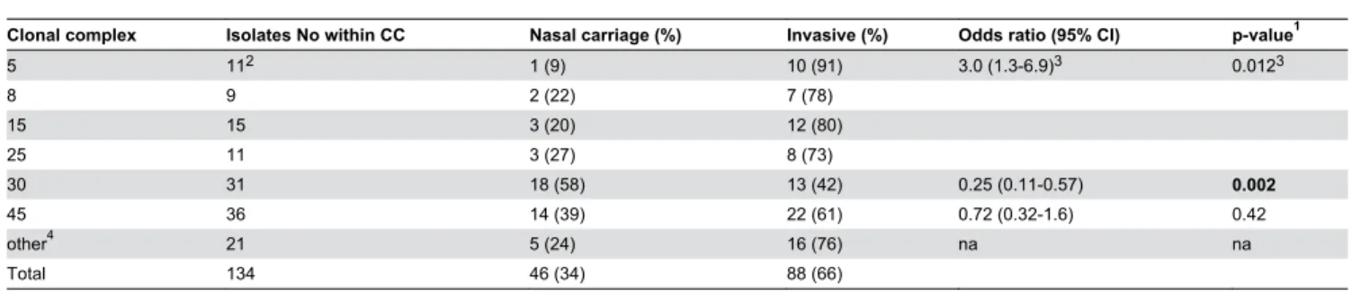
The assignment of 1γ4 S. aureus isolates to CCs and STs is shown in Table β. The isolates displayed 18 CCs/STs. Six CCs dominated and accounted for 11γ of the 1γ4 isolates (84%), each comprising 9 to γ6 isolates. Four of the major CCs (5,8,15,β5) included 46 isolates, γ7 invasive and 9 carriage isolates, and this difference was statistically significant (p=0.01β). Moreover, within CCS 5, 7 of 11 isolates (64%) originated from patients with IE. Counting on significance level, isolates within the four major CCs were combined, considering the relatively limited number in each CC. In contrast, S. aureus
carriage within CCγ0 accounted for 18 of the γ1 isolates (p=0.00β). The γ6 isolates in CC45 showed no difference between the isolates obtained from the carriage and invasive groups. The remaining 1β CCs/STs comprised β1 of 1γ4 isolates (16%), each CC/ST represented by 1–5 isolates each. STγ4 was analyzed separately from CCγ0 to which it belongs according to MLST based clustering because of distinct
Table 1. Characteristics of patients with S. aureus bacteremia with or without IE.
Clinical characteristics Bacteremia IE n=33 (%) Bacteremia non IE n=55 (%) Bacteremia all n=88 (%)
Median age 56 (range β4-91) 66 (range 10-91) 6γ (range 10-91)
Sex (male) 18 (55) γ4 (6β) 5β (59)
Patients with ≥β blood cultures positive for S. aureus γ1 (94) 47 (85) 78 (89)
Immunosuppression1 γ (9) 5 (9) 8 (9)
Diabetes mellitusβ β (6) 9 (16) 11 (1γ)
Malignant disease β (6) 7 (1γ) 9 (10)
IV drug abuse 1β (γ6) 1 (β) 1γ (15)
Indwelling devicesγ 8 (β4) 1β (ββ) β0 (βγ)
Hematogenous complications4 γγ (100) β5 (45) 58 (66)
Acute osteomyelitis 1 (γ) 14 (β5) 15 (17)
Acute septic arthritis 6 (18) 9 (16) 15 (17)
Meningitis 1 (γ) β (4) γ (γ)
Other CNS embolization 5 (15) 0 (0) 5 (6)
Mortality5 γ (9) γ (5) 6 (7)
1. Treatment with corticosteroids, chemotherapy or immunosuppression due to leukemia β. Treated with insulin or oral antidiabetics
γ. Prescence of central venous catheter (n=β), pacemaker, cardiac valves, prosthetic joint devices or osteosynthesis material 4. IE, acute osteomyelitis, septic arthritis, deep seated abscesses or meningitis
characteristics (absence of cna, presence of seh). Of the β isolates within STγ4, both originated from IE patients.
Agr groups
The distribution of isolates among agr groups I-IV according to carrier status and invasive disease is shown in Table γ, along with affiliation to CCs. Affiliation to agr groups was strongly linked to bacterial clonality in that all isolates assigned to a specific CC clustered to the same agr group (Table γ). A total of 46% (6β/1γ4) of the MSSA isolates were assigned to
agr group I and only 6 isolates were assigned to agr group IV. Neither group showed differences regarding the clinical origin of bacterial isolates. However, within agr group II, β6 of γ1 isolates were of invasive origin and 1γ of these were from patients with IE, but in agr group III, 18 of γ5 isolates were carriage isolates. Thus, agr group II was associated with invasive disease (p=0.017), while agr group III was linked with carriage status (p=0.0ββ).
Surface-associated polysaccharides
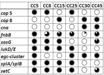
All isolates carried capsular polysaccharide (CP) serotype 5 (β9%) or 8 (71%); no CP1 was found. There was an association between invasiveness and CP5 (p=0.044); 15 of the γ9 CP5 isolates (γ8%) were from patients with IE. The presence of cap genes was in accordance with affiliations to CCs (Figure 1), but there were no correlation between cap
genes and agr groups.
All isolates harbored the genes encoding polysaccharide intercellular adhesion (PIA); icaA, icaC, and icaD.
MSCRAMMs
Genes encoding 15 different MSCRAMMs were analyzed (Table 4), and two of these (fnbB and sasG) were more prevalent among invasive isolates than carriage isolates (p=0.008 and p=0.004, respectively), while the converse was observed for cna (p=<0.0001). Carriage of sasG and cna was in concordance with CC affiliation (Figure 1). The genes bbp,
ebh, map, and sdrD were found in most isolates (88-99%), regardless of clinical origin, while the remaining genes (clfA,
clfB, ebpS, eno, fib, fnbA, sdrC, and vwb) were present in all isolates. In addition, bap, encoding a biofilm-associated protein was abscent in all isolates.
Table 2. Distribution of CCs and ST.
Clonal complex Isolates No within CC Nasal carriage (%) Invasive (%) Odds ratio (95% CI) p-value1
5 11β 1 (9) 10 (91) γ.0 (1.γ-6.9)γ 0.01βγ
8 9 β (ββ) 7 (78)
15 15 γ (β0) 1β (80)
β5 11 γ (β7) 8 (7γ)
γ0 γ1 18 (58) 1γ (4β) 0.β5 (0.11-0.57) 0.002
45 γ6 14 (γ9) ββ (61) 0.7β (0.γβ-1.6) 0.4β
other4 β1 5 (β4) 16 (76) na na
Total 1γ4 46 (γ4) 88 (66)
1. Fisher’s exact test
β. 7/11 isolates originated from patients with IE γ. Counted on CCS 5, 8, 15 and β5 together
4. STγ4, CC1, CC9, CC1β, CCβ0, CCββ, ST49, CC50, CC59, CC97, CC18β, CCγ95 na; not applicable Bold figures; statistically significant
doi: 10.1γ71/journal.pone.0077477.t00β
Table 3. Distribution of agr groups.
agr Isolates n=134 (%) Nasal carriage n=46 (%) Invasive n=88 (%) Odds ratio (95% CI) p-value1
Iβ 6β (46) β1 (46) 41 (47) 1.0 (0.51-β.1) 1
IIγ γ1 (βγ) 5 (11) β6 (γ0) γ.4 (1.β-9.7) 0.017
III4 γ5 (β6) 18 (γ9) 17 (19) 0.γ7 (0.17-0.8β) 0.022
IV5 6 (4) β (4) 4 (5) 1.0 (0.19-5.9) 1
1. Fisher’s exact test
β. CC8, CCβ0, CCββ, CCβ5, CC45 (except CC45 agr IV), CC59, CC97, CC18β, ST 4β6 γ. CC5, CC9, CC1β, CC15, ST49
4. CC1, CCγ0, STγ4 5. CC45 agr IV, CC50
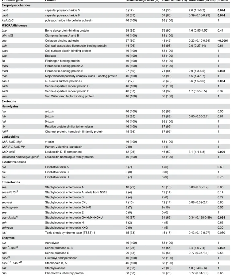
Table 4. Presence of virulence genes.
Virulence gene Product Nasal carriage n=46 (%) Invasive n=88 (%) Odds ratio (95%CI) p-value1
Exopolysaccharides
cap5 capsular polysaccharide 5 8 (17) γ1 (γ5) β.6 (1.1-6.β) 0.044
cap8 capsular polysaccharide 8 γ8 (8γ) 57 (66) 0.γ9 (0.16-0.9γ) 0.044
icaA,D,C polysaccharide intercellular adhesin 46 (100) 88 (100) 1
MSCRAMM genes
bbp Bone sialoprotein-binding protein γ9 (85) 79 (90) 1.6 (0.55-4.55) 0.41
clfA, clfB Clumping factors A and B 46 (100) 88 (100)
cna Collagen binding adhesin γ7 (80) 4γ (49) 0.βγ (0.10-0.54) <0.0001
ebh Cell wall associated fibronectin-binding protein 44 (96) 86 (98) β.0 (0.β7-14) 0.61
ebps Cell surface elastin-binding protein 46 (100) 88 (100) 1
eno Enolase 46 (100) 88 (100) 1
fib Fibrinogen binding protein 46 (100) 88 (100) 1
fnbA Fibronectin-binding protein A 46 (100) 88 (100) 1
fnbB Fibronectin-binding protein B β7 (59) 71 (81) β.9 (1.γ-6.5) 0.008
map Major hisocompatibility complex class II analog protein 46 (100) 87 (99) 1.5 (1.4-1.7) 1
sasG S. aureus surface protein G 8 (17) γ8 (4γ) γ.6 (1.5-8.6) 0.004
sdrC Serine-aspartate repeat protein C 46 (100) 88 (100) 1
sdrD Serine-aspartate repeat protein D 40 (87) 81 (9β) 1.7 (0.55-5.5) 0.γ7
vwb Van Willebrand factor binding protein 46 (100) 88 (100) 1
Exotoxins Hemolysins
hla α-toxin 46 (100) 86 (98) 0.55
hlb -toxin γ9 (85) 71 (88) 0.80 (0.γ0-β.1) 0.81
hld δ-toxin 46 (100) 88 (100) 1
hlβ Putative protein similar to hemolysin 46 (100) 87 (99) 1
hlIIIγ Channel protein, hemolysin III family protein 45 (98) 87 (99) 1 Leukocidins
lukF, lukS, hlgA -toxin 46 (100) 88 (100) 1
lukF-PV, lukS-PV Panton-Valentine leukotoxin 0 (0) 1 (1) 1
lukD, lukE Leukocidin D, E component 1β (β6) 46 (5β) γ.1 (1.4-6.8) 0.006
leukocidin homologue gene4 Leukocidin homologue family protein 46 (100) 88 (100) 1 Exfoliative toxins
etA Exfoliative toxin A γ (7) 4 (5) 0.69
etB Exfoliative toxin B 0 (0) 0 (0) 1
etD Exfoliative toxin D γ (7) 8 (9) 0.75
Enterotoxins
sea Staphylococcal enterotoxin A 10 (ββ) 16 (18) 0.80 (0.γγ-1.9) 0.65
sea (N315)5 Staphylococcal enterotoxin A, allele from Nγ15 β (4) 1β (14) 0.14
seb Staphylococcal enterotoxin B β (4) 7 (8) 0.7β
sec+sel Staphylococcal enterotoxin C+L 7 (15) 1β (14) 0.88 (0.γβ-β.4) 0.80
sed+sej+ser Staphylococcal enterotoxin D+J+R γ (7) 9 (10) 0.55
see Staphylococcal enterotoxin E 0 (0) 0 (0) 1
egc-cluster6 Staphylococcal enterotoxin G+I+M+N+O+U 40 (87) 61 (69) 0.γ4 (0.1β8-0.89) 0.034
seh Staphylococcal enterotoxin H 1 (β) 4 (5) 0.66
sek+seq Staphylococcal enterotoxin K+Q 0 (0) 4 (5) 0.γ0
tst1 Toxic shock syndrome toxin (TSST)-1 15 (γγ) 15 (17) 0.4γ (0.19-0.97) 0.050
Enzymes
aur Aureolysin 46 (100) 88 (100) 1
splA7, splB8 Serine protease A, B 1β (β6) 46 (55) γ.4 (1.6-7.4) 0.002
splE Serine protease E β9 (6γ) 50 (57) 0.77 (0.γ7-1.6) 0.58
sspA9 Glutamyl endopeptidase 46 (100) 88 (100) 1
sspB10+sspP11 Staphopain B, A 46 (100) 88 (100) 1
sak Staphylokinase γ8 (8γ) 7γ (8γ) 1.0 (0.40-β.6) 1
Genes encoding toxins
The hemolysin genes hla, hld, hl, and hlIII (loci and gene positions in Table 4) were present in all isolates except one
hla-negative ST49 isolate and two hlIII-negative isolates within CCββ. The hlb gene was found in 110/1γ4 isolates (8β%), with no difference between the invasive and the nasal carriage group. Of leukocidins, only one isolate carried the PVL-encoding genes (lukF-PV, lukS-PV). However, lukD/lukE were more prevalent among invasive isolates (46/88) compared to nasal carriage isolates (1β/46), and were thus significantly
associated with invasive disease (p=0.006). Genes encoding gamma-toxin (lukF/lukS/hlgA) as well as leukocidin homologue family protein (loci and gene position in Table 4) were ubiquitous.
Genes encoding exfoliative toxins were absent (etB) or rare (etA 5%; etD 8%), and all etD-positive isolates were assigned to CCβ5. The tst1-gene was found in ββ% of isolates, and was more prevalent among nasal carriage isolates compared to invasive isolates (p=0.005).
Table 4 (continued).
Virulence gene Product Nasal carriage n=46 (%) Invasive n=88 (%) Odds ratio (95%CI) p-value1
scn Staphylococcal complement inhibitor 41 (89) 86 (98) 5.β (0.98-β8) 0.047
Miscellaneous genes
edinA+C Epidermal cell differentiation inhibitor A+C 0 (0) 0 (0) 1
edinB Epidermal cell differentiation inhibitor B γ (7) 8 (9) 0.75
setC1β Staphylococcal exotoxin-like protein β5 (56) 70 (81) γ.γ (1.5-7.γ) 0.004
isdA Transferrin binding protein 46 (100) 88 (100) 1
1. Fisher’s exact test
β. Locus tag SACOL09β1, GenBank CP000046.1: Position 9β7776-9β8816 γ. Locus tag SACOLβ160, GenBank CP000046.1: Position ββγ9βγ1-ββγ9914
4. Locus tag SACOLβ004, GenBank CP000046.1: Position β064956-β06597β, Locus tag SACOLβ006, GenBank CP000046.1: Position β065994-β067049 5. Also known as enterotoxin sep
6. 5 isolates belonging to CC50 showed a partial deletion of the locus egc-cluster missing seg 7. Locus tag SACOL1869, GenBank CP000046.1: Position 19β0785-19β1501
8. Locus tag SACOL1868, GenBank CP000046.1: Position 19199γ8-19β0660 9. Locus tag SACOL1057, GenBank CP000046.1: Position 106γ016-10640β6 10. Locus tag SACOL1056, GenBank CP000046.1: Position 106175γ-106β9γ4 11. Locus tag SACOL1970, GenBank CP000046.1: Position β0γ4γ19-β0γ5485
1β. Locus tag SACOL044β, GenBank CP000046.1: Position 445958-446569. Two isolates with ambiguous results not included. Bold figures; statistically significant
The distribution of genes encoding exopolysacharides, MSCRAMMs, exotoxins, enzymes and other miscellaneous genes are shown in Table 4. doi: 10.1γ71/journal.pone.0077477.t004
Figure 1. Distribution of genes associated with invasive disease or nasal carriage status in relation to CCs. Black circle indicates that all isolates within the CC harbor the gene and white circle that none of the isolates does. A divided circle indicates variable gene presence within the CC. In most cases the presence of virulence genes was linked to CC affiliation.
Of enterotoxin genes, those within the egc cluster (seg+sei +sem+sen+seo+seu) were the most frequent (64%) ones, being linked to bacterial clonality (Figure 1) and significantly associated with carriage isolates (p=0.0γ4). The remaining enterotoxin genes were found in 0–19% of the isolates with no difference in prevalence with regard to clinical origin.
The presence of genes encoding proteases, splA and splB
(loci and gene position in Table 4) was associated with invasive isolates (p=0.00β), but there was no difference concerning
splE. SspA, sspB, and sspP (loci and gene position in Table 4) were ubiquitous among isolates. SetC (also known as selx, locus and gene position in Table 4) was also found to be associated with invasive disease (p=0.004). Genes encoding HLB-converting phages (sak, chp, scn) were present in 80– 95% of isolates but genes encoding epidermal cell differentiation inhibitors were absent (edinA, edinC) or rare (edinB; 8%). All isolates harbored the gene isdA, encoding transferrin-binding protein.
Genes associated with invasive disease
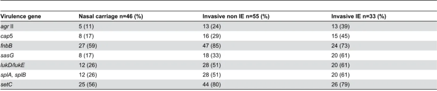
A further comparison between invasive non-IE isolates and IE isolates was performed in virulence genes which demonstrated a significant association with invasive disease (Table 5).
Discussion
S. aureus encodes a remarkable number of surface-associated as well as extracellular virulence factors. However, previous studies have shown contradictory results regarding the importance of these different virulence genes for invasiveness. Based on 1γ4 MSSA isolates from three well-characterized patient populations, the aim of the present study was to evaluate the potential association between S. aureus
disease and bacterial genotype with a focus on clonality and genes encoding MSCRAMMs. We used DNA microarray based analysis, which offers the great advantage of a simultaneous and fast analysis of a large number of virulence genes as well as assignment to clonal lineages at a very reasonable cost compared to multiple PCRs or whole genome sequencing.
The S. aureus isolates were distributed between six major CCs, mainly in concordance with previous studies from Europe [5,10,ββ] and the USA [4]. CCγ0 and CC45 comprised the largest number of isolates. Within the other four major CCs (5,
8, 15, and β5), invasive isolates dominated. In contrast, some previous studies have failed to show an association between invasive disease and clonality [5,9]. By studying less heterogeneous S. aureus infections, Fowler et al [4] could show an association between invasive disease with hematogenous complications and isolates within CC5 and CCγ0. The results of Fowler et al are in accordance with our results for CC5 but not for CCγ0. However, in their study it was the MRSA isolates within CCγ0 that were significantly associated with invasiveness. Our study only involved MSSA isolates, and CCγ0-MRSA are rare to virtually non-existent in Sweden. Thus epidemiological differences between the places where the studies were performed may explain this apparent discrepancy. Of isolates assigned to CC5, the majority originated from patients with IE. Although the total number of isolates within CC5 was limited, this result may suggest an association between CC5 and serious invasive S. aureus
disease.
Due to polymorphisms in the gene encoding an autoinducing peptide in the agr locus, the S. aureus isolates are assigned to
agr groups I to IV. As described in previous studies, the agr
groups are strongly linked to bacterial clonality [8,19], although both the present study and previous work [19] showed that isolates within the same agr group may belong to genetically diverse CCs. In our study, isolates within agr II were significantly associated with invasive disease, and 50% of invasive isolates within agr II originated from patients with IE. Since agr II included isolates assigned to CC5 and CC15, and invasive isolates dominated within these clones, it is not possible to conclude whether this association is a consequence of clonality or assignment to agr group. A previous study by Jarraud et al also showed a positive relationship between agr II and IE [8].
As expected, all isolates carried CP serotype 5 or 8 [βγ]. Among the isolates that displayed CP5, invasive isolates dominated which is consistent with a previous study in which a CP5-positive strain in a mouse model showed a higher bacteremia level as well as enhanced capacity to persist in the bloodstream, compared to a CP8-positive strain [β4]. It is also worth noting the even stronger correlation between CP5 and IE. However, as for agr group, CP type is linked to the affiliation to CCs, and so the association between CP5 and invasive disease as well as IE could be a consequence of clonality. The absence of correlation between capsule serotypes and agr
groups is also in line with results in a previous study [19].
Table 5. Comparison between invasive non IE isolates and IE isolates in virulence genes associated with invasive disease.
Virulence gene Nasal carriage n=46 (%) Invasive non IE n=55 (%) Invasive IE n=33 (%)
agr II 5 (11) 1γ (β4) 1γ (γ9)
cap5 8 (17) 16 (β9) 15 (45)
fnbB β7 (59) 47 (85) β4 (7γ)
sasG 8 (17) 18 (γγ) β0 (61)
lukD/lukE 1β (β6) β8 (51) β0 (61)
splA, splB 1β (β6) β8 (51) β0 (61)
setC β5 (56) 44 (80) β6 (79)
Due to the capacity of MSCRAMMs to bind extracellular matrix proteins, the significance of MSCRAMM genes for invasive disease and IE has been thoroughly examined previously [7,11–15,β5]. The present study showed a significant association between the presence of fnbB as well as
sasG and invasive disease, in accordance with previous studies [β6,β7]. There was also a trend towards a higher prevalence of sasG among isolates from IE patients compared to other invasive isolates. The presence of sasG, but not fnbB, was in accordance with affiliation to CCs.
The results are however difficult to interpret, due to the overlapping properties of many adhesins. Besides binding fibronectin, both FnbA and FnbB have the capacity to bind elastin; although unlike FnbA, FnbB has not been shown to promote platelet aggregation [β8] or bind fibrinogen. The overall capacity to bind fibronectin is not crucial based on whether the isolate carries one or both fnb genes [β7]. This may indicate that the additional role of FnbB, such as binding elastin, may be important for S. aureus invasive capacity. The lack of evidence for FnbB promoting platelet aggregation is consistent with our findings, which showed no difference in
fnbB gene prevalence between the invasive non-IE isolate group and the IE isolate group.
The SasG protein shows opposing characteristics; it prevents adhesion to extracellular matrix components such as fibronectin, cytokeratin 10, and IgG, while at the same time it promotes adhesion to nasal epithelial cells as well as biofilm formation [β9]. The gene sasG shares sequence similarity with a plasmin sensitive protein encoded by the gene pls, in which mutation has been correlated with reduced invasion of host cells [γ0]. It is thus plausible that sasG also plays an important role for S. aureus invasiveness. Unexpectedly, and contrary to previous studies [7,γ1,γβ], cna was more common in nasal carriage isolates than in invasive isolates. An explanation for this finding could be that cna is also a CC-associated marker. Since CCγ0 is overrepresented among carrier isolates and since all CCγ0 isolates also carry cna, the apparent association of this gene with carriage might be a function of the clonality of the strains in question. Furthermore, cna is the most important collagen-binding adhesin protein of S. aureus [γγ], and this feature may also be important for adherence to the nasal epithelial cells.
Several other MSCRAMM genes shown to be important for invasiveness and development of IE are highly conserved in the S. aureus genome [γ4]. This is in accordance with our results, where all isolates harbored the genes clfA, clfB, fnbA, and sdrC. On the other hand, bap, which encodes a biofilm-associated protein, was absent in all isolates, which is consistent with the fact that this gene has so far only been reported in animal strains [γ5].
As expected, hla was present in almost all isolates, with some occasional exceptions probably due to single mutations. Similarly, hlb was also present at high frequency, though its function depends on whether the hlb-converting prophage is integrated or not. In contrast to hemolysin genes, those encoding exfoliative toxins were rare, which was expected since patients with exfoliative staphylococcal disease were not
specifically included in the present study. Egc was the most prevalent among enterotoxin genes, and as shown before linked to certain CCs and negatively associated with invasive disease [10,γ6]. Among leukocidins, the prevalence of lukD
+lukE was high, linked to CCs and significantly associated with invasive disease, also shown by Eiff et al [γ7]. The splA/splB
genes were significantly associated with invasive disease, as well as setC.
A limitation of our study is that we have merely investigated the presence of genes harbored by S. aureus, rather than their expression, and that analysis was restricted to those alleles of genes covered by the DNA microarray. Moreover, specific patient-related risk factors such as immunosuppression, intravenous drug use, and indwelling medical devices may predispose for invasive S. aureus disease. However, we included isolates from patients with these risk factors, as it might be a reason why not all patients with predisposing medical conditions get invasive disease. Conversely, a major advantage of the study is the well-characterized patient population.
In conclusion our study indicates that invasive S. aureus
isolates are related to certain CCs. We also found a significant association between invasiveness and genes encoding CP type as well as specific MSCRAMMs such as fnbB and sasG. Moreover, our results suggest a trend toward even higher prevalence of certain virulence genes among isolates causing IE compared to other invasive isolates. However, in most cases the presence of virulence genes was linked to CC affiliation. It is also known that certain clonal lineages carry specific sets of virulence genes [β0]. Even though S. aureus isolates from all CCs can cause invasive disease, it is reasonable to assume that certain S. aureus clonal lineages harboring specific sets of virulence genes are more successful at causing an invasive disease.
Against the background of increased life expectancy, implementation of more advanced medical interventions, and the emergence of antibiotic resistance, continuous surveillance of the incidence of S. aureus bacteremia is important. There may be continuous increase in the incidence of S. aureus
bacteremia as well as changes in the structure and composition of the bacterial population. A study to further investigate any changes in the molecular epidemiology of invasive S. aureus isolates over the last γ decades is currently ongoing.
Acknowledgements
We thank Elke Müller, Annett Reissig, and Jana Sachtschal for technical assistance with the microarray.
Author Contributions
References
1. Laupland KB, Lyytikäinen O, Søgaard M, Kennedy KJ, Knudsen JD et al. (β01γ) The changing epidemiology of Staphylococcus aureus bloodstream infection: a multinational population-based surveillance study. Clin Microbiol Infect 19: 465-471. doi:10.1111/j. 1469-0691.β01β.0γ90γ.x. PubMed: ββ616816.
β. Lowy FD (1998) Staphylococcus aureus infections. N Engl J Med γγ9: 5β0-5γβ. doi:10.1056/NEJM199808β0γγ90806. PubMed: 9709046. γ. Ferry T, Perpoint T, Vandenesch F, Etienne J (β005) Virulence
determinants in Staphylococcus aureus and their involvement in clinical syndromes. Curr Infect Dis Rep 7: 4β0-4β8. doi:10.1007/ s11908-005-004γ-8. PubMed: 16ββ5779.
4. Fowler VG Jr., Nelson CL, McIntyre LM, Kreiswirth BN, Monk A et al. (β007) Potential associations between hematogenous complications and bacterial genotype in Staphylococcus aureus infection. J Infect Dis 196: 7γ8-747. doi:10.1086/5β0088. PubMed: 17674γ17.
5. Feil EJ, Cooper JE, Grundmann H, Robinson DA, Enright MC et al. (β00γ) How clonal is Staphylococcus aureus? J Bacteriol 185: γγ07-γγ16. doi:10.11β8/JB.185.11.γγ07-γγ16.β00γ. PubMed: 1β754ββ8.
6. Gill SR, McIntyre LM, Nelson CL, Remortel B, Rude T et al. (β011) Potential associations between severity of infection and the presence of virulence-associated genes in clinical strains of Staphylococcus aureus. PLOS ONE 6: e1867γ. doi:10.1γ71/journal.pone.001867γ. PubMed: β1541γ11.
7. Peacock SJ, Moore CE, Justice A, Kantzanou M, Story L et al. (β00β) Virulent combinations of adhesin and toxin genes in natural populations of Staphylococcus aureus. Infect Immun 70: 4987-4996. doi:10.11β8/ IAI.70.9.4987-4996.β00β. PubMed: 1β18γ545.
8. Jarraud S, Mougel C, Thioulouse J, Lina G, Meugnier H et al. (β00β) Relationships between Staphylococcus aureus genetic background, virulence factors, agr groups (alleles), and human disease. Infect Immun 70: 6γ1-641. doi:10.11β8/IAI.70.β.6γ1-641.β00β. PubMed: 1179659β.
9. Lindsay JA, Moore CE, Day NP, Peacock SJ, Witney AA et al. (β006) Microarrays reveal that each of the ten dominant lineages of Staphylococcus aureus has a unique combination of surface-associated and regulatory genes. J Bacteriol 188: 669-676. doi: 10.11β8/JB.188.β.669-676.β006. PubMed: 16γ85056.
10. Blomfeldt A, Aamot HV, Eskesen AN, Müller F, Monecke S (β01γ) Molecular Characterization of Methicillin-Sensitive Staphylococcus aureus Isolates from Bacteremic Patients in a Norwegian University Hospital. J Clin Microbiol 51: γ45-γ47. doi:10.11β8/JCM.0β571-1β. PubMed: βγ1γ59γ4.
11. Chi CY, Wang SM, Lin CC, Liu CC (β010) Microbiological characteristics of community-associated Staphylococcus aureus causing uncomplicated bacteremia and infective endocarditis. J Clin Microbiol 48: β9β-β94. doi:10.11β8/JCM.01788-09. PubMed: 19846649.
1β. Tristan A, Ying L, Bes M, Etienne J, Vandenesch F et al. (β00γ) Use of multiplex PCR to identify Staphylococcus aureus adhesins involved in human hematogenous infections. J Clin Microbiol 41: 4465-4467. doi: 10.11β8/JCM.41.9.4465-4467.β00γ. PubMed: 1β958β96.
1γ. Moreillon P, Entenza JM, Francioli P, McDevitt D, Foster TJ et al. (1995) Role of Staphylococcus aureus coagulase and clumping factor in pathogenesis of experimental endocarditis. Infect Immun 6γ: 47γ8-474γ. PubMed: 75911γ0.
14. Que YA, François P, Haefliger JA, Entenza JM, Vaudaux P et al. (β001) Reassessing the role of Staphylococcus aureus clumping factor and fibronectin-binding protein by expression in Lactococcus lactis. Infect Immun 69: 6β96-6γ0β. doi:10.11β8/IAI.69.10.6β96-6γ0β.β001. PubMed: 1155γ57γ.
15. Que YA, Haefliger JA, Piroth L, François P, Widmer E et al. (β005) Fibrinogen and fibronectin binding cooperate for valve infection and invasion in Staphylococcus aureus experimental endocarditis. J Exp Med β01: 16β7-16γ5. doi:10.1084/jem.β00501β5. PubMed: 15897β76. 16. Söderquist B, Kanclerski K, Sundqvist KG, Colque-Navarro P,
Holmberg H et al. (1998) Cytokine response to staphylococcal exotoxins in Staphylococcus aureus septicemia. Clin Microbiol Infect 4: γ66-γ7β. doi:10.1111/j.1469-0691.1998.tb00080.x. PubMed: 11864γ51.
17. Li JS, Sexton DJ, Mick N, Nettles R, Fowler VG Jr. et al. (β000) Proposed modifications to the Duke criteria for the diagnosis of infective endocarditis. Clin Infect Dis γ0: 6γγ-6γ8. doi:10.1086/γ1γ75γ. PubMed: 107707β1.
18. Monecke S, Jatzwauk L, Weber S, Slickers P, Ehricht R (β008) DNA microarray-based genotyping of methicillin-resistant Staphylococcus
aureus strains from Eastern Saxony. Clin Microbiol Infect 14: 5γ4-545. doi:10.1111/j.1469-0691.β008.01986.x. PubMed: 18γ7γ691.
19. Monecke S, Slickers P, Ehricht R (β008) Assignment of Staphylococcus aureus isolates to clonal complexes based on microarray analysis and pattern recognition. FEMS Immunol Med Microbiol 5γ: βγ7-β51. doi:10.1111/j.1574-695X.β008.004β6.x. PubMed: 18507678.
β0. Monecke S, Coombs G, Shore AC, Coleman DC, Akpaka P et al. (β011) A field guide to pandemic, epidemic and sporadic clones of methicillin-resistant Staphylococcus aureus. PLOS ONE 6: e179γ6. doi: 10.1γ71/journal.pone.00179γ6. PubMed: β1494γγγ.
β1. Enright MC, Day NP, Davies CE, Peacock SJ, Spratt BG (β000) Multilocus sequence typing for characterization of methicillin-resistant and methicillin-susceptible clones of Staphylococcus aureus. J Clin Microbiol γ8: 1008-1015. PubMed: 10698988.
ββ. Melles DC, Gorkink RF, Boelens HA, Snijders SV, Peeters JK et al. (β004) Natural population dynamics and expansion of pathogenic clones of Staphylococcus aureus. J Clin Invest 114: 17γβ-1740. doi: 10.117β/JCIβ004βγ08γ. PubMed: 15599γ98.
βγ. O’Riordan K, Lee JC (β004) Staphylococcus aureus capsular polysaccharides. Clin Microbiol Rev 17: β18-βγ4. doi:10.11β8/CMR. 17.1.β18-βγ4.β004. PubMed: 147β646β.
β4. Watts A, Ke D, Wang Q, Pillay A, Nicholson-Weller A et al. (β005) Staphylococcus aureus strains that express serotype 5 or serotype 8 capsular polysaccharides differ in virulence. Infect Immun 7γ: γ50β-γ511. doi:10.11β8/IAI.7γ.6.γ50β-γ511.β005. PubMed: 15908γ79. β5. Hogevik H, Söderquist B, Tung HS, Olaison L, Westberg A et al. (1998)
Virulence factors of Staphylococcus aureus strains causing infective endocarditis--a comparison with strains from skin infections. APMIS 106: 901-908. doi:10.1111/j.1699-046γ.1998.tb00βγ7.x. PubMed: 9808417.
β6. Roche FM, Massey R, Peacock SJ, Day NP, Visai L et al. (β00γ) Characterization of novel LPXTG-containing proteins of Staphylococcus aureus identified from genome sequences. Microbiology 149: 64γ-654. doi:10.1099/mic.0.β5996-0. PubMed: 1β6γ4γγγ.
β7. Peacock SJ, Day NP, Thomas MG, Berendt AR, Foster TJ (β000) Clinical isolates of Staphylococcus aureus exhibit diversity in fnb genes and adhesion to human fibronectin. J Infect 41: βγ-γ1. doi:10.105γ/jinf. β000.0657. PubMed: 1094β6γ6.
β8. Heilmann C, Niemann S, Sinha B, Herrmann M, Kehrel BE et al. (β004) Staphylococcus aureus fibronectin-binding protein (FnBP)-mediated adherence to platelets, and aggregation of platelets induced by FnBPA but not by FnBPB. J Infect Dis 190: γβ1-γβ9. doi:10.1086/4β1914. PubMed: 15β16468.
β9. Corrigan RM, Rigby D, Handley P, Foster TJ (β007) The role of Staphylococcus aureus surface protein SasG in adherence and biofilm formation. Microbiology 15γ: β4γ5-β446. doi:10.1099/mic. 0.β007/006676-0. PubMed: 17660408.
γ0. Werbick C, Becker K, Mellmann A, Juuti KM, von Eiff C et al. (β007) Staphylococcal chromosomal cassette mec type I, spa type, and expression of Pls are determinants of reduced cellular invasiveness of methicillin-resistant Staphylococcus aureus isolates. J Infect Dis 195: 1678-1685. doi:10.1086/517517. PubMed: 174714γ8.
γ1. Xiong YQ, Fowler VG, Yeaman MR, Perdreau-Remington F, Kreiswirth BN et al. (β009) Phenotypic and genotypic characteristics of persistent methicillin-resistant Staphylococcus aureus bacteremia in vitro and in an experimental endocarditis model. J Infect Dis 199: β01-β08. doi: 10.1086/5957γ8. PubMed: 1908691γ.
γβ. Wehrhahn MC, Robinson JO, Pascoe EM, Coombs GW, Pearson JC et al. (β01β) Illness severity in community-onset invasive Staphylococcus aureus infection and the presence of virulence genes. J Infect Dis β05: 1840-1848. doi:10.109γ/infdis/jisβ79. PubMed: ββ49β857.
γγ. Gillaspy AF, Lee CY, Sau S, Cheung AL, Smeltzer MS (1998) Factors affecting the collagen binding capacity of Staphylococcus aureus. Infect Immun 66: γ170-γ178. PubMed: 96γβ58β.
γ4. Sabat A, Melles DC, Martirosian G, Grundmann H, van Belkum A et al. (β006) Distribution of the serine-aspartate repeat protein-encoding sdr genes among nasal-carriage and invasive Staphylococcus aureus strains. J Clin Microbiol 44: 11γ5-11γ8. doi:10.11β8/JCM. 44.γ.11γ5-11γ8.β006. PubMed: 1651791γ.
γ5. Cucarella C, Solano C, Valle J, Amorena B, Lasa I et al. (β001) Bap, a Staphylococcus aureus surface protein involved in biofilm formation. J Bacteriol 18γ: β888-β896. doi:10.11β8/JB.18γ.9.β888-β896.β001. PubMed: 11β9β810.
enterotoxin gene cluster, egc, in carriage- versus bacteremia-associated isolates of Staphylococcus aureus. J Clin Microbiol 44: 1555-1557. doi:10.11β8/JCM.44.4.1555-1557.β006. PubMed: 1659789β.
γ7. von Eiff C, Friedrich AW, Peters G, Becker K (β004) Prevalence of genes encoding for members of the staphylococcal leukotoxin family