Adherence and virulence genes of
Escherichia coli
from children diarrhoea
in the Brazilian Amazon
Najla Benevides-Matos
1, Fabio A. Pieri
2, Marilene Penatti
3, Patrícia P. Orlandi
2 1Instituto de Pesquisas em Patologias Tropicais, Fundação Oswaldo Cruz, Porto Velho, RO, Brazil. 2
Instituto Leônidas e Maria Deane, Fundação Oswaldo Cruz, Manaus, AM, Brazil. 3
Hospital Infantil Cosme e Damião, Secretaria de Estado da Saúde, Porto Velho, RO, Brazil.
Submitted: September 7, 2013; Approved: June 6, 2014.
Abstract
The bacterial pathogen most commonly associated with endemic forms of childhood diarrhoea is Escherichia coli. Studies of epidemiological characteristics of HEp-2 cell-adherentE. coliin diar-rhoeal disease are required, particularly in developing countries. The aim of this study was evaluate the presence and significance of adherentEscherichia colifrom diarrhoeal disease in children. The prevalence of LA, AA, and DA adherence patterns were determined in HEp-2 cells, the presence of virulence genes and the presence of the O serogroups in samples obtained from 470 children with acute diarrhoea and 407 controls in Porto Velho, Rondônia, Brazil.E. coliisolates were identified by PCR specific for groups of adherentE. coli. Out of 1,156 isolates obtained, 128 (11.0%) were posi-tive foreaegenes corresponding to EPEC, however only 38 (29.6%) of these amplifiedbfpAgene. EAEC were isolated from 164 (14.1%) samples; of those 41(25%), 32 (19%) and 16 (9.7%) ampli-fiedeagg,aggAoraafAgenes, respectively and aggA was significantly associated with diarrhoea (P= 0.00006). DAEC identified by their adhesion pattern and there were few isolates. In conclusion, EAEC was the main cause of diarrhoea in children, especially when theaggAgene was present, fol-lowed by EPEC and with a negligible presence of DAEC.
Key words:EnteroadherentEscherichia coli, diarrhoea, children.
Introduction
Diarrhoea continues to be one of the most common causes of morbidity and mortality among infants and chil-dren in developing countries (Nakhjavaniet al., 2013). The bacterial pathogen most commonly associated with en-demic forms of childhood diarrhoea isEscherichia coli, which can be identified in about 50% of cases as presented by Chandraet al.(2012). At least six categories of diar-rhoeagenicE. colistrains are recognized on the basis of dis-tinct epidemiological and clinical features, specific virulence determinants, and an association with certain serotypes: enteropathogenic E. coli (EPEC), enterotoxi-genic E. coli (ETEC), enteroinvasive E. coli (EIEC), enterohemorrhagic E. coli (EHEC), enteroaggregativeE. coli(EAEC), and diffusely adherentE. coli(DAEC) (Tor-res et al., 2005). EPEC, EAEC, and DAEC isolates are
characterized by their distinct patterns of adherence to cul-tured epithelial cellsin vitro. EPEC strains are responsible for a large number of cases of infantile diarrhoea in several developing countries (Abbaet al., 2009; Chandra et al., 2012; Contreras et al., 2012; Nakhjavani et al., 2013). These strains belong to specific serotypes within different E. coliserogroups (O groups) and produce a characteristic adherence pattern in tissue culture cells called localized ad-herence (LA) (Humphries and Armstrong, 2010).
In the LA pattern, bacteria bind to localized areas of the cell surface, forming compact microcolonies (bacterial clusters) that can be visualized after 3 h of contact between the bacteria and the cells (Humphries and Armstrong, 2010). This phenomenon is associated with the presence of the EPEC plasmid adherence factor (EAF) (Bardiauet al., 2010). The central mechanism of EPEC pathogenesis is a lesion called attaching and effacing (A/E) characterized by
DOI: http://dx.doi.org/10.1590/S1517-838246120130917
Send correspondence to P.P. Orlandi. Leônidas and Maria Deane Institute, Oswaldo Cruz Foundation, Fiocruz Amazônia, Rua Terezina 476, 69057-070, Manaus, AM, Brazil. E-mail: patricia_orlandi@amazonia.fiocruz.br.
microvilli destruction, intimate adherence of bacteria to the intestinal epithelium, pedestal formation, and aggregation of polarized actin and other elements of the cytoskeleton at sites of bacterial attachment (Humphries and Armstrong, 2010; Torreset al., 2005). Atypical EPEC strains that do not carry the EAF plasmid can exhibit different adherence patterns including localized adherence like (LAL) pattern, and have been isolated from acute infantile diarrhoea in Brazil (Arenas-Hernández et al., 2012; Bardiau et al., 2010; Ochoa and Contreras, 2011).
EAEC strains are defined by their characteristic aggregative adherence (AA) to HEp-2 cells in bacterial cul-ture (Weintraub, 2007). EAEC produce at least three fim-brial adhesins encoded on a 60- to 65-MDa virulence plasmid required for expression of the AA pattern:aggA (AAF/I), aafA (AAF/II), and agg-3 (AAF/III) (Huang et al., 2006).
DAEC strains are characterized by their dispersing adherence (DA) pattern on cultured epithelial cells (Servin, 2005a). Two adhesins capable of mediating the diffuse-adherence phenotype have been characterized for DAEC strains. F1845 is a fimbrial adhesin that mediates the adher-ence of the DAEC strain C1845 to epithelial cells (Servin, 2005a, b). F1845 fimbriae are encoded by five genes, desig-nateddaaABCDE. The significance of F1845 in virulence remains unclear because DAEC strains of faecal origin rarely express F1845 adhesin (Camposet al., 1999; Torres et al., 2005). The adhesin involved in diffuse adherence (AIDA-I) is a plasmid-encoded protein of the clinical DAEC strain 2787. The AIDA-I precursor protein is en-coded by theaidAgene and its mature form mediates dif-fuse adherence to HeLa cells (Benz and Schmidt, 1992a, b). Studies evaluating the epidemiological characteris-tics of HEp-2 cell-adherentE. coliin diarrhoeal disease are required, particularly in developing countries. For this rea-son, and in order to determine the significance of the EAEC, EPEC, and DAEC strains as possible pathogenic microorganisms causing infantile diarrhoea, we
deter-mined the prevalence of the LA, AA, and DA patterns, the presence of virulence genes and the presence of O sero-groups in samples obtained from children with acute diar-rhoea and controls in Porto Velho, Rondônia, Brazil.
Material and Methods
Search for pathogens in the samples
Faecal samples from 470 diarrheic children (ages 0 to 72 months) and 407 children without diarrhoea (controls) were collected. The samples were collected at the Infantile Hospital Cosme and Damião in Porto Velho, Rondonia, Brazil, from March 2000 to March 2002.
Faecal samples were collected after natural excretion or through stimulation with a glycerin suppository. Sam-ples were divided into two fractions: one fraction was used for parasitological examination for helminths eggs and pro-tozoa cysts. The second aliquot was processed by routine microbiological and biochemical studies to identifyE. coli. Five fermenting colonies and up to three lactose-negative colonies from each child were selected from McConkey plates to be tested by conventional and PCR procedures. Reference strains used as positive controls in the PCR tests included the E. coli strains O44H18, EDL933, 6085, O157:H7, H19 and C600PEB1 provided by Pasteur Institute (Paris, France). The non-pathogenic E. coli strain HB101 was used as a negative control and to monitor for PCR contamination.
PCR analysis
All strains were evaluated by PCR for identification of virulence genes from a distinct category ofE. coli:eaeA and bfpA for EPEC; astA (encoding the toxin EAST1), eagg,aafAandaggAfor EAEC; anddaaEfor DAEC (Pass et al., 2000), using primer sequences presented in Table 1. PCR amplifications were performed as follows: 5.0mL of bacterial extract was added to a reaction mixture (Life Technologies, Carlsbad, CA, USA) with a final volume of
Table 1- Target genes and primers used to identify adherentEscherichia coliobtained from children diarrhoea in the Brazilian Amazon.
Target Gene Primer EP Reference
EPEC eaeA fp : 5’- TGAGCGGCTGGCATGAGTCATAC-3’ 241 Passet al.(2000)
bp : 5’- TCGATCCCCATCGTCACCAGAGG-3’
EPEC bfp fp : 5’- AATGGTGCTTGCGCTTGCTGC-3’ 324 Gunzburget al. (1995)
bp : 5’- GCCGCTTTATCCAACCTGGTA-3’
EAEC eagg fp : 5’- AGACTCTGGCGAAAGACTGTATC-3’ 194 Passet al.(2000)
bp : 5’- ATGGCTGTCTGTAATAGATGAGAAC-3’
EAEC aggA fb : 5’- GCTAACGCTGCGTTAGAAAGACC-3’ 352 Pivaet al.(2003)
bp : 5’- GGAGTATCATTCTATATTCGCC-3’
EAEC aafA fp : 5’ - GACAACCGCAACGCTGCGCTG-3’ 307 Pivaet al.(2003)
p : 5’- GATAGCCGGTGTAATTGAGCC-3’
50mL containing: 0.1 mM each of dATP, dCTP, dGTP and dTTP PCR buffer (10 mM Tris HCL, pH 8.3; 50 mM KCl; 2 mM MgCl2); 10 pMol of each PCR primer, and 1U of Taq DNA polymerase. Amplification was performed in a BioRad thermal cycler. The conditions used for amplifica-tion of the target genes are presented in Table 2. After the amplification process the tubes were cooled rapidly at 4 °C. The amplified DNA products were resolved by agarose gel electrophoresis (2%, Life Technologies, Carlsbad, CA, USA) and visualized by UV transillumination after ethi-dium bromide staining.
HEp-2 adherence test
AllE. coliisolates were subjected to HEp-2 adher-ence tests (Craviotoet al., 1991). HEp-2 cells were grown overnight to 50% confluence in Dulbecco’s Modified Ea-gle’s medium (Gibco BRL, Gaithersburg-MD, USA) con-taining penicillin, streptomycin, and 2% foetal bovine serum on eight-well chamber slides. Bacteria were grown for 16 h in Luria broth without shaking. The HEp-2 cells were washed five times with Phosphate Buffered Saline and then the medium was replaced with Dulbecco’s Mod-ified Eagle’s medium containing 1% mannose. A volume of 10mL of bacterial suspension was added per well and the slides were incubated at 37 °C in 5% CO2 for 3 h. The monolayers were washed five times with PBS, then fixed with 70% methanol and Giemsa. The strains were stained with solutions provided in the Panoptic Quick staining kit for one minute. Each strain was tested in duplicate, and ap-propriate controls were included in the test. Strains that ad-hered to the monolayers were recorded as adhering in localized, diffuse, or aggregative patterns.
Serotype methods
EnteroadherentE. coliisolates grown on nutrient agar were used for the identification of somatic (O) antigens by standard agglutination methods using specific polyvalent and monovalent antisera (Biomerieux, Craponne, Auverg-ne, France).
Statistical analysis
The prevalence of diarrhoeagenicE. coliin patient and control samples was compared by a two-tailed X2 test with Yates correction and Fisher’s exact test. When analys-ing the association of two or three pathogens in the same di-arrhoeal patient, the same test was used to evaluate the probability of association by random chance as a function of their respective individual frequencies in the population.
Results
A total of 1.156E. colistrains were isolated from fae-cal specimens from 470 diarrheic children (patients) and 407 children without diarrhoea (controls). The PCR screen-ing results for virulence factors of enteroadherent diar-rhoeagenicE. coliare presented in Table 3.
Screening for theeaesequence, which is specific for both EPEC and EHEC, there were 128 positive isolates comprising 11.7% of the total. Only 38eae-positive patient isolates amplified withbfpAprimers, indicating that they belonged to typical EPEC. Thus 90 isolates were consid-ered as atypical EPEC, once all isolates were negative for verotoxin genes in preview work (Orlandiet al., 2006). EAEC were isolated from 164 samples; of those 41(25%), 32 (19%) and 16 (9.7%) amplified foreagg,aggAandaafA, respectively. Of the isolated EAEC, 75 (45.7%) exhibited the AA adhesion pattern in HEp-2 cells, not amplifying for any specific markers for EAEC tested in that State. DAEC were mainly characterized by the pattern of HEp2 cell ad-hesion. Among the 68 DAEC isolates, only 6 (8.8%) pre-sented thedaaEgene that encodes biogenesis of the F1845 adhesin. The enteroadherentE. coli analysed had higher frequencies in children with acute gastroenteritis (Table 4).
The results show us that all samples belonging to the typical EPEC showed the LA pattern of adhesion after 3 h of incubation; they were distributed in 26 (68.4%) patients and 12 (31.5%) of the control samples. Of the 90 isolated atypical EPEC, we found that 55 (61.1%) of the cases pre-sented the LAL adhesion pattern after 6 h of incubation (Figure 1), while in the control samples 35 (38.9%) showed this adhesion profile. EAEC showed the AA adhesion pat-tern in 91 (55.5%) of the patients and 73 (44.5%) of the con-trol samples (Figure 1). Diffuse adhesion was characterized
Table 2- Amplification conditions for PCR reactions to identify adherentEscherichia coliobtained from children diarrhoea in the Brazilian Amazon.
Target gene First cycle Intermediaries conditions Final cycle
eaeA 94 °C, 5 min 25 cycles [94 °C, 2 min; 68 °C 1 min; 72 °C, 2 min] 72 °C, 5 min
eagg 94 °C, 5 min 25 cycles [94 °C, 2 min; 68 °C 1 min; 72 °C, 2 min] 72 °C, 5 min
bfpA 94 °C, 5 min 30 cycles [94 °C, 1 min; 56 °C 2 min; 72 °C, 1 min] 72 °C, 5 min
daaE 94 °C, 5 min 30 cycles [94 °C, 1 min; 56 °C 2 min; 72 °C, 1 min] 72 °C, 5 min
aggA 94 °C, 5 min 35 cycles [94 °C, 1 min; 60 °C 40 s; 72 °C, 30 s] 72 °C, 5 min
aafA 94 °C, 5 min 35 cycles [94 °C, 1 min; 60 °C 20 s; 72 °C, 15 s] 72 °C, 5 min
in 35 (51.4%) of the patients and 33 (48.6%) of the control samples (Figure 1). When in association with EAEC, we observed that atypical EPEC showed an atypical phenotype (LAL) at a high frequency (Figure 1). We also found that the presence of the aggA (AAF/I) gene was significantly associated with diarrhoea, because 31 isolates having the aggA gene (6.5%) were related to patients, while only (0.2%) of controls had this gene (p = 0.00006). The pres-ence of theastAgene in samples of EAEC showed no
corre-lation with diarrhoea, since it occurred in both patient and control isolates at almost the same frequency (Table 5).
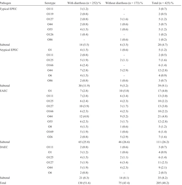
A total of 205/425 (48.2%) isolates were submitted to serotyping of somatic antigen (O), and the results are pre-sented in Table 6. We observed that the classic serogroups of EPEC and EAEC were the most frequently isolated, in agreement with the results obtained with the virulence fac-tors and especially with the phenotypes in cellular assays. The serogroups O125, O111, O44, O55 and O127 appeared as possibly associated with diarrhoea, because we found that most of these serogroups were isolated from children with acute gastroenteritis, in agreement with the distribu-tion of categories of diarrhoeagenicE. coliisolated (Ta-ble 6). EAEC isolates were mostly in the O44, O55, O111, O125 and O127 serogroups (Table 6).
Discussion
The pathogenic profile of enteroadherentE. colihas been studied throughout the world. We studied the frequen-cies of EPEC, EAEC and DAEC in faecal samples from children with and without diarrhoea in Porto Velho, Ron-donia Brazil. Screening for the genetic eaeA marker showed that 128E. coliisolates possessed this characteris-tic factor of both typical and atypical EPEC. This number of EPEC represented 11.4% of the total isolates, showing a presence of EPEC approximately twice that found preview study (Nakhjavaniet al., 2013) in a screen for thisE. coli group among 412 isolates obtained from 612 stool samples of children with diarrhoea in Tehran, Iran. The 90 samples that only had theeaeAgene belonged to the group of atypi-cal EPEC, representing about 70% of all EPEC, corroborat-ing the proportion of atypical EPEC presented by preview study (Nakhjavaniet al., 2013).
Virulence genes associated with EnteroadherentE. coli
Pathogen (nº of colonies) eaeA bfp eagg aggA aafA daaE
Typical EPEC (38) 38 (100%) 38 (100%) 0 (0%) 0 (0%) 0 (0%) 0 (0%)
Atypical EPEC (90) 90 (100%) 0 (0%) 10 (11.1%) 0 (0%) 0 (0%) 0 (0%)
EAEC (164) 0 (0%) 0 (0%) 41 (25%) 32 (19.5%) 16 (9.7%) 0 (0%)
DAEC (68) 0 (0%) 0 (0%) 0 (0%) 0 (0%) 0 (0%) 6 (8.8%)
Table 4- Adhesion pattern ofE. coliisolated from children with and without diarrhea in Porto Velho, RO, Brazil.
No. of isolates (%) in children
Adhesion pattern Total (n = 877) With diarrhoea (n = 470) Without diarrhoea (n = 407)
Typical EPEC (AL) 38 (4.3) 26 (5.5) 12 (2.9)
Atypical EPEC (ALL) 90 (10.2) 55 (11.7) 35 (8.5)
EAEC (AA) 164 (18.7) 91 (19.3) 73 (17.9)
DAEC (AD) 68 (7.7) 35 (7.4) 33 (8.1)
Total 360 (41.0) 207 (44.0) 153 (37.5)
Table 5- Phenotypic and genotypic frequencies found in enteroaggregativeE. colianalysed among children with (470) and without (407) diarrhoea from Porto Velho, RO, Brazil.
Virulence Markers Target With diarrhoea Without diarrhoea Value p
AA Adherent Pattern 91 (19.4%) 73 (17.8%) 0.5
aggA Fimbrial subunity AAF/I 31 (6.5%) 1 (0.2%) 0.00006
aafA Fimbrial subunity AAF/II 8 (1.7) 8 (1.9%) 0.7
astA East Toxin 9 (1.9%) 8 (1.9%) 0.9
Total 174 (37.0) 121 (29.7%) 0.02
Table 6- Serotyping of somatic antigen (O) of enteroaggregativeE. coliisolated from children with and without diarrhoea from Porto Velho, RO, Brazil.
Pathogen Serotype With diarrhoea (n = 252) % Without diarrhoea (n = 173) % Total (n = 425) %
Typical EPEC O111 3 (1.2) - 3 (0.7)
O119 2 (0.8) - 2 (0.5)
O127 2 (0.8) 3 (1.6) 5 (1.2)
O44 2 (0.8) 1 (0.6) 3 (0.7)
O55 4 (1.5) 1 (0.6) 5 (1.2)
O126 1 (0.4) - 1 (0.2)
O86 - 1 (0.6) 1 (0.2)
Subtotal 14 (5.5) 6 (3.5) 20 (4.7)
Atypical EPEC O1 4 (1.5) 1 (0.6) 5 (1.2)
O111 2 (0.8) - 2 (0.5)
O125 5 (1.9) 2 (1.1) 7 (1.6)
O166 6 (2.4) - 6 (1.4)
O44 7 (2.8) 5 (2.9) 12 (2.8)
O6 4 (1.5) - 4 (0.9)
O86 2 (0.8) 1 (0.6) 3 (0.7)
Subtotal 30 (11.9) 9 (5.2) 39 (9.1)
EAEC O1 7 (2.8) 10 (5.8) 17 (4.0)
O111 7 (2.8) 6 (3.4) 13 (3.0)
O125 6 (2.4) 4 (2.3) 10 (2.2)
O127 10 (3.9) 3 (1.7) 13 (3.0)
O166 6 (2.3) 4 (2.3) 10 (2.2)
O44 12 (4.8) 9 (5.2) 21 (4.8)
O55 6 (2.3) 3 (1.7) 12 (2.8)
O8 4 (1.5) 1 (0.6) 5 (1.2)
O169 5 (1.9) 1 (0.6) 6 (1.4)
O26 2 (0.8) 5 (2.9) 7 (1.6)
Subtotal 65 (25.8) 46 (26.6) 111 (26.2)
DAEC O111 2 (0.8) 1 (0.6) 3 (0.7)
O1 3 (1.2) 1 (0.6) 4 (0.9)
O125 4 (1.5) 2 (1.1) 6 (1.4)
O127 5 (1.9) 6 (3.4) 11 (2.5)
O44 5 (1.9) 4 (2.3) 9 (2.1)
O6 2 (0.8) - 2 (0.5)
Subtotal 21 (8.3) 14 (8.1) 35 (8.2)
The screen for the EAEC adherence factor showed that among 164 samples with the AA pattern, only 41 iso-lates were presented positive for at least one EAEC viru-lence marker; however, those 123 that were negative for these markers, were positive for the AA adhesion pattern in HEp-2 cells, demonstrating the specificity of the test cells. Within the EAEC samples, 32 possessed theaggAgene and 16 possessed theaafAgene.
We did not find an association between DAEC and diarrhoea. In contrast, studies conducted in the state of Espirito Santo and Northeast Brazil showed the diarrhoea-genic E. colimost prevalent in children with acute diar-rhoea was DAEC (Scaletsky et al., 2002b; Spano et al., 2008).
Regarding the DA adhesion pattern, the results showed low specificity for thedaaE gene. Studies have compared different methods to characterize enteroadherent E. coli. With regard to tests carried out for detection of DAEC, the use of a probe for detection of thedaaCgene se-quence showed low sensitivity (64.3%), confirming fre-quencies found by other investigators (Scaletsky et al., 2002a).
This study identified a large number of isolates that did not amplify with specific markers; however, the adhe-sion phenotypic test was able to identify the different ad-herence profiles, although more tests must be performed to identify possible adhesins associated with this pattern of adherence. The association between atypical EPEC and EAEC indicates a possible phenotype only observed when the two groups are associated with diarrhoea. In this study, the presence of theaggA(AAF/I) gene was associated with diarrhoea. We found 31 isolates positive for theaggAgene (6.5%) linked to patient cases, while only one patient (0.2%) of controls had this gene (p = 0.00006). A recent study has shown similar results, finding a higher frequency of theaggA(AAF/I) gene isolated from cases of diarrhoea (Pivaet al., 2003). We observed that the presence of the fimbriae AAF/I in our EAEC isolates is statistically associ-ated with diarrhoea. Studies performed in Spain showed that the presence of fimbriae AAF/I was not observed in EAEC samples; however, AAF/II was detected in 8.7% of the EAEC isolated (Vilaet al., 2000). Studies in southwest Nigeria showed the presence of AAF/I in 63% of isolated EAEC; 35% had AAF/II (Okekeet al., 2000). Similar re-sults were found in studies in India (Kahaliet al., 2004). In contrast other study (Eliaset al., 1999) detected AAF/I and AAF/II in 19% and 8%, respectively, of EAEC samples in São Paulo. Studies performed in Gabon showed that the presence of the AAF I and AAF II fimbriae was more likely in EAEC than non-EAEC isolates (Presterlet al., 2003). From these data we conclude that the prevalence of AAF seems to vary in relation to geographic region (Flores and Okhuysen, 2009).
The distribution of the serogroups found revealed a diversity of strains, which did not allow us to infer the
ac-tual prevalence of a specific serogroup in the population studied. In this study, we observed a higher frequency of the O111, O125, O44, O55 and O127 serogroups (Table 6); we emphasize the importance of serogroups since they are described as associated with pathogenic strains ofE. coli.
In conclusion, EAEC are the main cause of diarrhoea in children from Porto Velho, RO, Brazil and the presence ofaggAis highly correlated with this disease. EPEC is the second most common cause and when the same child is in-fected by both EAEC and atypical EPEC, a higher number of isolates of the latter present the LAL adherence pattern in HEp2 cells. In contrast to other regions in Brazil, DAEC was a significant contributor to diarrhoea cases in this city in the Brazilian western Amazon. In the studied city a more specific prevalent serogroup in paediatric diarrhoea was not identified. Therefore, the need to understand the mecha-nisms involved in the pathogenicity of these strains should be emphasized, especially when we consider the associa-tions identified, recalling that bacterial pathogenicity is a property contributed to by multiple factors that are ex-pressed in their natural environment, including horizontal gene transmission. Going forward, other experimental models should be used for complementary phenotypic and genomic studies.
Acknowledgments
The authors thank the Brazilian National Council for Scientific and Technological Development (CNPq).
References
Abba K, Sinfield R, Hart CAet al.(2009) Pathogens associated with persistent diarrhoea in children in low and middle in-come countries: Systematic review. BMC Infect Dis 9:88. Arenas-Hernández MMP, Martínez-Laguna Y, Torres AG (2012)
Clinical implications of enteroadherent escherichia coli. Curr Gastroen Rep 14:386-394.
Bardiau M, Szalo M, Mainil JG (2010) Initial adherence of EPEC, EHEC and VTEC to host cells. Vet Res 41:57.
Benz I, Schmidt MA (1992a) AIDA-I, the adhesin involved in dif-fuse adherence of the diarrhoeagenic Escherichia coli strain 2787 (O126:H27), is synthesized via a precursor molecule. Mol Microbiol 6:1539-1546.
Benz I, Schmidt MA (1992b) Isolation and serologic characteriza-tion of AIDA-I, the adhesin mediating the diffuse adherence phenotype of the diarrhea-associated Escherichia coli strain 2787 (O126:H27). Infect Immun 60:13-18.
Campos LC, Vieira MA, Trabulsi LRet al.(1999) Diffusely ad-hering Escherichia coli (DAEC) strains of fecal origin rarely express F1845 adhesin. Microbiol Immunol 43:167-170. Chandra BK, Singh G, Taneja Net al. (2012) Diarrhoeagenic
Escherichia coli as a predominant cause of paediatric noso-comial diarrhoea in India. J Med Microbiol 61:830-836. Contreras CA, Ochoa TJ, Ruiz Jet al.(2012) Genetic diversity of
Cravioto A, Tello A, Navarro Aet al.(1991) Association of Esch-erichia coli HEp-2 adherence patterns with type and dura-tion of diarrhoea. Lancet 337:262-264.
Elias WP, Suzart S, Trabulsi LRet al.(1999) Distribution of aggA and aafA gene sequences among Escherichia coli isolates with genotypic or phenotypic characteristics, or both, of enteroaggregative E. coli. J Med Microbiol 48:597-599. Flores J, Okhuysen PC (2009) Enteroaggregative Escherichia coli
infection. Curr Opin Gastroen 25:8-11.
Gunzburg ST, Tornieporth NG, Riley LW (1995) Identification of enteropathogenicEscherichia coliby PCR-bases detection of the bundle forming pilus gene. J Clin Microbiol 33:1375-1377.
Huang DB, Mohanty A, DuPont HLet al.(2006) A review of an emerging enteric pathogen: EnteroaggregativeEscherichia coli. J Med Microbiol 55:1303-1311.
Humphries RM, Armstrong GD (2010) Sticky situation: Local-ized adherence of enteropathogenic Escherichia coli to the small intestine epithelium. Future Microbiol 5:1645-1661. Kahali S, Sarkar B, Rajendran Ket al.(2004) Virulence
character-istics and molecular epidemiology of enteroaggregative
Escherichia coliisolates from hospitalized diarrheal patients in Kolkata, India. J Clin Microbiol 42:4111-4120.
Nakhjavani FA, Emaneini M, Hosseini Het al.(2013) Molecular analysis of typical and atypical enteropathogenic Esche-richia coli (EPEC) isolated from children with diarrhoea. J Med Microbiol 62:191-195.
Ochoa TJ, Contreras CA (2011) Enteropathogenic Escherichia coli infection in children. Curr Opin Infect Dis 24:478-483. Okeke IN, Lamikanra A, Czeczulin Jet al.(2000) Heterogeneous virulence of enteroaggregative Escherichia coli strains iso-lated from children in Southwest Nigeria. J Infect Dis 181:252-260.
Orlandi PP, Magalhães GF, Matos NBet al.(2006) Etiology of diarrheal infections in children of Porto Velho (Rondonia, Western Amazon region, Brazil). Braz J Med Biol Res 39:507-517.
Pass MA, Odedra R, Batt RM (2000) Multiplex PCRs for identifi-cation of Escherichia coli virulence genes. J Clin Microbiol 38:2001-2004.
Piva IC, Pereira AL, Ferraz LRet al.(2003) Virulence markers of enteroaggregative Escherichia coli isolated from children and adults with diarrhea in Brasília, Brazil. J Clin Microbiol 41:1827-1832.
Presterl E, Zwick RH, Reichmann Set al.(2003) Frequency and virulence properties of diarrheagenic Escherichia coli in children with diarrhea in Gabon. Am J Trop Med Hyg 69:406-410.
Scaletsky ICA, Fabbricotti SH, Aranda KRet al.(2002a) Com-parison of DNA hybridization and PCR assays for detection of putative pathogenic enteroadherent Escherichia coli. J Clin Microbiol 40:1254-1258.
Scaletsky ICA, Fabbricotti SH, Carvalho RLBet al.(2002b) Dif-fusely adherent Escherichia coli as a cause of acute diarrhea in young children in northeast Brazil: A case-control study. J Clin Microbiol 40:645-648.
Servin AL (2005a) Pathogenesis of Afa/Dr Diffusely Adhering Escherichia coli. Clin Microbiol Rev 18:264-292.
Servin AL (2005b) Pathogenesis of Afa/Dr diffusely adhering Escherichia coli. Clin Microbiol Rev 18:264-292.
Spano LC, Sadovsky ADI, Segui PNet al.(2008) Age-specific prevalence of diffusely adherent Escherichia coli in Brazil-ian children with acute diarrhoea. J Med Microbiol 57:359-363.
Torres AG, Zhou X, Kaper JB (2005) Adherence of diarrheagenic Escherichia coli strains to epithelial cells. Infect Immun 73:18-29.
Vila J, Vargas M, Henderson IRet al.(2000) Enteroaggregative
Escherichia coli virulence factors in traveler’s diarrhea strains. J Infect Dis 182:1780-1783.
Weintraub A (2007) Enteroaggregative Escherichia coli: Epide-miology, virulence and detection. J Med Microbiol 56:4-8. Yamamoto T, Echeverria P (1996) Detection of the
entero-aggregativeEscherichia coliheat-stable enterotoxin 1 gene sequences in enterotoxigenicE. colistrains pathogenic for humans. Infect Immun 61:1441-1445.
Associate Editor: Nilton Erbet Lincopan Huenuman