Secondary Metabolite Found in both Filamentous and
Unicellular Marine Strains
Pedro N. Lea˜o1,2*., Margarida Costa1., Vitor Ramos1
, Alban R. Pereira2, Virgı´nia C. Fernandes3, Valentina F. Domingues3, William H. Gerwick2,4, Vitor M. Vasconcelos1,5, Rosa´rio Martins1,6,7
1CIIMAR/CIMAR – Interdisciplinary Centre of Marine and Environmental Research, University of Porto, Porto, Portugal,2Scripps Institution of Oceanography, University of California, San Diego, La Jolla, California, United States of America,3REQUIMTE, Instituto Superior de Engenharia, Instituto Polite´cnico do Porto, Porto, Portugal,4Skaggs School of Pharmacy and Pharmaceutical Sciences, University of California, San Diego, La Jolla, California, United States of America,5Department of Biology, Faculty of Sciences, University of Porto, Porto, Portugal,6Centre of Health and Environmental Research – CISA, Superior School of Health Technology of Porto, Polytechnic Institute of Porto, Vila Nova de Gaia, Portugal,7Institute for Molecular and Cell Biology – IBMC, University of Porto, Porto, Portugal
Abstract
Cyanobacteria are widely recognized as a valuable source of bioactive metabolites. The majority of such compounds have been isolated from so-called complex cyanobacteria, such as filamentous or colonial forms, which usually display a larger number of biosynthetic gene clusters in their genomes, when compared to free-living unicellular forms. Nevertheless, picocyanobacteria are also known to have potential to produce bioactive natural products. Here, we report the isolation of hierridin B from the marine picocyanobacteriumCyanobiumsp. LEGE 06113. This compound had previously been isolated from the filamentous epiphytic cyanobacterium Phormidium ectocarpi SAG 60.90, and had been shown to possess antiplasmodial activity. A phylogenetic analysis of the 16S rRNA gene from both strains confirmed that these cyanobacteria derive from different evolutionary lineages. We further investigated the biological activity of hierridin B, and tested its cytotoxicity towards a panel of human cancer cell lines; it showed selective cytotoxicity towards HT-29 colon adenocarcinoma cells.
Citation:Lea˜o PN, Costa M, Ramos V, Pereira AR, Fernandes VC, et al. (2013) Antitumor Activity of Hierridin B, a Cyanobacterial Secondary Metabolite Found in both Filamentous and Unicellular Marine Strains. PLoS ONE 8(7): e69562. doi:10.1371/journal.pone.0069562
Editor:Brett Neilan, University of New South Wales, Australia
ReceivedFebruary 22, 2013;AcceptedJune 10, 2013;PublishedJuly 29, 2013
Copyright:ß2013 Lea˜o et al. This is an open-access article distributed under the terms of the Creative Commons Attribution License, which permits unrestricted use, distribution, and reproduction in any medium, provided the original author and source are credited.
Funding:PNL, MC and VR acknowledge funding from FCT – Foundation for Science and Technology (SFRH/BPD/70233/2010, BTI/PTDC/MAR/102638/2008/2010025 and SFRH/BD/80153/2011, respectively). This research was partially supported by the European Regional Development Fund (ERDF) through the COMPETE -Operational Competitiveness Programme and national funds through FCT, under the projects PEst-C/MAR/LA0015/2011, PTDC/MAR/102258/2008 and PTDC/ MAR/102638/2008. WHG acknowledges funding from the National Institutes of Health CA100851. The funders had no role in study design, data collection and analysis, decision to publish, or preparation of the manuscript.
Competing Interests:The authors have declared that no competing interests exist.
* E-mail: pleao@ciimar.up.pt
.These authors contributed equally to this work.
Introduction
Marine cyanobacteria have been shown to produce a diverse array of biologically significant natural products with activity in models for anticancer, neuromodulatory and anti-inflammatory drug discovery, and other areas [1]. Benthic, filamentous forms, in particular members of the classical (botanical) orders Oscillatoriales and Nostocales, have been the major sources of secondary metabolites reported from marine cyanobacteria [2]. This is in part explained by the fact that filamentous and colonial cyanobacteria appear to have larger genomes and thus can better accommodate sizable polyketide and non-ribosomal peptide pathways than picocyanobacteria [3,4]. These classes of biosynthetic products make up the majority of secondary metabolites isolated from cyanobacteria thus far [2], although a previously unrecognized capacity to produce ribosomally-encoded modified peptides has recently been described [5,6]. Another consideration is that some filamentous and colonial cyanobacteria grow to relatively high densities in coastal ecosystems, such as in mats or macroscopic tufts, thus yielding
enough biomass for chemical investigations from environmental samples.
Conversely, unicellular cyanobacteria usually need to be cultured in order to produce sufficient biomass for chemical and biological studies. The lack of ‘‘chemistry-ready’’ environmental samples and the difficulty to bring certain strains into laboratory culture may have skewed our perception of the richness of such smaller genome size cyanobacteria in terms of secondary metabolite production. As an example, the marine picocyanobac-teriumProchlorococcus, which is widespread in open ocean waters, produces a complex set of polycyclic lantipeptides [6]. The unicellular, not-yet-cultured symbiotic cyanobacterium Prochloron didemni is also known to produce a diverse array of secondary metabolites, including cyclic peptides and unusual fatty acids [7]. It should be noted that both of these examples report metabolites of a ribosomal origin, which may be a trend in picocyanobacteria as these more compact biosynthetic gene clusters may be better accommodated in small genomes.
Synechococcusand Synechocystisstrains isolated from the Portuguese coast [8]. A recent survey of cyanobacterial genomes [4] indicated the presence of biosynthetic gene clusters, mainly of the polyketide synthetase (PKS) and bacteriocin variety, among picocyanobac-teria generaCyanobium,SynechococcusandProchlorococcus.
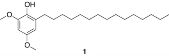
Here, we report the NMR-based isolation of hierridin B (1, Fig. 1) from the picocyanobacteriumCyanobiumsp. LEGE 06113 which had been isolated from the Atlantic coast of Portugal. The structure of1was confirmed by NMR and MS analyses. To our knowledge, this is the first report of a secondary metabolite from this cyanobacterial genus. Compound 1 had previously been isolated from the marine filamentous cyanobacteriumPhormidium ectocarpistrain SAG 60.90 and showed antiplasmodial activity [9]. We show that both cyanobacterial strains possess polyketide synthase (PKS) biosynthetic machinery, which is predicted to be involved in the biosynthesis of hierridin B. To further investigate the biological properties of this metabolite, we treated a panel of eight human cell lines to this compound. Interestingly, when tested up to a maximum concentration of 30mg mL21 (82.3mM),
hierridin B was only active against the colorectal adenocarcinoma cell line HT-29.
Results
Isolation and identification of hierridin B
A crude lipophilic extract fromCyanobium sp. LEGE 06113 was fractionated using vacuum-liquid chromatography (VLC). The1H NMR spectrum (500 MHz, CDCl3) of one of the most non-polar fractions contained two sharp singlets at d3.85 and d3.76, suggestive of aromatic methoxy groups, which led us to further investigate this fraction and ultimately obtain compound 1after purification by reversed-phase (RP) HPLC. Identification of the compound was initially based on comparison of the 1H NMR data with literature values for hierridin B and for one extended chain analogue, which had been previously reported [9] (Fig. S1). The length of the aliphatic chain of the isolated compound could not, however, be rigorously derived from the integration of the 1H NMR signals corresponding to the methylene envelope atd1.30-1.23. Consequently, GC-MS data
of the compound were acquired (no ionization was observed under our standard LC-ESI-MS conditions) and the identity of the purified metabolite was confirmed as hierridin B, due to the characteristic ions observed at m/z364 [C23H40O3], m/z168 [C9H12O3] (aromatic moiety following benzylic fragmentation and McLafferty rearrangement) and m/z 167 (benzylic moiety) (Fig. 2). Papendorf et al. [9] had used mass spectrometry data to aid in the structural characterization of metabolite1 following its partial purification from P. ectocarpi SAG 60.90. In the previous work withP. ectocarpi, the hierridins were obtained as a 1:1 mixture of 1 (pentadecyl-containing) and its heptadecyl analogue, both of which yielded the same fragment mass atm/z
168, but for which a molecular ion was observed atm/z392 for the larger compound. We saw no evidence for the
heptadecyl-containing metabolite in our materials fromCyanobiumsp. LEGE 06113, nor could we find evidence for the presence of any other structurally related compound.
Morphological and phylogenetic analyses
Cells fromCyanobiumsp. LEGE 06113 (Fig. 3A) are pale blue-green, spherical to shortly rod-shaped, with a diameter of 0.960.1mm (n = 20). Based on these morphological
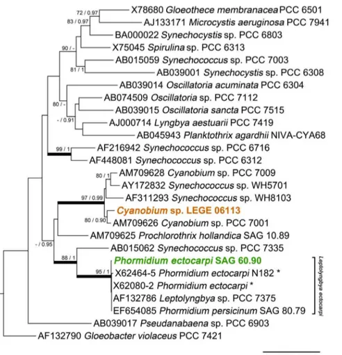
character-istics, the cyanobacterium was identified as Cyanobium sp., according to Komare´k and Anagnostidis [10]. This identifica-tion is also in accordance with the criteria of the Bergey’s Manual of Systematic Bacteriology [11]. Since hierridin B had been previously reported from the filamentous cyanobacterial strain P. ectocarpi SAG 60.90 (see morphology comparison in Fig. 3), we performed a 16S rRNA gene phylogenetic analysis to clarify the evolutionary relatedness between the two hierridin B producing cyanobacterial strains. From the phylogenetic tree (Fig. 4), it is clear thatCyanobium sp. LEGE 06113 is located in the same clade as other picocyanobacteria. This strain forms a sub-clade with the reference strain
Cyanobium sp. PCC 7001; interestingly, the PCC 7001 sample was also isolated primarily from intertidal sediments [11]. This analysis also identifies that Phormidium ectocarpi SAG 60.90 is very closely related to other marine, phycoerythrin-rich filamentous cyanobacteria, including the reference strain
Leptolyngbyasp. PCC 7375 (cluster 4, [11,12]; formerly assigned to P. ectocarpi [13]). However, it is also clear that all of the above strains are encompassed within a wider clade that also includes the picocyanobacteriumSynechococcussp. PCC 7335. It should be noted that the clustering of the two clades that comprise each of the two hierridin B producers has no phylogenetic strength, because it lacks statistical support (ML bootstrap value of 44%) (Fig. 4).
Presence of PKS genes
We have employed a PCR-based approach to detect the presence of PKS-related genes, by using degenerate primers targeting ketosynthase (KS) domains [14]. This allowed for the detection of one type I PKS gene fragment inCyanobiumsp. LEGE 06113 and two type I PKS gene fragments in P. ectocarpi SAG 60.90. The amino acid sequence identity between the two P. ectocarpiPKS genes was 54%. The identities between theCyanobium
sp. and each of the twoP. ectocarpiPKS gene sequences, also at the amino acid level, were of 45% and 47%, respectively. From NaPDoS phylogenetic analysis [15], the sequences fromP. ectocarpi
were found to bear homology and cluster with the KS domains from trans-AT type I PKSs VirF, CurF, JamJ and LnmJ (virginiamycin, curacin A, jamaicamides A and B, and leinamycin biosynthetic clusters, respectively). The sequence fromCyanobium
sp. was shown to cluster with the starter KS domains from the biosynthetic gene clusters of curacin A (CurA) and jamaicamides A and B (JamE).
Biological activity
While antiplasmodial activity was reported for 1 as a 1:1 mixture with its two-carbon longer analogue [9], any information regarding its cytotoxicity towards human cells was not available. Therefore, we tested pure hierridin B to a panel of eight human cell lines (Table 1). When evaluated up to a maximum concentration of 30mg mL21 (82.3mM), cytotoxicity was ob-served only to colon adenocarcinoma HT-29 cells (Table 1). A concentration-response assay (up to 100mg mL21) revealed its IC50as 100.2mM in this cell line (Fig. S2).
Discussion
Biogenesis
Feeding studies coupled with intermediate analysis by Horper and Marner [16,17] on miconidin and some of its derivatives (including miconidin methyl ether 2 (Fig. 5), a shorter chain analogue of1) provided evidence for the polyketide nature of these metabolites, isolated from the plant Primula obconica [17]. The biosynthesis of 2 is thus proposed [16] to involve a triketoacid
(2,4,6-trioxoundecanoic acid), which is cyclized, dehydrated and decarboxylated to yield 5-pentylbenzene-1,3-diol. This pathway intermediate leads to a variety of end products, including 2, following methylation and oxidation. Another end-product of this putative pathway is the benzoquinone primin (3, Fig. 5), produced by the same plant [17]. Analogous long-chain quinones have been reported from the plant kingdom; for example, 2-methoxy-6-pentadecyl-1,4-benzoquinone (4, Fig. 5) fromIris pseudacorusseeds [18] features a pentadecyl chain, as seen in1. Belamcandol A (5,
Figure 2. GC-MS analysis of purified 1.Top panel – total ion chromatogram (TIC). Bottom panel – mass spectrum of the main chromatographic peak (RT = 28.1 min), with the diagnostic ions for hierridin B atm/z364 and 168 clearly seen.
Fig. 5), found in the seeds of Belamcanda chinensis [19], is an unsaturated analogue of1. All of these long-chained metabolites generally can be explained by the biogenetic model put forward by Horper and Marner [16] for2and3. A similar biosynthetic origin is thus envisioned for4,5and1. In the present study, however, no evidence was found for the presence of any other end products or
intermediates that could originate from an analogous biosynthetic route to 1. Still, taking into account the small amount of compound1 that we were able to purify, such metabolites may not have been detected during our isolation procedures. The alkyl side chain in2 and3 has been proposed [16] to originate from acetyl-CoA which is polymerized by a PKS assembly line, rather than hexanoic acid being used as a starter unit. The recently reported biosynthetic route to the cylindrocyclophanes in the filamentous heterocystous cyanobacterium Cylindrospermum licheni-forme [20] involves an alkylresorcinol intermediate (6) that is structurally related to hierridin B. In this case, decanoic acid is used as the starter unit. The concerted action of two type I PKSs leads to a diketo intermediate, which is then cyclized by a type III PKS to6[20]. In the case of1, one can only speculate on whether palmitic acid primes this biosynthetic pathway, or whether a PKS is involved in the constructing the entire carbon framework. A polyketide origin for1is further supported by the fact that both hierridin B-producing cyanobacteria bear type I PKS machinery. Nevertheless, we were unable to find highly homologous KS domains shared by the two organisms, which would strengthen this hypothesis. It is unlikely that the PKS gene found inCyanobium
sp. LEGE 06113 is involved in the same biosynthetic transforma-tion as any of the PKS genes found in the P. ectocarpi strain. However, it is possible that the three recovered KS domains
Figure 3. Microphotographs of the two cyanobacterial strains known to produce 1.A –Cyanobiumsp. LEGE 06133; B –Phormidium ectocarpiSAG 60.90. Scale bar = 20mm.
doi:10.1371/journal.pone.0069562.g003
Figure 4. Maximum likelihood tree of 16S rRNA gene sequences from selected cyanobacteria and from the two strains known to produce metabolite 1.The nodal support values indicated near internal branches were determined by ML and BI methods, respectively; bootstrap values (for ML) below 70% and posterior probability values (for BI) below 0.90 were omitted. Thick branch lines indicate clades (or sub-clades) that are supported by simultaneous$85% bootstrap values and$0.95 bayesian posterior probabilities. The tree was rooted usingChloroflexus aurantiacus J-10-fl (D38365) as the outgroup, which was removed for clarity. Asterisks indicate concatenated sequences.
correspond to different portions of the putative hierridin B pathway. The fact that all of these domains show highest homology to KS domains involved in curacin A and jamaicamides biosynthesis is consistent with such a scenario.
Phylogenetic inferences
Our phylogenetic analysis, motivated by the aim of under-standing how related the two hierridin B producer strains are to one another, revealed that Cyanobium sp. LEGE 06113 and
Phormidium ectocarpiSAG 60.90 are clearly located in two distinct clades. Although these two clades emerged from the analysis as being more closely related to one another than with the other cyanobacterial lineages (Fig. 4), a common evolutionary lineage could not be established. Thus, it is improbable that the biosynthesis of this compound is deeply entrenched in the two lineages since their divergence. In fact, the findings of Shih et al. [4], point out that the genomes of some reference strains included in our phylogenetic study do not contain polyketide gene clusters, such as Synechococcus sp. PCC 7335 and the filamentous cyano-bacterium Prochlorothrix hollandica PCC 9006 (axenic strain, co-identical with SAG 10.89 [21]). Both of these latter strains are placed between the two sub-clades that include the hierridin B producers. By contrast, Synechococcussp. WH5701 andCyanobium
sp. 7001 (which belongs to the same clade asCyanobiumsp. LEGE 06113), andLeptolyngbyasp. PCC 7375 (closely related toP. ectocarpi
SAG 60.90) do possess PKS clusters. Hence, as hypothesized for other secondary metabolites produced by cyanobacteria [22,23,24], horizontal transfer of the entire gene cluster may be responsible for the presence of the hierridin biosynthetic machin-ery in these two distinct phylotypes. It will thus be of considerable interest, in particular from the perspective of secondary metabolite evolution, to study the distribution of hierridin B among cyanobacteria. However, to achieve this it will be necessary to identify the cluster of genes involved in its biosynthesis.
Biological activity
Papendorf et al. [9] isolated compound 1 guided by its antiplasmodial activity (low micromolar IC50). In the same study, the compound was tested but did not show antibacterial or anti-algal activity. It was also shown that1had no significant inhibitory activity in in vitro bioassays using HIV-1-reverse transcriptase or tyrosine kinase [9]. Antiplasmodial activity has been reported for the related metabolites3[25] and miconidin [26]. In this work, compound 1 showed selective activity, although of modest potency, towards the HT-29 cell line. The basis for this selectivity
is, at the moment, unclear, but may indicate the existence of a unique target in this cell line and the possibility of a favorable therapeutic window. Still, the related benzoquinone irisquinone [18] did not reveal any such selectivity in the NCI-60 cancer cell line screening program (NSC Number 614642), in which it showed an average potency in the low-to-mid micromolar range. Other benzoquinones, such as3and its longer-chain analogue, 2-methoxy-6-heptyl-1,4-benzoquinone, have been associated with anticancer activity [27]. To our knowledge, no putative natural roles have been put forward for1. It has been found, however, that naturally occurring concentrations of miconidin and3are able to effectively deter feeding by insects [28]. Cyanobacteria serve as prey for several insect larvae, and members of the cyanobacterial generaFischerellaandHapalosiphonhave been reported to produce insecticidal metabolites [29]. However, the active components of these latter cyanobacteria bear no structural relatedness to1.
In conclusion, we report here the isolation and cytotoxicity of a bioactive secondary metabolite, hierridin B (1), from a marine picocyanobacterium, Cyanobium sp. LEGE 06113. Interestingly, this compound had been previously isolated from a phylogenet-ically distant filamentous cyanobacterium. Future efforts to elucidate the biosynthetic route to 1 will be of importance not only in the study of the evolution and distribution of this and other
Figure 5. Selected natural products with structures related to that of compound 1. Miconidin methyl ether (2), primin (3), 2-methoxy-6-pentadecyl-1,4-benzoquinone (4), belamcandol A (5) and the resorcinol monomer of cylindrocyclophane F (6) are shown. doi:10.1371/journal.pone.0069562.g005
Table 1.Cytotoxicity of1towards a panel of human cell lines. (+growth inhibition observed,2no growth inhibition observed).
Cell line Type Growth inhibition (IC50)
HepG2 hepatocellular carcinoma 2(n/a)
HT-29 colon adenocarcinoma +(100.2mM)
MG63 osteosarcoma 2(n/a)
PNT2 normal prostate epithelium 2(n/a)
RKO colon adenocarcinoma 2(n/a)
SHSY5Y neuroblastoma 2(n/a)
SKBR3 breast adenocarcinoma 2(n/a)
T47D breast ductal carcinoma 2(n/a)
related natural products, but also to better understand the pathway cyanobacteria use to create these structures and to which end they are produced (i.e. from the eco-physiological point of view). BecauseCyanobiumspp. in general, and the strain used here in particular, exhibit high growth rates in laboratory cultures, and are thus being considered for biotechnological applications (e.g. [30,31]), metabolite1can be considered as an easily accessible by-product fromCyanobiumsp. LEGE 06113 biomass that may add to the value of its large scale culture.
Materials and Methods
Cyanobacterial strains
Cyanobiumsp. LEGE 06113 was isolated from a sand sample in a
Sabellariasp. reef collected in the lower tide zone of an intertidal rocky shore (Aguda beach, Portugal, N 41u02958,350, W 8u399
19,220). Field sampling at this location does not require, according to Portuguese law, any specific permission. In addition, the sampling did not involve any endangered or protected species. A monocyanobacterial culture of this strain is maintained in our culture collection (LEGE). For biomass production and subsequent chemical investigations, Cyanobium sp. LEGE 06113 cells were grown in 4 L cultures in Z8 medium [32] supplemented with 20 g L21NaCl, under a light/dark cycle of 14:10 h and a photon irradiance of approximately 30mmol m22s21. Cells were har-vested by centrifugation from nine of such cultures, rinsed with deionized water and freeze-dried.Phormidium ectocarpiSAG 60.90 was acquired from the Culture Collection of Algae at Goettingen University (SAG) and cultured in MN medium [33], under the light and temperature conditions described above. Microphoto-graphs and morphometric measures of cultures of the two cyanobacterial strains were acquired in a BX41 light microscope equipped with a DP72 digital camera and Cell B image analysis software (Olympus, Tokyo, Japan).
DNA extraction, amplification, sequencing and phylogenetic analysis
Biomass from exponential-phase, small-scale cultures of the two studied strains, Cyanobiumsp. LEGE 06113 andP. ectocarpi SAG 60.90 was harvested (2 mL) by centrifugation. Total genomic DNA (gDNA) was extracted from the resulting biomass pellets using a commercial kit (PureLinkTM Genomic DNA Mini Kit, Invitrogen, Carlsbad, CA, USA) and according to the manufac-turer’s instructions. A portion of the 16S rRNA gene was amplified by PCR with primers CYA 359F [34] and 1494Rc [35]. The primer pair 27F [35] and 781R [34] was used to amplify a different portion of the same gene inP. ectocarpiSAG 60.90, while we were unable to obtain this amplicon fromCyanobiumsp. LEGE 06113. To amplify polyketide synthase (PKS) genes, primers DKF and DKR [14], which target the ketosynthase (KS) domain, were used. The amplified fragments were purified from the excised agarose gel slices (Cut&Spin columns, GRiSP, Porto, Portugal), cloned (pGEM-TH Easy vector, Promega, Madison, WI, USA), and transformed into OneShotH TOP10 cells (Invitrogen). Following purification (GenEluteTM Plasmid Miniprep Kit, Sigma-Aldrich, St. Louis, MO, USA), plasmid DNA was sequenced (Macrogen, Inc., Seoul, Korea) using M13 primers. The resulting sequences were inspected for quality and deposited in GenBank (16S rRNA gene – accession numbers KC469577 and KC469578; PKS genes – accession numbers KF010866 to KF010868).
A 16S rRNA gene-based phylogenetic tree was constructed to infer the evolutionary relationship between the two hierridin B producing strains. Several available cyanobacterial sequences from
unicellular and filamentous non-heterocystous ‘‘Bergey’s reference strains’’ [11] were included in the analysis. Sequences from the best BLASTn hits in GenBank forPhormidium ectocarpiSAG 60.90 were also included in the multiple sequence alignment, which was performed in MEGA version 5 software package [36] using the ClustalW algorithm. The jModelTest 2.1.1 program [37] was used to evaluate which model of evolution best-fits our dataset. The corrected Akaike information criterion (AICc) was used to choose the optimal model of nucleotide substitution, which allowed us to select the GTR+C+I model (gamma shape parameter = 0.3860; number of gamma categories = 4; proportion of invariant sites = 0.3470). MrBayes version 3.1.2 [38] was then used for Bayesian inference (BI), using the same model and computing the parameter values indicated above. The phylogenetic tree recon-struction was performed using a random starting tree, while one cold and seven incrementally heated chains (temperature set 0.2) were run for 107generations, in two independent runs, with a tree sampling frequency of 100. The resulting consensus phylogeny was built from the last 75% of trees. MEGA 5 was also used to build a maximum-likelihood (ML) bootstrap tree (1000 replicates). The tree was reconstructed by using the same sequence alignment, model of evolution and parameters as the used for the Bayesian inferred tree. Gaps and ambiguously aligned positions were excluded (‘complete deletion’ option) from the analyses. The topologies retrieved from the two analyses were then evaluated using TreePuzzle 5.2 [39]. All the test comparisons indicated that the ML tree yielded the best topology for the dataset. The branch support values derived from the ML and BI analyses were then compared using TreeGraph 2 [40].
The PKS gene sequences obtained from both cyanobacterial strains were translated and submitted to the software tool NaPDoS (Natural Product Domain Seeker) [15], to identify the KS domains. The default parameters for KS domains were used in the analyses.
Purification and dereplication of 1
Freeze-dried biomass fromCyanobiumsp. LEGE 06113 (,10 g,
d.w.) was repeatedly extracted with a warm (,40uC) mixture of CH2Cl2:MeOH (2:1). The resulting crude extract (,2.7 g) was
subjected to normal-phase (silica gel 60, 0.015–0.040 mm, Merck KGaA, Damstadt, Germany) VLC, using a gradient from 100% hexane to 100% EtOAc to 100% MeOH. Nine fractions (A-I) were obtained and inspected by 1H NMR (500 MHz, Varian Inova). Fraction B (18.5 mg), putatively containing aromatic methoxy groups, was selected for further purification by semi-preparative RP-HPLC. The chromatographic system consisted of two 515 pumps and a 996 PDA detector (Waters, Milford, MA, USA), fitted with a Synergi Fusion-RP column (10mm,
250610 mm, Phenomenex, Torrance, CA, USA). A gradient program (flow of 3 mL min21) was used for the separation, from 85% MeCN (aq) to 100% MeCN for 30 min, then 100% MeCN for 13 min, to afford metabolite1(0.6 mg, pale yellow amorphous solid, RT = 24.8 min). The 1H NMR (500 MHz, CDCl3) spec-trum of1is available in Fig. S1.
Dereplication of known marine secondary metabolites was carried out with MarinLit (February 2011 update), by querying for the two methoxy groups and for a 1,2,3,5-tetrasubstituted aromatic moiety that was deduced from the meta-coupling (J= 2.8 Hz) of the two aromatic protons.
GC-MS analysis
an AS-3000 autosampler. A ZB-XLB capillary column (30 m60.25 mm60.25mm) from PhenomenexH (Torrance, CA, USA) was selected for chromatographic separation. Ultra-pure grade Helium (Linde So´gas, Lisbon, Portugal; purity$99.999%) was used as a carrier gas (flow 1.0 mL min21). The system was controlled by Xcalibur software, Version 1.3 (Thermo Fisher Scientific). Injections (2mL) of1at a concentration of 2 mg L21 were carried out in the splitless mode and injector temperature was 250uC. The compound was detected by mass spectrometry using full scan mode (m/z80-500).
The column oven temperature was programmed as follows: initial temperature of 150uC and increased by 6uC min21 to 295uC (held for 4 min). The mass spectrometer was operated in electron ionization (EI) mode at 70 eV with an external ionization source. A temperature of 285uC was used for the ion source and the electron multiplier was set at 1750 V (autotune to gain of 16107).
Cell lines and cytotoxicity assays
All cell lines included in the study are of human origin. Hepatocellular carcinoma cell line HepG2, colon adenocarcinoma cell line HT-29, neuroblastoma cell line SH-SY5Y, breast carcinoma cell line T47D and the normal prostate cell line PNT2 were purchased from Sigma-Aldrich. SKBR3 breast adenocarcinoma and RKO colon carcinoma (ATCC) were a kind gift from Helena Fernandes (Faculdade de Medicina Denta´ria, University of Porto) and MG-63 osteosarcoma was obtained from the ATCC. Tumor cells were cultured in Dulbecco’s modified Eagle medium (DMEM Glutamax) and the normal prostate cell line was cultured in minimal essential medium (a-MEM), supplemented with 10% fetal bovine serum (FBS), 2.5mg mL21 fungizone, penicillin-streptomycin (100 IU mL21 and 100mg mL21, respectively). Cells were incubated in a humidified atmosphere with 5% of CO2, at 37uC.
The cellular viability was evaluated by the reduction of the 3-(4,5-dimethylthiazole-2-yl)-2,5-diphenyltetrazolium bromide (MTT) [41]. Cells were seeded in 96-well culture plates at a concentration of 104cells cm22. After 24 h of adhesion, cells were exposed to 100mL fresh medium supplemented with1to a final concentration of 3 and 30mg mL21, for a period of either 24, 48
and 72 hours. After incubation, cells were exposed to 10mL of 0.5 mg/ml MTT. Following exposure, purple-colored formazan salts were dissolved in 100ml DMSO and the absorbance
measured at 550 nm in a microplate reader (Synergy HT, Biotek, USA). All tests were run in triplicate and averaged. A similar procedure was undertaken for determining the IC50value of1in the cell line HT-29. In this case, however, cells were incubated for 48 h and the final concentrations of 1 in the culture medium ranged from 0.03 to 100mg mL21. Dose-response data was used
to calculate the IC50 with the software GraphPad Prism v5.0 (GraphPad Software, La Jolla, CA, USA).
Supporting Information
Figure S1 1H NMR spectrum (500 MHz, CDCl3) of hierridin B (1). Insert shows comparison with previously reported 1H NMR data (300 MHz, CDCl3) for the compound (Papendorf et al., 1998,Phytochemistry49:2383–2386).
(TIF)
Figure S2 Cytotoxicity dose-response curve of HT-29 cells exposed to hierridin B (1) (IC50= 100.2mM).
(TIF)
Author Contributions
Conceived and designed the experiments: PNL MC VR ARP VCF VFD WHG VV RM. Performed the experiments: PNL MC VR ARP VCF. Analyzed the data: PNL MC VR ARP VCF VFD WHG VV RM. Wrote the paper: PNL MC VR ARP VCF VFD WHG VV RM.
References
1. Nunnery JK, Mevers E, Gerwick WH (2010) Biologically active secondary metabolites from marine cyanobacteria. Curr Opin Biotechnol 21: 787–793. 2. Tidgewell K, Clark BR, Gerwick WH (2010) The natural products chemistry of
Cyanobacteria. In: Mander L, Lui H-W, editors. Comprehensive Natural Products II Chemistry and Biology. Oxford: Elsevier. pp. 141–188. 3. Jones AC, Monroe EA, Eisman EB, Gerwick L, Sherman DH, et al. (2010) The
unique mechanistic transformations involved in the biosynthesis of modular natural products from marine cyanobacteria. Nat Prod Rep 27: 1048–1065. 4. Shih PM, Wu D, Latifi A, Axen SD, Fewer DP, et al. (2013) Improving the
coverage of the cyanobacterial phylum using diversity-driven genome sequenc-ing. Proc Natl Acad Sci USA 110: 1053–1058.
5. Donia MS, Ravel J, Schmidt EW (2008) A global assembly line for cyanobactins. Nat Chem Biol 4: 341–343.
6. Li B, Sher D, Kelly L, Shi Y, Huang K, et al. (2010) Catalytic promiscuity in the biosynthesis of cyclic peptide secondary metabolites in planktonic marine cyanobacteria. Proc Natl Acad Sci USA 107: 10430–10435.
7. Donia MS, Fricke WF, Partensky F, Cox J, Elshahawi SI, et al. (2011) Complex microbiome underlying secondary and primary metabolism in the
tunicate-Prochloronsymbiosis. Proc Natl Acad Sci USA 108: E1423–E1432.
8. Martins R, Fernandez N, Beiras R, Vasconcelos V (2007) Toxicity assessment of crude and partially purified extracts of marineSynechocystisandSynechococcus
cyanobacterial strains in marine invertebrates. Toxicon 50: 791–799. 9. Papendorf O, Ko¨nig GM, Wright AD (1998) Hierridin B and
2,4-dimethoxy-6-heptadecyl-phenol, secondary metabolites from the cyanobacteriumPhormidium ectocarpiwith antiplasmodial activity. Phytochemistry 49: 2383–2386. 10. Koma´rek J, Anagnostidis K (1998) Cyanoprokaryota, 1.Teil: Chroococcales; Ettl
H, Ga¨rtner G, Heynig H, Mollenhauer D, editors. Jena-Stuttgart-Lu¨beck-Ulm: Gustav Fischer. 548 p.
11. Castenholz RW (2001) Phylum BX. Cyanobacteria: Oxygenic Photosynthetic Bacteria. In: Boone DR, Castenholz RW, editors. Bergey’s Manual of Systematic Bacteriology. New York: Springer. pp. 473–553.
12. Wilmotte A, Herdman M (2001) Phylogenetic Relationships Among the Cyanobacteria Based on 16S rRNA Sequences. In: Boone DR, Castenholz RW, editors. Bergey’s Manual of Systematic Bacteriology. New York: Springer. pp. 487–493.
13. Rippka R, Deruelles J, Waterbury JB (1979) Generic assignments, strain histories and properties of pure cultures of cyanobacteria. J Gen Microbiol 111: 1–61. 14. Moffitt MC, Neilan BA (2001) On the presence of peptide synthetase and
polyketide synthase genes in the cyanobacterial genus Nodularia. FEMS Microbiol Lett 196: 207–214.
15. Ziemert N, Podell S, Penn K, Badger JH, Allen E, et al. (2012) The Natural Product Domain Seeker NaPDoS: A Phylogeny Based Bioinformatic Tool to Classify Secondary Metabolite Gene Diversity. PLoS ONE 7: e34064 16. Horper W, Marner FJ (1996) Biosynthesis of primin and miconidin and its
derivatives. Phytochemistry 41: 451–456.
17. Horper W, Marner FJ (1995) Phenols and Quinones from Leaves ofPrimula obconica. Nat Prod Lett 6: 163–170.
18. Marner FJ, Horper W (1992) Phenols and Quinones from Seeds of DifferentIris
Species. Helv Chim Acta 75: 1557–1562.
19. Fukuyama Y, Okino J, Kodama M (1991) Structures of Belamcandol A and B Isolated from the Seed ofBelamcanda chinensis. Chem Pharm Bull (Tokyo) 39: 1877–1879.
20. Nakamura H, Hamer HA, Sirasani G, Balskus EP (2012) Cylindrocyclophane Biosynthesis Involves Functionalization of an Unactivated Carbon Center. J Am Chem Soc 134: 18518–18521.
21. Pinevich A, Velichko N, Ivanikova N (2012) Cyanobacteria of the genus
Prochlorothrix. Front Microbiol 3: 173.
22. Mikalsen B, Boison G, Skulberg OM, Fastner J, Davies W, et al. (2003) Natural variation in the microcystin synthetase operon mcyABC and impact on microcystin production inMicrocystisstrains. J Bacteriol 185: 2774–2785. 23. Gu L, Wang B, Kulkarni A, Geders TW, Grindberg RV, et al. (2009)
Metamorphic enzyme assembly in polyketide diversification. Nature 459: 731– 735.
24. Tidgewell K, Engene N, Byrum T, Media J, Doi T, et al. (2010) Evolved diversification of a modular natural product pathway: Apratoxins F and G, two cytotoxic cyclic depsipeptides from a palmyra collection ofLyngbya bouillonii. ChemBioChem 11: 1458–1466.
26. SepulvedaBoza S, Cassels BK (1996) Plant metabolites active againstTrypanosoma cruzi. Planta Med 62: 98–105.
27. Gunatilaka AAL, Berger JM, Evans R, Miller JS, Wisse JH, et al. (2001) Isolation, synthesis, and structure-activity relationships of bioactive benzoqui-nones fromMiconia lepidotafrom the Suriname rainforest. J Nat Prod 64: 2–5. 28. Bernays E, Lupi A, Bettolo RM, Mastrofrancesco C, Tagliatesta P (1984)
Antifeedant Nature of the Quinone Primin and Its Quinol Miconidin from
Miconiaspp. Experientia 40: 1010–1011.
29. Lea˜o PN, Engene N, Antunes A, Gerwick WH, Vasconcelos V (2012) The chemical ecology of cyanobacteria. Nat Prod Rep 29: 372–391.
30. Mendes LBB, Cunha PCR, D’oca MGM, Abreu PC, Primel EG (2011) Method for removing pollutants from produced water. U.S. Patent 7955505 31. Blank CE, Hinman NW (2012) Production of Cyanobacterial or Algal Biomass
Using Chitin as a Nitrogen Source. U.S. Patent Application 20120244603 A1 32. Kotai J (1972) Instructions for the preparation of modified nutrient solution Z8
for algae. Norwegian Institute for Water Research, Blindern, Oslo 11/69: 5 pp. 33. Rippka R (1988) Isolation and purification of cyanobacteria. In: Packer L, Glazer AN, editors. Methods Enzymol. San Diego: Academic Press. pp. 3–27. 34. Nu¨bel U, GarciaPichel F, Muyzer G (1997) PCR primers to amplify 16S rRNA
genes from cyanobacteria. Appl Environ Microbiol 63: 3327–3332.
35. Neilan BA, Jacobs D, DelDot T, Blackall LL, Hawkins PR, et al. (1997) rRNA sequences and evolutionary relationships among toxic and nontoxic cyanobac-teria of the genusMicrocystis. Int J Syst Bacteriol 47: 693–697.
36. Tamura K, Peterson D, Peterson N, Stecher G, Nei M, et al. (2011) MEGA5: Molecular Evolutionary Genetics Analysis Using Maximum Likelihood, Evolutionary Distance, and Maximum Parsimony Methods. Mol Biol Evol 28: 2731–2739.
37. Darriba D, Taboada GL, Doallo R, Posada D (2012) jModelTest 2: more models, new heuristics and parallel computing. Nat Methods 9: 772–772. 38. Huelsenbeck JP, Ronquist F (2001) MRBAYES: Bayesian inference of
phylogenetic trees. Bioinformatics 17: 754–755.
39. Schmidt HA, Strimmer K, Vingron M, von Haeseler A (2002) TREE-PUZZLE: maximum likelihood phylogenetic analysis using quartets and parallel comput-ing. Bioinformatics 18: 502–504.
40. Stover BC, Muller KF (2010) TreeGraph 2: Combining and visualizing evidence from different phylogenetic analyses. BMC Bioinformatics 11: 7.