Crystal Structure of Allophycocyanin from
Marine Cyanobacterium
Phormidium
sp.
A09DM
Ravi Raghav Sonani1☯, Gagan Deep Gupta2, Datta Madamwar1
*, Vinay Kumar2☯
*
1BRD School of Biosciences, Vadtal Road, Satellite Campus, Sardar Patel University, Vallabh Vidyanagar, India,2Protein Crystallography Section, Solid State Physics Division, Bhabha Atomic Research Centre, Mumbai, India
☯These authors contributed equally to this work.
*[email protected](DM); [email protected](VK)
Abstract
Isolated phycobilisome (PBS) sub-assemblies have been widely subjected to X-ray crystal-lography analysis to obtain greater insights into the structure-function relationship of this light harvesting complex. Allophycocyanin (APC) is the phycobiliprotein always found in the PBS core complex. Phycocyanobilin (PCB) chromophores, covalently bound to conserved Cys residues ofα- andβ- subunits of APC, are responsible for solar energy absorption from phy-cocyanin and for transfer to photosynthetic apparatus. In the known APC structures, hetero-dimers ofα- andβ- subunits (known asαβmonomers) assemble as trimer or hexamer. We here for the first time report the crystal structure of APC isolated from a marine cyanobacteri-um (Phormidiumsp. A09DM). The crystal structure has been refined against all the observed data to the resolution of 2.51Åto Rwork(Rfree) of 0.158 (0.229) with good stereochemistry of the atomic model. ThePhormidiumprotein exists as a trimer ofαβmonomers in solution and in crystal lattice. The overall tertiary structures ofα- andβ- subunits, and trimeric quaternary fold of thePhormidiumprotein resemble the other known APC structures. Also, configuration and conformation of the two covalently bound PCB chromophores in the marine APC are same as those observed in fresh water cyanobacteria and marine red algae. More hydropho-bic residues, however, constitute the environment of the chromophore bound toα-subunit of thePhormidiumprotein, owing mainly to amino acid substitutions in the marine protein.
Introduction
Phycobiliproteins (PBPs) and linker proteins (LPs) are mutually arranged in cyanobacteria and red algae to form multi-molecular assemblies of around 4–8 MDa called phycobilisomes (PBS) [1–3]. Morphologically, PBS are composed of two major sub-structures with core sit-uated on outer surface of thylakoid membrane and rods radiating out from core [4–6]. Rods contain phycocyanin (PC, absorptionλmax*610–620 nm) and/or phycoerythrin (PE, absorp-tionλmax*540–570 nm), and associated LPs. The core invariantly contains allophycocyanin
a11111
OPEN ACCESS
Citation:Sonani RR, Gupta GD, Madamwar D, Kumar V (2015) Crystal Structure of Allophycocyanin from Marine CyanobacteriumPhormidiumsp. A09DM. PLoS ONE 10(4): e0124580. doi:10.1371/ journal.pone.0124580
Academic Editor:Alexander Wlodawer, NCI-Frederick, UNITED STATES
Received:January 31, 2015
Accepted:March 3, 2015
Published:April 29, 2015
Copyright:© 2015 Sonani et al. This is an open access article distributed under the terms of the
Creative Commons Attribution License, which permits unrestricted use, distribution, and reproduction in any medium, provided the original author and source are credited.
Data Availability Statement:The coordinates and structure factors have been deposited in the Protein Data Bank with the accession no. 4RMP Partial amino acid sequence of the subunits have been submitted to GenBank (GenBank accession nos., CDY72720, CDY72721).
Funding:The authors have no support or funding to report.
(APC, absorptionλmax*653 nm) and associated LPs [3]. Phycobilisomes harvest the sunlight and exhibit efficient energy transfer in the direction from PE!PC!APC!chlorophyll[7]. The energy absorbed bychlorophyllis used to split water molecules generating electrons and protons in the photosynthetic reaction centers. It has recently been shown that phycobilisomes supply energy to both the photosystems I and II [8].
Structural assemblies of different phycobiliproteins within PBS were found to be quite similar, despite divergence in the amino acid sequence [9]. The heterodimer (αβmonomer) of two homologousα- andβ- subunits of PE, PC and APC proteins is the building block of PBS. Each subunit contains covalently attached non-cyclic tetrapyrrole chromophore(s) en-abling the PBPs to absorb and emit light within specific wavelength range across solar spec-trum. Threeαβmonomers associate into the disc shaped trimers, which have often been observed to form hexamers forming the rods and core cylinders [9,10]. Linker peptides occu-py the central cavity of 25 to 50 Å diameter within the rods/core cylinders and play central role in tethering up the PBPs together in a way that governs the expression of unique spectral properties of chromophores within PBS [11,12]. The energy absorbed by the chromophores of PC and PE is funneled through APC discs and linker proteins tochlorophyll. The unique red-shifted absorbances of APC chromophores and their fluorescence emission overlapping to that ofchlorophyllhas been found to play major connecting role in this light funneling phenomenon [13].
Allophycocyanin is bound to the same chromophore as that of phycocyanin, namely phyco-cyanobilin (PCB), and these interactions result in distinct 650 nm absorption band of APC. The red shift in the absorption of allophycocyanin bound PCB has been proposed to occur due to its surrounding protein micro-environment within the quaternary structure of the protein [14]. Also, configuration and conformation of the chromophores are thought to contribute in modulating the chromophore energies bound to different phycobiliproteins [3]. To elucidate the molecular interactions of chromophores and APC, a number of X-ray diffraction studies have been reported for diverse APC proteins isolated from fresh water cyanobacteria and ma-rine red algae [15–18].
Potential applications of phototrophic cyanobacteria for the generation of renewable energy by optimizing their photosynthetic pathways have been discussed [19–20]. Clearly, the spec-tral range over which organism is able to capture sunlight and efficiency of transfer to the pho-tosynthetic apparatus could play critical role in exploring its utility in capturing solar energy. It has also been established that PBS using cyanobacteria are more abundant in nutrient rich waters with highchlorophyllconcentrations [21,22]. Intuitively, a marine cyanobacterium, adapted to low light, could be expected to be efficient in light capture and energy transfer to the downstream molecular machinery.
In the present paper, we report the first crystal structure of APC isolated from the marine cyanobacterium,Phormidiumsp. A09DM (formerly known asLyngbyasp. A09DM). The overall trimeric quaternary fold of thePhormidiumprotein resembles the other known APC structures from red algae and fresh water cyanobacteria. However, more hydrophobic residues in the microenvironment of the PCB chromophore bound toα-subunit of the marine cyano-bacterium are observed in the crystal structure.
Materials and Methods
Cultivation of organism for APC production
Phormidiumsp. A09DM, isolated from rocky shores of Okha, Gujarat, India, was used as a source of APC. Set of standard laboratory conditions described elsewhere [23–26] were used for the cultivation ofPhormidiumsp. A09DM. In brief, they were grown in artificial salt nutri-ent (ASN)—III medium under cool white light (36 W, 130μmol photons m-2s-1, 12:12 hours light: dark cycles) at 27 ± 2°C.
Purification and characterization of
Phormidium
APC
The exponentially growing cyanobacterial cells (28 days after inoculation) were harvested by centrifugation at 3000 ×g for 15 min (Kubota 6500, Bunkyo-Ku, Tokyo, Japan) at 20°C. Allophycocyanin was isolated and purified from pelleted cell mass using the method de-scribed earlier [27,28] with slight modification. Briefly, pelleted cell mass was washed and re-suspended in five volumes of the extraction buffer (10 mM Tris-HCl, pH 8.1) for freeze-thaw cycles from -25°C to 4°C. The brick red supernatant, obtained after centrifugation, was subjected to 40% ammonium sulfate precipitation in the presence of 0.01% Triton X-100 to precipitate non-targeted proteins. Saturation of supernatant was further increased to 70% to pellet down the APC protein. Protein pellet was suspended in 10 mM Tris—HCl buffer (pH, 8.1) and APC was purified by passing through gel permeation column (350 × 10 mm) packed with Sephadex G-150 by using 10 mM Tris-HCl buffer (pH, 8.1) as mobile phase. Allophycocyanin rich fractions were pooled and loaded onto DEAE-cellulose anion ex-changer (Column dimension: 40 × 10 mm) to achieve crystallization grade purity. Bound proteins were eluted with NaCl step gradient in 10 mM Tris-HCl buffer (pH, 8.1). Highly pure APC fractions were desalted and concentrated by ultra-filtration using a Macrosep (10 kDa MWCO centrifugal device, Pall Corporation). Purification was carried out at 4°C. Buffers used in the protocol were prepared in mili-Q water supplemented with 0.01% sodi-um azide.
Purified APC was assessed for purity, homogeneity, integrity and functionality by well-established standard biochemical methods [29–31]. Polyacrylamide gel electrophoresis (PAGE) was performed in non-denaturing as well as denaturing conditions as described by Singh et al. [29]. The resolved gels were stained according to Garfin [32]. Presence of APC was confirmed by bilin specific zinc-acetate staining [33], which appeared as orange fluorescence bands under UV trans-illumination (AlphaEase FC Imaging System, Alpha Innotech Corp., USA). The mas-ses of APC component proteins were verified by MALDI-TOF mass spectrometry.
Spectroscopic properties of purified APC were investigated by UV-visible absorption and fluorescence emission spectroscopy as described previously [34]. Absorbance of the protein was recorded over 250–750 nm wavelength range on UV-Visible Spectrophotometer (Analy-tik Jena AG Specord 210, Germany) at 25°C. Protein purity was inferred from UV-visible spectrum using‘purity ratio’that was calculated from absorbance ratio, A653/A280. Protein concentration was estimated according to the method of Lowry et al. [35] with BSA used as the standard. Allophycocyanin content was estimated from UV—visible spectrum by using the equation of Bennet and Bogorad [36]. To adjudge the functional integrity, fluorescence emission spectrum of APC was traced upon excitation at 645 nm by fluorescence spectropho-tometer (F-7000, Hitachi High Technologies, Japan).
oligomeric status of the protein was estimated from the molecular weight calculated from the elution profiles of the APC protein and molecular weight markers.
Sequence determination
Genes of allophycocyaninα- andβ- subunits (apcAandapcB, respectively) were amplified in 30μL PCR reactions consisting of 1X buffer (10 mM Tris pH 9.0, 50 mM KCl, 1.5 mM MgCl2, 0.1% Triton X-100), 0.33 mM each of dNTPs, ~100 ng of template DNA, 1.5 U of Taq DNA poly-merase and 0.66 pmoles each of primers (Table 1). The primers used were designed on the basis of conserved nucleotide sequences ofapcAandapcBgenes available in UniProt database. Ampli-fication program was set with 35 cycles of 1 min denaturation step (94°C), 1 min annealing step (55°C), and 1 min elongation step (72°C) using Biorad iCycler version 4.006 (Biorad, CA, USA). The 35 cycles were appended with initial denaturation step at 94°C of 5 min and a final extension step at 72°C for 20 min. The ~0.5 kb PCR products ofapcAandapcBwere sequenced by the auto-mated DNA Analyzer 3730xl using BigDye Terminator v3.1 sequencing chemistry (Applied Bio-systems, Foster City, CA). Partial gene sequences were submitted to NCBI GenBank with the accession nos.LM993862, LM993863. Partial amino acid sequences of theα- andβ- subunits were deduced from these gene sequences (GenBank accession nos., CDY72720, CDY72721).
Crystallization of the protein and data collection statistics
Crystallization ofPhormidiumAPC (4 mg/mL) was attempted using sitting—drop vapor
diffusion method and formulated crystallization screens, including PEG, JCSG+, PACT and MBClass suites from Qiagen. The protein yielded crystals with a number of conditions con-taining PEG 6000 (16–20%) at pH of about 8. Crystals of different morphologies were ob-served to grow in the same wells of JCSG+ conditions containing additives like ZnCl2and MgCl2. The diffraction experiments showed a strong tendency of twinning in some of these crystals, while other hexagonal plates diffracted poorly. Diffraction quality crystals of the protein were obtained by sitting-drop method using Intelli-well (ARI, Hampton) crystalliza-tion plates and JCSG+ crystallizacrystalliza-tion condicrystalliza-tion containing 100 mM bicine (pH, 8.5) and 20% (w/v) PEG 6000 at 15°C. The crystals grew to the size of about 120x100x60μm3in 15 days. For diffraction experiments at 100 K, these crystals were flash cooled after immersing in 20% glycerol. These crystals belonged to space groupH32 and diffracted to about 2.5 Å resolution using fine-focus X-ray source (Microstar, Bruker) under cryo-conditions. The 3-dimensional diffraction intensity data from the crystals ofPhormidiumAPC were acquired on an image plate detector (MARRESEARCH) using Cu Kαradiation with 1.0° oscillation per image. The data were processed using XDS/autoPROC [37].
Structure solution and refinement
The initial phases forPhormidiumAPC crystals were obtained by the molecular replacement method using Phaser suite [38] and atomic coordinates of anαβmonomer of the red algae
Table 1. List of designed primers for amplification ofapcA andapcB genes.
Gene Primer Sequence (5’–3’)
apcA (485 bp) Forward primer ATGAGTATCGTCACTAAATCCATCG
Reverse primer TACTGCATTGCACCGACAAC
apcB (485 bp) Forward primer ATGCAAGACGCAATTACTTCCG
Reverse primer TAGCTTAAGCCAGAGCAGATGTAG
APC (PDB ID: 1KN1), without solvents and phycocyanobilin chromophores, as search model. The initial phases with the molecular replacement solution were reasonably accurate such that electron density for most of the known amino acids ofα- andβ- subunits (GenBank accession nos., CDY72720, CDY72721) could be identified in the electron density maps. The electron density could also be seen for the PCB chromophores (CYC) at lower contour levels. Only partial amino acid sequences ofα(9–161) andβ(1–155) subunits ofPhormidium allophyco-cyanin were available from nucleotide sequencing experiments. Amino acid sequences for the N-terminal eight amino acids of theα-subunit and for the C-terminal six amino acids of theβ -subunit were derived from the consensus sequence of allophycocyanin proteins available in NCBI. These residues and four corrections in the nucleotide derived amino acid sequence were subsequently validated in the refined structure. The individual atomic coordinates were refined by Phenix [39] with maximum likelihood target and with model building using COOT [40]. The covalent bonds between the Cys residues and CYC ligands were restrained to distance of 1.80Å in Phenix cycles of refinement. The progress in the refinement of the atomic model was monitored by Rworkand Rfree. The atomic coordinates and structure factors have been deposit-ed in the Protein Data Bank with the accession no.4RMP.
Analysis for chromophore-protein interactions
The 3D coordinates of the trimer assembly of the known APC structures, available in PDB, were generated by PISA/COOT. The 2D projection diagrams of chromophore-protein interac-tions for thePhormidiumAPC protein and other known APC structures were generated from the 3D coordinates of trimer assembly for each of the protein using LigPlot+ software [41]. De-fault parameters (distance of 3.35Å for identifying H-bonds and maximum distance of 3.9 Å for non-bonded contacts) were used to portray H-bond interactions and hydrophobic contacts between the chromophores and protein amino acid residues. The identified protein contact residues for a chromophore were marked in the multiple sequence alignment of known struc-tures achieved using Clustal Omega [42]. The differences in the microenvironments of PCB chromophores in allophycocyanin proteins from different sources were elucidated from this alignment. In parallel, multiple sequence alignment of APC proteins available in NCBI was also carried out using COBALT [43]. The top 100 sequences homologous toPhormidiumAPC were analyzed for conservation of identified protein contact residues.
Results and Discussion
Purification and characterization
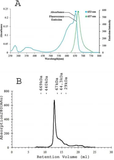
well with previously described APC absorption pattern [30,31]. APC specific peaks dominating over the protein specific peak (at 280 nm) in UV-visible spectrum again signified the absence of linker as well as other cellular protein (Fig 3A). Purity ratio, total APC content and total protein content (Table 2) collectively substantiated the success of purification protocol. Peaks at 18056 and 17987 Da were observed in MALDI-TOF experiments that were expected to be due toα -andβ- subunits (S1 Fig). The molecular mass of thePhormidiumAPC oligomer was determined to be 118 kDa based on its elution profile on the molecular sieve column (Fig 3B). Given the ob-served mass of 36043 Da for anαβmonomer, thePhormidiumprotein is expected to form tri-mer in the buffer containing 10 mM Tris-HCl (pH, 8.0) and 100 mM sodium chloride.
Crystallographic analysis
The APC crystal belonged to space groupH32, with unit-cell parameters a = b = 101.2 Å, c = 193.0 Å. Data statistics are shown inTable 3. Based on determined molecular mass ofαβ
Fig 2. PAGE analysis of the purified allophycocyanin. A) Silver stained and zinc acetate stained 15% SDS-PAGE analyses of purifiedPhormidiumAPC. Protein molecular mass standard are shown in lane M. Only two bands were observed on silver stained and zinc acetate stained SDS-PAGE. These correspond to α- andβ- subunits of the purified APC, suggesting also absence of linker peptide in purified APC complex. Nearly 10μg of APC protein was loaded in each lane.B) Silver stained and zinc acetate stained 12% Native-PAGE of purifiedPhormidiumAPC further confirm homogeneity of the purified protein.
doi:10.1371/journal.pone.0124580.g002
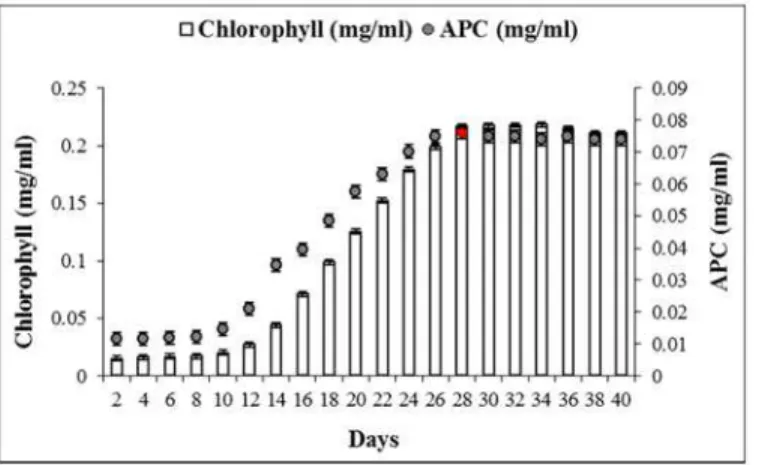
Fig 1. Growth pattern (in terms of chlorophyll, vertical bars) and APC production profile (filled circles) ofPhormidiumsp. A09DM, grown for 40 days.Allophycocyanin content was calculated using the Bennet and Bogorad [36] equations.
monomer (36043 Da) and the volume of the asymmetric unit, a Matthews parameter [44] of 2.64 Å3Da-1and a solvent content of 53.4% suggested the highest normalized probability (Ptot 1.0) for oneαβmonomer in the asymmetric unit. The molecular replacement calculations yielded a unique solution with LLG value of 313 (TFZ, 15.9). The initial molecular replacement phases were accurate to show electron density for the known amino acids and covalently bound chromophores. A total of 14 amino acid residues, those were deduced from the Fig 3.PhormidiumAPC exists as trimer ofαβmonomers in solution. A) UV-visible absorption spectrum (cyan) of purifiedPhormidiumAPC showed major band at 653 nm that suggests formation of trimer in the solution. The fluorescence emission spectrum (green) of purified APC was measured upon excitation at 645 nm.B) The Superdex 200 gel-filtration profile of thePhormidiumprotein. The APC protein elutes at volume corresponding to an oligomer of 118 kDa. The column was calibrated with gel filtration molecular weight markers (Carbonic anhydrase, 29 kDa; Ovalbumin, 44.3 kDa; Bovine serum albumin, 67 kDa; Apoferritin, 440 kDa; Bovine thyroglobulin, 669 kDa). Elution volumes of the marker proteins are shown in the Figure.
doi:10.1371/journal.pone.0124580.g003
Table 2. PhormidiumAPC protein purification progress and yield.
Purification step Total protein content (mg) APC content (mg) APC (%) Impurities (%) Purity ratio, A653/A280 Yield (%)
Crude extract 88.76 6.55 7.38 92.62 0.09 100.00
Ammonium sulfate precipitation 8.20 5.31 64.75 35.25 2.08 81.20
Chromatographic purification 4.84 4.71 97.31 2.69 5.43 71.91
consensus sequence, were also found to fit well in the electron density maps. Also, four correc-tions in the partial amino acid sequence (GenBank accession nos., CDY72720, CDY72721)
were deduced based on refined atomic parameters, electron density maps, and conservation of amino acids in the multiple sequence alignment of APC orthologs. These were also confirmed from the nucleotide sequence chromatograms using different primers. The mis-annotation of 9-VNA-11 to 9-VKA-11, 48-ERI-50 to 48-ECI-50 and 144-EDA-146 to 144-ENA-146 of theα-subunit were mainly due to weak signals in the chromatogram. While mis-annotation of 34-GEL-36 to 34-GKL-36 of theβ-subunit was due to overlapping strong signal in the chro-matogram. The molecular masses of chromophore bound subunits with improved amino acid sequences were calculated to be 18064 and 18011 Da forα- andβ- subunits, respectively. The theoretical molecular masses match well with molecular mass of 18056 and 17987 Da observed in MALDI-TOF analysis.
The refined structural model of the APC protein has Rwork(Rfree) of 0.16 (0.23) against all the observed data with F/σ(F)0 (Table 3). The electron density for all the residues and both the chromophores is clearly defined (Fig 4). The evaluation using MOLPROBITY [46] revealed good stereochemistry of the structure, with nearly 98% residues in the most favored regions of the Ramachandran plot. Residue Thr-74 of theβ-subunit lies close to disallowed region of the Ramachandran plot (withF,Cvalues of 78.3°, 137.3°) in all the known APC structures available in PDB. This residue resides on the loop region and its peptide amide accepts H-bond from the OB atom of the chromophore covalently linked with theα-subunit. Its main-chain carbonyl oxygen also accepts H-bonds from amide nitrogen atoms of Arg-77 and Tyr-88. It has been suggested that in the case of Thr-74 ofβ-subunit the specific requirements of the pro-tein fold and ligand interaction override the usual Ramachandran constrains [47]. Interestingly Table 3. Crystallographic data statistics forPhormidiumAPC.
Unit Cell 101.2, 101.2, 193.0 (Å), 90, 90, 120(°)
Space group H3 2
Matthews coefficient (Å3
/Da) 2.64
Solvent content (%)a 53.4
Resolution limits (Å) 30–2.51 (2.64–2.51)b
Unique reflections 13362
Redundancy 10.8 (10.7)b
Completeness (%) 99.9 (99.8)b
Rmerge 0.109 (0.557)b
Mean I/ meanσ(I) 22.8 (5.3)b
Refinement statistics
Resolution range (Å) 30–2.51
Wilson B (Å2) 30.6
Final Rwork/ Rfree 0.158/0.229
Ramachandaran plotc 98.7/1.0/0.3
Rotamer outliers (%) 0.4
Number of non-hydrogen atoms 2713
Root-mean-square deviation from ideality
Bond lengths (Å) 0.008
Bond angles (°) 1.65
a. Estimated from Matthews coef
ficient with one (αβ) monomer per asymmetric unit.
b. Values in the highest resolution shell.
c. Percentage residues in the Ramachandran plot, Favored/Allowed/Outliers.
also, the post-translationally modified Asn-71 (γ-N-methyasparagine, MEN) of theβ-subunit also resides in the same loop. The methylation of this asparagine contributes to the efficiency of directional energy transfer in phycobilisomes [48]. The N-methyl group of MEN is clearly seen in the electron density maps and its side chain interacts with the chromophore ofβ -sub-unit (dOδ1-NC~3Å).
Structure analysis
Theα- andβ- subunits share nearly 35% sequence identity. The structures ofα- andβ- sub-units are comprised of sevenα-helices adopting globin-like fold (SCOP, 46457) and their folds are identical to that of other known APC structures. The subunit structures match with the rms deviation of 2.5 Å for 638 equivalent main chain atoms. The two subunits form a stable dimer (αβmonomer) with burial of nearly 5090 Å2(~34%) of the solvent accessible area at the interface. The protein interfaces, surfaces and assemblies (PISA) service at EBI [49] estimated a gain of nearly 55.3 kcal/M in solvation free energy on the formation ofαβmonomer. A large number of potential H-bonds and salt-bridges at the interface also contribute towards the sta-bility ofαβmonomer.
Although only oneαβmonomer is in the asymmetric unit, a trimer of thePhormidium protein exists in the crystal structure. Also the formation of trimer in solution is expected from the observation of the prominent 653 nm absorbance band and APC elution profile on the molecular sieve column as the protein maintained in low salt buffer (10 mM Tris-HCl and 100 mM sodium chloride) migrates as 118 kDa oligomer on the molecular sieve column. It has been recently suggested that hexamers are intrinsically less stable than trimers and are easily disrupted by small changes in the structure in solution lacking stabilizing phosphate [18]. The trimer in the crystals is generated by the crystallographic symmetry. Threeαβ monomers of APC align side-by-side to form a hollow disc (Fig 5A) with an estimated gain of nearly 211 kcal/M of solvation free energy on trimer formation. The trimeric quaternary fold ofPhormidiumprotein resembles the trimers of APC observed from fresh water cyano-bacteria and red algal species.
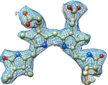
Fig 4. Representative electron density map.Shown is the fit of a PCB chromophore (ball-and-stick) in 2Fo —Fc,σA-weighted electron-density map drawn at 1.2σcontour level. The figure was prepared with Chimera
suite [45].
Two trimers interact face-to-face to form hexamers in the crystals of APC fromP.yezoensis andG.violaceus(PDB IDs: 1KN1 and 2VJT), and assembly of such hexamers into core cylinders is expected to be present in active phycobilisomes. However,PhormidiumAPC does not form ex-tended assembly in the crystals. Also, packing of trimers in the crystal lattice ofPhormidiumAPC differs from the APC structures ofS.platensis(PDB ID: 1ALL),T.vulcanus(PDB ID: 3DBJ) and S.elongatus(PDB ID: 4F0U). Its C-termini helices from threeα-chains of a trimer in a layer inter-act with three different laterally displaced trimers in another layer. A bilayer of laterally displaced trimers further forms loose association with other bilayers through their respectiveβ-subunits. The packing of trimers in the structure ofPhormidiumAPC thus matches with the interaction of trimers observed in the crystal structure ofT.elongatusAPC (PDB ID: 2V8A; [47]) resolved at 3.5Å resolution. It is interesting that crystals ofPhormidiumandT.elongatusAPC proteins were obtained under totally different conditions. Crystals ofT.elongatesAPC were obtained from 1 M ammonium sulphate, while PEG 6000 was used for crystallization of thePhormidiumprotein.
One PCB chromophore is covalently bonded to the conserved Cys-81 ofα-subunit (α -chromophore) and other chromophore is covalently linked to conserved Cys-81 ofβ-subunit. The two chromophores are separated by a distance of about 51Å (center-to-center) in anαβ monomer. The configuration and conformation of PCB chromophores in the two subunits of PhormidiumAPC do not differ from each other, as well as from the other known structures of APC from different species (Fig 6). However, microenvironments around chromophores differ markedly inα- andβ-subunits (Fig 7). Trimer formation packs the amino acid residues ofβ -subunit closer toα-chromophore, while atoms ofβ-subunit only interact with its bound chro-mophore. Theβ-subunit contributes three Tyr, three Thr and one Leu residues to the microen-vironment of theα-chromophore. Of these, Tyr-62 and Thr-66 form hydrogen bonds with the chromophore atoms (Fig 7). The bathochromic shift observed in APC upon trimerization has been attributed due to the coupling of the hydrophobicity of theα-chain chromophore envi-ronment and charged residues contributed by theβ-chain [50]. The chromophores ofα- and β- subunits in a trimer also interact with each other at shorter distance of about 10.5Å (center-to-center distance of ~21Å) (Fig 5B). Detailed analyses of the chromophore-protein interac-tions observed in thePhormidiummarine APC, and other fresh water cyanobacteria and ma-rine red algae suggest that hydrophobic contacts of theα-chromophore with Thr-75 and Tyr-78 residues ofβ-subunit and with Met-85 and Ala-129 ofα-subunit are not conserved in all the known APC structures (Fig 8). These amino acid residues approach theα-chromophore of Fig 5. Trimer formation ofαβmonomers ofPhormidiumAPC results in close interactions of the chromophores of two subunits. A)Ribbon model of the trimer. Eachαβmonomer is shown in two shades of the same color. The bound chromphores are shown as spheres.B)Chromophores (shown as spheres) are placed at 51Åapart (center-to-center distance) in anαβmonomer. Closer packing of the chromphores (dcenter-to-center~21Å) from differentαβmonomers results due to oligomerization. The figure was prepared
using Chimera suite.
PhormidiumAPC within a distance of 3.9Å. Thr-75 and Tyr-78 residues are conserved in the known APC structures, while Met-85 and Ala-129 of thePhormidiumprotein are mostly substituted by Leu and Gly in other APC orthologs. The Sδatom of Met-85 is placed at about 4.3Å from the Sγatom of Cys-81 that forms a covalent bond with theα-chromophore. The Cβ atom of Ala-129 interacts with the chromophore directly, and this interaction is not found in other known APC structures due to presence of Gly residue at the equivalent position in the alignment.
Fig 6. Configuration and conformation of the chromophores in the known APC structures.The atomic coordinates of PCB chromophores in the known APC structures (PDB IDs: 1ALL, 1KN1, 1B33, 2V8A, 2VJT, 3DBJ and 4F0U) were superposed onto the PCB chromophore ofPhormidiumAPC by Chimera suite.A) Superposed chromophores bound toα-subunitsB) Superposed chromophores bound toβ-subunits.
The protein sequence database search reveals that Met-85 is conserved inSynechococcus andCyanothecemarine cyanobacteria as well, whereas position 129 is occupied by Ala/Ser resi-dues. Interaction between theα-chromophore and 129thresidue could be expected to be pres-ent in both the marine cyanobacteria. A database resource for marine cyanobacterial sequences is not presently available. We looked at the APC homologous sequences available at NCBI. Using thePhormidiumAPC as query sequence, top 100 most closely related sequences to Phor-midiumAPC were aligned using COBALT. We observed that Met/Leu and Gly/Ala/Ser are the only residues found at positions 85 and 129 ofα-subunit, respectively. Also, several species car-rying Met and Ala/Ser at these positions were identified. These species are found both in fresh water (F) and marine water niches (M), and includeNodosilinea nodulosa(F),Leptolyngbya sp. Heron Island J (M),Leptolyngbyasp. PCC 7375 (M),Synechococcussp. PCC 7335 (M), Gloeocapsasp. PCC 7428 (F),Synechococcussp. JA-3-3Ab (F),Halothecesp. PCC 7418, Gloeo-capsasp. PCC 73106 (F),Synechococcussp. JA-2-3B'a(2–13) (F),Dactylococcopsis salina(M), Arthrospira jenneri fb(F),Stanieria cyanosphaera(F),Pleurocapsasp. PCC 7319 (M),Spirulina subsalsa(F, M),Rubidibacter lacunae(M),Synechococcussp. PCC 7336 (M),Mastigocoleus tes-tarum(M),Synechococcus sp.NKBG15041c(M),Synechococcussp. PCC 7002 (M), Cyanobac-terium stanieri(F),Cyanothecesp. PCC 7424 (F),filamentous cyanobacterium ESFC-1(M) and Cyanothece(F). Clearly structural information on phycobiliproteins from organisms adapted to different sunlight niches shall be required to elucidate if the chromophore microenviron-ment alone affects the spectral properties and energy transfer efficiency of APC in cyanobacte-ria, and if microenvironment could be targeted for engineering energy capture and energy transfer efficiency.
Conclusions
We have resolved the crystal structure of APC of marine cyanobacterium,Phormidiumsp. A09DM. The protein is observed to exist as a trimer both in solution and in crystals. The over-all tertiary structures ofα- andβ- subunits, trimeric quaternary fold, and configuration and conformation of the chromophores of the marine protein resemble the other known APC structures from red algae and fresh water cyanobacteria. However, microenvironment of the PCB chromophore bound toα-subunit is enriched by hydrophobic residues, owing to the pres-ence of Met-85 and Ala-129 residues, in thePhormidiumAPC protein.
Fig 7. A LigPlus schematic 2D representation of the chromophore-protein interactions.Residues from α-subunit (shown as XXXnnn(A)) and fromβ-subunit (shown as XXXnnn(B)) within a distance of 3.9Åfrom the chromophore atoms are displayed. Non-chromophore residues involved in hydrophobic contact(s) are shown with residue labels. Side chains of residues forming covalent and H-bonds with the chromophore atoms are shown as sticks. H-bonds are shown in green dashed lines. Chromophore bonds are shown in purple.A)Microenvironment around theα-chromophore (labeled Cyc181(A) in the figure).B)
Microenvironment around chromophore (Cyc181(B)) covalently bound to Cys-81 ofβ-subunit. The figure was prepared using LigPlus suite [41].
Supporting Information
S1 Fig. MALDI-TOF spectrum of the Phormidium APC.Two overlapping peaks were re-solved in the expanded m/z scale and these showed masses of the two APC subunits to be 17988.3 and 18055.6 Da.
(PDF)
Acknowledgments
We thank Dr. Ashok Varma, ACTREC, for the help in diffraction data acquisition and MALDI-TOF analysis.
Fig 8. Multiple sequence alignment ofα(A) andβ(B) subunits of APC orthologs, and
microenvironments of PCB chromophores in the known APC structures.APCS,Phormidium; 1ALL,
Spirulina platensis; 1KN1,Porphyra yezoensis; 2V8A,Thermosynechococcus Elongatus; 2VJT,Gloeobacter Violaceus; 3DBJ,Thermosynechococcus vulcanus; 4F0U,Synechococcus elongatusPCC 7942. Multiple sequence alignment was achieved with Clustal Omega and residues which constitute microenvironments of PCB chromophores were detected using LigPlus suite. Blue shaded residues ofα-subunit and cyan shaded residues ofβ-subunit are within non-bonded distance of 3.9Åfrom the chromophore atoms covalently linked toα-subunit. Theβ-subunit residues shaded in red are within non-bonded distance of 3.9Åfrom any of the chromophore atom covalently linked toβ-subunit. Secondary structure of the Phormidium APC, as estimated using STRIDE (http://webclu.bio.wzw.tum.de/stride/; [51]), is also shown (α,α-helices; G, 310helices; T,
Turns). The figure was prepared using Jalview [52]. Theγ-N-methylasparagine residue of theβ-subunit is marked with a black square.
Author Contributions
Conceived and designed the experiments: DM VK. Performed the experiments: RRS GDG VK. Analyzed the data: VK. Contributed reagents/materials/analysis tools: DM VK. Wrote the paper: RRS VK.
References
1. Glazer AN. Light guides Directional energy transfer in a photosynthetic antenna. J Biol Chem. 1989; 264: 1–4. PMID:2491842
2. Huber R. Nobel lecture. A structural basis of light energy and electron transfer in biology. EMBO J. 1989; 8: 2125–2147. PMID:2676513
3. MacColl RJ. Cyanobacterial phycobilisomes. Struct Biol. 1998; 124: 311–334. PMID:10049814 4. Adir N. Elucidation of the molecular structures of components of the phycobilisome: reconstructing a
giant. Photosynth Res. 2005; 85: 15–32. PMID:15977057
5. Adir N. Structure of the phycobilisome antennae in cyanobacteria and red algae. In: Fromme P, editors. Photosynthetic Protein Complexes: A Structural Approach. Wiley-VCH Verlag GmbH & Co KGaA, Weinheim; 2008. pp. 243–274.
6. Watanabe M, Ikeuchi M. Phycobilisome: architecture of a light harvesting supercomplex. Photosynth Res. 2013; 116: 265–276. doi:10.1007/s11120-013-9905-3PMID:24081814
7. Gantt E, Lipschultz CA. Energy transfer in phycobilisomes from phycoerythrin to allophycocyanin. Bio-chim Biophys Acta. 1973; 292: 858–861. PMID:4705459
8. Liu H, Zhang H, Niedzwiedzki DM, Prado M, He G, Gross ML, et al. Phycobilisomes supply excitations to both photosystems in a megacomplex in cyanobacteria. Science. 2013; 342: 1104–1107. doi:10.
1126/science.1242321PMID:24288334
9. Anderson LK, Toole CM. A model for early events in the assembly pathway of cyanobacterial phycobili-somes. Mol Microbiol. 1998; 30: 467–474. PMID:9822813
10. MacColl RJ. Allophycocyanin and energy transfer. Biochim Biophys Acta. 2004; 1657: 73–81. PMID:
15238265
11. Lundell DJ, Williams RC, Glazer AN. Molecular architecture of a light-harvesting antenna In vitro as-sembly of the rod substructures of Synechococcus 6301 phycobilisomes. J Biol Chem. 1981; 256: 3580–3592. PMID:6782105
12. David L, Marx A, Adir N. High-resolution crystal structures of trimeric and rod phycocyanin. J Mol Biol. 2011; 405: 201–213. doi:10.1016/j.jmb.2010.10.036PMID:21035460
13. Marx A, Adir N. Structural characteristics that stabilize or destabilize different assembly levels of phycocyanin by urea. Photosynth Res. 2014; 121: 87–93. doi:10.1007/s11120-014-9996-5PMID:
24687534
14. Murakami A, Mimuro M, Ohki K, Fujita Y. Absorption spectrum of allophycocyanin isolated from Ana-baena cylindrica: variation in absorption spectrum induced by changes in physico-chemical environ-ment. J Biochem. 1981; 89: 79–86. PMID:6783641
15. Brejc K, Ficner R, Huber R, Steinbacher S. Isolation, crystallization, crystal structure analysis and re-finement of allophycocyanin from the cyanobacterium Spirulina platensis at 23 A resolution. J Mol Biol. 1995; 249: 424–440. PMID:7783202
16. Reuter W, Wiegand G, Huber R, Than ME. Structural analysis at 2.2 A of orthorhombic crystals pres-ents the asymmetry of the allophycocyanin-linker complex, APLC78, from phycobilisomes of Mastigo-cladus laminosus. Proc Natl Acad Sci USA. 1999; 96: 1363–1368. PMID:9990029
17. Liu JY, Jiang T, Zhang JP, Liang DC. Crystal structure of allophycocyanin from red algaePorphyra yezoensisat 2.2 A resolution. J Biol Chem. 1999; 274: 16945–16952. PMID:10358042
18. Marx A, Adir N. Allophycocyanin and phycocyanin crystal structures reveal facets of phycobilisome assembly. Biochim Biophys Acta. 2013; 1827: 311–318. doi:10.1016/j.bbabio.2012.11.006PMID:
23201474
19. Quintana N, Van der Kooy F, Van de Rhee MD, Voshol GP, Verpoorte R. Renewable energy from Cya-nobacteria: energy production optimization by metabolic pathway engineering. Appl Microbiol Biotech-nol. 2011; 91: 471–490. doi:10.1007/s00253-011-3394-0PMID:21691792
21. Kettler GC, Martiny AC, Huang K, Zucker J, Coleman ML, Rodrigue S, et al. Patterns and implica-tions of gene gain and loss in the evolution of Prochlorococcus. PLoS Genet. 2007; 3: e231. PMID: 18159947
22. Bibby TS, Zhang Y, Chen M. Biogeography of photosynthetic light-harvesting genes in marine phyto-plankton. PLoS One. 2009; 4: e4601. doi:10.1371/journal.pone.0004601PMID:19240807
23. Waterbury JB, Stanier RY. Isolation and growth of cyanobacteria from marine and hypersaline environ-ments. In: Starr MP, Stolp H, Trüper HG, Balows A, Schlegel HG, editors. The Prokaryotes. Springer-Verlag, Berlin; 1981. pp. 221–223.
24. Soni B, Kalavadia B, Trivedi U, Madamwar D. Extraction, purification and characterization of phycocya-nin fromOscillatoria quadripunctulata- Isolated from the rocky shores of Bet-Dwarka, Gujarat, India. Process Biochemistry. 2006; 41: 2017–2023.
25. Soni B, Trivedi U, Madamwar D. A novel method for single step hydrophobic interaction chromatogra-phy for the purification of chromatogra-phycocyanin fromPhormidium fragileand its characterization for antioxidant property. Bioresour Technol. 2008; 99: 188–194. PMID:17234404
26. Parmar A, Singh NK, Kaushal A, Sonawala S, Madamwar D. Purification, characterization and compari-son of phycoerythrins from three different marine cyanobacterial cultures. Bioresour Technol. 2011; 102: 1795–1802. doi:10.1016/j.biortech.2010.09.025PMID:20889334
27. Sonani RR, Singh NK, Kumar J, Thakar D, Madamwar D. Concurrent purification and antioxidant activi-ty of phycobiliproteins fromLyngbyasp A09DM: An antioxidant and anti-aging potential of phycoery-thrin inCaenorhabditis elegans. Process Biochem. 2014a; 49: 1757–1766.
28. Sonani RR, Singh NK, Awasthi A, Prasad B, Kumar J, Madamwar D. Phycoerythrin extends life span and health span ofCaenorhabditis elegans. AGE. 2014b; 36: 1–14.
29. Singh NK, Parmar A, Madamwar D. Optimization of medium components for increased production of C-phycocyanin from Phormidium ceylanicum and its purification by single step process. Bioresour Technol. 2009; 100: 1663–1669. doi:10.1016/j.biortech.2008.09.021PMID:18954974
30. Su HN, Xie BB, Chen XL, Wang JX, Zhang XY, Zhou BC, et al. Efficient separation and purification of allophycocyanin from Spirulina (Arthrospira) platensis. J Appl Phycol. 2010; 22: 65–70.
31. Parmar A, Singh NK, Madamwar D. Allophycocyanin from a local isolateGeitlerinemasp A28DM (Cya-nobacteria): a simple and efficient purification process. J Phycol. 2010; 46: 285–289.
32. Garfin D. One-dimensional gel electrophoresis. In: Deutscher MP, Abelson JN, Simon MI, editors. Guide to protein purification. Academic Press; 1990. pp. 425–441.
33. Berkelman TR, Lagarias JC. Visualization of bilin-linked peptides and proteins in polyacrylamide gels. Anal Biochem. 1986; 156: 194–201. PMID:3526971
34. Singh NK, Parmar A, Sonani RR, Madamwar D. Isolation, identification and characterization of novel thermotolerantOccillatoriasp N9DM: change in pigmentation in response to temperature. Process Bio-chem. 2012; 47: 2472–2479.
35. Lowry OH, Rosebrough NJ, Farr AL, Randall RJ. Protein measurement with the Folin phenol reagent. J Biol Chem. 1951; 193: 265–275. PMID:14907713
36. Bennet A, Bogorad L. Complementary chromatic adaptation in filamentous blue—green algae. J Cell
Biol. 1973; 58: 419–435. PMID:4199659
37. Vonrhein C, Flensburg C, Keller P, Sharff A, Smart O, Paciorek W, et al. Data processing and analysis with the autoPROC toolbox. Acta Crystallogr D Biol Crystallogr. 2011; 67: 293–302. doi:10.1107/
S0907444911007773PMID:21460447
38. McCoy AJ, Grosse-Kunstleve RW, Adams PD, Winn MD, Storoni LC, Read RJ. Phaser crystallographic software. J Appl Cryst. 2007; 40: 658–674.
39. Adams PD, Afonine PV, Bunkóczi G, Chen VB, Davis IW, Echols N, et al. PHENIX: a comprehensive Python-based system for macromolecular structure solution. Acta Crystallogr D Biol Crystallogr. 2010; 66: 213–221. doi:10.1107/S0907444909052925PMID:20124702
40. Emsley P, Cowtan K. Coot: model-building tools for molecular graphics. Acta Crystallogr D Biol Crystal-logr. 2004; 60: 2126–2132. PMID:15572765
41. Laskowski RA, Swindells MB. LigPlot+: multiple ligand-protein interaction diagrams for drug discovery. J Chem Inf Model. 2011; 51: 2778–2786. doi:10.1021/ci200227uPMID:21919503
42. Sievers F, Wilm A, Dineen D, Gibson TJ, Karplus K, Li W, et al. Fast, scalable generation of high-quality protein multiple sequence alignments using Clustal Omega. Mol Syst Biol. 2011; 7: 539. doi:10.1038/ msb.2011.75PMID:21988835
43. Papadopoulos JS, Agarwala R. COBALT: constraint-based alignment tool for multiple protein se-quences. Bioinformatics. 2007; 23: 1073–1079. PMID:17332019
45. Pettersen EF, Goddard TD, Huang CC, Couch GS, Greenblatt DM, Meng EC, et al. UCSF Chimera—a
visualization system for exploratory research and analysis. J Comput Chem. 2004; 25: 1605–1612.
PMID:15264254
46. Chen VB, Arendall WB, Headd JJ, Keedy DA, Immormino RM, Kapral GJ, et al. MolProbity: all-atom structure validation for macromolecular crystallography. Acta Crystallogr D Biol Crystallogr. 2010; 66: 12–21. doi:10.1107/S0907444909042073PMID:20057044
47. Murray JW, Maghlaoui K, Barber J. The structure of allophycocyanin from Thermosynechococcus elongatus at 3.5 A resolution. Acta Crystallogr Sect F Struct Biol Cryst Commun. 2007; 63: 998–1002.
PMID:18084078
48. Swanson RV, Glazer AN. Phycobiliprotein methylation. Effect of the gamma-N-methylasparagine resi-due on energy transfer in phycocyanin and the phycobilisome. J Mol Biol. 1990; 214: 787–796. PMID:
2117667
49. Krissinel E, Henrick K. Inference of macromolecular assemblies from crystalline state. J Mol Biol. 2007; 372: 774–797. PMID:17681537
50. McGregor A, Klartag M, David L, Adir N. Allophycocyanin trimer stability and functionality are primarily due to polar enhanced hydrophobicity of the phycocyanobilin binding pocket. J Mol Biol. 2008; 384: 406–421. doi:10.1016/j.jmb.2008.09.018PMID:18823993
51. Heinig M, Frishman D. STRIDE: a web server for secondary structure assignment from known atomic coordinates of proteins. Nucleic Acids Res. 2004; 32: W500–502. PMID:15215436
52. Waterhouse AM, Procter JB, Martin DM, Clamp M, Barton GJ. Jalview. Version 2—a multiple sequence
alignment editor and analysis workbench. Bioinformatics. 2009; 25: 1189–1191. doi:10.1093/