Effects of Intraosseous Erythropoietin during
Hemorrhagic Shock in Swine
Vesna Borovnik-Lesjak1, Kasen Whitehouse1, Alvin Baetiong1, Yang Miao1, Brian M. Currie1, Sathya Velmurugan1, Jeejabai Radhakrishnan2, Rau´l J. Gazmuri3*
1Resuscitation Institute at Rosalind Franklin University of Medicine and Science, North Chicago, Illinois, United States of America,2Department of Medicine and Resuscitation Institute at Rosalind Franklin University of Medicine and Science, North Chicago, Illinois, United States of America,3Department of Medicine, Department of Physiology and Biophysics, and Resuscitation Institute at Rosalind Franklin University of Medicine and Science, and Critical Care Medicine at the Captain James A. Lovell Federal Health Care Center, North Chicago, Illinois, United States of America
Abstract
Objective: To determine whether erythropoietin given during hemorrhagic shock (HS) ameliorates organ injury while improving resuscitation and survival.
Methods:Three series of 24 pigs each were studied. In an initial series, 50% of the blood volume (BV) was removed in 30 minutes and normal saline (threefold the blood removed) started at minute 90 infusing each third in 30, 60, and 150 minutes with shed blood reinfused at minute 330 (HS-50BV). In a second series, the sameHS-50BVprotocol was used but
removing an additional 15% of BV from minute 30 to 60 (HS-65BV). In a final series, blood was removed as inHS-65BVand
intraosseous vasopressin given from minute 30 (0.04 U/kg min21) until start of shed blood reinfusion at minute 150
(HS-65BV+VP). Normal saline was reduced to half the blood removed and given from minute 90 to 120 in half of the animals. In
each series, animals were randomized 1:1 to receive erythropoietin (1,200 U/kg) or control solution intraosseously after removing 10% of the BV.
Results:InHS-50BV, O2consumption remained near baseline yielding minimal lactate increases, 88% resuscitability, and 60%
survival at 72 hours. InHS-65BV, O2 consumption was reduced and lactate increased yielding 25% resuscitability. In
HS-65BV+VP, vasopressin promoted hemodynamic stability yielding 92% resuscitability and 83% survival at 72 hours.
Erythropoietin did not affect resuscitability or subsequent survival in any of the series but increased interleukin-10, attenuated lactate increases, and ameliorated organ injury based on lesser troponin I, AST, and ALT increases and lesser neurological deficits in theHS-65BV+VPseries.
Conclusions:Erythropoietin given during HS in swine failed to alter resuscitability and 72 hour survival regardless of HS severity and concomitant treatment with fluids and vasopressin but attenuated acute organ injury. The studies also showed the efficacy of vasopressin and restrictive fluid resuscitation for hemodynamic stabilization and survival.
Citation:Borovnik-Lesjak V, Whitehouse K, Baetiong A, Miao Y, Currie BM, et al. (2014) Effects of Intraosseous Erythropoietin during Hemorrhagic Shock in Swine. PLoS ONE 9(11): e110908. doi:10.1371/journal.pone.0110908
Editor:Raghavan Raju, Georgia Regents University, United States of America
ReceivedMay 30, 2014;AcceptedSeptember 21, 2014;PublishedNovember 3, 2014
This is an open-access article, free of all copyright, and may be freely reproduced, distributed, transmitted, modified, built upon, or otherwise used by anyone for any lawful purpose. The work is made available under the Creative Commons CC0 public domain dedication.
Data Availability:The authors confirm that all data underlying the findings are fully available without restriction. All relevant data are within the paper and its Supporting Information files.
Funding:This research was supported by the Telemedicine and Advanced Technology Research Center (TATRC) at the U.S. Army Medical Research and Materiel Command (USAMRMC) Fort Detrick, MD under contract number: W81XWH-11-2-0019. Funding was received by RJG. The funders had no role in study design, data collection and analysis, decision to publish, or preparation of the manuscript.
Competing Interests:The authors have declared that no competing interests exist. * Email: raul.gazmuri@rosalindfranklin.edu
Introduction
Acute hemorrhage resulting from traumatic injury is responsible for a high percentage of death in military personnel engaged in combat operations [1]. A recent report including 4,596 battlefield fatalities from Operation Iraqi Freedom and Operation Enduring Freedom between October 2001 and June 2011 showed that 87.3% of all injury related deaths occurred before arriving to a medical treatment facility [2]. Of these deaths, 24.3% were deemed potentially survivable with acute mortality associated with hemorrhage in 90.9%. The current acute management of
hemorrhage focuses on hemostasis, hemodynamic stabilization, and rapid transfer to a medical treatment facility.
[20]. We hypothesized that similar benefits could be elicited in other low-flow states such as hemorrhagic shock (HS) and ameliorate organ injury improving resuscitability and survival. This hypothesis was supported by rat models of HS in which pretreatment with EPO improved survival associated with lesser reductions in mean aortic pressure and lesser increases in lactic acid, tumor necrosis factor (TNF)-a, and interleukin (IL)-6 [14] along with lesser injury to the liver and kidneys [13,14], and by studies – also in rats – showing that EPO given during HS attenuated intestinal mucosal injury and bacterial translocation [22] along with maintaining intestinal microcirculatory blood flow [23]. Although – to the best of our knowledge – the effects of EPO during HS have not been investigated in large animal models (i.e., swine, sheep, and dog), EPO has been shown to exert tissue protection in swine models of liver [15] and spinal cord [11] ischemia.
We developed a model of HS in swine – an animal higher in the phylogenic scale and thus of greater translational relevance – and investigated the effects of EPO incorporating logistic constraints expected to limit care in far forward combat operations. We used a protocol of controlled bleeding as the initial approach in a multi-year project to first characterize the effects of the proposed interventions without the confounding elements of uncontrolled bleeding and tissue injury (to be incorporated in future series). We conducted three successive series of 24 animals each in which animals were randomized 1:1 to receive EPO (1,200 U/kg) or control solution. The series had in common (a) removal of blood to a target percentage of the estimated blood volume (simulating bleeding and hemostasis in the field); (b) delivery of EPO through the intraosseous route upon removal of 10% of the animal’s blood volume (simulating early drug delivery using a low-skill technique); (c) fluid resuscitation with 0.9% NaCl (normal saline) initiated after a period of untreated HS (simulating delayed access to rescuers); (d) shed blood reinfusion at the end of HS (simulating arrival to a medical post), and (e) contingent on the series, recovery from anesthesia and 72 hour observation. The first series modeled low severity HS; the second series modeled high severity HS; and the third series modeled high severity HS with use of vasopressin to augment resuscitability while examining the role of limited fluid resuscitation.
Materials and Methods
The studies were approved by the Institutional Animal Care and Use Committee (IACUC) at Rosalind Franklin University of Medicine and Science (approval number 12–23) and by the United States Army Medical Research and Materiel Command Animal Care and Use Review Office (ACURO) and were conducted according to institutional guidelines.
Animal Housing and Husbandry
Animals were group housed in pens at the Biological Resource Facility (AAALAC accredited facility) at the Rosalind Franklin University of Medicine and Science in which lights are set at the recommended illumination levels of a 12/12-hour cycle controlled via automatic timers. Temperature was maintained at 61–81uF. Resting mats were provided and Aspen Sani-Chip bedding from a certified vendor (Harlan Laboratories, Indiana) was used. Health assessment for general health and well-being, possible injuries, or death was performed daily by animal care technicians and the day before/during/after the experiment by the investigator.
Animal Preparation
Basic Preparation. Male domestic pigs (32–48 kg, age,11 weeks) were sedated with ketamine hydrochloride (30 mg?kg21
intramuscularly). Anesthesia was induced with propofol (2 mg?kg21
through an ear vein) and the animal intubated with a size 8 tracheal tube initiating positive pressure ventilation with a volume controlled ventilator (840 Ventilator System, Nellcor Puritan Bennett, Boulder, CO) set to deliver a tidal volume of 10 mL?kg21, peak flow of 60 l
?min21, and FiO
2 of 0.5.
Respiratory rate was adjusted at baseline to maintain the end-expired PCO2(PETCO) between 35 and 45 mmHg (Capnogard,
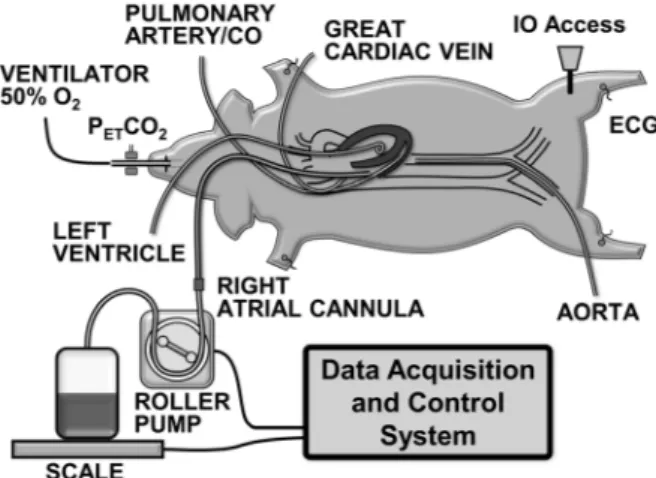
Novometrix Medical Systems, Wallingford, CT). Anesthesia was continued using isoflurane (1.75% to 2.75%) and a 1:1 mixture of nitrous oxide and oxygen. The electrocardiogram was recorded through defibrillator adhesive skin pads. All procedures were performed using sterile technique. A 7 F high-fidelity micro-tip catheter transducer (Millar Instruments, Houston, TX) was advanced through the right femoral artery into the descending thoracic aorta for pressure measurement (Figure 1). A 7 F thermodilution balloon-tipped pulmonary artery catheter was advanced through the left cephalic vein or through the left internal jugular vein (when the cephalic vein was used to access the great cardiac vein as described under Experimental Series) into the pulmonary artery for measuring core temperature and thermodi-lution cardiac output along with pressures in the right atrium and pulmonary artery. A 6 F high-fidelity micro-tip pressure transducer pigtail catheter (Millar Instruments, Houston, TX) was advanced through the surgically exposed left carotid artery for measuring left ventricular pressure. A 23 F cannula (Bio-Medicus, Medtronic, Minneapolis, MN) was advanced through the left external jugular vein into the right atrium and used for blood withdrawal into a blood transfer bag. Core temperature was maintained between 37.5uC and 38.5uC with water-circulated blanket (Blanketrol II, Cincinnati SubZero, Cincinnati, OH).
Hemorrhagic Shock Protocol
The animal’s blood volume was estimated at 60 ml/kg-body weight and a predetermined percentage was withdrawn into a heparinized transfer bag (heparin 10 U?ml21
of blood) using a roller pump (model 313S, Watson Marlow, Inc., Wilmington, MA) controlled by a custom-developed software in LabVIEW 6.0. The heparinized transfer bag was placed on an electronic scale (model 2200, Doran Scales, Inc., Batavia, IL) connected to the LabVIEW
software to gravimetrically monitor the rate of blood withdrawal (blood density = 1.06 g/ml) and automatically adjust the pump rate as needed (Figure 1). The withdrawn blood was kept in a water bath at 37.5uC until reinfusion. Resuscitation was subse-quently attempted by administration of normal saline followed by reinfusion of the shed blood using a blood transfusion filter (PALL Biomedical, Port Washington, NY). The volume, timing, and use of additional drugs varied as described underExperimental Series. In each series, pigs were randomized 1:1 to receive a 1,200 U/kg bolus of erythropoietin (Epogen [epoetin alpha]; 20,000 U/ml, Amgen) or normal saline vehicle (control) into the left tibia upon 10% removal of the blood volume (6 minutes from the start of blood removal). The investigators were blind to the treatment assignment and the group identification was revealed only after completion of the data analysis in each series.
At the completion of resuscitation in the first and third series, all catheters were removed, vessels ligated, and the skin wounds stapled, all under sterile conditions. The animal was allowed to recover from anesthesia and the endotracheal tube removed after resumption of spontaneous breathing and returned to its pen. The animal was then monitored every 60 minutes until it was able to
right itself to sternal recumbency and thereafter every 4 hours for the initial 24 hours and at a minimum interval of 8 hours until completion of the 72 hours. A fentanyl dermal patch was used for analgesia throughout the 72 hour post-resuscitation period. If additional analgesia was needed, 2.2 mg/kg of flunixin meglumine was administered intramuscularly. The neurological status was evaluated at 24, 48, and 72 hours post-resuscitation using a neurological deficit score (0 = best; 420 = worst) [24]. The pig was euthanized at 72 hours by intravenous injection of euthanasia solution (pentobarbital sodium and phenytoin sodium; 5 ml, Vedco Inc., St Joseph, MO), or earlier – for humanitarian reason – in the event of moderate to severe of pain and distress unalleviated by analgesic agents, inability to eat or drink unassisted after 24 hours post-surgery, non-weight bearing or paralysis after 24 hours, depression or lethargy after 48 hours, profuse diarrhea, infection not resolved with antimicrobial therapy, lack of righting reflex, or cyanosis with difficulty breathing. The choice of drugs, route of administration, surgical preparation, and method of euthanasia were based on the recommendations by ACLAM board certified DVMs.
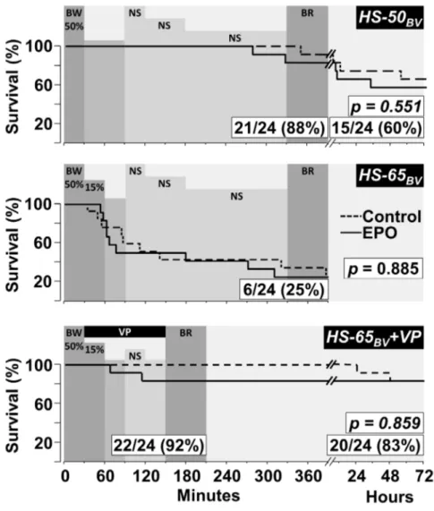
Figure 2. Survival curves comparing pigs treated with EPO and controls.SeriesHS-50BVandHS-65BV+VPinclude 72 hour survival. The
p-values for survival differences between groups were calculated using the Gehan-Breslow test and are shown within each graph along with the resuscitation and survival rates for the combined EPO and control groups. The shaded horizontal bars successively represent; the percentage of blood withdrawn (BW), the interval of hemorrhagic shock after blood withdrawal without fluid administration, the administration of normal saline (NS) as described in the Method, and blood reinfusion (BR). Shown in65BV+VPis the vasopressin infusion (VP).
doi:10.1371/journal.pone.0110908.g002
After euthanasia, the whole left lung was weighed before and after drying in the oven at 70uC for at least 72 hours for calculations of the wet/dry ratio in the last series.
Sample Size
The sample size of 12 pigs per group was based on extrapolation from work in similar animal models intended to identify biologically robust differences in survival effects and continuous variables with a power.0.60 at analevel of 0.05.
Experimental Series
Experiments were performed between 9 AM to 5 PM in a large animal surgical suite located inside the university Biological Resource Facility. Three consecutive series of 24 experiments each were conducted. The sequence of interventions are described below and depicted in Figure 2. In the first series, 50% of the estimated blood volume was withdrawn in 30 minutes (HS-50BV).
Animals remained untreated for 60 minutes after which normal saline – threefold the blood volume removed – was infused delivering sequentially a third of each in 30, 60, and 150 minutes followed by infusion of the shed blood in 60 minutes. The HS-50BVprotocol triggered a vigorous adaptive response that enabled
maintaining oxygen consumption close to baseline resulting in minimal lactate increases and high resuscitability and survival
without differences between EPO and control. To test EPO under greater HS severity, a second series was conducted withdrawing an additional 15% of the blood volume in 30 minutes after completing the initial 50% of blood volume removal for a total of 65% of blood volume removed (HS-65BV). Animals remained
untreated for 30 minutes after which the sameHS-50BVprotocol
for fluid resuscitation and blood reinfusion was applied. In this series, an additional 7 F angiographic catheter was advanced with the aid of fluoroscopy from the left cephalic vein into the great cardiac vein to assess effect on myocardial metabolism [25]. The
HS-65BV protocol was indeed severe, reducing resuscitability to
only 25%, but again showing no difference between EPO and control. A third series was then conducted using the same HS-65BV protocol for blood withdrawal but infusing arginine
vasopressin to prevent death by maintaining a higher coronary perfusion (HS-65BV+VP). Vasopressin (Pitressin, JHP
Pharma-ceuticals, Rochester, MI) was given intraosseously as a bolus (0.04 U?kg21
) upon completion of the initial 50% of blood volume removal followed by a continuous infusion (0.04 U?kg21
?min21
) using a syringe pump (PHD 2000 Syringe Pump Series, Harvard Apparatus, Holliston, MA) until start of blood reinfusion.
InHS-65BV+VP, we also assessed the effect of less or no fluid
resuscitation [26] under conditions of shorter HS duration. Thus, animals were also randomized 1:1 to receive either normal saline infusion – half of the blood volume withdrawn in 30 minutes – or no fluid at all. Blood was reinfused starting at 150 minutes (Figure 2). The addition of vasopressin dramatically improved resuscitability, allowing examination of survival and impact on organ function by blood sampling every 24 hours from the superior vena cava after sedation with ketamine hydrochloride (30 mg?kg21
intramuscularly). Pigs were euthanized at 72 hours.
Experimental Outcomes
The primary outcome was survival at 390 minutes in series HS-65BV(without recovery from anesthesia) and survival at 72 hours
in seriesHS-50BVand in seriesHS-65BV+VP(with recovery from
anesthesia). Secondary outcomes included: (1) hemodynamic and metabolic function, (2) myocardial function, (3) organ injury including the heart, brain, lung, liver, and kidney, (4) plasma cytokines, and (5) blood cell count.
Table 1.Baseline Characteristics.
HS-50BV HS-65BV HS-65BV+VP
Variable CTR EPO CTR EPO CTR EPO
n 12 12 12 12 12 12
Weight (kg) 39.663.7 37.564.2 34.661.5 35.461.2 39.462.4 38.162.5
Preparation Time (min) 170636 174641 123659 114621 119622 113621
Temperature (6C) 38.160.4 38.060.4 38.060.3 38.060.2 38.260.3 38.260.2
Respiration Rate (bpm) 3165 3164 3562 3661 3662 3562
End-tidal CO2(mmHg) 3862 3862 4062 4262 3863 3763
Mean Arterial Pressure (mmHg) 6268 6268 5966 6369 6167 6869
Cardiac Index (ml/min?m-2) 4.6
60.6 4.461.1 3.960.6 4.261.1 4.860.8 4.960.8
Heart Rate (bpm) 98620 9269 98610 106616 105620 100612
Blood Withdrawal Index (ml/m2) 1398670 1370643 19096162 19726316 1814639 1796639
Values are mean6SD.HS-50BV, blood volume withdrawal 50%;HS-65BV, blood volume withdrawal 65%;HS-65BV+VP, blood volume withdrawal 65% and vasopressin infusion. CTR, control; EPO, erythropoietin. There were no statistically significant differences between groups within each series.
doi:10.1371/journal.pone.0110908.t001
Measurements
Blood analysis. Blood samples were collected from the aorta and pulmonary artery in all three series with the addition of great cardiac vein inHS-65BV. Blood samples were processed on site for
pH, PO2, PCO2, hemoglobin, and lactate using a cartridge based
device (OPTI CCA-TS Blood Gas and Electrolyte Analyzer, OPTI Medical Systems, Roswell, GA) and for common hemo-globin types (oxy-, met-, carboxy-, and reduced-) using a co-oximeter (AVOXimeter 4000, AVOX systems Inc., San Antonio, TX). O2content in the aorta (CaO2), pulmonary artery (CvO2),
and great cardiac vein (CgcvO2) was calculated according to the
following equation:
O2Content ml
dl
~Hemoglobin g dl
|1 :39
ml g
|SFO2
z0 :003
ml dl:mmHg
{1
|PO2ðmmHgÞ
where 1.39 denotes ml of O2 bound to 1 g of hemoglobin
(Hufner’s number), SFO2the fraction of oxyhemoglobin relative to
the four hemoglobin types, and 0.003 the O2solubility coefficient.
Aortic blood samples were also taken and processed for complete
blood count and chemistry (blood urea nitrogen [BUN], creati-nine, alanine aminotransferase [ALT], aspartate aminotransferase [AST], and troponin I) at the Captain James A. Lovell Federal Health Care Center, North Chicago, IL.
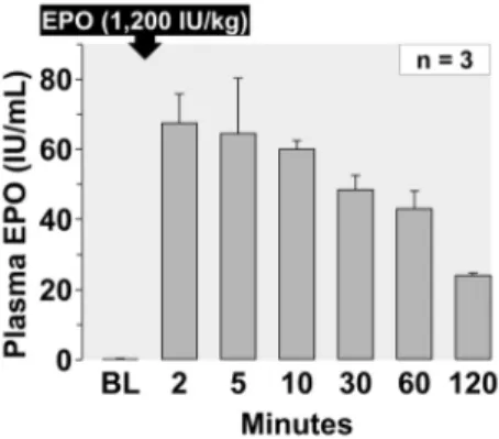
Plasma EPO. In seriesHS-50BV, the serum level of EPO was
measured in three animals that received EPO and in one control using a double-antibody ‘‘sandwich’’ enzyme-linked immunosor-bent assay kit (MD Bioproducts, St Paul, MN) targeted to human EPO according to the manufacturer instructions. The EPO level in serum samples (diluted 100 times) was calculated using a standard curve generated with EPO calibrators included in the kit (0, 10.3, 24.8, 48, 156, and 523 mU/ml). The final plasma concentration was determined by applying the dilution factors and a conversion factor whereby one U/ml of EPO [Epogen (epoetin alpha), Amgen] equaled 0.798 U/ml of the calibrator.
Hemodynamic Measurements. Thermodilution cardiac output was measured in duplicate after bolus injection of normal saline (5 ml) into the right atrium (HP-Philips M012AT cardiac output module, Amsterdam, The Netherlands). Cardiac output was normalized to body surface area using the Kelley equation (body surface area [m2] = 0.073?body-weight2/3[kg]) [27]. Aortic and left ventricular pressure signals were calibrated with a built-in calibration system (PCU-2000, Millar). Other pressure signals were zeroed to mid-cavity level. All signals were sampled and digitized at 250 Hz using a 16-bit data acquisition board
Figure 4. Hemodynamic and myocardial effects of EPO (open circles, n = 12) and vehicle control (closed circles, n = 12) in series HS-50BV.Numbers in brackets indicate when the number of animals decreased from the preceding time point consequent to death of the animal. BL, baseline; BW, blood withdrawal; HS, hemorrhagic shock; NS, normal saline; BR, blood reinfusion; Ao, aortic pressure; SVRI, systemic vascular resistance index; LVSWI, left ventricular stroke work index; RVWI, right ventricular stroke work index. Values are shown as mean6SEM. Differences between groups were analyzed by two-way repeated measures ANOVA. There were no overall significant treatment effects. However, there were overall statistically significant interactions between treatment and time for Ao mean (p= 0.033), cardiac index (p,0.001), LVSWI (p= 0.001), and RVSWI (p, 0.001). *p#0.05,{p#0.01, and`p#0.001 denote statistically significant differences between groups at the specified time points.ap#0.05,bp#0.01, andcp#0.001 denote significant differencesvsbaseline using the Holm-Sidak test for multiple comparisons showing the differences only when they occurred in one of the two groups.
doi:10.1371/journal.pone.0110908.g004
(AT-MIO-16XE-50; National Instruments, Austin, TX) and analyzed using custom developed software (Labview 6.0, National Instruments).
Cytokine Measurements. In the HS-65BV+VP series,
plasma levels of IL-6, IL-8, IL-10, and TNF-a were measured by a prototype 4-plex porcine cytokine electrochemiluminescence assay kit (Lot# Z00X2801, Meso Scale Discovery) using a QuickPlex SQ 120 multiplex imager (Meso Scale Discovery). The assay was performed as recommended by the manufacturer. Standards were prepared using IL-6, IL-8, IL-10, and TNF-a calibrators provided in the assay kit after a series of dilutions representing concentrations of 10,000 pg/ml, 2500 pg/ml, 625 pg/ml, 156.3 pg/ml, 39.1 pg/ml, 9.8 pg/ml, 2.4 pg/ml, and 0 pg/ml. Plasma collected at baseline, end of blood withdrawal, 24 hours after resuscitation, and 72 hours after resuscitation previously stored at280uC was thawed in ice and centrifuged at 2,320gfor 10 minutes. Twenty five microliters of the plasma was used for the assay. All standards and samples were run in duplicates. Concentrations were calculated from a 4-parameter logistic equation standard curve using Discovery Workbench software (Meso Scale Discovery). Lower limit of detection (LLOD) of the assay was 2 pg/ml for IL-6, 5 pg/ml for IL-8, 1 pg/ml for IL-10, and 4 pg/ml for TNF-a. The coefficient of variation between the duplicate samples was,5%.
Cardiac Function. Indices of cardiac function were derived from left ventricular pressures, reporting the maximal rate of left ventricular pressure increase (dP/dtmax) and pressure decrease
(dP/dtmin), the stroke volume index (SVI), and the left and right
ventricular stroke work (LVSWI and RVSWI), corresponding to SVI times the difference between the systolic and end-diastolic left ventricular pressures (LVSWI) and SVI times the difference between the mean pulmonary and right atrial pressures (RVSWI) expressed in centijoules (cJ) by multiplying by 0.013332 [28].
Statistical Analysis
SigmaPlot 11.0 (Systat Software, Point Richmond, CA) was used for statistical analysis. For all repetitive variables, two-way repeated measures ANOVA was used to test for the treatment effect between groups and their interaction over time identifying differences at specified time points when present. For clarity, statistical results are presented only at time points shown in tables and figures, but reflect analysis of all available time points. Kaplan-Meier survival curves were plotted and statistical differ-ences assessed using the Gehan-Breslow test. The hematological data from survivors in theHS-50BVandHS-65BV+VP serieswere
pooled; analyzing changes from baseline to 72 hours post-resuscitation within each treatment group by paired t-test and differences between groups by unpaired t-test. The data were
presented as means 6 SD unless otherwise stated. A two-tailed probability value ofp,0.05 was considered significant.
Results
No unexpected adverse events occurred. Demise occurred attributed to hemorrhagic shock consequent to hemodynamic compromise during the acute phase and to organ dysfunction during the 72 hour observation interval.
Baseline
No significant differences between EPO and control groups were observed at baseline within each series as shown on Table 1 and on each successive tables and figures, except for RVSWI in
HS-50BV.
EPO plasma levels
Plasma levels of EPO averaging 3 representative experiments from series HS-50BV are shown in Figure 3 confirming the
adequacy of the intraosseous route yielding levels in excess of 20 U/ml for at least 120 minutes after administering 1,200 U/kg.
Resuscitation and survival
The initial resuscitation rate for all three series and subsequent 72 hour survival forHS-50BV and HS-65BV +VPare shown in
Figure 2. For the EPO and the control group combined;HS-50BV
resulted in 88% initial resuscitation and 60% survival;HS-65BV
resulted in only 25% initial resuscitation (noticing that demise started after removing more than 50% of the blood volume); and
HS-65BV +VP resulted in 92% initial resuscitation (preventing
demise after exceeding 50% of blood volume withdrawal) and 83% survival. EPO had no effect on initial resuscitation or subsequent survival in any of the three series.
Hemodynamic and myocardial function
Blood removal triggered a chronotropic response that attenu-ated reductions in cardiac index and aortic blood pressure yielding a relatively stable hemodynamic state after completion of blood removal despite marked reduction in left and right ventricular work indexes, attributed mainly to reduced preload (Figures 4–6). Administration of normal saline, three-fold the volume of blood removed in series HS-50BV and HS-65BV, markedly increased
cardiac index (to levels higher than baseline), normalized left and right ventricular work indexes, and reduced the chronotropic response without substantial change in aortic pressure (Figures 4
Figure 6. Hemodynamic and myocardial effects of EPO (open circles, n = 12) compared with vehicle control (closed circles, n = 12) in
HS-65BV+VP.Numbers in brackets indicate when the number of animals decreased from the preceding time point consequent to death of the animal. BL, baseline; BW, blood withdrawal; HS, hemorrhagic shock; NS, normal saline; BR, blood reinfusion; Ao, aortic pressure; SVRI, systemic vascular resistance index; LVSWI, left ventricular stroke work index; RVWI, right ventricular stroke work index. Values are shown as mean6SEM. Differences between groups were analyzed by two-way repeated measures ANOVA. There was an overall statistically significant treatment effect for LVSWI (p= 0.035) and an overall statistically significant interaction between treatment and time for Ao mean (p= 0.002). *p#0.05,{p#0.01, and`p# 0.001 denote statistically significant differences between groups at the specified time points.ap#0.05,bp#0.01, andcp#0.001 denote significant differencesvsbaseline using the Holm-Sidak test for multiple comparisons showing the differences only when they occurred in one of the two groups.
doi:10.1371/journal.pone.0110908.g006
and 5). Administration of vasopressin in HS-65BV+VP with or
without normal saline (equal to half the volume of blood removed) increased systemic vascular resistance and aortic pressure with a modest increase in cardiac index and left and right stroke work indexes (Figure 6). Blood reinfusion maintained or increased cardiac index, aortic pressure, and work indexes inHS-50BVand
HS-65BV; whereas inHS-65BV+VPthe predominant effect was
increase in cardiac index and work indices (Figure 4–6). Relatively minor effects that varied contingent on the series were observed in relation to EPO. In HS-50BV, EPO appeared to blunt the
hemodynamic and myocardial response to HS and the subsequent fluid resuscitation and blood reinfusion interval (Figure 4). The opposite effect was observed in HS-65BV and HS-65BV+VPin
which favorable hemodynamic and myocardial response were more prominent in the EPO group during HS and the subsequent fluid resuscitation and blood reinfusion intervals (Figure 5 and 6).
Oxygen metabolism and lactatemia
The chronotropic response along with the expected increase in systemic oxygen extraction in HS-50BV allowed adequate
adaptation to HS evidenced by minimal lactatemia and high resuscitation and survival rates (Figure 7 and Figure 2,HS-50BV).
Greater blood volume removal in HS-65BV exhausted the
adaptive response evidenced by higher systemic oxygen extraction, higher levels of lactic acid, and substantial demise after removing
more than 50% of the blood volume (Figure 7 and Figure 2, HS-65BV). Use of vasopressin in HS-65BV+VP enabled survival
despite similar exhaustion of the adaptive response and was attributed to maintaining a higher aortic pressure required for coronary perfusion (Figure 7 and Figure 2, HS-65BV+VP).
Relatively minor metabolic effects related to EPO were observed, highlighting an attenuation of lactate increase inHS-65BV+VP
(Figure 7).
Myocardial metabolism
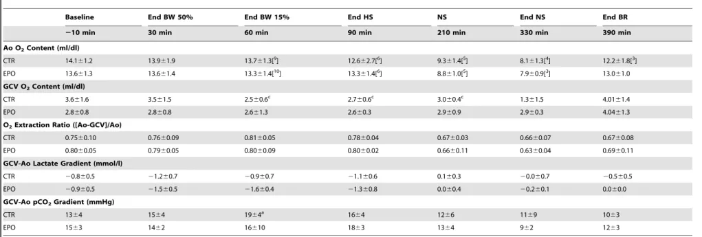
In HS-65BV, potential myocardial metabolic effects produced
by severe HS were assessed measuring myocardial oxygen, lactate, and pCO2 differences across the coronary circuit. As shown in
Table 2, HS was not associated with myocardial ischemia (despite ischemia in other organs evidenced by lactatemia), with EPO and control groups behaving similarly.
Effects of fluid resuscitation
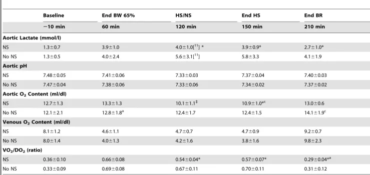
The effect of low-volume fluid administration was assessed in theHS-65BV+VP seriesand shown in Figure 8 and Table 3. Fluid
administration promoted an increase in cardiac index, mean aortic pressure, and left ventricular dP/dtmax(Figure 8) accompanied by
attenuation of systemic oxygen extraction and faster normalization of lactic acidosis (Table 3). Of the 12 animals that received fluids, 11 were resuscitated and remained alive at 72 hours. Of the 12
Figure 7. Metabolic effects of EPO (open circles) compared with vehicle control (closed circles) in seriesHS-50BV,HS-65BV, and HS-65BV+VP. Numbers in brackets indicate when the number of animals decreased from the preceding time point. BW, blood withdrawal; HS, hemorrhagic shock; NS, normal saline; BR, blood reinfusion. Values are shown as mean6SEM. Differences between groups were analyzed by two-way repeated measures ANOVA for each series separately. There were no overall significant treatment effects. However, there was an overall statistically significant interaction between treatment and time for lactate in seriesHS-65BV+VP(p= 0.007). *p#0.05 denotes statistically significant
differences between groups at the specified time points.ap#0.05 andbp#0.01 denote significant differencesvsbaseline using the Holm-Sidak test for multiple comparisons showing the differences only when they occurred in one of the two groups.
Table 2.Myocardial Metabolic Effects of EPO inHS-65BV.
Baseline End BW 50% End BW 15% End HS NS End NS End BR
210 min 30 min 60 min 90 min 210 min 330 min 390 min
Ao O2Content (ml/dl)
CTR 14.161.2 13.961.9 13.761.3[9] 12.662.7[6] 9.361.4[5] 8.161.3[4] 12.261.8[3]
EPO 13.661.3 13.661.4 13.361.4[10] 13.361.4[6] 8.861.0[5] 7.960.9[3] 13.061.0
GCV O2Content (ml/dl)
CTR 3.661.6 3.561.5 2.560.6c 2.760.6c 3.060.4c 1.361.5 4.0161.4
EPO 2.860.8 2.860.8 2.661.3 2.660.3 2.960.9 2.960.3 4.0461.3
O2Extraction Ratio ([Ao-GCV]/Ao)
CTR 0.7560.10 0.7660.09 0.8160.05 0.7860.04 0.6760.03 0.6660.07 0.6760.08
EPO 0.8060.05 0.7960.05 0.8060.09 0.8060.02 0.6660.11 0.6360.04 0.6960.11
GCV-Ao Lactate Gradient (mmol/l)
CTR 20.860.5 21.260.7 20.960.7 21.160.6 0.160.3 20.060.7 20.560.5
EPO 20.960.5 21.560.5 21.660.4 21.360.8 0.060.4 20.260.1 0.060.0
GCV-Ao pCO2Gradient (mmHg)
CTR 1364 1564 1964a 16
64 1266 1169 1063
EPO 1563 1462 16610 1863 1364 962 1263
Numbers in brackets indicate when the sample size decreased from the initial twelve animals. BW, blood withdrawal; HS, hemorrhagic shock; NS, normal saline; BR, blood reinfusion; EPO, erythropoietin; CTR, control; Ao, aorta; GCV, great cardiac vein. Values are mean6SD. The data was analyzed using two-way repeated measures ANOVA. There were no overall significant treatment effects and no overall statistically significant interactions between treatment and time.ap
#0.05;cp
#0.001 denote significant differencesvsbaseline using the Holm-Sidak test for multiple comparisons showing the differences only when they occurred in one of the two groups. doi:10.1371/journal.pone.0110908.t002
Intraosseou
s
Erythrop
oietin
during
Hemorrha
gic
Shock
PLOS
ONE
|
www.ploson
e.org
9
November
2014
|
Volume
9
|
Issue
11
|
Figure 8. Hemodynamic effects of fluid resuscitation (open symbols, n = 12) and no fluid resuscitation (closed symbols, n = 12) in seriesHS-65BV+VP.Numbers in brackets indicate when the number of animals decreased from the preceding time point consequent to death of the animal. BL, baseline; BW, blood withdrawal; HS, hemorrhagic shock; NS, normal saline; BR, blood reinfusion; Ao, aortic pressure; SVRI, systemic vascular resistance index. Values are shown as mean6SEM. Differences between groups were analyzed by two-way repeated measures ANOVA. There was an overall statistically significant treatment effect for cardiac index (p= 0.021). There were also overall statistically significant interactions between treatment and time for cardiac index (p,0.001) and SVRI (p,0.001). *p#0.05, {p#0.01, and`p#0.001 denote statistically significant differences between groups at the specified time points.ap#0.05, andcp#0.001 denote significant differencesvsbaseline using the Holm-Sidak test for multiple comparisons showing the differences only when they occurred in one of the two groups.
doi:10.1371/journal.pone.0110908.g008
Table 3.Metabolic Effect of Fluid Resuscitation inHS-65BV+VP.
Baseline End BW 65% HS/NS End HS End BR
210 min 60 min 120 min 150 min 210 min
Aortic Lactate (mmol/l)
NS 1.360.7 3.961.0 4.061.0[11] * 3.960.9* 2.761.0*
No NS 1.360.5 4.062.4 5.663.1[11] 5.863.3 4.161.9
Aortic pH
NS 7.4860.05 7.4160.06 7.3360.03 7.3760.04 7.4060.03
No NS 7.4760.04 7.3860.06 7.3360.06 7.3460.02 7.3760.02
Aortic O2Content (ml/dl)
NS 12.761.3 13.361.3 10.161.1`
10.961.0*c 13.0
60.6
No NS 12.162.1 12.861.8a 12.461.7 12.461.5 14.161.9c
Venous O2Content (ml/dl)
NS 8.161.2 4.661.1 4.760.7 4.760.9 9.260.7
No NS 8.061.4 4.061.3 4.261.6 3.861.6 9.862.3
VO2/DO2(ratio)
NS 0.3660.10 0.6660.08 0.5460.04* 0.5760.07* 0.2960.04*a
No NS 0.3360.09 0.6960.08 0.6760.11 0.7060.11 0.3160.12
Numbers in brackets indicate when the sample size decreased from the initial twelve animals. Values are mean6SD. BW, blood withdrawal; HS, hemorrhagic shock; NS, normal saline; BR, blood reinfusion; VO2/DO2, oxygen consumption divided by oxygen delivery. The data was analyzed using two-way repeated measures ANOVA. There were overall statistically significant interactions between treatment and time for aortic pH (p= 0.043), aortic O2content (p#0.001), and VO2/DO2ratio (p#0.001). There was no overall statistically significant treatment effect. *p#0.05 and`
p#0.001 denote statistically significant differences between groups at the specified time point. ap
#0.05 andcp
#0.001 denote statistically significant differencesvsbaseline using the Holm-Sidak test for multiple comparisons showing the differences only when they occurred in one of the two groups.
Table 4.Effect of EPO on Organ Function inHS-65BV+VP.
Baseline Post-Resuscitation
210 min 24 h 48 h 72 h
Creatinine (mg/dl)
CTR 1.360.2 1.561.4 1.461.7 [11] 0.960.1b[10]
EPO 1.060.2 0.960.2 [10] 0.960.2 0.960.1
Blood Urea Nitrogen (mg/dl)
CTR 1065 22622 15624 762
EPO 963 963 763 762
AST (U/l)
CTR 3567 4006501c 188
6204 1136118
EPO 3266 1686101{
85643 67626
ALT (U/l)
CTR 51610 1266109c 107
651c 100
636
EPO 5368 82624* 86626 58623c
Troponin I (ng/ml)
CTR 0.2260.18 1.4762.40c 0.48
61.06 0.2260.24
EPO 0.1560.09 0.4160.29 0.2060.47 0.1260.09*
Neurologic Deficit Score
CTR 060 26645a 15
645 266
EPO 060 13635 060 060
Numbers in brackets indicate when the sample size decreased from the initial twelve animals. Values are mean6SD. CTR, control; EPO, erythropoietin. The data was analyzed using two-way repeated measures ANOVA. There was no overall significant treatment effect and no overall statistically significant interactions between treatment and time. *p#0.05 and{p
#0.01 denote statistically significant differences between groups at the specified time points.ap#0.05,bp#0.01, andcp#0.001 denote statistically significant differencesvsbaseline using the Holm-Sidak test for multiple comparisons showing the differences only when they occurred in one of the two groups.
doi:10.1371/journal.pone.0110908.t004
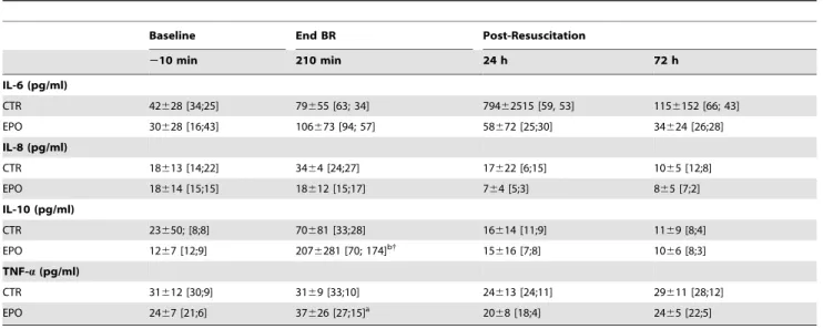
Table 5.Effect of EPO on Plasma Cytokines inHS-65BV+VP.
Baseline End BR Post-Resuscitation
210 min 210 min 24 h 72 h
IL-6 (pg/ml)
CTR 42628 [34;25] 79655 [63; 34] 79462515 [59, 53] 1156152 [66; 43]
EPO 30628 [16;43] 106673 [94; 57] 58672 [25;30] 34624 [26;28]
IL-8 (pg/ml)
CTR 18613 [14;22] 3464 [24;27] 17622 [6;15] 1065 [12;8]
EPO 18614 [15;15] 18612 [15;17] 764 [5;3] 865 [7;2]
IL-10 (pg/ml)
CTR 23650; [8;8] 70681 [33;28] 16614 [11;9] 1169 [8;4]
EPO 1267 [12;9] 2076281 [70; 174]b{ 15
616 [7;8] 1066 [8;3]
TNF-a(pg/ml)
CTR 31612 [30;9] 3169 [33;10] 24613 [24;11] 29611 [28;12]
EPO 2467 [21;6] 37626 [27;15]a 20
68 [18;4] 2465 [22;5]
Samples were available for each of the 12 control (CTR) pigs including the 10 that survived at 72 hours; but, for only 10 of the erythropoietin (EPO) treated pigs, which included all the survivors. Values are mean6SD showing also the median with interquartile range in brackets as values for several time events were not normally distributed. BR, blood reinfusion; IL, interleukin; TNF-a, tumor necrosis factor-a. The data was analyzed using two-way repeated measures ANOVA. There was no overall
statistically significant treatment effect. There was a statistically significant time effect for IL-8 (p =0.011), IL-10 (p#0.001) and TNF-a(p =0.006) and there was a borderline statistically significant interactions between treatment and time for IL-10 (p =0.062).{p
#0.003 denotes a statistically significant difference between groups at the specified time point.ap
#0.05,bp
#0.001 denote statistically significant differences from baseline within each group using the Holm-Sidak test for multiple comparisons.
doi:10.1371/journal.pone.0110908.t005
animals that did not receive fluids, 11 were also resuscitated but only 9 were alive at 72 hours; a difference however that was not statically significant.
Organ injury and function
The post-resuscitation effect on various organs was assessed daily inHS-65BV+VP. As shown in Table 4, EPO treated animals
had an attenuated increase in AST, ALT, troponin I, and less neurological deficit at some point during the post-resuscitation phase. There were statistically insignificant differences suggesting less kidney injury in the EPO group. There was no difference in the percentage of lung water at 72 hours between EPO and control pigs (81.060.6%vs81.861.0%).
Plasma cytokines
Plasma IL-6, IL-8, IL-10, and TNF-a were measured in the
HS-65BV+VPseries, analyzing the effects of EPO (Table 5) and
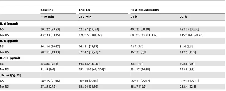
the effects of fluid resuscitation (Table 6). Overall there was a time effect with increases in IL-8 and IL-10 by the end of blood reinfusion reversing to baseline by 72 hours. Most prominently, EPO was associated with an increase in IL-10 by the end of blood reinfusion (Table 5). Administration of normal saline blunted increases in IL-8 and in IL-10 (Table 6).
Hematological effects
Pooled data from animals that survived in the HS-50BV and
HS-65BV+VPseries (18 animals in control group and 17 in EPO
group) showed no differences between treatment groups at baseline with cell counts within normal for swine [29]. Red blood cell count and hematocrit increased relative to baseline in both
Table 6.Effect of Fluid Resuscitation on Plasma Cytokines inHS-65BV+VP.
Baseline End BR Post-Resuscitation
210 min 210 min 24 h 72 h
IL-6 (pg/ml)
NS 30622 [23;23] 62627 [57; 24] 40623 [38;20] 42625 [38;33]
No NS 43633 [33;45] 120677 [101; 68] 88062620 [83; 132] 1156164 [69; 61]
IL-8 (pg/ml)
NS 16614 [10;17] 16611 [17;17] 969 [5;4] 864 [6;5]
No NS 20611 [19;13] 37642 [32;27] * 16623 [5;9] 1165 [11;9]
IL-10 (pg/ml)
NS 25653 [9;11] 846120 [38;35] 864 [7;4] 1066 [9;3]
No NS 1165 [9;6] 1816262 [67; 206]a* 23617 [14;28] 1269 [8;3]
TNF-a(pg/ml)
NS 28615 [21;16] 30610 [29;10] 26613 [25;17] 30611 [27;13]
No NS 2765 [27;5] 38624 [31;16] 1867 [19;5] 2364 [22;3]
Samples were available for all 11 pigs in each group that survived the initial 24 hours and for all the 9 that survived at 72 hours from the group without fluid resuscitation. Values are mean6SD showing also the median with interquartile range in brackets as values for several time events were not normally distributed. BR, blood reinfusion; NS, normal saline; IL, interleukin; TNF-a, tumor necrosis factor-a. The data was analyzed using two-way repeated measures ANOVA. There was no
overall statistically significant treatment effect. There was a statistically significant time effect for IL-8 (p= 0.008), IL-10 (p#0.001), and TNF-a(p= 0.008). *p#0.05 denotes a statistically significant difference between groups at the specified time points.ap
#0.001 denotes a statistically significant difference from baseline using the Holm-Sidak test for multiple comparisons.
doi:10.1371/journal.pone.0110908.t006
Table 7.Hematological Effects of EPO inHS-50BVandHS-65BV+VP.
Baseline 72 h Post-Resuscitation
Red Blood Cells (106/ml) CTR 5.560.4[18] 6.0
60.8a
EPO 5.660.6[17] 6.860.7*c
Hematocrit (%) CTR 31.561.7 34.464.8a
EPO 31.662.1 39.364.5*c
White Blood Cells (103/
ml) CTR 18.064.0 20.964.9
EPO 20.063.5 21.065.5
Platelets (103/ml) CTR 312
668 3326106
EPO 304685 3976164a
Values are mean6SD. CTR, control; EPO, erythropoietin. Data was pooled fromHS-50BVandHS-65BV+VP series. Numbers in bracket indicate pooled sample size. Unpairedt-test was used to compare differences in the pooled hematological data between treatment groups at given time points. *p#0.05. Pairedt-test was used to compare pooled hematological data fromHS-50BVandHS-65BV+VP seriesat baseline and post-resuscitation within each treatment group.ap
#0.05 andcp
#0.001. There were no statistically significant differences between groups at baseline.
groups at 72 hours post-resuscitation but to a greater extent in the EPO group (Table 7). Over the same time interval, platelet count increased but only in the EPO group whereas the white blood cell count remained unchanged.
Discussion
The present study failed to demonstrate a beneficial (or detrimental) effect of EPO on initial resuscitability or subsequent survival in a swine model of HS regardless of its severity. The work was conducted in three consecutive HS series modeling mild severity, high severity with high fatality despite aggressive fluid resuscitation, and high severity with low fatality associated with vasopressin infusion and low-fluid or no-fluid resuscitation. EPO in the last series featuring high severity with low fatality increased plasma levels of the anti-inflammatory cytokine IL-10, attenuated lactatemia, and lessened transient injury to the liver, heart, and brain based on enzyme release and clinical neurological deficit. In addition, the study addressed several current aspects of HS management showing the efficacy of vasopressin infusion and restrictive fluid resuscitation.
EPO Effect
The dose of EPO chosen for the present experiments (1,200 U/ kg) was extrapolated from a dose previously used in a study targeting sudden cardiac arrest victims (90,000 U) [21]. A lower dose (12,000 U) was deemed effective in a pilot human assessing potential protection after acute myocardial infarction [30]. In the present study, we confirmed that the chosen EPO dose – delivered through the intraosseous route – reached the bloodstream (Figure 3) attaining plasma levels that exceeded 30 U/ml for the initial 60 minutes decreasing to approximately 20 U/ml after 120 minutes. Only a brief exposure to EPO is required to trigger a sustained protective effect during HS [31] and levels above 5 U/ ml are sufficient to elicit cytoprotection [32]. Accordingly, the lack of effects of EPO on resuscitability and survival occurred despite a sustained presence of EPO in the bloodstream and evidence of biological activity given the significant increase in hematocrit and platelets after 72 hours (Table 5).
The lack of impact on initial resuscitability from HS likely reflected the mechanism of death. Animals died preceded by progressive reductions in blood pressure that at some point precipitously compromised coronary perfusion and cardiac function. However, there was no evidence of protracted myocar-dial ischemia before demise upon which EPO could have exerted an acute ‘‘protective’’ effect, as previously reported during resuscitation from cardiac arrest [20]. The lack of myocardial ischemia was shown in seriesHS-65BV(the series with the highest
HS severity and mortality), in which the lactate gradient across the coronary circuit was negative (i.e., lactate utilization) and the PCO2gradient was not increased [25], both indicating absence of
myocardial ischemia (Table 2). At the same time, substantial systemic lactic acidosis developed in series HS-65BV and series
HS-65BV+VP, indicating critical reductions in oxygen delivery
prompting anaerobic metabolism in other tissues. In HS-65BV+VP, EPO attenuated increases in lactic acid, consistent
with a beneficial effect at the mitochondrial level as we have reported in a rat model of cardiac arrest [20]. Likewise, there was attenuation of markers of organ injury in EPO treated pigs in the
HS-65BV+VP series. The protective effects at the cell level
reported by us [20] and others [32] in rats involve at the very least Akt activation (phosphorylation); a kinase that plays a key role in cell survival and other adaptive responses including mitochondrial bioenergetic function and enhanced myocardial contractility
[33,34]. In addition, plasma IL-10 was higher in pigs that received EPO at the end of blood reinfusion. IL-10 mediates anti-inflammatory effects and EPO can increase production of IL-10 [35–37]; pointing to the pleiotropic effect of EPO and mediation of additional effects that could be potentially beneficial for HS. The levels of IL-6 and IL-8 (pro-inflammatory cytokines) were lower at 24 and 72 hours in pigs treated with EPO but the differences were not statistically significant.
Studying the potential clinical relevance of these effects at the organ level was beyond the scope of the present study. In clinical settings, patients are exposed to repetitive injuries after stabiliza-tion (i.e., biological, infectious, sterile, etc.) and informastabiliza-tion on whether organ susceptibility to subsequent injuries could be ameliorated by EPO would be of substantial clinical interest.
Hemodynamic Response to Hemorrhagic Shock
A prominent (adaptive) chronotropic response maintained the cardiac index inHS-50BVat,80% of baseline, enabling increases in oxygen extraction to preserve aerobic metabolism and thereby yielding only minor increases in lactic acid. In seriesHS-65BVand
HS-65BV+VP, however, the cardiac index was maintained at
,62% of baseline and the oxygen extraction reached its maximum level prompting systemic lactic acidosis; most likely from tissues excluded from the circulation following redistribution of blood flow towards vital organs. Accordingly, in this swine model, the critical transition from ‘‘compensated’’ to ‘‘uncompen-sated’’ HS occurred after removing 50% of the blood volume.
Vasopressin
Hemodynamic crises, including HS, trigger the release of vasopressin as part of a prominent neuroendocrine stress response. However, the endogenous vasopressin response is time-limited and exogenous administration has been suggested for hemodynamic stabilization in sepsis [38,39] and HS [40,41]. In models of HS, vasopressin was comparably superior to fluid administration or epinephrine [42,43] and has been proposed as the ‘‘preferred’’ vasopressor agent for hemodynamic stabilization during HS [41]. In a small clinical study in civilians suffering traumatic injury with hypotension, vasopressin infusion reduced the need of resuscita-tion fluids [44]. Clinicaltrials.gov lists two studies examining the effects of vasopressin in civilians with HS; the AVERT Shock study by Sims, C and the VITRIS study by Wenzel, V. [45].
These reasons prompted us to select vasopressin in our last series (HS-65BV+VP), with the dose chosen according to a
previous study by Voelckelet al., also in swine [42]. The timing for initiation of vasopressin infusion was based on seriesHS-65BV,
noticing that demise began to occur after removing greater than 50% of the blood volume (Figure 2). The effect of vasopressin infusion on resuscitability was impressive, increasing from 25% (in
HS-65BV) to 92% (inHS-65BV+VP) for the same degree of blood
volume removal (Figure 2). Moreover, 83% of the animals in HS-65BV+VPwere alive at 72 hours and with essentially no organ
dysfunction (Table 4).
Fluid Resuscitation
There is growing consensus that aggressive fluid resuscitation during HS can be detrimental [46]. Coagulation is compromised consequent to dilution of clotting factors and reduced activity by hypothermia and acidosis. Concomitantly, the endogenous fibri-nolytic system is activated, tilting the hemostatic balance toward bleeding [47]. Excessive fluid also drives edema formation, which may affect the lungs and other tissues [48]. Large amounts of normal saline can precipitate hyperchloremic acidosis which has been associated with increased risk of renal injury [49] and
activation of inflammatory cascades [50]. For this and other (logistic) reasons, resuscitation with minimal or no fluid is gaining acceptance. The Tactical Combat Casualty Care Guidelines recommend fluid administration during HS only when there is altered mental status (in the absence of head injury) and weak or absent peripheral pulses.
In our initial two series (i.e., HS-50BV and HS-65BV), we
administered normal saline 3 times the amount of blood removed according to the old paradigm resulting in a substantial increase in cardiac index (via preload increase) exceeding baseline levels. However, despite high initial resuscitability, the 72 hour survival was less than in the more severeHS-65BV+VPseries suggesting
that excess fluid could have been detrimental. In theHS-65BV+VP
series, we examined the current paradigm and administered normal saline half the amount of blood removed in half of the animals and no fluids in the other half observing no differences in resuscitability or survival. Normal saline, however, had as positive effect on cardiac index, aortic pressure, and left ventricular dP/ dtmax while reducing systemic oxygen extraction and the
generation of lactic acid (Figure 8, Table 3). Thus, there was hemodynamic and metabolic benefit elicited by fluid administra-tion which could be critical under condiadministra-tions of more severe HS and when vasopressin alone is not sufficient.
Limitations
Extrapolation of the current findings to real-life battlefield resuscitation from hemorrhagic shock is limited by several factors. Upmost is modeling HS without additional tissue injury. Although HS can occur in military and (more so) in civilian settings with low-grade or unassociated tissue injury, HS in the battlefield occurs typically accompanied by substantial tissue injury. Tissue injury compounds the severity of HS partly by the specific organ dysfunction, wound contamination, and amplification of the inflammatory response. In addition, the rate of blood withdrawal was controlled by a predetermined algorithm and did not follow the natural profile of bleeding after injury. The present studies, however, allowed assessing the effects of the studied interventions on severe HS without the confounding effects of tissue injury and uncontrolled bleeding. In subsequent series we incorporated the elements of tissue injury and uncontrolled bleeding by using a liver laceration model. Other limitations include the use of anesthesia, masking some of the physiologic responses to hypovolemic shock. The naturally occurring neuropeptide in swine is lysine vasopres-sin whereas in humans it is arginine vasopresvasopres-sin (the one used in the present studies). Thus, potency may vary contingent on specific
receptors and vascular beds and translation of these findings to humans will need to consider human dose-responses. Despite these limitations, our model reproduced key characteristics of resusci-tation from hemorrhagic shock and provided mechanistic infor-mation under highly controlled conditions that would be difficult to obtain in a more realistic model.
Conclusions
EPO given during HS in a swine model of HS failed to alter resuscitability and 72 hour survival regardless of HS severity and concomitant treatment with fluids and vasopressin. EPO, howev-er, attenuated lactate increases and acute injury to the livhowev-er, heart, and brain based on enzyme release and neurological deficit scores. The studies also showed that vasopressin infusion with restrictive fluid administration is highly effective for hemodynamic stabiliza-tion and subsequent survival.
Supporting Information
Datafile S1 Hemodynamic and metabolic data from the three hemorrhagic shock series.
(XLSX)
Checklist S1 ARRIVE guidelines checklist. (PDF)
Acknowledgments
This research was supported by the Telemedicine and Advanced Technology Research Center (TATRC) at the U.S. Army Medical Research and Materiel Command (USAMRMC) Fort Detrick, MD under contract number: W81XWH-11-2-0019.
We also thank the Pathology Laboratory and the Pharmacy Services at Captain James A. Lovell Federal Health Care Center for their support processing blood samples and facilitating the purchase of the various drugs used in the project.
Non-Endorsement Disclaimer: The views, opinions and/or findings contained in this report are those of the author(s) and should not be construed as an official Department of the Army position, policy or decision unless so designated by other documentation.
Author Contributions
Conceived and designed the experiments: RJG. Performed the experi-ments: VBL KW BMC. Analyzed the data: VBL KW BMC RJG. Contributed reagents/materials/analysis tools: AB YM SV JR. Wrote the paper: VBL RJG.
References
1. Kauvar DS, Lefering R, Wade CE (2006) Impact of hemorrhage on trauma outcome: an overview of epidemiology, clinical presentations, and therapeutic considerations. J Trauma 60: S3–11.
2. Eastridge BJ, Mabry RL, Seguin P, Cantrell J, Tops T, et al. (2012) Death on the battlefield (2001-2011): implications for the future of combat casualty care. J Trauma Acute Care Surg 73: S431–S437.
3. Cai Z, Manalo DJ, Wei G, Rodriguez ER, Fox-Talbot K, et al. (2003) Hearts from rodents exposed to intermittent hypoxia or erythropoietin are protected against ischemia-reperfusion injury. Circulation 108: 79–85.
4. Moon C, Krawczyk M, Ahn D, Ahmet I, Paik D, et al. (2003) Erythropoietin reduces myocardial infarction and left ventricular functional decline after coronary artery ligation in rats. Proc Natl Acad Sci U S A 100: 11612–11617. 5. Parsa CJ, Matsumoto A, Kim J, Riel RU, Pascal LS, et al. (2003) A novel protective effect of erythropoietin in the infarcted heart. J Clin Invest 112: 999– 1007.
6. Tramontano AF, Muniyappa R, Black AD, Blendea MC, Cohen I, et al. (2003) Erythropoietin protects cardiac myocytes from hypoxia-induced apoptosis through an Akt-dependent pathway. Biochem Biophys Res Commun 308: 990–994.
7. Vilarinho KA, de Oliveira PP, Saad MJ, Eghtesady P, Filho LM, et al. (2013) Erythropoietin protects the systolic function of neonatal hearts against ischaemia/reperfusion injury. Eur J Cardiothorac Surg 43: 156–162. 8. Brines ML, Ghezzi P, Keenan S, Agnello D, de Lanerolle NC, et al. (2000)
Erythropoietin crosses the blood-brain barrier to protect against experimental brain injury. Proc Natl Acad Sci U S A 97: 10526–10531.
9. Siren AL, Fratelli M, Brines M, Goemans C, Casagrande S, et al. (2001) Erythropoietin prevents neuronal apoptosis after cerebral ischemia and metabolic stress. Proc Natl Acad Sci U S A 98: 4044–4049.
10. Celik M, Gokmen N, Erbayraktar S, Akhisaroglu M, Konakc S, et al. (2002) Erythropoietin prevents motor neuron apoptosis and neurologic disability in experimental spinal cord ischemic injury. Proc Natl Acad Sci U S A 99: 2258– 2263.
11. Simon F, Scheuerle A, Groger M, Vcelar B, McCook O, et al. (2011) Comparison of carbamylated erythropoietin-FC fusion protein and recombinant human erythropoietin during porcine aortic balloon occlusion-induced spinal cord ischemia/reperfusion injury. Intensive Care Med 37: 1525–1533. 12. Vesey DA, Cheung C, Pat B, Endre Z, Gobe G, et al. (2004) Erythropoietin
13. Abdelrahman M, Sharples EJ, McDonald MC, Collin M, Patel NS, et al. (2004) Erythropoietin attenuates the tissue injury associated with hemorrhagic shock and myocardial ischemia. Shock 22: 63–69.
14. Wu WT, Lin NT, Subeq YM, Lee RP, Chen IH, et al. (2010) Erythropoietin protects severe haemorrhagic shock-induced organ damage in conscious rats. Injury 41: 724–730.
15. Shimoda M, Sawada T, Iwasaki Y, Mori S, Kijima H, et al. (2009) Erythropoietin strongly protects the liver from ischemia-reperfusion injury in a pig model. Hepatogastroenterology 56: 470–475.
16. Buemi M, Vaccaro M, Sturiale A, Galeano MR, Sansotta C, et al. (2002) Recombinant human erythropoietin influences revascularization and healing in a rat model of random ischaemic flaps. Acta Derm Venereol 82: 411–417. 17. Bohr S, Patel SJ, Shen K, Vitalo AG, Brines M, et al. (2013) Alternative
erythropoietin-mediated signaling prevents secondary microvascular thrombosis and inflammation within cutaneous burns. Proc Natl Acad Sci U S A 110: 3513–3518.
18. Singh D, Kolarova JD, Wang S, Ayoub IM, Gazmuri RJ (2007) Myocardial protection by erythropoietin during resuscitation from ventricular fibrillation. Am J Ther 14: 361–368.
19. Borovnik-Lesjak V, Whitehouse K, Baetiong A, Artin B, Radhakrishnan J, et al. (2013) High-dose erythropoietin during cardiac resuscitation lessens postresusci-tation myocardial stunning in swine. Transl Res 162: 110–121.
20. Radhakrishnan J, Upadhyaya MP, Ng M, Edelheit A, Moy HM, et al. (2013) Erythropoietin facilitates resuscitation from ventricular fibrillation by signaling protection of mitochondrial bioenergetic function in rats. Am J Transl Res 5: 316–326.
21. Grmec S, Strnad M, Kupnik D, Sinkovic A, Gazmuri RJ (2009) Erythropoietin facilitates the return of spontaneous circulation and survival in victims of out-of-hospital cardiac arrest. Resuscitation 80: 631–637.
22. Kao NR, Xenocostas A, Driman DK, Rui T, Huang W, et al. (2011) Recombinant human erythropoietin improves gut barrier function in a hemorrhagic shock and resuscitation rat model. J Trauma 71: S456–S461. 23. Kao RL, Xenocostas A, Rui T, Huang W, Martin CM (2010) The effect of
erythropoietin on microcirculation perfusion and tissue bioenergetics of the small intestine in a hemorrhagic shock and resuscitation rat model. J Trauma 68: 1342–1348.
24. Berg RA, Otto CW, Kern KB, Sanders AB, Hilwig RW, et al. (1994) High-dose epinephrine results in greater early mortality after resuscitation from prolonged cardiac arrest in pigs: a prospective, randomized study. Crit Care Med 22: 282– 290.
25. Gudipati CV, Weil MH, Gazmuri RJ, Deshmukh HG, Bisera J, et al. (1990) Increases in coronary vein CO2 during cardiac resuscitation. J Appl Physiol 68: 1405–1408.
26. Morrison CA, Carrick MM, Norman MA, Scott BG, Welsh FJ, et al. (2011) Hypotensive resuscitation strategy reduces transfusion requirements and severe postoperative coagulopathy in trauma patients with hemorrhagic shock: preliminary results of a randomized controlled trial. J Trauma 70: 652–663. 27. Kelley KW, Curtis SE, Marzan GT, Karara HM, Anderson CR (1973) Body
surface area of female swine. J Anim Sci 36: 927–930.
28. Faybik P, Lahner D, Schramm W (2010) A longstanding error by Ernest Henry Starling (author reply 1584–5). Resuscitation 81: 1584–1585.
29. Jonasson R, Johannisson A, Jacobson M, Fellstrom C, Jensen-Waern M (2004) Differences in lymphocyte subpopulations and cell counts before and after experimentally induced swine dysentery. J Med Microbiol 53: 267–272. 30. Ozawa T, Toba K, Suzuki H, Kato K, Iso Y, et al. (2010) Single-dose
intravenous administration of recombinant human erythropoietin is a promising treatment for patients with acute myocardial infarction - randomized controlled pilot trial of EPO/AMI-1 study -. Circ J 74: 1415–1423.
31. Brines M, Cerami A (2008) Erythropoietin-mediated tissue protection: reducing collateral damage from the primary injury response. J Intern Med 264: 405–432.
32. Nandra KK, Collino M, Rogazzo M, Fantozzi R, Patel NS, et al. (2013) Pharmacological preconditioning with erythropoietin attenuates the organ injury and dysfunction induced in a rat model of hemorrhagic shock. Dis Model Mech 6: 701–709.
33. Beiser DG, Wojcik KR, Zhao D, Orbelyan GA, Hamann KJ, et al. (2010) Akt1 genetic deficiency limits hypothermia cardioprotection following murine cardiac arrest. Am J Physiol Heart Circ Physiol 298: H1761–H1768.
34. Huang HH, Shao ZH, Li CQ, Vanden Hoek TL, Li J (2014) Baicalein protects cardiomyocytes against mitochondrial oxidant injury associated with JNK inhibition and mitochondrial Akt activation. Am J Chin Med 42: 79–94. 35. Bryl E, Mysliwska J, Debska-Slizien A, Rachon D, Bullo B, et al. (1998) The
influence of recombinant human erythropoietin on tumor necrosis factor alpha and interleukin-10 production by whole blood cell cultures in hemodialysis patients. Artif Organs 22: 177–181.
36. Liu X, Xie W, Liu P, Duan M, Jia Z, et al. (2006) Mechanism of the cardioprotection of rhEPO pretreatment on suppressing the inflammatory response in ischemia-reperfusion. Life Sci 78: 2255–2264.
37. Jia HB, Jin Y, Ji Q, Hu YF, Zhou ZQ, et al. (2009) Effects of recombinant human erythropoietin on neuropathic pain and cerebral expressions of cytokines and nuclear factor-kappa B. Can J Anaesth 56: 597–603.
38. Guzman JA, Rosado AE, Kruse JA (2003) Vasopressin vs norepinephrine in endotoxic shock: systemic, renal, and splanchnic hemodynamic and oxygen transport effects. J Appl Physiol 95: 803–809.
39. Russell JA, Fjell C, Hsu JL, Lee T, Boyd J, et al. (2013) Vasopressin compared with norepinephrine augments the decline of plasma cytokine levels in septic shock. Am J Respir Crit Care Med 188: 356–364.
40. Voelckel WG, Convertino VA, Lurie KG, Karlbauer A, Schochl H, et al. (2010) Vasopressin for hemorrhagic shock management: revisiting the potential value in civilian and combat casualty care. J Trauma 69 Suppl 1: S69–S74.
41. Anand T, Skinner R (2012) Arginine vasopressin: The future of pressure-support resuscitation in hemorrhagic shock. J Surg Res 178: 321–329.
42. Voelckel WG, Raedler C, Wenzel V, Lindner KH, Krismer AC, et al. (2003) Arginine vasopressin, but not epinephrine, improves survival in uncontrolled hemorrhagic shock after liver trauma in pigs. Crit Care Med 31: 1160–1165. 43. Raedler C, Voelckel WG, Wenzel V, Krismer AC, Schmittinger CA, et al.
(2004) Treatment of uncontrolled hemorrhagic shock after liver trauma: fatal effects of fluid resuscitation versus improved outcome after vasopressin. Anesth Analg 98: 1759–1766.
44. Cohn SM, McCarthy J, Stewart RM, Jonas RB, Dent DL, et al. (2011) Impact of low-dose vasopressin on trauma outcome: prospective randomized study. World J Surg 35: 430–439.
45. Lienhart HG, Wenzel V, Braun J, Dorges V, Dunser M, et al. (2007) [Vasopressin for therapy of persistent traumatic hemorrhagic shock: The VITRIS.at study]. Anaesthesist 56: 145–8, 150.
46. Bougle A, Harrois A, Duranteau J (2013) Resuscitative strategies in traumatic hemorrhagic shock. Ann Intensive Care 3: 1.
47. Nunez TC, Young PP, Holcomb JB, Cotton BA (2010) Creation, implemen-tation, and maturation of a massive transfusion protocol for the exsanguinating trauma patient. J Trauma 68: 1498–1505.
48. Madigan MC, Kemp CD, Johnson JC, Cotton BA (2008) Secondary abdominal compartment syndrome after severe extremity injury: are early, aggressive fluid resuscitation strategies to blame? J Trauma 64: 280–285.
49. Shaw AD, Bagshaw SM, Goldstein SL, Scherer LA, Duan M, et al. (2012) Major complications, mortality, and resource utilization after open abdominal surgery: 0.9% saline compared to Plasma-Lyte. Ann Surg 255: 821–829.
50. Volta CA, Trentini A, Farabegoli L, Manfrinato MC, Alvisi V, et al. (2013) Effects of two different strategies of fluid administration on inflammatory mediators, plasma electrolytes and acid/base disorders in patients undergoing major abdominal surgery: a randomized double blind study. J Inflamm (Lond) 10: 29.