DOI: 10.1590/0004-282X20150039
ARTICLE
Effect of volume replacement during
combined experimental hemorrhagic shock
and traumatic brain injury in prostanoids,
brain pathology and pupil status
Efeito da reposição volêmica durante o choque hemorrágico combinado com traumatismo
craniencefálico sobre prostanóides, patologia cerebral e
status
pupilar
Fernando Campos Gomes Pinto1, Matheus Fernandes de Oliveira2, Ricardo Prist3, Maurício Rocha e Silva4, Luiz Fernando Ferraz da Silva5,6, Antonio Capone Neto7
Head injury is the main cause of trauma-related deaths in over 60% of cases1,2,3,4,5. Hemorrhage and shock are
ob-served in up to 20% of patients with head injuries6,7,8,9,10,11,12.
Hypotension, even for very brief periods, is a well-established cause of secondary brain injury, and con-tributes to worse outcomes13,14,15.
1Universidade de São Paulo, Hospital das Clínicas, Instituto de Psiquiatria, Divisão de Neurocirurgia Funcional, Sao Paulo SP, Brazil;
2Hospital do Servidor Público Estadual de São Paulo, IAMSPE, Departamento de Neurocirurgia, Sao Paulo SP, Brasil;
3Universidade de São Paulo, Faculdade de Medicina, Divisão de Pesquisa, Sao Paulo SP, Brazil;
4Universidade de São Paulo, Departamento de Fisiologia, Sao Paulo SP, Brazil;
5Universidade de São Paulo, Departamento de Patologia, Sao Paulo SP, Brazil;
6Hospital Israelita Albert Einstein, Sao Paulo SP, Brazil;
7Hospital Israelita Albert Einstein, Unidade de Terapia Intensiva, Sao Paulo SP, Brazil.
Correspondence: Matheus Fernandes de Oliveira; Rua Luís Góis, 1333/apto 23; 04043-150 Sao Paulo SP, Brasil; E-mail: [email protected] Conflict of interest: There is no conlict of interest to declare.
Received 10 January 2015; Accepted 03 February 2015. ABSTRACT
Traumatic brain injury (TBI) is the main cause of trauma-related deaths. Systemic hypotension and intracranial hypertension causes cerebral ischemia by altering metabolism of prostanoids. We describe prostanoid, pupilar and pathological response during resuscitation with hypertonic saline solution (HSS) in TBI. Method: Fifteen dogs were randomized in three groups according to resuscitation after TBI (control group; lactated Ringer’s (LR) group and HSS group), with measurement of thromboxane, prostaglandin, macroscopic and microscopic pathological evaluation and pupil evaluation. Result: Concentration of prostaglandin is greater in the cerebral venous blood than in plasma and the opposite happens with concentration of thromboxane. Pathology revealed edema in groups with the exception of group treated with HSS.
Discussion and conclusion: There is a balance between the concentrations of prostaglandin and thromboxane. HSS prevented the formation of cerebral edema macroscopically detectable. Pupillary reversal occurred earlier in HSS group than in LR group.
Keywords: traumatic brain injury, volemic resuscitation, pathology, inlammation.
RESUMO
O traumatismo cranioencefálico (TCE) é a principal causa de morte relacionada ao trauma. O choque hemorrágico e hipertensão intracraniana causam isquemia cerebral alterando o metabolismo de prostanóides. Neste estudo, relatamos o comportamento dos prostanóides, resposta pupilar e patologia durante a reposição volêmica com solução salina hipertônica (SSH) no TCE. Método: Quinze cachorros foram randomizados em três grupos (controle, grupo de Ringer lactato e grupo de SSH) e foram avaliados tromboxane, prostaglandina, avaliação patológica macroscópica e microscópica e status pupilar. Resultado: A concentração de prostaglandina é maior no sangue cerebral em comparação ao plasma, e o inverso ocorre com o tromboxane. A patologia revelou edema em todos os grupos, com exceção do grupo tratado com SSH. Discussão e conclusão: Existe um equilíbrio entre concentrações cerebrais e plasmáticas de prostaglandina e tromboxane. A SSH protegeu o cérebro da formação de edema pós traumático.
Several studies have demonstrated that elevated intracra-nial pressure (ICP) impairs protective mechanisms against blood loss and hypotension, creating a substantial treat-ment challenge1,2,3,13,14. he type of luid used for volume re
-placement can have a signiicant impact on ICP. As hyper
-osmolar therapy has been used to reduce elevated ICP due to a variety of causes, including traumatic brain injury (TBI), the use of hypertonic saline solution (HSS) for resuscitation of head-injured patients has been considered an appealing strategy6,7,8,9,10.
Additionally, hemorrhagic hypotension associated with
intracranial hypertension causes decreased cerebral blood
low and cerebral ischemia by altering the synthesis, activa
-tion, and release of potent vasoactive substances-vasoactive prostanoids, such as prostacyclin (PGI2) and
thrombox-ane (TXA2)—and may interfere with cerebrovascular
reactivity16,17,18,19,20,21.
A previous report by our group evaluated the systemic and
cerebral, hemodynamic, and oxygenation responses to difer
-ent solutions used for volume replacem-ent during the early phases of hemorrhagic shock (HS) associated with TBI1. We
now report the results of further evaluations, including assess-ment of prostanoid, pupillary, and pathological response,
dur-ing luid replacement with HSS in experimental TBI.
METHODS
his study was conducted at the Experimental Division
of the Heart Institute (InCor), Hospital das Clinicas, Faculty of Medicine, University of São Paulo (HC-FMUSP), in ac-cordance with international standards for the use of ex-perimental animals22, after being approved by the Scientiic
Committee of the Heart Institute and the FMUSP Research
Ethics Committee.
Experiments were performed on 15 mongrel dogs (weight
16.3 ± 1.9 kg), which were fasted overnight with free access
to water. Anesthesia was provided with ketamine (10 mg/kg)
and halothane (1.5% during preparation and insult, 0.5%
during treatment). After orotracheal intubation, mechani
-cal ventilation was initiated (Harvard Apparatus 708, South
Natick, MA) to keep end-tidal CO2 (ETCO2) between 32 and
37 mmHg (Dixtal DX 1265 capnometer, Manaus, AM, Brazil). he femoral artery and vein were cannulated with P240 catheters. A pulmonary artery catheter (93A-131H-7F, Baxter Healthcare Corporation, CA) was inserted through the right
jugular vein and connected to a cardiac output monitor
(mod-el 9520A, American Edwards Lab, CA). Cardiac output param
-eters were acquired by averaging two measures. he hemody
-namic catheters were connected through pressure transducers
(P23XL Viggo-Spectramed, Statham, CA) to a multichannel acquisition system (MP100, Biopac System, CA). Measured variables were recorded and analyzed on a personal computer using Acknowledge 3.0 for Windows software.
he left transverse sinus was cannulated through a 2-mm
burr hole to obtain cerebral venous blood samples. An intra
-parenchymal iberoptic ICP sensor (model 110-4B, Camino
NeuroCare Inc., San Diego, CA) was placed in the right pa
-rietal lobe through an 2-mm burr hole drilled in the
pari-etal bone and connected to a ICP monitor (V420-7, Camino Laboratories, San Diego, CA). A 1-cm diameter hole was drilled into the left parietal bone for delivery of luid percussion by a speciic device (Fluid Percussion Machine, Bioengineering
Division, InCor/HC-FMUSP, São Paulo, Brazil); a delated epi
-dural balloon (#5 Foley catheter) was also placed in the epi-dural space through this hole. Core temperature was kept in the 36ºC-38ºC range with heated blankets.
Insult
HS was induced by withdrawal of blood through an aor-tic catheter over 5 to 10 minutes to a mean arterial pressure
(MAP) of 40 mmHg. he shed blood was heparinized and
stored. TBI was induced by luid percussion (4 atm). After lu
-id percussion, the ep-idural balloon was inlated with approxi
-mately 5 mL to a target ICP of 20 to 25mmHg. he experi
-mental design simulated the clinical scenario of prehospital and early hospital resuscitation phases, including intracrani-al hematoma drainage (Figure 1).
Time in minutes
Preparation Insult
T0 T1 T2 T3 T4 T5
Observation “Early hospital”
“Pre-hospital” 20’
0’ 20’ 40’ 60’ 90’ 180’
20’ 20’ 120’
Groups
Fifteen mongrel male dogs, weight 13 to 20 kg, were
ran-domized to one of three groups: HS + TBI + HSS (n = 5), simu
-lating prehospital treatment with 3% saline solution (HSS),
8 mL/kg over 10 minutes; HS+TBI+LR (n = 5), simulating prehospital treatment with lactated Ringer’s (LR) solution, 16 mL/kg over 10 minutes; and HS+TBI (n = ), a control group in which no luids were provided.
During a simulated early in-hospital treatment phase,
the two treatment groups received shed whole blood
trans-fusion targeted to a hematocrit of 30% and HSS or LR, ac
-cording to the speciic group, to maintain mean arterial pressure (MAP) greater than 70 mmHg. After 60 minutes, the epidural balloon was delated in the treatment-group animals, simulating intracranial hematoma drainage. he balloon was kept inlated throughout the experiment in
control animals.
MAP (mmHg) and ICP (mmHg) were measured or cal
-culated using standard formulas. Biochemical and blood gas
analyses were performed using a STAT Proile Ultra analyzer (Nova Biomedical, Waltham, MA).
Statistical analysis
Results were expressed as mean plus standard
devia-tion. Analysis of variance (ANOVA) was used for between-group comparisons. Diferences were evaluated by Student’s
t-test and accepted as statistically signiicant when p-values
were < 0.05.
Measurement of thromboxane B2 and 6-keto prostaglandin F1 alpha
At three distinct time points—T0’ (baseline), T40’
(40 minutes, end of prehospital treatment), and T90’ (30 min-utes after the end of the hospital treatment period), brain ve-nous blood and central arterial blood samples were collected for immunoassay measurement of the stable metabolites of
TXA2 and PGI2: thromboxane B2 (TXB2) and 6-keto prosta
-glandin F1 alpha respectively.
Samples (10 mL blood each) were collected into dry
tubes and centrifuged at 3000 rpm for 15 minutes. Serum
ali-quots (2 mL) were then pipetted into microcentrifuge tubes (Eppendorf ®) and stored at -70°C.
In due course, the samples were thawed and subjected
to chemical extraction in speciic columns of commercial
-ly available kits (hromboxane B2 Enzymeimmunoassay
Biotrak System - code RPN221, and 6-Keto_Prostaglandin F1
Alpha Enzymeimmunoassay Biotrak System - code RPN220; Amersham Biosciences UK Limited). After elution with ethyl acetate, the samples were dried by centrifugation (Speed-Vac,
model SC 210a, Manufacturer Institute Inc. Savant, Holbrook,
New York, USA) in a vacuum system for 60 minutes. he ab
-sorbance data obtained from the assays were applied to a
speciic formula and the concentration in pg/mL was deter
-mined after plotting a standard curve.
Pathology
Gross examination
After euthanasia (T180’), through an extended calvarec
-tomy, the dura was carefully opened and the supratentorial portion of the brain removed en bloc to the level of the
mid-brain; the cerebellum was not removed. A biparietal coronal
section at the level of the lateral ventricles was performed to evaluate the cerebral cortex, white matter, and the
in-terior of the ventricles. Brains were identiied and kept in
closed containers with 10% formalin for at least 1 month before microscopic evaluation. We considered macroscop-ic cerebral edema the nacreous and swollen aspect of the brain parenchyma.
Microscopic examination
After removal, brains were ixed in 10% formalin for
20 days. To identify brain structures, several sequential
coro-nal sections were made across the brain. he following seg
-ments were evaluated: right and left frontal cortex, right and
left parietal cortex, right and left temporal cortex, right and left occipital cortex, right and left basal ganglia (putamen, caudate, and thalamus), and right and left hippocampus.
hese sections were cut into 4-μm slices and stained with he
-matoxylin and eosin.
To evaluate ischemic injury in each region, a semiquan-titative analysis was performed by two blinded
patholo-gists. he degree of ischemic injury was graded on a scale
of 0 to 4, based on the presence of neuronal shrinkage,
pres-ence of red neurons, and neuropil atrophy. he score ranged from zero to four, as more ischemic features were detected.
Diferences between means in the study groups were ana
-lyzed using ANOVA.
Pupillary examination
Before starting the experiment, all dogs were evaluated and found to exhibit equal pupils and symmetrical
pupil-lary light relex. During the experiment, pupils were assessed every 10 minutes. he presence of anisocoria or mydriasis
and the progression of pupillary changes during the exper-iment – with respect to reversal of pupillary change to the
normal pattern – were considered signiicant.
RESULTS
Systemic inflammatory response and brain release of thromboxane B2 and 6-keto prostaglandin F1 alpha
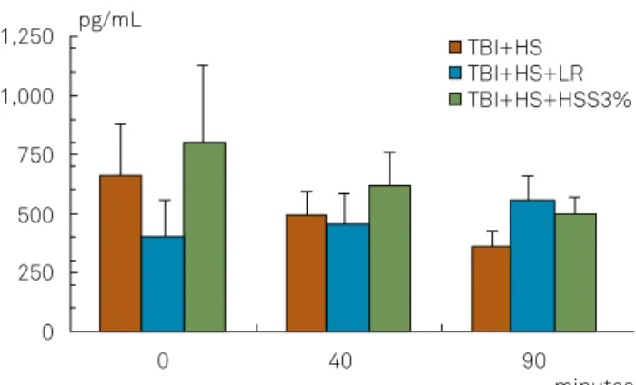
Cerebral thromboxane
1,250 pg/mL
minutes TBI+HS
TBI+HS+LR TBI+HS+HSS3% 1,000
500
250 750
0
0 40 90
TBI: traumatic brain injury; HS: hemorrhagic shock; LR: lactated Ringer’s; HSS: hypertonic saline solution.
Figure 2. Cerebral thromboxane.
Systemic thromboxane
2,500 pg/mL
minutes TBI+HS
TBI+HS+LR TBI+HS+HSS3%
* 2,000
1,000
50 1,500
0
0 40 90
TBI: traumatic brain injury; HS: hemorrhagic shock; LR: lactated Ringer’s; HSS: hypertonic saline solution.
Figure 3. Systemic thromboxane (* = signiicative statistical difference).
Cerebral prostaglandin
2,500 pg/mL
minutes TBI+HS
TBI+HS+LR TBI+HS+HSS3% 1,600
800
400 1,200
0
0 40 90
TBI: traumatic brain injury; HS: hemorrhagic shock; LR: lactated Ringer’s; HSS: hypertonic saline solution.
Figure 4. Cerebral prostaglandin.
Systemic prostaglandin
800 pg/mL
minutes TBI+HS
TBI+HS+LR TBI+HS+HSS3%
*
600
200 400
0
0 40 90
TBI: traumatic brain injury; HS: hemorrhagic shock; LR: lactated Ringer’s; HSS: hypertonic saline solution.
Figure 5. Systemic prostaglandin.
Control group
In this group, systemic thromboxane levels increased
progressively, showing a statistically signiicant diference at T90, with the highest value (1,800 pg/mL) compared to the
treatment groups. Systemic prostaglandin levels remained
stable throughout the experiment (statistically signiicant at T90), with the highest value (600 pg/mL) compared to the
treatment groups.
Cerebral thromboxane levels decreased progressively,
but with no signiicant diference as compared with the oth
-er groups. C-erebral prostaglandin levels increased up to T40
and declined thereafter until T90, without signiicant difer
-ences in relation to the other groups.
Effects of lactated Ringer’s solution
During early in-hospital treatment phase (40’-60’), in
-fused blood volume was 12.9 ± 4.8 ml/kg. In hospitalar phase, LR volume infused was 5.9 ± 9.6 ml/kg.
ICP presented with progressive increasing and was high-er than in control group and HSS 3% group. When epidural
balloon was delated, ICP decreased and became stable at
around 20 mmHg (Figures 6 and 7).
In the LR-treated group, systemic levels of thrombox
-ane and prostaglandin decreased progressively. In the brain, thromboxane levels increased while prostaglandin
levels decreased, although the diferences were not statis
-tically signiicant.
Effects of hypertonic saline solution
During early in-hospital treatment phase (40’-60’), in
-fused blood volume was 11.3 ± 4.0 ml/kg. In hospitalar phase, HSS 3% volume infused was 3.8 ± 4.3 ml/kg. Diferences in
infused blood and cristaloid volumes between groups were
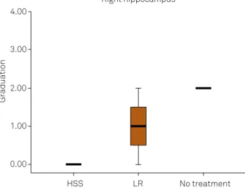
Microscopic examination of cerebral tissue
Microscopic examination of the brain ( frontal, temporal,
parietal, occipital, and hippocampal regions) showed signii
-cant diferences across groups, particularly in the presence
of ischemic lesions in the right and left hippocampi when comparing the HSS-treated group versus the untreated
con-trol group; such injuries were most evident in the untreated
group (Figures 9 and 10).
Pupillary examination
he majority of the dogs showed pupillary changes
(11 of 15 dogs). Findings were anisocoria, with the left pupil being larger than the right (7 of 11 dogs), or bilateral mydria-sis (4 of 11 dogs). Most pupillary changes were manifested within 20 minutes, immediately before the start of the pre-hospital treatment phase.
In the HSS group, two of four animals with anisocoria had early reversal to normal pupillary response at T40. Conversely,
in the LR-treated group, in the two cases in which there was
reversal of mydriasis, this occurred at T90.
Discussion
Systemic inflammatory response and brain release of thromboxane B2 and 6-keto prostaglandin F1 alpha)
Various mammalian cells and tissues oxidize arachi
-donic acid from cell membranes into physiologically ac-tive components. These components include prostacy-clin, thromboxanes, prostaglandins, and leukotrienes. Regardless of its etiology, cerebral ischemia stimulates the synthesis, activation, and release of vasoactive and immu-nologically active substances16,17,18,19,23,24,25,26.
he biological efects of prostacyclin are opposite to those of thromboxane A2. Prostacyclin is a vasodilator and potent platelet aggregation inhibitor, whereas thromboxane A2 is a
vasoconstrictor and platelet aggregation promoter. he phys
-iological balance between the activities of prostacyclin and
50 mm Hg
minutes 40
30
20
10
0
-10 010203040506070 90 120 150 180
Intracranial Pressure (ICP)
TBI+HS TBI+HS+LR TBI+HS+HSS3%
****
TBI: traumatic brain injury; HS: hemorrhagic shock; LR: lactated Ringer’s; HSS: hypertonic saline solution.
Figure 6. Intracranial pressure (ICP) for each group in treatment protocol.
50 mm Hg
minutes 40
30
20
10
0
-10 0 10203040506070 90 120 150 180
Mean Arterial Pressure (PAM)
TBI+HS TBI+HS+LR TBI+HS+HSS3%
***
*
* * *
* *
TBI: traumatic brain injury; HS: hemorrhagic shock; LR: lactated Ringer’s; HSS: hypertonic saline solution.
Figure 7. Mean arterial pressure (MAP) for each group in treatment protocol.
ICP after HSS 3% was the lowest in 60 initial minutes.
When epidural balloon was delated, ICP decreased and be
-came stable at around 20 mmHg (Figures 6 and 7).
In the HSS-treated group, systemic thromboxane levels
increased slightly up to T40 and declined thereafter; system
-ic prostaglandin levels behaved similar, but both the increase and the subsequent decline were more pronounced. Cerebral thromboxane levels decreased progressively while cerebral prostaglandin levels increased slightly up to T40 and then
de-creased, although with no statistically signiicant diferences.
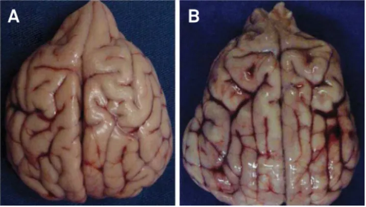
Gross examination of cerebral tissue
he most striking result was the identiication of edema in the brains of all control (untreated) animals and in the LR group; in the HSS-treated group, no edema was identiied. he
other gross indings were similar across the three groups: trau
-matic subarachnoid hemorrhage (TSAH), two cases of acute
subdural hematoma (ASDH), three left frontotemporal contu
-sions, and two cases of intraventricular hemorrhage (Figure 8).
TBI: traumatic brain injury; HS: hemorrhagic shock; LR: lactated Ringer’s. Figure 8. Superior view of two experimental brains. A is an example of normal brain and B is an example of bihemispheric edema, with diffuse hemorrhage (dog number 14,
HS + TBI + LR group).
thromboxane A2 is probably important for maintaining a
healthy vascular bed23,24,25,26.
Widespread cerebral ischemia causes release of TXB2,
the principal metabolite of thromboxane A2 (TXA2), an eico
-sanoid with potent vasoconstrictive and platelet aggregating
properties. his increase in TXB2 in the cerebral venous cir
-culation, which persists for up to 2 hours after reperfusion, is associated with cerebral hypoperfusion23,24,25,26.
Hemorrhagic shock associated with increased
intracrani-al pressure (20 mmHg) causes decreased cerebrintracrani-al blood low
and a signiicant increase in the release of TXB2 into the ce
-rebral venous circulation. Isolated HS suicient to reduce ce
-rebral perfusion pressure to levels found in combined HS and
TBI, however, does not increase cerebral venous TXB2. his
suggests that both incomplete and complete cerebral
isch-emia are associated with generation of TXA2 in the brain; how
-ever, it does not explain whether the increase in TXA2 is a sec
-ondary phenomenon to ischemia or whether TXA2 produces
cerebral ischemia through its vasoconstrictor efects16,17,18,19.
In our experiment, plasma concentrations of thrombox-ane in central blood samples were always higher than those
found in cerebral blood, regardless of the analyzed group. he
opposite pattern was observed for prostaglandin concentra-tions, which were consistently higher in the cerebral venous
circulation. his suggests the existence of a balance between
prostaglandin and thromboxane concentrations.
here were no statistically signiicant diferences be
-tween the treated groups (LR and HSS) regarding cerebral and
plasma concentrations of thromboxane and prostaglandin.
However, the control group (no treatment) exhibited signii
-cantly higher thromboxane and prostaglandin concentrations
after 90 minutes. he progressive increase in central arterial
plasma concentrations of thromboxane in the control group
shows the inluence of untreated hemorrhagic shock and pul
-monary circulation ischemia on levels of this mediator.
Gross examination of cerebral tissue
he presence of edema in the brains of all control and LR-treated animals and the absence of cerebral edema in all
HSS-treated animals suggests that HSS prevented the forma-tion of macroscopically detectable cerebral edema.
Other indings, such as traumatic subarachnoid hemor
-rhage, acute subdural hematoma, cerebral contusion, intra-ventricular hematoma, and corpus callosum and subcortical
hemorrhages simply conirm that the experimental model of
traumatic brain injury used in this study induces both focal
and difuse cerebral lesions.
Microscopic examination of cerebral tissue
Considering the selective vulnerability of some areas of the central nervous system to ischemia, we found that there
was a signiicant diference between the control group and
the HSS-treated group regarding presence of ischemia in the right and left hippocampi.
A larger sample would probably have allowed statistical conirmation of a potential diference between LR and HSS
in this aspect.
Pupils
It is known that shock itself can lead to pupillary changes, especially bilateral mydriasis, due to a vigorous adrenergic re-sponse that culminates in pupillary dilation1,2,3,4,5. However,
in-tracranial injuries with mass efect, which cause uncal hernia
-tion, can trigger anisocoria with ipsilateral pupillary dilation
secondary to compression of parasympathetic ibers in the
third cranial nerve against the tentorium cerebelli. Reversal of
shock with luid resuscitation might be able to reverse pupil
-lary changes generated by this adrenergic mechanism1,2.
Graduation
Right hippocampus 4.00
3.00
2.00
1.00
0.00
HSS LR No treatment
LR: lactated Ringer’s; HSS: hypertonic saline solution.
Figure 9. Histological analysis of right hyppocampus. There was signiicant statistical difference in this topography between HSS 3% group and control.
Graduation
Left hippocampus 3.00
1.50 2.00 2.50
1.00
0.50
0.00
HSS LR No treatment
LR: lactated Ringer’s; HSS: hypertonic saline solution.
We noticed earlier reversal of the pathological pupillary pattern in the HSS-treated group (at 40 minutes) than in the
LR-treated group (reversal at 90 minutes). hus, HSS can pro
-vide early beneits secondary to reversal of brain herniation if
administered in the prehospital environment.
In conclusion, treated groups (LR or HSS) presented no statistically signiicant diferences between them in cerebral
and plasma concentrations of thromboxane and prostaglan-din. However, control group (no treatment) showed values of
systemic plasma concentration of thromboxane and
prosta-glandin signiicantly higher at 90 minutes, suggesting more aggressive inlammatory response after TBI. hen, the group
treated with 3% HSS presented no cerebral edema detected macroscopically and did not disclose ischemic hippocampus like control group. Finally, pupillary reversal occurred earlier
in HSS group than in LR group. hose indings suggest HSS to
be a valuable therapeutic intervention to prevent inlamma
-tory events following TBI.
References
1. Pinto FC, Capone-Neto A, Prist R, E Silva MR, Poli-de-Figueiredo LF. Volume replacement with lactated Ringer’s or 3% hypertonic saline solution during combined experimental hemorrhagic shock and traumatic brain injury. J Trauma. 2006;60(4):758-64. http://dx.doi.org/10.1097/01.ta.0000214581.89316.73
2. Rocha-e-Silva M, Poli de Figueiredo LF. Small volume hypertonic resuscitation of circulatory shock. Clinics (São Paulo). 2005;60(2):159-72. http://dx.doi.org/10.1590/S1807-59322005000200013
3. Balbino M, Capone Neto A, Prist R, Ferreira AT, Poli-de-Figueiredo LF. Fluid resuscitation with isotonic or hypertonic saline solution avoids intraneural calcium inlux after traumatic brain injury associated with hemorrhagic shock. J Trauma. 2010;68(4):859-64. http://dx.doi.org/10.1097/TA.0b013e3181af69d3
4. Tan PG, Cincotta M, Clavisi O, Bragge P, Wasiak J, Pattuwage L et al. Prehospital luid management in traumatic brain injury. Emerg Med Australas. 2011;23(6):665-76. http://dx.doi.org/10.1111/j.1742-6723.2011.01455.x
5. Exo JL, Shellington DK, Bayir H, Vagni VA, Janesco-Feldman K, Ma L et al. . Resuscitation of traumatic brain injury and hemorrhagic shock with polynitroxylated albumin, hextend, hypertonic saline, and lactated Ringer’s: effects on acute hemodynamics, survival, and neuronal death in mice. J Neurotrauma. 2009;26(12):2403-8. http://dx.doi.org/10.1089/neu.2009.0980
6. Gantner D, Moore EM, Cooper DJ. Intravenous luids in traumatic brain injury: what’s the solution? Curr Opin Crit Care. 2014;20(4):385-9. http://dx.doi.org/10.1097/MCC.0000000000000114
7. Dekker SE, Sillesen M, Bambakidis T, Jin G, Liu B, Boer C et al. Normal saline inluences coagulation and endothelial function after traumatic brain injury and hemorrhagic shock in pigs. Surgery. 2014;156(3):556-63. http://dx.doi.org/10.1016/j.surg.2014.04.016
8. Shein SL, Shellington DK, Exo J, Jackson TC, Wisniewski SR, Jackson EK et al. Hemorrhagic shock shifts the serum cytokine proile from pro- to anti-inlammatory after experimental traumatic brain injury in mice. J Neurotrauma. 2014;31(6):1386-95. http://dx.doi.org/10.1089/neu.2013.2985
9. Wang JW, Li JP, Song YL, Tan K, Wang Y, Li T et al. Hypertonic saline in the traumatic hypovolemic shock: meta-analysis. J Surg Res. 2014;191(2):448-54. http://dx.doi.org/10.1016/j.jss.2014.04.027
10. Van Aken HK, Kampmeier TG, Ertmer C, Westphal M. Fluid resuscitation in patients with traumatic brain injury: what is a SAFE approach?. Curr Opin Anaesthesiol. 2012;25(5):563-5. http://dx.doi.org/10.1097/ACO.0b013e3283572274
11. Phillips CR, Vinecore K, Hagg DS, Sawai RS, Differding JA, Watters JM et al. Resuscitation of haemorrhagic shock with normal saline vs. lactated Ringer’s: effects on oxygenation, extravascular lung water and haemodynamics. Crit Care. 2009;13(2):R30. http://dx.doi.org/10.1186/cc7736
12. Schackford SR, Mackersie RC, Holbrook TL, Davis JW, Holingsworth-Fridlund P, Hoyt DB et al. The epidemiology of traumatic death. A population-based analysis. Arch Surg. 1993;128(5):571-5. http://dx.doi.org/10.1001/archsurg.1993.01420170107016
13. Czosnyka M, Smielewski P, Piechnik S, Steiner LA, Pickard JD. Cerebral autoregulation following head injury. J Neurosurg. 2001;95(5):756-63. http://dx.doi.org/10.3171/jns.2001.95.5.0756
14. Anderson JT, Wisner DH, Sullivan PE, Matteucci M, Freschman S, Hildreth J et al. Initial small-volume hypertonic resuscitation of shock and brain injury: short- and long-term effects. J Trauma. 1997;42:592-600. http://dx.doi.org/10.1097/00005373-199704000-00003
15. Doyle JA, Davis DP, Hoyt DB. The use of hypertonic saline in the treatment of traumatic brain injury. J Trauma. 2001;50(2):367-83. http://dx.doi.org/10.1097/00005373-200102000-00030
16. Aibiki M, Maekawa S, Yokono S. Moderate hypothermia improves imbalances of thromboxane A2 and prostaglandin I2 production after traumatic brain injury in humans. Crit Care Med. 2000;28(12):3902-6. http://dx.doi.org/10.1097/00003246-200012000-00029
17. Al-Turki A, Armstead WM. Altered release of prostaglandins by opioids contributes to impaired cerebral hemodynamics following brain injury. Crit Care Med. 1998;26(5):917-25. http://dx.doi.org/10.1097/00003246-199805000-00029
18. Armstead WM, Lefler CW, Busija DW, Beasley DG, Mirro R. Adrenergic and prostanoid mechanisms en control of cerebral blood low in hypotensive newborn pigs. Am J Physiol. 1988;254(4 Pt 2):H671-7.
19. DeWitt DS, Prough DS. Vasoactive prostanoids and traumatic brain injury. Crit Care Med. 1998;26(5):819-21. http://dx.doi.org/10.1097/00003246-199805000-00008
20. Lefler CW. Prostanoids: intrinsic modulators of cerebral circulation. News Physiol Sci. 1997;12(2):72-7.
21. Grice SC, Chappell ET, Prough DS, Whitey JM, Su M, Watkins WD. Ibuprofen improves cerebral blood low after global cerebral ischemia in dogs. Stroke. 1987;18(4):787-91. http://dx.doi.org/10.1161/01.STR.18.4.787
22. Eliastam M, Safar P, Shapiro M, Gorovitz S. Ethical aspects of cardiopulmonary-cerebral resuscitation research. Ann Emerg Med. 1984;13(9):874-5. http://dx.doi.org/10.1016/S0196-0644(84)80464-3
23. Holcroft JW, Vassar MJ, Turner JE, Derlet RW, Kramer GC. 3% saline and 7% dextran: 70 in the resuscitation of severely injuried patients. Ann Surg. 1987;206(3):279-85. http://dx.doi.org/10.1097/00000658-198709000-00006
24. Kong DL, Prough DS, Whitley JM, Taylor C, Vines S, Deal DD et al. Hemorrhage and intracranial hypertension in combination increase cerebral production of thromboxane A2. Crit Care Med. 1991;19(4):532-8. http://dx.doi.org/10.1097/00003246-199104000-00013
25. Bulger EM, Cuschieri J, Warner K, Maier RV. Hypertonic resuscitation modulates the inlammatory response in patients with traumatic hemorrhagic shock. Ann Surg. 2007;245(4):635-41. http://dx.doi.org/10.1097/01.sla.0000251367.44890.ae