Encodes a Structural HMT,
SMYD1b, with Chaperone-Like Function
during Fast Muscle Sarcomere Assembly
Kendal Prill☯, Pamela Windsor Reid☯, Serene L. Wohlgemuth¤, David B. Pilgrim*
Department of Biological Sciences, University of Alberta, Edmonton, Alberta, Canada
☯These authors contributed equally to this work.
¤ Current address: Centre for Prions and Protein Folding Diseases, University of Alberta, Edmonton, Alberta, Canada
Abstract
The vertebrate sarcomere is a complex and highly organized contractile structure whose assembly and function requires the coordination of hundreds of proteins. Proteins require proper folding and incorporation into the sarcomere by assembly factors, and they must also be maintained and replaced due to the constant physical stress of muscle contraction. Zebrafish mutants affecting muscle assembly and maintenance have proven to be an ideal tool for identification and analysis of factors necessary for these processes. Thestill heart mutant was identified due to motility defects and a nonfunctional heart. The cognate gene for the mutant was shown to besmyd1band thestill heartmutation results in an early non-sense codon. SMYD1 mutants show a lack of heart looping and chamber definition due to a lack of expression of heart morphogenesis factorsgata4,gata5andhand2. On a cellular level, fast muscle fibers in homozygous mutants do not form mature sarcomeres due to the lack of fast muscle myosin incorporation by SMYD1b when sarcomeres are first being assembled (19hpf), supporting SMYD1b as an assembly protein during sarcomere formation.
Introduction
The sarcomere is a complex and highly organized structure where distinct protein filaments rely on the involvement and coordination of hundreds of other proteins for proper patterning, assembly and maintenance. Sarcomere proteins fall into three broad categories including con-tractile proteins, such as actin and myosin; structural attachment and anchoring components, such as integrins,α-actinin and myomesin, and patterning and folding chaperones such as Hsp90 [1–3]. While the function and activity of these proteins is well established in the mature sarcomere, the mechanisms by which the sarcomere proteins are assembled and initially pat-terned as the myocyte differentiates are still a matter of conjecture.
OPEN ACCESS
Citation:Prill K, Windsor Reid P, Wohlgemuth SL, Pilgrim DB (2015)Still HeartEncodes a Structural HMT, SMYD1b, with Chaperone-Like Function during Fast Muscle Sarcomere Assembly. PLoS ONE 10(11): e0142528. doi:10.1371/journal.pone.0142528
Editor:Eric A Shelden, Washington State University, UNITED STATES
Received:January 9, 2015
Accepted:October 22, 2015
Published:November 6, 2015
Copyright:© 2015 Prill et al. This is an open access article distributed under the terms of theCreative Commons Attribution License, which permits unrestricted use, distribution, and reproduction in any medium, provided the original author and source are credited.
Data Availability Statement:All relevant data are within the paper and its Supporting Information files. All raw data is available at the figshare database: http://dx.doi.org/10.6084/m9.figshare.1454588.
Many human myopathies are due to muscle weakness or progressive degeneration of the muscle due to the inability to maintain or replace sarcomere proteins [4,5]. The sarcomere is a continuously active structure with high levels of protein turnover. When a structural sarco-meric protein begins losing integrity, a newly formed and folded protein replaces it; a process facilitated by sarcomere chaperones and their cofactors [6]. Without chaperones for mainte-nance of sarcomere proteins, this complex contractile structure would denature and muscle function would cease [7–10]. Some chaperones are required for assembly of the sarcomere, others for maintenance (SMYD2) and some have been proposed to perform both functions throughout muscle development (UNC45b and HSP90a1)[6,11–13], however many factors remain unidentified [14]. Of those assembly and maintenance factors that have been isolated, molecular function and clients of these proteins often remain unclear.
A powerful approach to understanding this patterning process has been to knock down known or suspected factors in cell culture or animal models and determine the earliest detect-able defect, but this can only test components identified by criteria such as gene expression or biochemical purification to be enriched in the developing sarcomere. Embryonic lethality of mammalian embryos bearing null mutations in many sarcomeric proteins hampers gene dis-covery screens in mice. The zebrafish provides a powerful platform for such forward screens as the embryo is able to survive for several days without cardiac or trunk muscle function. An extensive genetic analysis of developmental mutants published in 1996 identified hundreds of mutations affecting locomotive behavior [14]. In the majority of mutants, defects are seen either in trunk muscle, as seen in motility mutantssloth,twitch onceandtechno trousers, or car-diac muscle in mutantsvalentine,weak atriumandbeach bum[14,15]. However, a small sub-set (less than 15%) of motility mutants have defects in both heart and trunk muscle [14], suggesting that there may be only a small number of shared factors that work in both tissues. These dual cardiac and skeletal mutants suggest a model where one pathway responsible for patterning or sarcomere assembly is shared between both muscle types early muscle development.
We have been particularly interested in those non-myofilament proteins, such as myosin-specific chaperone UNC-45, that function early in the patterning of striated muscle, as they are likely candidates for factors necessary for establishing the patterning, alignment and register of the early myofibrils [10,16–19]. Those zebrafish mutants affecting both cardiac and skeletal muscle, and whose myofibers show early disorganization without obvious defects to somite morphogenesis would be excellent candidates for factors in a rudimentary chaperone-depen-dent assembly process common to all sarcomeres, or alternatively to a common mechanism for sarcomere homeostasis.
The SMYD family of proteins has been recently identified as contributing to sarcomere assembly and maintenance [20,21]. While the presence of MYND and SET domains in the SMYD protein suggests a role as a histone methyltransferase (HMT) [22,23], recent work has demonstrated that there are other protein binding domains within this family that are consis-tent with chaperone-like behavior [20,21,24]. SMYD1 is expressed in the heart and skeletal muscle in both mouse and zebrafish; knockdown in mice leads to a fatal cardiomyopathy while a knockdown ofsmyd1bin zebrafish leads to a dual-skeletal and cardiomyopathy that is lethal at 7 days past fertilization [22,25]. Zebrafish SMYD1 mutants share similar phenotypes to other muscle chaperone mutants,steifandsloth, that have mutations inunc45bandhsp90a1, respectively [2,10,13]. Mutations in either of these assembly/maintenance proteins results in paralysis (sloth/hsp90a1) or a dual myopathy of both cardiac and skeletal tissues (steif/unc45b) [2,10,13].
One of the mutants identified in the 1996 screen,still heart(sth) [14] had characteristics (a nonfunctional heart and reduced motility) similar to existing mutants known to affect early Competing Interests:The authors have declared
sarcomere patterning and assembly (steif/unc45bandsloth/hsp90a1) [2,10]. Here we show that fast muscle fibers insthembryos do not form mature sarcomeres as a result of the lack of fast muscle myosin. Additionally, fast myosin is never incorporated into the early premyofibers when sarcomeres are first being assembled (19hpf). Genetic mapping and complementation of thesthmutant shows that the phenotype is due to a nonsense mutation in thesmyd1bgene. The expression of muscle chaperonesunc45bandhsp90a1do not increase until 24hpf, suggest-ing that these proteins do not increase their expression in response to an absence of SMYD1b (at 19hpf) but are rather responding to increased levels of misfolded myosins[26–28]. This data, along with existing expression data from other systems, suggests that SMYD1b has the role of a thick-filament assembly protein during sarcomere formation.
Materials and Methods
Ethics Statement
All protocols were performed according to the guidelines stated by the Canadian Council for Animal Care and the protocols approved by the Animal Care and Use Committee of the Uni-versity of Alberta (Fish Research License: 14–0101 FR).
Zebrafish Line Maintenance
The Nuüsslein-Volhard lab identified thestill hearttm123a(sth) zebrafish mutant in the
Tubingen (TB) genetic background from an ENU mutagenesis screen [14]. Thesthtm123astrain was crossed to the wild-type AB strain in our lab and further bred to the WIK (Wild Indian Karyotype) strain for microsatellite mapping [29,30]. Theflatlinezf340mutant strain, with a T to A transversion mutant in exon 2, was generated from a ENU mutagenesis screen [14] and studied/maintained by the Rottbauer lab at the University of Ulm, Germany [20]. This mutant strain was a generous gift from Dr. Wolfgang Rottbauer. Thestill heartstrain was maintained as adult heterozygotes in a recycling water aquatic facility and bred and maintained according to standard protocols [31]. Embryos were collected and maintained at 28.5°C in standard Zeb-rafish Embryo Medium for up to 7 days before collected for experiment preparation.
Complementation Crossing
Adult heterozygousstill heartand adult heterozygousflatlinemutants were crossed in three separate pairings (1 heterozygous malestill heartto 1 heterozygous femaleflatline) and the fer-tilized embryos were collected for phenotype scoring. Crosses betweenstill heartandflatline
heterozygotes gave broods with wild type and mutant phenotypes, consistent with Mendelian ratios.
Touch Test
Mutant Sequencing
Homozygousstill heartmutants and their siblings were collected at 2 dpf, after the phenotype becomes apparent, for RNA extraction using TRIzol (Life Technologies, Burlington, ON, Can-ada).smyd1b(GenBank:CT027631) cDNA was sequenced from homozygous mutant and wild-type sibling RNA extractions that were reverse transcribed. The mutation present in cDNA was confirmed by genomic sequencing of an exon 1 RT/PCR product in homozygous
sthmutants and their wild type siblings. Genomic sequencing was performed 10 times from 10 individual embryos. cDNA sequencing was performed 10 times from RNA extractions of 30 embryos. All sequencing was performed at the Molecular Biology Service Unit at the University of Alberta on a Sanger DNA Sequencer (ABI 3730). Primer sequences are available inS1 Table.
In situ hybridization and immunostaining
In situhybridization was performed on whole-mount zebrafish embryos fixed overnight in 4% paraformaldehyde (PFA), as previously published [32], using antisense RNA probes synthe-sized by in vitro transcription using SP6, T7 or T3 RNA polymerase. Probes used were zebra-fishunc45b[13] (GenBank:CR847826),hsp90a1[33] (GenBank:BC075757),smyhc1[34] (GenBank:CU929259) andmyhc4[35] (GenBank:BX511237). For immunostaining, embryos were staged morphologically, fixed for 1 hour at room temperature in 4% PFA. Immunostain-ing was carried out as previously described [33] with antibodies against fast-muscle myosin (1/ 10 F310, DSHB, Iowa, USA), slow-muscle myosin (1/10 F59, DSHB, Iowa, USA). All experi-ments were carried out 3 separate times, from 3 individual broods with a minimum of 15 embryos per condition (total = 45 wt and 45sthembryos per probe/antibody). Primer sequences are available inS1 Table.
RNA extractions and qPCR
RNA extractions were performed on collections of a minimum of 30 embryos that were flash frozen in liquid nitrogen before using a TRIzol RNA extraction. RNA was standardized to one concentration for all samples before proceeding to cDNA synthesis using a qScript cDNA syn-thesis kit (Quanta Biosciences, Gaithersberg, MD, USA). cDNA was diluted to 5ng/ul for Quantitative PCR.ef1awas used as the endogenous control for normalization of samples and expression of genes of interest. qPCR and data analysis was carried out using the Illumina Eco Real-Time PCR system. Three replicates of expression for each gene were generated for each N number of WT andstill heartembryo collections. Error bars represent the standard deviation of the WT andstill heartgene expression replicates generated from each qPCR. Primer sequences are available inS1 Table.
Raw data can be found athttp://dx.doi.org/10.6084/m9.figshare.1454588
Results
Still heart
has defects in both heart and skeletal muscle structure and
function as a result of a mutation in
smyd1b
Fig 1.Still heart, asmyd1bmutant, has defects in heart and fast skeletal muscle tissue.A lateral view of 48hpf wild type (A) andstill heart(sth) mutants (B), which have pericardial edema, small eyes, malformed head and reduced motility. Black arrowheads highlight the pericardial edema in sth mutants and the absence of edema in wild type. White arrowheads indicate blood pooling in the mutant and the absence of pooling in wild type. (C&D)Sthmutant hearts are underdeveloped and do not beat. (E-G) Examination of lateral myofibers at 5dpf under DIC microscopy revealed striations, indicative of fully formed sarcomeres, are visible in the myofibers of wild type muscle (E, black arrowheads), while absent in the fast muscle ofstill heartmutants (F); striations are present insthslow muscle (G, black arrowhead) but are disturbed by nuclei and fluid-filled spaces (G, white arrowhead). Sequencing ofsmyd1bcDNA from wild type embryos (H) andsthmutant embryos (I) revealed a 9 nucleotide insertion between exon 1 and 2 in thesmyd1bmRNA, creating an in-frame stop codon (I, underlined sequence). The insertion is the first 9 nucleotides of intron one as sequenced from wild type smyd1b genomic sequence (J). This is a result of a transition mutation in the splice donor site of intron 1 (I, green letter in sequence, J, outlined letter in sequence). This results in a premature truncation of the SMYD1b protein after exon 1 (K).
mutants, at 48 hpf (hours post fertilization), do not swim away after being touched but are only capable of a vibrational response. A heartbeat is never seen during development but two cham-bers do form although smaller than the wild type (Fig 1C and 1D). The progressive effect of the mutant is also visible in skeletal muscle tissue. At 24hpf,sthfast muscle tissue lacks striation in myofibrils, which suggests a defect in sarcomere assembly; slow-twitch myofibrils exhibit stria-tion and appear normal. By 5 dpf, fast-twitch myofibrils are disorganized and both fast and slow muscle tissue is riddled with fluid-filled vacuoles (Fig 1E–1G). Part of the progressive nature of thesthphenotype may be due to the increasing accumulation of fluids in the body due to the lack of circulation.
Thesthmutation was mapped at low resolution to a region on chromosome 8 [36], however none of the published markers in the region proved polymorphic for microsatellite analysis in our genetic backgrounds to allow finer resolution mapping. The more recent correlation of genetic and physical maps for zebrafish indicated that the minimal region contained a candi-date locus,smyd1b, based on similarity to a morpholino knockdown phenotype [22] and the phenotype of asmyd1bmutation,flatline[20]. A complementation cross between heterozygous
still heartandflatlineadults produced mutant progeny, identifyingflatlineas allelic tostill heartas a mutant at thesmyd1blocus. Sequence analysis ofsmyd1bcDNA fromsthmutant embryos revealed an insertion of 9 nucleotides that contain an in-frame stop codon between exon 1 and exon 2 (Fig 1H&1I). Genomic sequencing ofsth smyd1bshowed that the splice donor site of intron 1 has a transition mutation (Fig 1I&1J) that abolishes the function of this site and incorporates the stop codon that causes a premature truncation of theSmyd1bprotein, effectively removing all of the functional domains (Fig 1K).
SMYD1b is involved in the assembly of the sarcomeres in cardiac and
fast-twitch muscle
While contraction of the heart insthmutants is not seen, skeletal muscle is capable of some vibrational movement. One possibility for the reduced motility is that a lack of nutrient circula-tion due to a nonfunccircula-tional heart could disturb the development of normal skeletal muscle. The other possibility for the motility phenotype is that it is a direct effect of thesthmutation. To distinguish between these possibilities, we performed touch tests on young embryos (before the heart forms and begins beating at 24 hpf) derived fromsthheterozygous parents and sorted them based on motility response. Of 548 embryos tested at 20 hpf, 156 (28%) were scored as deficient in touch response. After the sorted embryos were allowed to develop for several more hours, they were re-tested for the motility defect as well as for heartbeat. Of the embryos origi-nally scored with a motility defect, 13% showed normal movement and heartbeat at the later time point, while none of the embryos scored as wild-type showed subsequent movement defect. Overall, 138 of the 548 (25%) embryos showed both motility and heart defects at the later time point, consistent with a single recessive locus with complete penetrance. Therefore, the vibrational movement and motility defect ofstill heartmutants is present before the heart-beat begins and is a defect of thesthmutation, not a secondary effect of a lack of circulation. In addition, it appears that development of the vascular system to the skeletal muscle is superfi-cially unaffected insthmutants (S1 Fig).
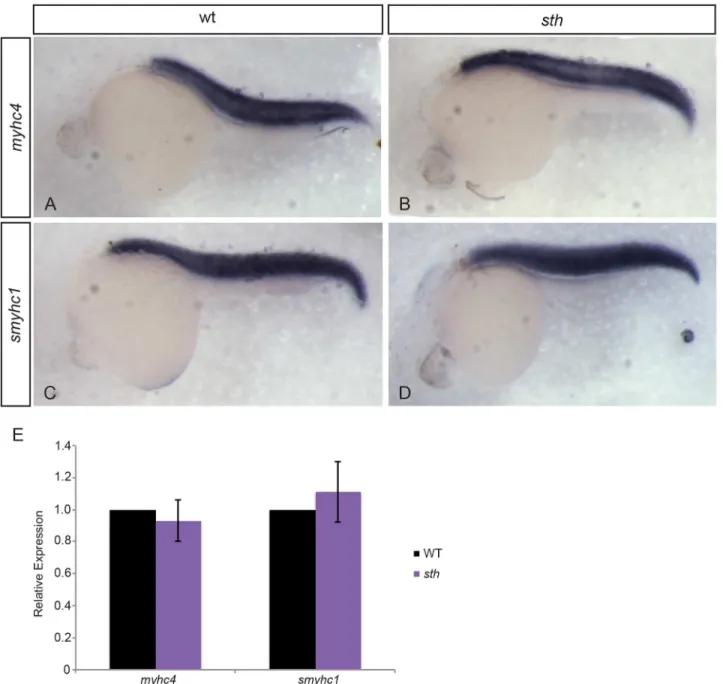
sarcomere.Still heartmutants do not swim away after being touched, a normal evasion response to predators that is seen in their wild type siblings. The lack of an avoid response sug-gests that thesthmutation is affecting fast skeletal muscle fibers that are used for rapid swim-ming in response to an acute stimulus. We analyzed the expression levels of fast and slow-twitch myosin mRNA insthmutants and wild type siblings and found that there were no sig-nificant differences in myosin expression between homozygous mutants and sibs (Fig 2A–2E).
Comparison of fast and slow muscle fibers in wild type andstill heartembryos shows that fast muscle fibers lack striations, indicative of sarcomeres, when compared to fast muscle in
Fig 2. Myosin expression is unaffected by the absence of SMYD1b.At 24hpf, fast myosin (myhc4) expression in wild type (A) andstill heartembryos (B) is similar within the trunk muscle. Slow myosin (smyhc1) expression in wild type (C) andstill heartembryos (D) is also similar at 24hpf. Quantitative PCR on wild type andsthmutants for fast and slow myosin expression shows myosin expression is normal when compared to wild type embryos at 24hpf (E). (qPCR: n = 3, 30 embryos each time/phenotype. Error bars are standard deviation.).
wild type siblings at 48 hpf (Fig 3A-3H). Slow muscle maintains striations throughout develop-ment although myofibrils eventually become disrupted as a secondary effect from the increas-ing disorganization surroundincreas-ing fast muscle fibers (Fig 1G). Even though fast myosin is not incorporated into the sarcomere, fast myosin protein is still abundant in the muscle tissue [21]. This supports a model where SMYD1b is a chaperone-like protein that is involved in the assembly and maintenance of the sarcomere. To determine if SMYD1b is an assembly factor in
Fig 3. SMYD1b is required for fast myosin incorporation during sarcomere assembly.At 48hpf, fast myosin (F310—green) and actin (phalloidin—red) staining is visible in the premyofibrils in wt zebrafish tails (A&B), while absent from the premyofibrils insthfast muscle tissue (C&D). Slow muscle (F59) develops normally in both wild type andstill heartzebrafish at 48hpf (E&F, G&H). At 19hpf, in wild type embryos, fast myosin (F310) is beginning to be incorporated into the maturing myofibril (I) and overlaps (white arrowheads) with the developing actin (phalloidin) (J) fibers in trunk muscle (K, merge, K’inset, white arrowhead). Fast myosin is not incorporated into the maturing premyofibril (L), although actin fibers are still present (M&N, N’inset).
sarcomere development, we examined early time points (15 and 19 hpf) in fast muscle sarco-mere formation. Sarcosarco-meres are absent in fast muscle insthmutants at later stages [20], but it is not clear whether this is due to sarcomere breakdown in the absence of a maintenance role of SMYD1b, or if the sarcomeres never formed due to a lack of assembly by SMYD1b. If sarco-meres are present at the time when myosin is first incorporated, then SMYD1b is involved in the maintenance of sarcomeric proteins that later breakdown from stress of contraction and are never replaced. If myosin is not present at the beginning of sarcomere formation, then SMYD1b is an assembly factor that is required for the proper folding and incorporating of fast myosin into the sarcomere; if SMYD1b is an assembly factor, this does not rule out the possibil-ity that it also has a role in maintenance of sarcomere proteins. At the 20 somite stage, the embryo contains both mature somites with formed sarcomeres and developing somites that have yet to build their sarcomeres. We examined fast myosin immunostaining at 14 and 20 somites in wild type andstill heartembryos. At 14 somites, fast myosin was not detectable in any of the somites. However at 20 somites, premature actin/myosin fibers form between myo-septa in wild type embryos (Fig 3I-3K). Instill heartembryos, at 20 somites, actin fibers are present, but no fast myosin fibers can be detected in fast muscle tissue (Fig 3L-3N). The lack of fast myosin in the premature actin/myosin fiber during early sarcomere formation supports the model that SMYD1b is an assembly factor rather than a maintenance protein.
SMYD1b works in tandem with myosin chaperones HSP90a1 and
UNC45b to fold and incorporate fast myosin into the sarcomere
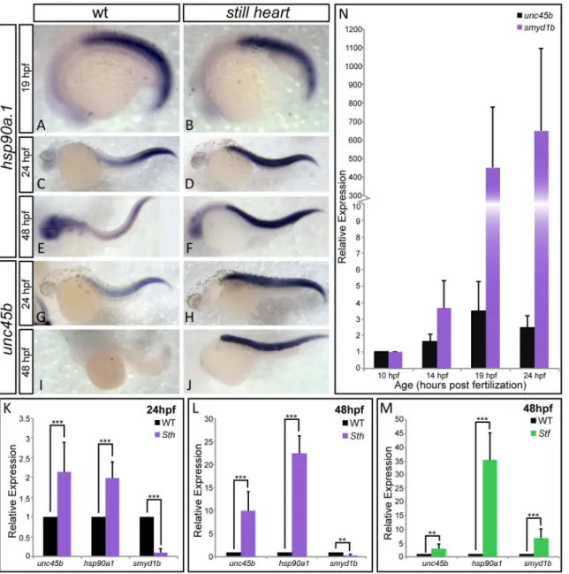
The absence of fast myosin incorporation suggests that SMYD1b is involved in incorporation of myosin into fast muscle sarcomeres. To test whether thesmyd1bsthmutation is affecting sar-comere formation at the level of protein folding and assembly, and not transcriptional activa-tion, we examined the mRNA expression levels of myosin chaperonesunc45bandhsp90a1. These muscle chaperones will transcriptionally respond to the presence of misfolded client pro-teins but not the complete absence of sarcomeric structural propro-teins [20,37].Still heart
mutants show a significant increase inunc45b/hsp90a1expression when compared to wild type siblings at 24 and 48 hpf (Fig 4C-4M). This increased expression is not due directly to the lack of SMYD1b, as at 19hpf the expression ofhsp90a1resembles wild type expression (Fig 4A and 4B), suggesting that these chaperones are not co-regulated at the onset of fast myosin develop-ment. The increasing expression of these chaperones could be due the accumulation of mis-folded myosins. We have observed the same trend of increasing chaperone expression of
smyd1bandhsp90a1inunc45b/steifmutants (Fig 4N). Taken together, this supports a model where SMYD1b, UNC45b and HSP90a1 work together to properly fold and incorporate fast myosin into the sarcomere and exhibit a transcriptional up-regulation when myosin is not incorporated/folded in mutant myofibers.
Although the lack of SMYD1b predominantly shows defects in fast myosin incorporation, we analyzed the expression ofsmyd1bin comparison tounc45bin wild type embryos.Smyd1b
is first detectable byin situ hybridizationat 10hpf, at the time whenunc45bis expressed and working with non-muscle myosin to build the premature sarcomere [16]. The expression of
smyd1b9 hours before fast muscle begins to develop (Fig 4O), suggests that SMYD1b may have an earlier muscle-myosin independent function during muscle development but such an early role remains unclear.
Discussion
muscle factors that have a role in both cardiac and skeletal sarcomere formation, suggests both muscle types have early development components in common, but later diverge and specialize during development. However, the precise molecular role of the majority of the factors in the formation and maintenance of the sarcomere remain unknown [14,15]. Here we demonstrate that the histone methyltransferase, SMYD1b, is a chaperone-like assembly protein that is cru-cial to the assembly of the sarcomere with regards to fast but not slow muscle myosin in stri-ated muscle.
Fig 4. SMYD1b is co-regulated with other myosin chaperones HSP90a1 and UNC45b.In-situ hybridization staining of wild type andstill heartembryos shows thathsp90a1expression is normal instill heartmutants, when compared to wild type embryos at 19hpf (A&B). However, the expression ofhsp90a1 andunc45bdramatically increases instill heartmutants at 24hpf (C-F & K) and 48hpf throughout the somites (G-J & M). Additionally,smyd1bexpression increases significantly when UNC45b is absent insteifmutants (N), supporting co-regulation of these three genes. (O) A time course ofsmyd1bexpression during muscle formation reveals thatsmyd1bis expressed early at 10hpf whenunc45bis expressed and increases in its expression as muscle development progresses. Due to the rapid development ofunc45bstaining in the somites of the embryo in panel J, the head while present, has no background staining and is not clear in this focal plane. (qPCR: n = 3, 30 embryos each time/phenotype. Error bars are standard deviation.).
It has been clear over the last few years that the Smyd family of proteins plays a role in mus-cle development but their molecular role and potential cofactors are relatively unknown [11, 25,38]. SMYD2 is indirectly responsible for TITIN stability in the sarcomere by methylating HSP90a1 and forming a complex with the two during sarcomere formation [11]. We show that SMYD1b does not affect myosin mRNA expression, as would be expected from a chromatin modifying protein, but instead has a role in fast myosin incorporation into the sarcomere. It has been suggested that SMYD1b has a chaperone-like function and may be acting as an assembly or maintenance protein [20,21]. This speculation generates three models for SMYD1b function. The first is that SMYD1b is involved early for establishment of the sarco-mere, the second is that it is involved later for maintenance and the third model requires SMYD1b for both assembly and maintenance. To determine which model is most likely, we examined the earliest stages of myosin incorporation as an indirect measure of SMYD1b neces-sity. Our results show that fast, but not slow, muscle myosin is not incorporated into the sarco-mere in the absence of SMYD1b, and it is at least an assembly protein specific to fast muscle tissue.
In bothflatlineandstill heart, an increased expression in HSP90 and UNC45b is seen where SMYD1b is absent [20]. Although myosin chaperones HSP90, UNC45b and SMYD1b show evidence of co-regulation and have been suggested to work together to fold and incorporate myosin, little is known about whether SMYD1b interacts directly with the others, or functions independently at the same stage of assembly [20,21]. Two models may explain how these three chaperones work together during sarcomere development. The first model is that HSP90, UNC45b and SMYD1b all physically interact and work together to fold and incorporate fast myosin into the sarcomere. In this case, we hypothesize that the MYND domain of SMYD1b is important for binding other proteins such as UNC45b or HSP90a1. The second model is that HSP90a1 and UNC45b interact with SMYD1b indirectly through contacts with fast myosin, and that myosin is passed on in an assembly line type fashion. We can conclude based on the increased expression ofhsp90a1,unc45borsmyd1bin the absence of eithersmyd1borunc45b, that these three chaperones are required for fast myosin folding and incorporation. The increased expression of these chaperones when myosin is not incorporation is an unfolded pro-tein response that would eventually lead to a negative feedback loop on myosin if myosin can-not be properly folded. We see evidence of this regulation when either of these chaperones is missing; there is a decrease in the target myosin protein levels when the expression of the myo-sin chaperones is elevated [20]. Overexpression ofunc45bleads to similar thick filament defects with decreased levels of myosin protein in the developing myofibers, however the mechanism behind how abundant UNC45b levels disrupts the myofibers is unknown [7,39].
It is interesting to note thatsmyd1bis expressed hours before fast muscle begins to develop, but at the same time point whereunc45bis expressed and has been suggested to be involved in assembly of the non-muscle myosin (NMM) scaffold of the premature sarcomere. It is unclear what SMYD1b’s function may be early in development, but perhaps SMYD1b aids UNC45b in folding and setting up the organization of NMM in the early sarcomere. The dramatic increase insmyd1bat 19hpf could be due to the onset of developing fast muscle where SMYD1b is required for sarcomere assembly whileunc45bfunctions in both fast and slow muscle.
order of their function, may reveal a coordinated chaperone system with SMYD proteins hav-ing early roles that stimulate the function of other chaperone proteins in muscle development.
Supporting Information
S1 Fig. Vasculature is normal instill heartmutants at 48hpf.Lateral view of 48hpf zebrafish vasculature demonstrates a repeating network of arteries and veins that highlight each somite in the trunk. Comparing vasculature organization between wild type (A) andstill heart(B), there are no visible differences. (n = 10 embryos)
(TIF)
S1 Table. Primer Sequences used for analysis of SMYD1b’s role in muscle development. (PDF)
Acknowledgments
We would like to express thanks to Dr. Andrew Waskiewicz for allowing us use of his zebrafish aquatic facility. We would also like to thank Dr. Wolfgang Rottbauer for generously providing us withflatlinezebrafish embryos. The F310 and F59 antibody developed by Stockdale, F.E was obtained from the Developmental Studies Hybridoma Bank developed under the auspices of the NICHD and maintained by the University of Iowa, Department of Biology, Iowa City, IA 52242.
Author Contributions
Conceived and designed the experiments: DBP KP. Performed the experiments: KP PWR. Analyzed the data: DBP KP PWR. Contributed reagents/materials/analysis tools: KP PWR SLW. Wrote the paper: KP DBP PWR.
References
1. Clark KA, McElhinny AS, Beckerle MC, Gregorio CC. Striated muscle cytoarchitecture: an intricate web of form and function. Annual Review Cell Developmental Biology. 2002; 18:637–706.
2. Hawkins TA, Haramis AP, Etard C, Prodromou C, Vaughan CK, Ashworth R, et al. The ATPase-depen-dent chaperoning activity of Hsp90a regulates thick filament formation and integration during skeletal muscle myofibrillogenesis. Development. 2008; 135(6):1147–56. doi:10.1242/dev.018150PMID: 18256191
3. Sparrow JC, Schock F. The initial steps of myofibril assembly: integrins pave the way. Nat Rev Mol Cell Biol. 2009; 10(4):293–8. doi:10.1038/nrm2634PMID:19190670
4. Bonnemann CG, Laing NG. Myopathies resulting from mutations in sarcomeric proteins. Curr Opin Neurol. 2004; 17(5):529–37. PMID:15367857
5. Sato Y, Probst HC, Tatsumi R, Ikeuchi Y, Neuberger MS, Rada C. Deficiency in APOBEC2 leads to a shift in muscle fiber type, diminished body mass, and myopathy. The Journal of biological chemistry. 2010; 285(10):7111–8. doi:10.1074/jbc.M109.052977PMID:20022958
6. Etard C, Roostalu U, Strahle U. Shuttling of the chaperones Unc45b and Hsp90a between the A band and the Z line of the myofibril. J Cell Biol. 2008; 180(6):1163–75. doi:10.1083/jcb.200709128PMID: 18347070
7. Bernick EP, Zhang PJ, Du S. Knockdown and overexpression of Unc-45b result in defective myofibril organization in skeletal muscles of zebrafish embryos. BMC Cell Biol. 2010; 11:70. doi: 10.1186/1471-2121-11-70PMID:20849610
8. Codina M, Li J, Gutierrez J, Kao JP, Du SJ. Loss of Smyhc1 or Hsp90alpha1 function results in different effects on myofibril organization in skeletal muscles of zebrafish embryos. PLoS One. 2010; 5(1): e8416. doi:10.1371/journal.pone.0008416PMID:20049323
10. Etard C, Behra M, Fischer N, Hutcheson D, Geisler R, Strahle U. The UCS factor Steif/Unc-45b inter-acts with the heat shock protein Hsp90a during myofibrillogenesis. Developmental biology. 2007; 308 (1):133–43. PMID:17586488
11. Donlin LT, Andresen C, Just S, Rudensky E, Pappas CT, Kruger M, et al. Smyd2 controls cytoplasmic lysine methylation of Hsp90 and myofilament organization. Genes & Development. 2012; 26(2):114–9.
12. Geach TJ, Zimmerman LB. Paralysis and delayed Z-disc formation in the Xenopus tropicalis unc45b mutant dicky ticker. BMC Dev Biol. 2010; 10:75. doi:10.1186/1471-213X-10-75PMID:20637071
13. Wohlgemuth SL, Crawford BD, Pilgrim DB. The myosin co-chaperone UNC-45 is required for skeletal and cardiac muscle function in zebrafish. Developmental biology. 2007; 303(2):483–92. PMID: 17189627
14. Granato M, van Eeden FJM, Schach U, Trowe T, Brand M, Furutani-Seiki M, et al. Genes controlling and mediating locomotion behavior of the zebrafish. Development. 1996; 123:399–413. PMID: 9007258
15. Stainier DY, Fouquet B, Chen JN, Warren KS, Weinstein BM, Meiler SE, et al. Mutations affecting the formation and function of the cardiovascular system in the zebrafish embryo. Development. 1996; 123:285–92. PMID:9007248
16. Myhre JL, Pilgrim DB. At the start of the sarcomere–a previously unrecognized role for myosin chaper-ones and associated proteins during early myofibrillogenesis. Biochemistry Research International. 2012;Article ID 712315:1–16.
17. Ao W, Pilgrim D. Caenorhabditis elegans UNC-45 is a component of muscle thick filaments and coloca-lizes with myosin heavy chain B, but not myosin heavy chain A. J Cell Biology. 2000; 148:375–84.
18. Price MG, Landsverk ML, Barral JM, Epstein HF. Two mammalian UNC-45 isoforms are related to dis-tinct cytoskeletal and muscle-specific functions. J Cell Science. 2002; 115:4013–23. PMID:12356907
19. Ferrante MI, Kiff RM, Goulding DA, Stemple DL. Troponin T is essential for sarcomere assembly in zeb-rafish skeletal muscle. Journal of cell science. 2011; 124(Pt 4):565–77. doi:10.1242/jcs.071274PMID: 21245197
20. Just S, Meder B, Berger IM, Etard C, Trano N, Patzel E, et al. The myosin-interacting protein SMYD1 is essential for sarcomere organization. Journal of cell science. 2011; 124(Pt 18):3127–36. doi:10.1242/ jcs.084772PMID:21852424
21. Li H, Zhong Y, Wang Z, Gao J, Xu J, Chu W, et al. Smyd1b is required for skeletal and cardiac muscle function in zebrafish. Mol Biol Cell. 2013; 24(22):3511–21. doi:10.1091/mbc.E13-06-0352PMID: 24068325
22. Tan X, Rotllant J, Li H, De Deyne P, Du SJ. SmyD1, a histone methyltransferase, is required for myofi-bril organization and muscle contraction in zebrafish embryos. Proceedings of the National Academy of Sciences of the United States of America. 2006; 103(8):2713–8. PMID:16477022
23. Brown MA, Sims RJ 3rd, Gottlieb PD, Tucker PW. Identification and characterization of Smyd2: a split SET/MYND domain-containing histone H3 lysine 36-specific methyltransferase that interacts with the Sin3 histone deacetylase complex. Molecular cancer. 2006; 5:26. PMID:16805913
24. Li H, Xu J, Bian YH, Rotllant P, Shen T, Chu W, et al. Smyd1b_tv1, a key regulator of sarcomere assembly, is localized on the M-line of skeletal muscle fibers. PLoS One. 2011; 6(12):e28524. doi:10. 1371/journal.pone.0028524PMID:22174829
25. Gottlieb PD, Pierce SA, Sims RJ, Yamagishi H, Weihe EK, Harriss JV, et al. Bop encodes a muscle-restricted protein containing MYND and SET domains and is essential for cardiac differentiation and morphogenesis. Nat Genet. 2002; 31(1):25–32. PMID:11923873
26. Melkani GC, Bodmer R, Ocorr K, Bernstein SI. The UNC-45 chaperone is critical for establishing myo-sin-based myofibrillar organization and cardiac contractility in the Drosophila heart model. PLoS One. 2011; 6(7):e22579. doi:10.1371/journal.pone.0022579PMID:21799905
27. Melkani GC, Lee CF, Cammarato A, Bernstein SI. Drosophila UNC-45 prevents heat-induced aggrega-tion of skeletal muscle myosin and facilitates refolding of citrate synthase. Biochemical and biophysical research communications. 2010; 396(2):317–22. doi:10.1016/j.bbrc.2010.04.090PMID:20403336
28. Yu Q, Bernstein SI. UCS proteins: managing the myosin motor. Current Biology. 2003; 13:R525–7. PMID:12842031
29. Streisinger G, Walker C, Dower N, Knauber D, Singer F. Production of clones of homozygous diploid zebra fish (Brachydanio rerio). Nature. 1981; 291(5813):293–6. PMID:7248006
30. Johnson SL, Zon LI. Genetic backgrounds and some standard stocks and strains used in zebrafish developmental biology and genetics. Methods in cell biology. 1999; 60:357–9. PMID:9891347
32. Gongal PA, Waskiewicz AJ. Zebrafish model of holoprosencephaly demonstrates a key role for TGIF in regulating retinoic acid metabolism. Human molecular genetics. 2008; 17(4):525–38. PMID:17998248
33. Myhre J, Hills J, Prill K, Wohlgemuth S, Pilgrim D. The titin A-band rod domain is dispensable for initial thick filament assembly in zebrafish. Developmental biology. 2014.
34. Bryson-Richardson RJ, Daggett DF, Cortes F, Neyt C, Keenan DG, Currie PD. Myosin heavy chain expression in zebrafish and slow muscle composition. Developmental dynamics: an official publication of the American Association of Anatomists. 2005; 233(3):1018–22.
35. Maves L, Waskiewicz AJ, Paul B, Cao Y, Tyler A, Moens CB, et al. Pbx homeodomain proteins direct Myod activity to promote fast-muscle differentiation. Development. 2007; 134(18):3371–82. PMID: 17699609
36. Postlethwait JH, Johnson SL, Midson CN, Talbot WS, Gates M, Ballinger EW, et al. A genetic map for the zebrafish. Science. 1994; 264:699–703. PMID:8171321
37. Rottbauer W, Wessels G, Dahme T, Just S, Trano N, Hassel D, et al. Cardiac myosin light chain-2: a novel essential component of thick-myofilament assembly and contractility of the heart. Circ Res. 2006; 99(3):323–31. PMID:16809551
38. Phan D, Rasmussen TL, Nakagawa O, McAnally J, Gottlieb PD, Tucker PW, et al. BOP, a regulator of right ventricular heart development, is a direct transcriptional target of MEF2C in the developing heart. Development (Cambridge, England). 2005; 132(11):2669–78.