FACULDADE DE MEDICINA
Cristiane Neves Alessi Pissulin
Avaliação da miotoxicidade e neurotoxicidade (com
foco na junção neuromuscular) após aplicação de
bupivacaina seguida de laser terapia
Tese apresentada à Faculdade de Medicina, Universidade Estadual Paulista “Júlio de Mesquita Filho”, Campus de Botucatu, para obtenção do título de Doutora em Bases Gerais de Cirurgia.
Orientadora: Profa. Dra. Selma Maria Michelin Matheus
Avaliação da miotoxicidade e neurotoxicidade (com
foco na junção neuromuscular) após aplicação de
bupivacaina seguida de laser terapia
Tese apresentada à Faculdade de Medicina, Universidade Estadual
Paulista “Júlio de Mesquita Filho”,
Campus de Botucatu, para obtenção do título de Doutora em Bases Gerais de Cirurgia.
Orientadora: Profa. Dra. Selma Maria Michelin Matheus
Avaliação da miotoxicidade e neurotoxicidade (com foco na
junção neuromuscular) após aplicação de bupivacaina
seguida de laser terapia
Tese apresentada à Faculdade de Medicina da Universidade Estadual
Paulista “Júlio de Mesquita Filho”, Campus de Botucatu, para obtenção do título de Doutor.
Orientadora: Profa. Dra. Selma Maria Michelin Matheus
Comissão examinadora
_____________________________________________________ Dra. Maeli Dal Pai
Universidade Estadual Paulista Júlio de Mesquita Filho/ Botucatu
_____________________________________________________ Prof. Dr. Fausto Viterbo
Universidade Estadual Paulista Júlio de Mesquita Filho/ Botucatu
_____________________________________________________ Profa. Dra. Elaine Minatel
Universidade Estadual de Campinas (UNICAMP)
_____________________________________________________ Prof. Dr. Carlos Eduardo Assumpção de Freitas
Universidade do Oeste Paulista (Unoeste)
Dedicatória
Eu dedico a Deus que não me permitiu fraquejar, chorar, adoecer, sem que antes me fortalecesse.
Aos meus pais Marly e Dirceu, que nunca mediram esforços para me amar e me fazer feliz, sempre incentivando o meu crescimento e amadurecimento.
Ao meu amado esposo Flávio Danilo, que me sustenta a cada minuto, me ensina a superar as barreiras, e alegra os meus dias.
Louvo a Deus por ter me dado o privilégio de chegar onde cheguei com muita honestidade e dignidade.
Agradeço a minha família por me sustentar e lembrar a cada minuto que eu sou capaz, e que as batalhas são para os sábios e tementes a Deus, que mesmo em caminhos tortuosos e escuros, sempre enxergam a luz.
Agradeço a Instituição que trabalho, Universidade do Oeste Paulista (Unoeste), que me ajudou neste processo de aprimoramento.
Agradeço aos diretores que souberam entender as minhas ausências. Deus foi generoso comigo! Me presenteou com amigos que em momentos difíceis, não mediram esforços para me apoiar. Agradeço por todo incentivo e companheirismo nesta jornada.
Agradeço a Dra. Maeli Dal Pai e ao Dr. Carlos Eduardo Assumpção de Freitas pelas valiosas contribuições dadas no meu exame de qualificação.
Agradeço ao laboratório do Departamento de Morfologia, coordenado pela Profa. Maeli Dai Pai pelas valiosas oportunidades de aprendizado e análises realizadas.
Ao Dr. Carlos Roberto Padovani do Departamento de Bioestatítica da Unesp/Botucatu, agradeço pelas contribuições estatísticas realizadas no meu estudo.
A minha Orientadora Selma Maria Michelin Matheus, obrigada por todo carinho e delicadeza que conduziu o meu estudo, por toda ajuda nesta jornada. Agradeço pelo imenso conhecimento compartilhado e transmitido.
Epígrafe
A vereda do justo é como a luz da alvorada, que brilha cada vez mais até a plena claridade do dia.
A bupivacaina é um anestésico utilizado na prática médica e odontológica para bloqueio do nervo periférico e alívio da dor pré e pós-operatória devido à sua ação analgésica de longa duração. Suas principais limitações são a miotoxicidade, a neurotoxicidade e a inflamação. A laserterapia de baixa potência (LBP) tem sido utilizada para várias propostas terapêuticas, apresentando ação anti-inflamatória, regenerativa e analgésica. O objetivo do estudo foi avaliar o efeito do laser de Arseneto de Gálio (AsGa) sobre a morfologia das junções neuromusculares, fibras musculares e nervo associados ao músculo esternomastóideo de ratos após injeção de bupivacaina. 32 ratos Wistar machos adultos foram divididos em 2 grupos: Grupo Controle (C: n=16) e Grupo Laser (L: n= 16). Os grupos foram subdivididos seguindo os antímeros e substâncias injetadas: direito (bupivacaina 0,5%), esquerdo (Cloreto de sódio 0,9%). Após 24 horas houve aplicação de LBP (AsGa 904nm, 50mW, 4,8J) durante 5 dias consecutivos. A seguir, os animais foram eutanasiados, o sangue foi coletado para determinação da creatina kinase (CK); a porção superficial dos músculos esternomastóideos e os nervos associados foram dissecados, removidos e submetidos às seguintes análises: análise histopatológica e ultraestrutural; análises morfológica e morfométrica das JNMs (reação Esterase inespecífica), microscopia confocal de varredura a laser e análise ultraestrutural; e os nAChRs (alpha, beta e gama) e os níveis de TNF foram quantificados pelo Western Blotting; foi ainda realizada morfometria dos nervos e quantificação da CK muscular. Não foram observadas alterações no antímero que recebeu o cloreto de sódio com ou sem aplicação de laser. Os músculos que receberam bupivacaina apresentaram maiores níveis de inflamação, atrofia e necrose e dos valores de CK muscular; maior número de núcleos central e porcentagem de colágeno, o diâmetro máximo das junções foram menores quando comparado aos músculos que receberam cloreto de sódio, não havendo efeito neurotóxico. Após aplicação do laser terapia houve redução nos scores histopatológicos, no número de núcleos centrais, na porcentagem de colágeno, havendo aumento no diâmetro máximo das JNM. Ultraestruturalmente foi observada redução da mionecrose, havendo recuperação nas dobras juncionais e zona ativa. Através da microscopia confocal houve aumento no perímetro dos nAChR, bem como aumento na área relativa planar. A análise da expressão proteica do nAChR1 mostrou similaridade nos grupos estudados. Houve aumento da expressão proteica da subunidade após aplicação de LLLT. Os valores de TNF mantiveram-se constantes. A LBP, na dose utilizada, reduziu a fibrose e mionecrose no músculo esternomastóideo desencadeada pela bupivacaina, acelerando o processo de regeneração muscular. Também reduziu as alterações estruturais da JNM e molecular dosnAChRs desencadeadas pela bupivacaina, fornecendo dados importantes para indicação da LBP em protocolos terapêuticos de lesões desencadeadas pelos anestésicos locais.
neuromuscular junction) after application of bupivacaine
followed by laser therapy
Bupivacaine is an anesthetic used in medical and dental practice as a peripheral nerve block and for relief of pre and postoperative pain due to its long duration analgesic action. Its principle limitations are myotoxicity, neurotoxicity, and inflammation. Low-level laser therapy (LBP) has been used for various therapeutic approaches, presenting an anti-inflammatory, regenerative, and analgesic action. The aim of the study was to evaluate the effects of the Arsenide Gallium laser (GaAs) on the morphology of neuromuscular junctions, muscle fibers, and the nerve associated with the sternomastoid muscle of rats after injection with bupivacaine. In total, 32 adult male Wistar rats were divided into 2 groups: Control group (C: n = 16) and Laser Group (L: n = 16). The groups were subdivided according to the antimeres and injected substances: right (0.5% bupivacaine), left (sodium chloride 0.9%). Twenty-four hours after the injection, LBP was applied (GaAs 904nm, 50mW, 4.8J) for 5 consecutive days. Subsequently, the animals were euthanized; blood was collected for determination of creatine kinase (CK); the surface portion of the sternomastoid muscles and associated nerves were dissected, removed, and submitted to the following tests: histopathological and ultrastructural analysis; morphological and morphometric analysis of the JNMs (nonspecific esterase reaction), confocal laser scanning microscopy and ultrastructural analysis; and the nAChRs (alpha, beta, and gamma) and TNF levels were quantified by Western blotting; in addition, nerve morphometry and quantification of muscle CK were performed. No alterations were observed in the antimere which received the sodium chloride, with or without laser application. The muscles receiving bupivacaine presented higher levels of inflammation, atrophy and necrosis, and muscle CK values; a greater number of central nuclei and percentage of collagen; and the maximum diameter of the junctions was lower when compared to the muscles that received sodium chloride, without a neurotoxic effect. After application of laser therapy there was a reduction in histopathology scores, number of central nuclei, and percentage of collagen, with an increase in the maximum diameter of the JNM. Ultrastructurally, a reduction in myonecrosis was observed, with recovery in the junctional folds and active zone. Through the confocal microscopy, there was an increase in the perimeter of nAChR, as well as a relative increase in planar area. Analysis of nAChR1 protein expression demonstrated similarity in the groups studied. There was increased protein expression of the subunit after application of LBP. The TNF values remained constant. LBP, at the dose used, reduced fibrosis and myonecrosis triggered by bupivacaine in the sternomastoid muscle, accelerating muscle regeneration. It also reduced structural alterations in the JNM and molecular alterations in nAChRs triggered by bupivacaine, providing important data for the indication of LBP in therapeutic protocols for injuries triggered by local anesthetics.
Figura 01- Morfologia da junção neuromuscular. Desde a origem do neurônio motor na medula espinhal até a inervação na fibra muscular com as especializações dos 3 compartimentos da junção neuromuscular...15
Figura 02- Imagem do receptor nicotínico de acetilcolina (nAChR). Adaptado de Unwin, 1995...16
Figura 03- Esquema dos comprimentos de onda e profundida. Fonte: adaptado de Barolet, 2008...22
Paper 1
Table 1. Morphological comparison of general pathological processes in experimental models exposed to bupivacaine and the laser ...………..… 58
Figure 1. Ventral view of the right and left sternomastoid muscles. After 7 days of bupivacaine and chloride injections (A) CBupi () and CCl (); and applying LLLT, (B) LBupi () and CCl (). ………..………...58
Figure 2. A- Photomicrography and electromicrography of cross sections of the sternomastoid muscle from the Control and Laser groups (HE). (a) The presence of mononuclear cell infiltration (), edema (), blood vessels with hyperemia (), muscle fibers in a degenerative process, necrotic and with total loss of polygonal characteristic (). (b) Muscle cells in the process of regeneration (), and fibroblasts ( ). (c) Partial loss of the polygonal characteristic of the muscle fibers (). (d) Muscle fibers with preserved histological architecture. Electromicrography: Mitochondria (m), Z line (Z), triads (T), sarcomeres ([). e: areas in regeneration (*), Nuclei (N), areas of myonecrosis (*), normal myofibrils (white arrows). f:blood vessels (VS). B- Quantification of the number of central and peripheral nuclei in the subgroups, nonparametric analysis of variance for repeated measures model in the independent groups complemented with the Dunn test (Zar, 2009),* p<0.05; ** p< 0.01. C- Protein expression of TNFthrough western blot of the CBupi, CCl, LBupi and LCL subgroups, analysis of variance complemented by the Bonferroni test (Zar, 2009)………..……...59
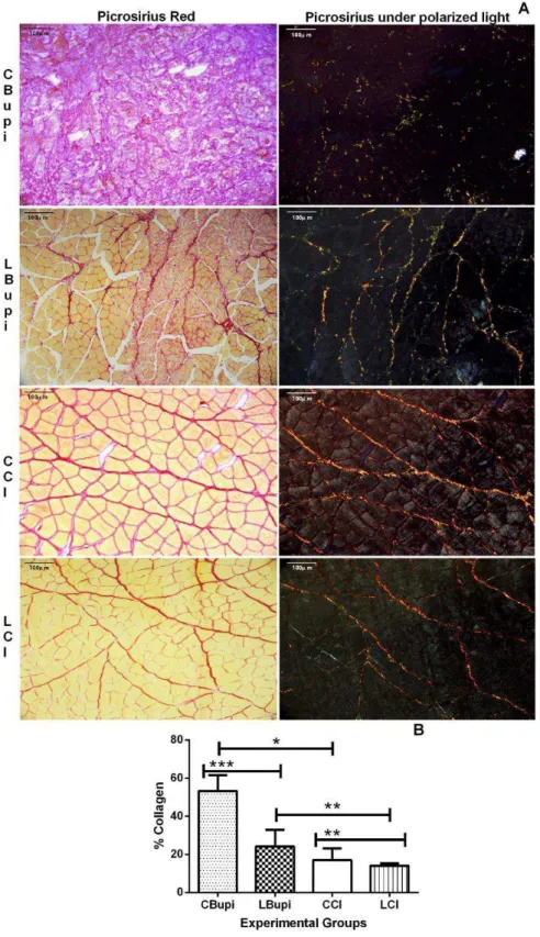
Figure 3. A- Photomicrographs of cross sections of the sternomastoid muscle (Picrosirius Red). B- Percentage of collagen area (analysis of variance, complemented by the Bonferroni test) (Zar, 2009). * p<0.05; ** p< 0.01; ***
p<0.001………..………..……….…60
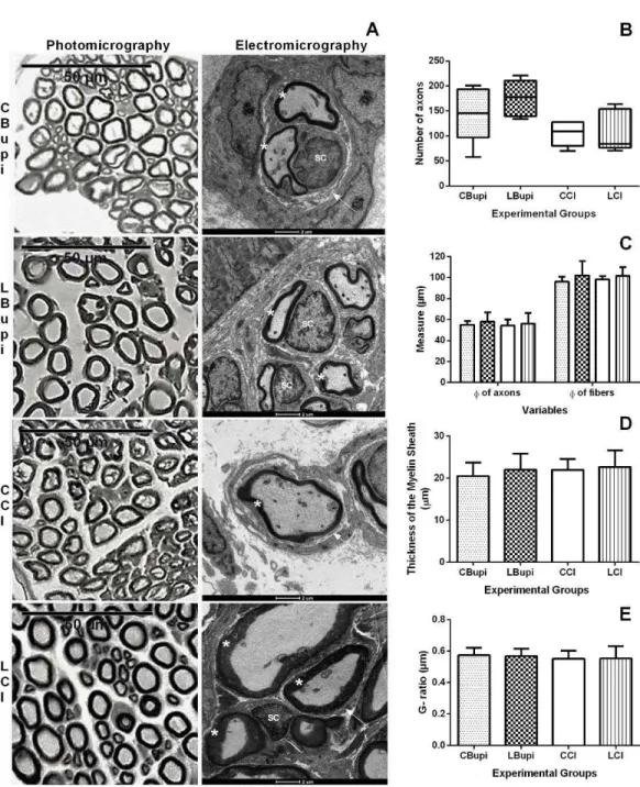
Figure 1. A- Light microscopy findings following the nonspecific esterase reaction (Full preparation) B- Electromicrography of the NMJs of the various groups: () active zone; (▶) presynaptic membrane; (*) synaptic cleft; () postsynaptic membrane; (⇨) junctional folds; () apex of a junctional fold; (M) mitochondria (vs) synaptic vesicles, (N) nucleus C- The maximum diameter (µm) of the NMJs of the subgroups studied. The significance of differences was determined using analysis of variance for the model with two factors, together with the multiple comparison Tukey test, considering a 5% level of significance, (*) p<0.05 (Zar, 2009) ……...…89
Figure 2. A- Confocal microscopy findings of the surface portion of the sternomastoid muscle of the experimental group. The acetylcholine receptors (red), nerve terminals (green), and nuclei (blue) are shown. B- Morphometry of the area (A), perimeter (P), and relative planar area. The significance of differences was analyzed using an analysis of variance for repeated measures model for independent groups complemented with the Bonferroni test (**) p< 0.01, (Zar, 2009). The protein expression of nicotinic acetylcholine receptors AChR (C), AChR (D), AChR1 (E). The significance of differences was determined using nonparametric analysis of variance for repeated measures model for independent groups complemented with the Dunn test (Zar, 2009). (*) p<0.05, and (**) p<0.01………..….……90
Acetylcholine (ACh)
Acetylcholinesterase (AChE) Adenosine triphosphate (ATP) Alpha ()
Cytochrome c oxidase (COX) Creatine Kinase (CK)
Diameter ()
Duchenne muscular dystrophy (mdx) Epsilon ()
Extracellular signal-regulated kinase (ERK2)
GAllium Arsenide (GaAs)
Gallium Arsenide Aluminum (GaAsAl) Gamma ()
Gliceraldeído-3-fosfato desidrogenase (GAPDH) Helium-Neon (HeNe)
Hematoxylin-Eosin (HE)
Indium-gallium-aluminum-phosphide (InGaAIP) Joules (J)
Kilogram (Kg) Laser Therapy (LT)
Light Emitting Diode Therapy (LEDT) Low-Level laser therapy (LLLT) Meter (m)
Myogenic regulatory factors (MRFs) Nanometer (nm)
Neuromuscular junction (NMJ)
Nicotinamide adenine nucleotide (NADH) Nicotinic acetylcholine receptor (nAChR) Ribonucleic acid (RNA)
Transmission Electron Microscopy (TEM) Tumor Necrosis Factor (TNF)
Objetivo ...26
Referência Bibliográfica ...26
Paper 1. LOW-LEVEL LASER THERAPY (LLLT) ACCELERATES THE STERNOMASTOID MUSCLE REGENERATION PROCESS AFTER MYONECROSIS DUE TO BUPIVACAINE Abstract ...38
Introduction...38
Methodology...39
Results ... 42
Discussion ... 45
Conclusion... 49
References ... 49
Table and Figures ... 58
Paper 2. GaAs LASER THERAPY REESTABLISHES THE MORPHOLOGY OF THE NMJ AND nAChRs AFTER INJURY DUE TO BUPIVACAINE Abstract ...64
Introduction ...65
Methodology ...66
Results ... 71
Discussion ...74
Conclusion ...80
References ...80
Figures ...89
1. Introdução
Os anestésicos locais são drogas que atuam como um bloqueador reversível da condução nervosa, impedindo a transmissão da informação de dores, sensoriais, motoras e autonômicas para o sistema nervoso central e a partir dele (Carvalho, 1994). São utilizados em procedimentos cirúrgicos médicos e odontológicos, dando ao paciente mais segurança e conforto (Cobb et al., 2016; Kishore et al., 2016; Balga et al., 2013; Horn et al., 2015; Jaichandran et al., 2015; Ilfeld et al., 2015; Shapiro & Schroeck, 2016). Têm ação no bloqueio da transmissão de estímulos em nervos periféricos no alívio da dor pós-operatória (Nouette-Gaulainet al., 2011; Abdolhossein-Davoodabadi, 2015; Yang et al., 2015; Beck et al., 2015; Kalchofner Guerrero et al., 2016; Yu et al.,2016) e na terapêutica da dor crônica.
Os anestésicos locais são substâncias que podem interferir na transmissão neuromuscular e seus efeitos são potencializados quando associados aos bloqueadores neuromusculares (Wang et al., 2010; Locks et al., 2015).
Os anestésicos locais atuam tanto nos canais de sódio voltagem-dependentes (axônio), quanto nos receptores nicotínicos de acetilcolina (nAChRs) da junção neuromuscular (JNM) (Rossman, 2011; Goodman & Gilman, 2012).
A JNM é uma sinapse química, anatômica e funcionalmente especializada por transferir as informações de um neurônio motor para a fibra muscular (Engel, 2003; Malomouzh, 2012). Utiliza a acetilcolina como neurotransmissor, sendo envolvida diretamente com o processo de contração muscular (Willmann & Fuhrer, 2002). O funcionamento, a manutenção e a regeneração do músculo estriado esquelético dependem da integridade da JNM (Koirala et al., 2003).
A morfologia da JNM contempla três compartimentos: o compartimento pré-sináptico, com as terminações nervosas e a célula de Schwann; o compartimento extracelular, com a lâmina basal; e o compartimento pós-sináptico, com o sarcolema juncional com as dobras juncionais e o sarcoplasma que propicia apoio estrutural e metabólico para a região pós-sináptica (Engel,2003; Malomouzh, 2012) (Figura 01).
É, portanto, no compartimento pós-sináptico que estão localizadas as dobras juncionais. Nelas, estão presentes os nAChRs, que são proteínas integrais de membrana heteroligoméricas, com peso molecular de 290KDa (Lindstrom, 2000).
De morfologia rosácea, possui uma estrutura pentamérica em formato de canal iônico (Rossman, 2011). Existem dois tipos de receptores do tipo muscular, sendo o primeiro presente na fibra muscular embrionária ou desnervada com as subunidades 1 (número de 2), 1, e . O segundo receptor está na forma juncional madura, localizado na fibra muscular inervada, composto pelas subunidades 1 (número de 2), 1, e (Shuetze & Role, 1987; Rossman, 2011).
O sítio de ligação para a Acetilcolina (ACh) ocorre na interface das subunidades e (Rossman, 2011) (Figura 2A e 2B). A duração do estado aberto do receptor depende da duração da ocupação da ACh no local (Naguib et al., 2002).
Figura 02- Imagem do receptor nicotínico de acetilcolina (nAChR). Adaptado de Unwin, 1995
A bupivacaina, por exemplo, é um anestésico local que age como antagonista não competitivo, e pode bloquear a abertura do canal iônico
ou de um sítio alostérico dos nAChRs, inibindo-o (Rossman, 2011).
A maioria dos estudos utilizam abordagem com protocolos mistos e seguem uma hierarquia de potência dos anestésicos locais que são:
bupivacaina ≥ levobupivacaína > ropivacaína > lidocaína = mepivacaína ≥
prilocaína> procaína (Mather, 2010).
A maior limitação dos anestésicos locais são as reações adversas incluindo, dentre outras, a inflamação, a neurotoxicidade (McAlvin et al, 2013) e a miotoxicidade (McAlvin et al, 2013; Plank et al., 2016).
A avaliação da miotoxicidade de um anestésico local de uso clínico é de fundamental importância, pois o dano muscular pode ser considerado uma complicação em potencial na anestesia local (Plank et al., 2016). No entanto, o tecido muscular apresenta uma capacidade de se adaptar a diversas ocorrências (alta plasticidade), inclusive às lesões decorrentes dos anestésicos locais (Harridge, 2007).
A bupivacaina tem sido utilizada em protocolos experimentais, como modelo de estudo para mionecrose (Danieli-Betto et al., 2010; Wen et al., 2013; Otrocka-Domagala et al., 2015) ou mesmo para caracterização da desnervação (Çalgüner et al., 2003). Este anestésico acarreta mudanças nas características morfológica e estrutural das JNMs durante os processos de degeneração e regeneração da fibra muscular, processo este semelhante à diferenciação que ocorre na fibra muscular normal (Nishizawa et al., 2003).
Foster & Carlson, em 1980, relataram que a lidocaína produz uma menor lesão muscular quando comparada à bupivacaina, que promove mionecrose (Peraz-Castro et al., 2009).
A bupivacaina age com excelência na analgesia pós-operatória, mas induz à mionecrose e alterações no metabolismo e estrutura do retículo sarcoplasmático e mitocondrial, ocasionando dor muscular e disfunções musculares. As miopatias induzidas pela bupivacaina são tempo e concentração-dependentes, devendo ter mudanças adaptativas no protocolo de forma individualizada, para cada paciente (Nouette-Gaulain, et al., 2011).
Sua miotoxicidade foi avaliada na concentração-dependente de 0,75%, 0,38% e 0,19% em músculos extrínsecos do olho de coelho. Foi observado que, após cinco dias na concentração de 0,75%, ocorreram mionecrose e degeneração aguda com estágio inicial de regeneração da fibra muscular e, em estágios avançados, o aparecimento de cicatrizes. Já, nas concentrações de 0.38% e 0.19% não foram observados efeito a longo prazo e aparecimento de cicatrizes (Zhang et al, 2010).
Quando o processo de inflamação é excessivo e prolongado, ele interfere na etapa de regeneração, resultando até mesmo na formação de fibrose muscular (Porter et al., 2002; Liu et al., 2015).
As soluções de anestésicos locais podem estar associadas à inflamação local, e o músculo estriado esquelético parece ser muito sensível a esta reação. Renton et al. (2010) investigaram as lesões decorrentes de injeções de anestesias locais, e observaram grandes lesões iatrogênicas após administração destas soluções no bloqueio do nervo alveolar inferior e lingual, tendo como consequência para o paciente, sintomas de dor e manifestações importantes na função do aparelho estomatognático, como a presença de trismo (rigidez muscular), limitando assim a abertura da boca (Sanchez, Takara & Alonso, 2010).
anestésicas em estudo é um desafio atual nos procedimentos cirúrgicos médico e odontológico, a fim de aumentar o tempo de bloqueio e diminuir o processo inflamatório e a toxicidade local, como na associação de carreadores como lipossomas, ciclodextrinas, géis e polímeros (Cereda, et al., 2012; McAlvin et al., 2013).
Segundo Yang et al., 2011, as lesões no nervo após anestésicos locais é uma complicação, podendo levar à dormência local ou fraqueza muscular. Hinton, Dechow & Carlson, em 1985, já relatavam efeitos miotóxicos como a presença de fraqueza muscular com redução de 30 a 40% da força de mordida após injeção de anestésico local, com efeitos persistentes por até duas semanas.
Em experimentos, Ji et al. (2015) observaram lesões neurológicas na raiz e coluna posterior da medula após anestesia intratecal com o anestésico bupivacaina. Na ultraestrutura identificaram edema, vacuolização e descontinuidade da bainha de mielina e apoptose de neurônios. Alterações morfológicas semelhantes à atrofia muscular é uma alteração evidenciada também após injeção de bupivacaina (Scott, Miller & Shieh, 2009) como verificadas em estudos de desnervação (Benoit e Belt, 1970; Carlson, 1976; Çalgüner et al., 2003).
Diversos recursos terapêuticos são utilizados para a saúde funcional do tecido muscular esquelético. Dentre esses destacamos, a Laserterapia de Baixa Intensidade (LBI), que tem sido empregada como ação anti-inflamatória, regenerativa e analgésica (Genovese, 2007).
Todas essas modalidades de tratamento estão respaldadas na relação que a luz tem com as funções biológicas e com a saúde dos seres vivos. É bem conhecida a importância da luz na síntese de substâncias vitais para os animais, e na fotossíntese dos vegetais, da qual provém grande parte da energia orgânica existente no planeta. A luz tem a capacidade de destruir microrganismos patogênicos, e por outro lado pode induzir doenças como o câncer de pele e as queimaduras. O laser com todas as suas similaridades e diferenças da luz convencional, também tem o efeito de modificar funções biológicas, positiva ou negativamente (Goodman & Kaufman, 1997).
É bem estabelecido que a laserterapia atua na cadeia de transporte de elétrons mitocondrial, aumentando o espaço entre as membranas externa e interna da mitocôndria e também dilatando a crista mitocondrial (Iyomasa et al., 2013).
Os lasers são formados por uma energia de fótons com propriedades específicas, com o mesmo comprimento de onda eletromagnética, direção, frequência e cor (Maiman, 1960). São capazes de levar grande quantidade de energia aos tecidos, com precisão. Essas características conferem a essa luz as propriedades de monocromaticidade, colimação e coerência (temporal e espacial) que acrescentam benefícios aos procedimentos em que é associado (Goodman & Kaulfman, 1997; Robertson et al., 2009).
Bem tolerada pelo organismo, a laserterapia é uma forma de energia não-invasiva, de fácil aplicação, baixo custo, não possui efeito mutagênico e pode ser utilizada sem risco (Brugnera-JR & Pinheiro, 1998; Ferraresi, Hamblin & Parizotto, 2012; Alves et al., 2014).
é uma modalidade terapêutica estabelecida. Estes parâmetros incluem: tipo de aparelho, meio semicondutor, comprimento de onda, densidade de energia, potência, tempo de exposição, número de aplicações e duração total do tratamento (Vieira, 2004; Bjordal, 2012).
Baseado na teoria proposta por Albert Einstein em 1916 sobre “os
princípios da amplificação da luz por emissão estimulada de radiação”
(Takac & Stojanovic, 1999; Geiges, 2011), o primeiro laser foi desenvolvido na década de 1960, tendo sido utilizado para clínica somente em 1978 por uma escola Alemã, no Departamento de Dermatologia da Luawing, em Munique.
A primeira diretriz da World Association for Laser Therapy (WALT) sobre a dosagem do laser terapia de baixa intensidade (LBI) para as desordens musculoesqueléticas foi publicada em agosto de 2005. A Walt recomenda a utilização de energia final de 2 a 4 Joules (J) no tratamento de diversos tecidos humanos, dentre eles, o músculo estriado esquelético (WALT, 2006).
Estudos laboratoriais relatam que o efeito positivo do LBI pode ser perdido, quando se faz uma overdose do laser, alterando a morfologia dos fibroblastos, danificando e diminuindo a atividade mitocondrial, e alteração do DNA e da membrana plasmática. Portanto, a fototerapia é capaz de alterar células normais e lesionadas (Hawkins & Abrahamse, 2006). Freitas et al. (2015) não observaram alterações na porcentagem de colágeno, mesmo utilizando alta dosagem da LBI (4,8J).
Apenas para a analgesia e reparação do nervo periférico, WALT recomenda que a dose seja maior que a recomendada para o músculo esquelético (Bjordal, 2012).
Vários são os lasers utilizados na prática clínica nas lesões teciduais, sendo mais frequentes os de Hélio-Neon (HeNe), Indio-Gálio-Alumínio-Fosforeto (InGaAIP), Arseneto de Gálio (AsGa) e Arseneto de Gálio Alumínio (AsGaAl) (Silva et al., 2010). A faixa de radiação eletromagnética do LBI está entre 1mW a 500mW, e o comprimento de onda (penetração no tecido) situa-se entre 660 a 1000 nanômetros (nm) (Huang et al., 2011).
As radiações vermelhas apresentam comprimento de onda abaixo de 700nm, enquanto a infravermelha, acima de 700nm. Quanto maior o comprimento de onda, maior é a penetração da radiação no tecido. No tecido muscular, por exemplo, o infravermelho tem maior penetração, e por este motivo, é utilizado com maior frequência nas práticas clínicas fisioterápicas (Turner & Hode, 2003; Freitas et al., 2015) (Figura 04).
Muitas são as respostas biológicas relatadas pela literatura. É benéfica na redução de processos inflamatórios após lesões (Freitas et al., 2015; Otrocka-Domagala et al., 2015; Zagatto et al., 2016), têm ação analgésica nas disfunções temporomandibulares e dores miofaciais (Shirani et al., 2009; Arduino et al., 2016; Pires de Sousa et al., 2016), agem como miorrelaxantes (Núñez et al., 2006), atuam na redução da fadiga muscular (Lopes-Martins et al., 2006; Borsa et al., 2013; Zagatto et al., 2016). A LBI também melhora a parestesia pós-cirúrgica (Simões, 2007), promovendo a regeneração muscular (de Sousa et al., 2011; Silva et al., 2012; Aranha de Sousa et al., 2013; Freitas et al., 2015), e prevenindo a formação da fibrose muscular (de Sousa et al., 2011). A LBI de 808nm obteve uma resposta efetiva, quando aplicada extra oral para trismo, edema pós-exodontia e reparo tecidual (Aras & Güngörmüs, 2010).
Segundo Silva et al. (2012), as aplicações de laser de Arseneto de Gálio (AsGa) 904nm, potência de 5mW, a uma dose de 3J, mostrou-se eficaz na melhora da regeneração muscular de ratos. Dias et al. em 2012, verificou que após a laserterapia no músculo masseter, houve modificação no fenótipo da fibra muscular, melhorando sua capacidade oxidativa.
Em 2011. Mandic & Rancié, utilizando uma energia final de 15J por 10 sessões, 60 mW potência, laser AsGa 904nm, observaram diminuição da dor e o espasmo muscular, quando comparado ao grupo controle.
Após criolesão no músculo tibial anterior de ratos, Souza et al. (2011), aplicaram laser InGaAIP (660nm), potência 20mW, 1,6J de dose final e observaram aumento da síntese de colágeno do tipo I e III e diminuição da área de mionecrose.
904nm, Leal-Júnior et al. (2014) verificaram a diminuição do processo inflamatório usando uma energia final de 1J.
Na avaliação do potencial eletrofisiológico da junção neuromuscular do músculo diafragma de camundongos, a irradiação do LBI de 830nm, com energia de 12 j/cm² nos elementos da junção neuromuscular, provocou uma diminuição da liberação de acetilcolina, mas apenas a nível fisiológico da liberação dos neurotransmissores. Já, o LBI de 655nm, com energia de 1 – 12 j/cm² não teve efeitos eletrofisiológicos detectados na liberação de neurotransmissores da junção neuromuscular de camundongos (Nicolau et al., 2004).
O LBI de AsGa é um laser infravermelho pulsado (904nm) capaz de penetrar alguns centímetros (0,5 a 2,5 cm) a mais, quando comparado ao laser HeNe (624nm), sendo, portanto, mais efetivo em tecidos mais profundos, como o músculo esquelético, além de proporcionar maior analgesia, enquanto que o laser HeNe age com mais eficácia no tratamento de úlceras da pele (Basford, 1993; Carvalho et al., 2006).Os lasers infravermelhos reduzem o espasmo muscular e aumentam a mobilidade muscular (Nicolau, 2004).
Na clínica, pacientes têm experimentado o uso do laser após anestesia local para alívio da dor, sem o uso de drogas (Aras et al., 2010; Yang et al. 2011). Clockie et al., 1991, aplicaram a LBI na dose de 0.97 j/cm², 3 minutos após a extração do 3˚ molar e obtiveram redução da dor pós-operatória. Já, Markovic & Todorovic, 2006, relataram que doses menores de 4 j/cm² não obtiveram resultados significativos.
Segundo os achados de Alcântara et al. (2013), é recomendável que a LBI deve ser iniciada o mais rapidamente possível após a lesão do nervo periférico, pois sua aplicação aumenta a atividade das Metaloproteinases, especialmente de MMP-9, e o nível da proteína TNFα
atenuação do processo inflamatório, minimiza a formação de fibrose e estimula a regeneração muscular (Assis et al., 2012). Segundo França et al (2013), a LBI pode ter um efeito positivo no músculo esquelético durante seu processo de reparo muscular ao promover uma melhora na qualidade da reorganização das miofibrilas e no perimísio, reduzindo assim a fibrose (França et. al., 2013).
Na regeneração muscular, a LBI atua na analgesia, estimulando a proliferação das células musculares e satélites, intensificando, desta forma, a síntese de proteínas de mioblastos, aumentando a área da fibramuscular e a densidade mitocondrial (Genovese, 2007; Shirani et al., 2009; Dias et al., 2012; Silva et al., 2012; Aranha de Sousa et al., 2013; Borsa et al., 2013; Mantineo, Pinheiro & Morgado, 2014; Freitas et al., 2015). A irradiação com o laser ainda atua como terapêutica complementar em lesões neuronais (Liang et al., 2008), alívio da dor crônica (Masoumipoor et al., 2014; Erthal & Nohama,2015), e redução da apoptose de mionúcleos e células miogênicas decorrentes de desordens músculo esqueléticas (Sergio et al., 2016).
2. Objetivo
Avaliar os efeitos da laserterapia sobre as junções
neuromusculares, fibras musculares e nervo associados ao músculo esternomastóideo após injeção de bupivacaina.
Objetivos Específicos:
a) Níveis de inflamação: Análise morfopatológica das fibras musculares e
quantificação de TNF e da área de colágeno;
b) Miotoxicidade: Análise da Quantificação da Creatina kinase (CK) sérica e muscular, Análise morfológica e ultraestrutural das fibras musculares;
c) Neurotoxicidade: Análises morfológicas, morfométricas e
ultraestruturais do nervo esternomastóideo e das junções
neuromusculares associadas; Análise da distribuição dos receptores de acetilcolina (nAChRs) por meio de microscopia confocal, e quantificação proteica dos nAChRs.
3. Referências Bibliográficas
Abdolhossein-Davoodabadi, Reza-Fazel M, Reza-Vafaei H, Parviz S. Comparison of the effects of intrapleural bupivacaine and morphine on post-thoracotomy pain. Middle East J Anaesthesiol. 2015 Oct;23(3):267-72.
Alcântara CC, Gigo-Benato D, Salvini TF, Oliveira AL, Anders JJ, Russo TL. Effect of low-level laser therapy (LLLT) on acute neural recovery and inflammation-related gene expression after crush injury in rat sciatic nerve. Lasers Surg Med. 2013 apr; 45(4):246-52.
Alves AN, Fernandes KPS, Melo CAV, Yamaguchi RY, França CM, Teixeira DF, Bussadori SK, Nunes FD, Mesquita-Ferrari RA. Modulating effect of low level-laser therapy on fibrosis in the repair process of the tibialis anterior muscle in rats.Lasers Med. Sci. 2014 Mar;29 (2):813–21.
local pathological alterations induced by Bothrops moojeni snake venom. Photochem Photobiol Sci. 2013 Oct;12(10):1895-902.
Aras MH, Güngörmüs M. Placebo-Controlled randomized clinical trial of the effect two different low-level laser therapies (LLLT) - intraoral and extraoral – on trismus and facial swelling following surgical extraction of the lower third molar. Lasers Med Sci. 2010 Sep; 25(5):641-5.
Aras MH, Ömezli MM, Güngörmüs M. Does low-level laser therapy have na antianesthetic effect? : a review. Photomedicine and Laser Surgery 2010; 28(6):719-22.
Arduino PG, Cafaro A, Garrone M, Gambino A, Cabras M, Romagnoli E, Broccoletti R. A randomized pilot study to assess the safety and the value of low-level laser therapy versus clonazepam in patients with burning mouth syndrome. Lasers Med Sci. 2016 Feb 12.
Assis L, Moretti AI, Abrahão TB, Cury V, Souza HP, Hamblin MR, Parizotto NA. Low-level laser therapy (808nm) reduces inflammatory response and oxidative stress in rat tibialis anterior muscle after cryolesion. Lasers Surg Med. 2012 Nov; 44(9):726-35.
Assis LR. Laser de baixa intensidade (830 nm) na regeneração do músculo tibial anterior em ratos. [Dissertação]. São Carlos: Universidade Federal de São Carlos; 2012.
Balga I, Gerber H, Schorno XH, Aebersold Keller F, Oehen HP. Bupivacaine crystal deposits after long-term epidural infusion. Anaesthesist. 2013 Jul;62(7):543-8.
Basford JR. Laser Therapy: scientific basis and clinical role. Orthopedics 1993 may; 16 (5):541-7.
Beck DE, Margolin DA, Babin SF, Russo CT. Benefits of a Multimodal Regimen for Postsurgical Pain Management in Colorectal Surgery. Ochsner J. 2015 Winter;15(4):408-12.
Benoit PW, Belt WD. Destruction and regeneration of skeletal muscle after treatment with a local anesthetic, bupivacaine (Marcaine).J Anat. 1970;107:547-56.
Bjordal JM. Low-level laser therapy (LLLT) and World association for laser therapy (WALT) dosage recommendations. Photomedicine and Laser Surgery 2012; 30(2):61-2.
muscle contractile function and postexercise recovery? A systematic review. J Athl Training 48(1):57–67
Brugnera-Jr A, Pinheiro ALB. Lasers na odontologia moderna. São Paulo: Pancast; 1998.
Carlson BM. A quantitative study of muscle fiber survival and regeneration in normal, predenervated, and Marcaine-treated free muscle grafts in the rat. Exp Neurol. 1976 Sep;52(3):421-32.
Carvalho JCA. Farmacologia dos anestésicos locais. Rev Bras Anestesiol.1994; 44(1):75-82.
Carvalho PT, Mazzer N, dos Reis FA, Belchior AC, Silva IS. Analysis of the influence of low-power HeNe laser on the healing of skin wounds in diabetic and non-diabetic rats.Acta Cir Bras. 2006 May-Jun;21(3):177-83
Cereda CMS, Tofoli GR, Maturana LG, Piericci A, Nunes LAS, Franz-Montan M, Oliveira ALR, Arana S, Araujo DR, Paula E. Local neurotoxicity and myotoxicity evalution of cyclodextrin complexes of bupivacaine and ropivacaine. International Anesthesia Research Society 2012; 115(5):1234-41.
Clokie C, Bentley KC, Head TW. The effects of the helium-neon laser on postsurgical discomfort: a pilot study. J Can Dent Assoc. 1991; 57: 584-6.
Cobb B, Cho Y, Hilton G, Ting V, Carvalho B. Active Warming Utilizing Combined IV Fluid and Forced-Air Warming Decreases Hypothermia and Improves Maternal Comfort During Cesarean Delivery: A Randomized Control Trial. Anesth Analg. 2016 May;122(5):1490-7.
Çalgüner E, GözilR,Erdogân D, Kurt I, KeskiL S, Elmas Ç, Sabuncuoglu H. Atrophic and Regenerative Changes in Rabbit Mimic Muscles after Lidocaine and Bupivacaine Application. Anat. Histol. Embryol.2003, 32:54–9.
Danieli-Betto D, Peron S, Germinario E, Zanin M, Sorci G, Franzoso S, Sandonà D, Betto R. Sphingosine 1-phosphate signaling is involved in skeletal muscle regeneration.Am J Physiol Cell Physiol. 2010 Mar;298(3):C550-8.
de Souza TO, Mesquita DA, Ferrari RA, Dos Santos Pinto D Jr, Correa L, Bussadori SK, Fernandes KP, Martins MD. Phototherapy with low-level laser affects the remodeling of types I and III collagen in skeletal muscle repair. Lasers Med Sci. 2011 Nov; 26(6):803-14.
Dias FJ, Issa JPM, Barbosa APA, Vasconcelos PB, Watanabe L, Mizusakilyomasa M. Effects of low-level laser irradiation in ultrastructural morphology, and immunoexpression of VEGF and VEGFR-2 of rat masseter muscle. Micron. 2012;43:237-44.
Engel AG. The neuromuscular junction. In: Engel, AG; Franzini-Armstrong C. Myology: basic and clinical. 3. ed. New York: International Edition, 2003;1:325-372.
Erthal V, Nohama P. Treatment for neuropathic pain and chronic inflammation using LASER in animal models. Conf Proc IEEE Eng Med Biol Soc. 2015 Aug;2015:1315-8.
França CM, de Loura Santana C, Takahashi CB, Alves AN, De Souza Mernick AP, Fernandes KP, de Fatima Teixeira da Silva D, Bussadori SK, Mesquita-Ferrari RA. Effect of laser therapy on skeletal muscle repair process in diabetic rats. Lasers Med Sci. 2013 Sep;28(5):1331-8.
Foster AH, Carlson BM. Myotoxicity of local anesthetics and regeneration of the damaged muscle fibers. Anesth Analg. 1980; 59:727-36.
Freitas CEA, Bertaglia RS, Junior IJV, Mareco EA, Salomão RAS, de Paula TG, Nai GA, Carvalho RF, Pacagnelli FL, Dal-Pai-Silva M. High Final Energy of Low-Level Gallium Arsenide Laser Therapy Enhances Skeletal Muscle Recovery without a Positive Effecton Collagen Remodeling. Photochemistry and Photobiology, 2015, 91: 957–65.
Geiges ML. History of lasers in desmatology. Curr Probl Dermatol. 2011; 42:1-6.
Genovese WJ. Laser de baixa intensidade: aplicações terapêuticas em odontologia. São Paulo: Ed. Santos, 2007.
Goodman & Gilman. As Bases Farmacológicas da Terapêutica. 12 Ed. Editora McGrallHill, 2012; 757-69.
Goodman JD, Kaufman HW. Effects of an Argon Laser on the Crystalline Proprieties and Rate of Dissolution in Acid of Tooth Enamel in the Presence of Sodium Fluoride. J Dent Res. 1977; 56 (10): 1201-7.
Harridge SD. Plasticity of human skeletal muscle: gene expression to in vivo function. Exp Physiol. 2007 Sep; 92(5):783-97.
Hinton RJ, Dechow PC, Carlson DS. Recovery of jaw muscle function following injection of a myotoxic agent (lidocaine-epinephrine). Oral Surg Oral Med Oral Pathol. 1985 Mar;59(3):247-51.
Horn BJ, Cien A, Reeves NP, Pathak P, Taunt CJ Jr. Femoral Nerve Block vs Periarticular Bupivacaine Liposome Injection After Primary Total Knee Arthroplasty: Effect on Patient Outcomes. J Am Osteopath Assoc. 2015 Dec 1;115(12):714-9.
Huang YY, Chen AC, Carrol JD Hamblim MR. Biphasic dose response in low-level light therapy. Dose Response. 2009; 7:358-83.
Ilfeld BM, Viscusi ER, Hadzic A, Minkowitz HS, Morren MD, Lookabaugh J, Joshi GP. Safety and Side Effect Profile of Liposome Bupivacaine (Exparel) in Peripheral Nerve Blocks.Reg Anesth Pain Med. 2015 Sep-Oct;40(5):572-82.
Iyomasa MM, Rizzi EC, Leão JC, Issa JP, Dias FJ, Pereira YC, Fonseca MJ, Vicentini FT, Watanabe IS. Zymographic and ultrastructural evaluations after low-level laser irradiation on masseter muscle of HRS/J strainmice. Lasers Med Sci. 2013 May; 28 (3):777-83.
Jaichandran VV, Raman R, Gella L, Sharma T. Local anesthetic agents for vitreoretinal surgery: no advantage to mixing solutions. Ophthalmology. 2015 May;122(5):1030-3.
Ji J, Yan X, Li Z, Lai Z, Liu J. Therapeutic effects of intrathecal versus intravenous monosialoganglioside against bupivacaine-induced spinal neurotoxicity in rats. Biomed Pharmacother. 2015 Feb; 69:311-6
Kalchofner Guerrero KS, Campagna I, Bruhl-Day R, Hegamin-Younger C, Guerrero TG. Intraperitoneal bupivacaine with or without incisional bupivacaine for postoperative analgesia in dogs undergoing ovariohysterectomy. Vet Anaesth Analg. 2016 Feb 12.
Karu TI, Pyatibrat LV, Kalendo GS. Photobiological modulation of cell attachment via cytochrome c oxidase, Photochem. Photobiol. Sci. 2004; 3:211–216.
Kishore N, Payal YS, Kumar N, Chauhan N. In Spinal Anaesthesia for Cesarean Section the Temperature of Bupivacaine Affects the Onset of Shivering but Not the Incidence: A Randomized Control Trial. J Clin Diagn Res. 2016 Jan;10(1):UC18-21.
Koirala, S.; Reddy, L. V.; KO, C. P. Roles of glial cells in the formation, function, and maitenance of the neuromuscular junction. J Neurocytol., 2003;32:987-1002.
therapy protects skeletal muscle of mdx mice against damage, inflammation and morphology changes delaying dystrophy progression. PLoS One. 2014 Mar 5;9(3):e89453.
Liang HL, Whelan HT, Eells JT, Wong-Riley MT. Near-infrared light via light-emitting diode treatment is therapeutic against rotenone- and 1-methyl-4-phenylpyridinium ion-induced neurotoxicity. Neuroscience. 2008 Jun 2;153(4):963-74.
Lindstrom JM. Acetylcholine receptors and myasthenia. Muscle Nerve. 2000 Apr;23(4):453-77.
Liu X, Wu G, Shi D, Zhu R, Zeng H, Cao B, Huang M, Liao H. Effects of nitric oxide on notexin-induced muscle inflammatory responses. Int J Biol Sci. 2015 Jan 5; 11(2):156-67.
Locks Gde F, Cavalcanti IL, Duarte NM, Cunha RM, Almeida MC. [Use of neuromuscular blockers in Brazil]. Rev Bras Anestesiol. 2015 Sep-Oct;65(5):319-25.
Lopes-Martins RAB, Marcos RL, Leonardo PS, Prianti AC JR, Muscará MN, Aimbire F, Frigo L, Iversen VV, Bjordal JM. Effect of low-level laser (Ga-Al-As 655nm) on skeletal muscle fatigue induced by electrical stimulation in rats. J Appl Physiol. 2006 jul; 101(1): 283-8.
Maiman TH. Stimulated optical radiation in ruby.Nature 1960;187:492-4.
Malomouzh AI. Non-Cholinergic Signaling Pathways at Vertebrate Neuromuscular Junctions. In: Cseri J. (Ed.) Skeletal muscle: from myogenesis to clinical relations. Kazan: InTech, 2012;380.
Mandić M, Rancié N. [Low power laser in the treatment of the acute low back pain].Vojnosanit Pregl. 2011 Jan;68(1):57-61.
Mantineo M, Pinheiro JP, Morgado AM. Low-level laser therapy on skeletal muscle inflammation: evaluation of irradiation parameters. J Biomed Opt. 2014 Sep; 19(9):98002.
Markovic A, Todorovic LI. Postoperative analgesia after lower third molar surgery: contribution of the use of long-acting local anesthetics, low-power laser, and diclofenac. Oral Surg, Oral Med, Oral Pathol, Oral Radiol Endod. 2006; 102(5): 4-8.
Masoumipoor M, Jameie SB, Janzadeh A, Nasirinezhad F, Soleimani M, Kerdary M. Effects of 660- and 980-nm low-level laser therapy on neuropathic pain relief following chronic constriction injury in rat sciatic nerve. Lasers Med Sci. 2014 Sep;29(5):1593-8.
6(11):1313-32.
Minatel E, Santo Neto H, Marques MJ. Acetylcholine receptors and neuronal nitric oxide synthase distribution at the neuromuscular junction of regenerated muscle fibers. Muscle Nerve. 2001 Mar; 24(3):410-6.
Miyashita E, Fonseca AS. Odontologia Estética, Artes Médicas, p. 739-68, 2004.
Nicolau RA, Martinez MS, Rigau J, Tomàs J. Effect of low power 655nm diode laser irradiation on the neuromuscular junctions of the mouse diaphragm. Lasers Surg Med. 2004; 34(3):277-84.
Nicolau RA, Martinez MS, Rigau J, Tomàs J. Neurotransmitter release changes induced by low power 830nm diode laser irradiation on the neuromuscular junctions of the mouse. Lasers Surg Med. 2004; 35(3):236-41.
Nishizawa T, Tamaki H, Kasuga N, Takekura H. Degeneration and regeneration of neuromuscular junction architecture in rat skeletal muscle fibers damaged by bupivacaine hydrochloride. J Muscle Res Cell Motil. 2003; 24(8):527-37. Nouette-Gaulain K, Jose C, Capdevila X, Rossignol R. From analgesia to
myopathy: when local anesthetics impair the mitochondrion. Int J Biochem Cell Biol. 2011 jan; 43(1):14-19.
Núñez SC, Garcez AS, Suzuki SS, Ribeiro MS. Management of mouth opening in pacients with temporomandibular disorders through low-level laser therapy and transcutaneus electrial neural stimulation. Photomed Laser Surg. 2006 Feb; 24(1):45-9.
Otrocka-Domagała I, Mikołajczyk A, Paździor-Czapula K, Gesek M, Rotkiewicz T, Mikiewicz M. Effect of low-energy laser irradiation and antioxidant supplementation on cell apoptosis during skeletal muscle post-injury regeneration in pigs.Pol J Vet Sci. 2015;18(3):523-31.
Perez-Castro R, Patel S, Garavito-Aguilar ZU, Rosenberg A, Recio-Pinto E, Zhang J, Blanck TJJ, Xu F. Cytotoxicity of local anesthetics in human neuronal cells. Anesthesia & Analgesia 2009 mar; 108(3):997-1007.
Pires de Sousa MV, Ferraresi C, Kawakubo M, Kaippert B, Yoshimura EM, Hamblin MR. Transcranial low-level laser therapy (810 nm) temporarily inhibits peripheral nociception: photoneuromodulation of glutamate receptors, prostatic acid phophatase, and adenosine triphosphate. Neurophotonics. 2016 Jan;3(1):015003.
Plank C, Hofmann P, Gruber M, Bollwein G, Graf BM, Zink W, Metterlein T. Modification of Bupivacaine-Induced Myotoxicity with Dantrolene and Caffeine In Vitro. Anesth Analg. 2016 Feb; 122(2):418-23.
P, Li J, Guo W, Andrade FH. A chronic inflammatory response dominates the skeletal muscle molecular signature in dystrophin-deficient mdx mice. Hum Mol Genet. 2002 Feb 1; 11(3):263-72.
Renton T, Adey-Viscuso D, Meechan JG, Yilmaz Z. Trigeminal nerve injuries in relation to the local anaesthesia in mandibular injections. Br Dent J.2010 nov; 209(9): E15.
Robertson V, Ward A, Low J, Reed A. Eletroterapia Explicada: Princípios e Práticas. 5ª edição, São Paulo, Elsevier. 2009.
Rossman AC. The physiology of the nicotinic acetylcholine receptor and its importance in the administration of anesthesia. AANA J. 2011 oct; 79(5):433-40.
Sanchez GA, Takara D, Alonso GL. Local anesthetics inhibit Ca-ATPase in masticatory muscles. J Dent Res. 2010 apr; 89(4):372-7.
Scott AB, Miller JM, Shieh KR. Treating strabismus by injecting the agonist muscle with bupivacaine and the antagonist with botulinum toxin. Trans Am Ophthalmol Soc. 2009 Dec;107:104-9.
Sergio LP1, Campos VM1, Vicentini SC2, Mencalha AL1, de Paoli F3, Fonseca AS.
Low-Intensity red and infrared lasers affect mRMA expression of DNA nucleotide excision repair in skin andmuscle tissue. Lasers Med Sci. 2016 Apr;31(3):429-35.
Shapiro P, Schroeck H. Seizure After Abdominal Surgery in an Infant Receiving a Standard-Dose Postoperative Epidural BupivacaineInfusion. AA Case Rep. 2016 Jan 28.
Shirani AM, Gutknecht N, Taghizadeh M, Mir M. Low-level laser therapy and myofacial pain dysfunction syndrome: a randomized controllers clinical trial. Lasers Med Sci. 2009; 24(5):715-20.
Silva JP, Silva MA, Almeida APF, Júnior IL, Matos AP. Laser Therapy in the Tissue Repair Process: A Literature Review. Photomed Laser Surg. 2010; 28(1):17-21.
Silva LH, Silva MT, Gutierrez RM, Conte TC, Toledo CA, Aoki MS, Liebano RE, Miyabara EH. GaAs 904-nm laser irradiation improves myofiber mass recovery during regeneration of skeletal muscle previously damaged by crotoxin. Lasers Med Sci. 2012 Sep; 27(5):993-1000.
Simões A. Laser em baixa intensidade para profissionais da área da saúde: aula. São Paulo: Laboratório Especial de Laser em Odontologia da Universidade de São Paulo. 25 de outubro de 2007.
Schuetze SM, Role LW. Developmental regulation of nicotinic acetylcholine receptors. Annu Rev Neurosci. 1987;10:403-57.
de Souza TO, Mesquita DA, Ferrari RA, Dos Santos Pinto D Jr, Correa L, Bussadori SK, Fernandes KP, Martins MD. Phototherapy with low-level laser affects the remodeling of types I and III collagen in skeletal muscle repair. Lasers Med Sci. 2011 Nov;26(6):803-14.
Takac S, Stojanovic S. Characteristics of laser light. Med Pregl. 1999 jan-feb; 52(1-2): 29-34.
Turner J, Hode L. Laser Therapy: clinical practice and scientific background. [S.l.]: Primas Book. 2003.
Unwin N. Acetylcholine receptor channel imaged in the open state. Nature. 1995 Jan 5;373(6509):37-43.
Vieira WHB. Efeitos do laser terapia de baixa intensidade em 780nm sobre a performance muscular aeróbia de ratos em treinamento físico em esteira. [Dissertação]. São Carlos: Universidade Federal de São Carlos; 2004.
Wang H, Zhang Y, Li ST. The effect of local anesthetics on the inhibition of adult muscle-type nicotinic acetylcholine receptors by nondepolarizing muscle relaxants.Eur J Pharmacol. 2010 Mar 25;630(1-3):29-33.
Wen X, Xu S, Liu H, Zhang Q, Liang H, Yang C, Wang H. Neurotoxicity induced by bupivacaine via T-type calcium channels in SH-SY5Y cells. PLoS One. 2013 May 2;8(5):e62942.
Willmann R, Fuhrer C. Neuromuscular synaptogenesis: clustering of acetylcholine receptors revisited. Cell Mol Life Sci. 2002 Aug;59(8):1296-316.
World Association of Laser Therapy (WALT). Consensus agreement on the design and conduct of clinical studies with low-level laser therapy and light therapy for musculoskeletal pain and disorders. Photomed Laser Surg. 2006;24:761-2.
Yang S, Abrahams MS, Hurn PD, Grafe MR, Kirsch JR. Local anesthetic schwann cell toxicity is time and concentration-dependent. Reg Anesth Pain Med. 2011; 36(5):444-51.
injection able to replace the intravenous patient controlled analgesia? J Thorac Dis. 2015 Nov;7(11):1960-9.
Yu SW, Szulc AL, Walton SL, Davidovitch RI, Bosco JA, Iorio R. Liposomal Bupivacaine as an Adjunct to Postoperative Pain Control in Total Hip Arthroplasty. J Arthroplasty. 2016 Jan 21. pii: S0883-5403(16)00064-4.
Zagatto AM, de Paula Ramos S, Nakamura FY, de Lira FS, Lopes-Martins RÁ, de Paiva Carvalho RL. Effects of low-level laser therapy on performance, inflammatory markers, and muscle damage in young waterpolo athletes: a double-blind randomized, placebo-controlled study. Lasers Med Sci. 2016 Apr;31(3):511-21.
Zhang C, Phamonvaechavan P, Rajan A, Poon DY, Topcu-Yilmaz P, Guyton DL. Concentration-dependent bupivacaine myotoxicity in rabbit extraocular muscle. J AAPOS 2010 aug; 14(4):323-7.
Paper 1: Submetido ao Journal of Photochemistry and Photobiology B: Biology (impact factor: 3.188)
Paper 1
LOW-LEVEL LASER THERAPY (LLLT) ACCELERATES THE STERNOMASTOID MUSCLE REGENERATION PROCESS AFTER
MYONECROSIS DUE TO BUPIVACAINE
Cristiane Neves Alessi Pissulin M.D.1, Ana Angélica Henrique
Fernandes M.D,PhD 2, Alejandro Manuel Sanchez Orellana3, Renata
Calciolari Rossi e Silva PhD. 4, Selma Maria Michelin Matheus M.D.,
PhD.5
1Department of Anatomy, Universidade do Oeste Paulista (UNOESTE), Presidente Prudente, SP, Brazil; General Bases of Surgery, Botucatu Medical School; Unesp, Botucatu, SP, Brasil,crispissulin@gmail.com 2Department of Chemistry and Biochemistry, Instituto de Biociências, Unesp, Instituto de Biociências, Botucatu, SP, Brasil; angelica@ibb.unesp.br
3Biological Sciences Student, Unesp, Instituto de Biociências, Botucatu, SP, Brasil,alesanc96@outlook.com
4Department of Pathology, Universidade do Oeste Paulista (UNOESTE), Presidente Prudente, SP, Brasil, renata@unoeste.br
5Department of Anatomy, Instituto de Biociências; General Bases of Surgery, Botucatu Medical School; Unesp, Botucatu, SP, Brasil, micmath@ibb.unesp.br
Correspondence
Address correspondence to Dr. Selma M. M. Matheus, Departamento de Anatomia,Instituto de Biociências, Rubião júnior s/n, Cep 18618000, Unesp, Campus de Botucatu/SP/Brasil,micmath@ibb.unesp.br; +55(014)38800025
Keywords: Bupivacaine, Low-Level Light Therapy, Muscle Regeneration, Fibrosis.
Highlights: LLLT decreased fibrosis and myonecrosis (sternomastoid muscle) caused by bupivacaine.
Competing Interests
The authors declare no competing interests.
Abstract
Background: Because of its long-lasting analgesic action, bupivacaine is an
anesthetic used for peripheral nerve block and relief of postoperative pain. Muscle degeneration and neurotoxicity are its main limitations. There is strong evidence that low-level laser therapy (LLLT) assists in muscle and nerve repair. The authors evaluated the effects of a Gallium Arsenide laser (GaAs), on the regeneration of muscle fibers of the sternomastoid muscle and accessory nerve after injection of bupivacaine. Methods: In total, 30
Wistar adult rats were divided into 2 groups:control group (C: n = 15) and laser group (L: n = 15). The groups were subdivided by antimere, with 0.5% bupivacaine injected on the right and 0.9% sodium chloride on the left. LLLT (GaAs 904nm, 50mW, 4.8J) was administered for 5 consecutive days, starting 24 hours after injection of the solutions. Seven days after the trial period, blood samples were collected for determination of creatine kinase (CK).The sternomastoid nerve was removed for morphological and morphometric analyses; the surface portion of the sternomastoid muscle was used for histopathological and ultrastructural analyses. Muscle CK and TNF protein levels were measured. Results:The anesthetic promoted
myonecrosis and increased muscle CK without neurotoxic effects. The LLLT reduced myonecrosis, characterized by a decrease in muscle CK levels, inflammation, necrosis, and atrophy, as well as the number of central nuclei in the muscle fibers and the percentage of collagen.TNF values remained constant. Conclusions:LLLT, at the dose used, reduced
fibrosis and myonecrosis in the sternomastoid muscle triggered by bupivacaine, accelerating the muscle regeneration process.
1. Introduction
Local anesthetics are generally used in clinical, medical and dental practices to reduce operative pain1-7and as co-adjuncts in postoperative analgesia8-13.
Many adverse effects have been reported as a consequence of the injection of local anesthetics; these include numbness, muscle weakness14,15, lockjaw16,17, inflammation18, paresthesia15 and muscle degeneration19-22.
Considering these alterations, bupivacaine has also been used in experimental protocols as a model for myonecrosis20-22as well as for the characterization of denervation19.
Used for epidural and spinal anesthesia, bupivacaine is considered a long-lasting local anesthetic, having the potential to trigger medical complications such as cauda equina syndrome, permanent spinal nerve injury, edema, vacuolization and rupture in the myelin sheath, degeneration of neurons and nuclei fragmentation21,23,24,30, transient neurologic syndrome31, and neuronal apoptosis21,23,24,30,32-34.
Laser therapy (LT) has been reported to play a positive role in muscle regeneration. LT acts as an analgesic, stimulating the proliferation of muscle and satellite cells, thus enhancing protein synthesis in myoblasts and increasing the area of muscle fibers and mitochondrial density35-42. Laser irradiation also acts as an adjunctive therapy in neuronal injury43 and, chronic pain relief44-45, and reduces myonuclear apoptosis and myogenic cells resulting from musculoskeletal disorders46.
Because local anesthetics are of fundamental importance in clinical practice and the deleterious effects of bupivacaine on muscle and nerve tissue are well described in the literature, LLLT emerges as a non-invasive co-adjunctive procedure to minimize tissue damage, which is a potential complication in clinical practice.
Thus, the objective of the present study was to investigate the effects of low-level laser therapy on the healing process of muscle fibers of the sternomastoid muscle and associated nerve after injection of bupivacaine.
2. Methodology
Thirty adult male Wistar rats were kept in individual cages with food and water ad libitum in an environment with controlled temperature (24 ± 2°C) and photoperiod (12h:12h). All experiments and procedures were approved by the Animal Use Ethics Committee (São Paulo State University, UNESP, CEUA- Protocol 509).
3.0 black nylon (Brasutura®). The solutions were deposited in the open muscle field, subfascially in the middle third47 and distal muscle belly48.
After 24 hours, the animals were randomly divided into two groups: control and laser.
The laser group received laser treatment with a GaAs diode laser (Endofthoton, KLD Biosystems, Amparo, Brazil) with a pulsed emission wavelength of 904nm, output power (average) of 50 mW, and 0.035 cm² emitting area of the beam for 5 consecutive days. The previously calibrated laser was directly applied to the skin (direct contact) in the injection areas in both antimeres. The application was performed with the laser pen held at an angle of 90° to the irradiated surface. The laser treatment lasted48 seconds with an energy of 2.4J per point, for a final total energy 4.8J.
Four groups were formed based on the antimere and treatment: right antimeres- CBupi (without LLLT), LBupi (with LLLT); left antimeres CCl (without LLLT), LCl (with LLLT).
Seven days after application of the solutions, the animals were anesthetized with ketamine/xylazine (90 mg/kg and 10 mg/kg, respectively) intraperitoneally and then decapitated. Blood samples were collected, centrifuged at 936RCF, and the serum was frozen.
The sternomastoid muscle used in this study consisted of two macroscopically clearly distinguishable even portions, white and red49;based on the bupivacaine diffusion, only the superficial muscle portions were removed with the associated nerve and processed for analyses.
2.1- Creatine Kinase (CK)
Serum samples and approximately 100 mg of muscle tissue were used from each group (n = 5 in each group) for the determination of creatine kinase activity (Kit Labtest – CK-nac liquiform 117-1/60)50.
The absorbance of the samples was read at 25°C using a UV spectrophotometer with a wavelength of 340nm and quartz cells with a 1-cm optical path length.
2.2- Histopathological analysis of muscle tissue and collagen quantification
CM1800 cryostat (-25°C). Two slides were prepared; the first was stained with Hematoxylin-Eosin (HE) and the second with Picrosirius Red51. The slides stained with HE were analyzed using a photomicroscope (BX 41-2) with a digital camera (model SIS-SC30) and objectives of 20x and 40x for image capture. Double-blind semi-quantitative analysis of the degree of tissue components (inflammatory infiltrate, atrophy and necrosis) was described as absent (grade 0), mild (grade 1), moderate (grade 2) and high (grade 3)52. These images were also used to count the central and peripheral nuclei with the "ImageJ" software program (http://rsbweb.nih.gov/ij/).
To quantify intramuscular collagen, 5 random images of each slide were obtained (20X). The percentage of collagen was calculated using Leica QWin software (Wetzlar, Germany).
2.3- Transmission Electron Microscopy (TEM)
Muscle fragments (n = 5 in each group), fixed in Karnovsky solution, were processed according to the routine for Transmission Electron Microscopy analysis by the Electron Microscopy Center IB/Unesp/Botucatu. Ultra-thin sections were obtained from longitudinal cuts in muscle fragments using an ultramicrotome (Ultratome 880 LKB III) and stained with uranyl acetate solution in 50% alcohol and subsequently with lead citrate. The material was analyzed and photographed with a transmission electron microscope (TECNAI Spirit Fei Company).
2.4- Morphological and Morphometric Analyses of the Sternomastoid Nerve
2.5- Western Blotting
Frozen muscle samples (third medium)from 5 animals from each group were homogenized with a tissue homogenizer (IKA UltraTurrax/T-25) in 0.5 ml of lysis buffer (1% Triton X-100, 10 mM sodium pyrophosphate, 100 mM sodium fluoride, aprotinin 10 µg/ml, 1 mMPMSF, sodium orthovanadate - 0.25 mMNa3VO4,150 mM NaCl and 50 mMTris-HCl, pH 7.5). The samples were centrifuged at 11,000 rpm for 20 min and the supernatant collected. A 100-µL aliquot of the homogenate was treated with 100µL of Laemmli sample buffer (2% SDS, 20% glycerol, bromophenol blue 0.04 mg/ml, 0.12 M Tris-HCl, pH 6.8, and 0.28 M β -mercaptoethanol).The samples were then incubated at 97°C for 5 minutes and stored in a freezer at -20°C until use.
One aliquot of the pure extract from each sample (not treated with Laemmli) was used to quantify total protein using the Bradford method (Bradford, 1976). Exact quantities of the total protein from each sample (70 µg) were subjected to electrophoresis in 4-15% polyacrylamide gel (SDS-PAGE) and then transferred to nitrocellulose membranes (Bio-Rad Laboratories, Hercules, California) in the wet system.
The proteins transferred to the membranes were blocked with 5% skimmed milk diluted in TBS-Tween for 1h at room temperature and incubated with different TNFprimary antibodies (abcam – ab66579; 1 mg/ml) (Gapdh – Cell Signaling - 14C10; 1:1000), overnight at 4ºC. After incubation with the primary antibody, the membranes were washed in TBS-Tween and incubated with specific anti-rabbit secondary antibody (Cell Signaling - 7074s) 1:5000 for 1 h at room temperature.Again, the membranes were washed, and the ECLTM Selected Western Blotting Detection Reagent (GE Healthcare, Uppsala, Sweden)detection system was used. After image capture in a transilluminator G-Box, densitometric quantification of the bands was performed using ImageJ software (version 1.71, 2006, Austria). The protein expression values were normalized to the values obtained for the GAPDH protein, which was used as a reference.
3. Results
3.1- Creatine Kinase (CK)
The results of the quantification of CK serum levels revealed no statistically significant differences between the groups: Control (216.69 ± 30.54) and Laser (205.63 ± 40.50), p> 0.05.
We observed that muscle CK levels were higher in the control group that received bupivacaine (401.55 ± 40.41) compared to the group that received sodium chloride (331.90 ± 41.51), p <0.01.After application of laser therapy, there was a statistically significant decrease in CK levels (310.20 ± 38.51) (p <0.01).
3.2 - Histopathological, Electron Micrograph, and Quantification of Collagen and Western Blot Analysis
The general morphological analysis of the sternomastoid muscles stained with HE demonstrated the presence of inflammatory cells with basophilic nuclei (mononuclear cell infiltration), blood vessels with hyperemia, edema in the connective tissue, muscle fibers in a degenerative and necrotic process and total loss of its polygonal characteristic in the control group that received bupivacaine (Figure 2Aa).When it was possible to identify the muscle fibers, central nuclei were associated. After laser application, a lower quantity of inflammatory infiltrate was observed; areas in the process of regeneration were present, characterized by the presence of muscle fibers with basophilic cytoplasm containing central and peripheral nuclei (Figure 2Ab).
In the control group that received sodium chloride, there was loss of the polygonal characteristic in only a few muscle fibers; the mononuclear cell infiltration was slight when compared to the group that received bupivacaine (Figure 2Ae). After application of LLLT, the presence of inflammatory infiltrate was not detected and the histological architecture of the muscle fibers was preserved (Figure 2Af). The presence of fibroblasts in the endomysium of the animals that received LLLT in both antimeres was common.
were no alterations (Table 1) (values in scores).
There was an increase in the quantification of central nuclei in the control group that received bupivacaine (277) (p<0.01) and a decrease in the number of peripheral nuclei (53) (p<0.05) compared to the groups that received sodium chloride (Figures 2B e C). In the group that received bupivacaine and LLLT, there was a statistically significant increase in the number of peripheral nuclei (287) (p<0.05) (Figure 2C).
The evaluation of TNF protein expression in all experimental groups revealed no statistically significant differences, although a decrease in TNF expression was observed in the group receiving bupivacaine and LLLT application (Figure 2D).
The ultrastructural analysis found comparatively normal myofibrils, without signs of alteration, Z rectilinear line, inter-myofibrillar mitochondria of varying sizes and T tubules associated with the cisternae of the sarcoplasmic reticulum forming triads in the group that received sodium chloride (Figures 2Ag and 2Ah).
In the group that received bupivacaine, degeneration regions and myonecrosis were present, containing disorganized myofibrils with fragmented and discontinuous Z line and abundant central nuclei. Inflammatory cells, areas of fibrosis, and signs of edema were present (Figure 2Ae).
After application of LLLT, organized sarcomeres and scarce degeneration regions were detected. The nuclei were for the most part peripheral, and blood vessels and central nuclei were present (Figure 2Af). The Picrosirius Red coloring enabled analysis of the collagen fibers present in the endomysium and perimysium, which were marked in red, and the muscle fibers in yellow. This analysis was performed using conventional optical light microscopy and optical polarized light microscopy (Figure 3A). In the group that received sodium chloride, an increase in red tone staining was identified in polarized light, suggesting the presence of mature collagen - type I. In the bupivacaine group, the birefringence of collagen fibers tended towards yellow to green, suggestive of the presence of type III collagen fibers (newly synthesized) (Figure 3A).