Larissa Daniele Bobermin., Andre´ Quincozes-Santos*., Maria Cristina Guerra, Marina Concli Leite, Diogo Onofre Souza, Carlos-Alberto Gonc¸alves, Carmem Gottfried
Department of Biochemistry, Institute of Basic Health Sciences, Federal University of Rio Grande do Sul, Porto Alegre, Rio Grande do Sul, Brazil
Abstract
Ammonia is implicated as a neurotoxin in brain metabolic disorders associated with hyperammonemia. Acute ammonia toxicity can be mediated by an excitotoxic mechanism, oxidative stress and nitric oxide (NO) production. Astrocytes interact with neurons, providing metabolic support and protecting against oxidative stress and excitotoxicity. Astrocytes also convert excess ammonia and glutamate into glutamine via glutamine synthetase (GS). Resveratrol, a polyphenol found in grapes and red wines, exhibits antioxidant and anti-inflammatory properties and modulates glial functions, such as glutamate metabolism. We investigated the effect of resveratrol on the production of reactive oxygen species (ROS), GS activity, S100B secretion, TNF-a, IL-1band IL-6 levels in astroglial cells exposed to ammonia. Ammonia induced oxidative stress, decreased GS activity and increased cytokines release, probably by a mechanism dependent on protein kinase A (PKA) and extracellular signal-regulated kinase (ERK) pathways. Resveratrol prevented ammonia toxicity by modulating oxidative stress, glial and inflammatory responses. The ERK and nuclear factor-kB (NF-kB) are involved in the protective effect of resveratrol on cytokines proinflammatory release. In contrast, other antioxidants (e.g., ascorbic acid and trolox) were not effective against hyperammonemia. Thus, resveratrol could be used to protect against ammonia-induced neurotoxicity.
Citation:Bobermin LD, Quincozes-Santos A, Guerra MC, Leite MC, Souza DO, et al. (2012) Resveratrol Prevents Ammonia Toxicity in Astroglial Cells. PLoS ONE 7(12): e52164. doi:10.1371/journal.pone.0052164
Editor:Ken Arai, Massachusetts General Hospital/Harvard Medical School, United States of America ReceivedMay 10, 2012;AcceptedNovember 15, 2012;PublishedDecember 21, 2012
Copyright:ß2012 Bobermin et al. This is an open-access article distributed under the terms of the Creative Commons Attribution License, which permits unrestricted use, distribution, and reproduction in any medium, provided the original author and source are credited.
Funding:This work was supported by the Conselho Nacional de Desenvolvimento Cientı´fico e Tecnolo´gico (CNPq), Coordenac¸a˜o de Aperfeic¸oamento de Pessoal de Nı´vel Superior (CAPES), FINEP/Rede IBN 01.06.0842-00 and INCTEN National Institute of Science and Technology for Excitotoxicity and Neuroprotection. The funders had no role in study design, data collection and analysis, decision to publish, or preparation of the manuscript.
Competing Interests:The authors have declared that no competing interests exist. * E-mail: andrequincozes@yahoo.com.br
.These authors contributed equally to this work.
Introduction
Ammonia is implicated as a neurotoxin in brain metabolic disorders associated with hyperammonemia, including hepatic encephalopathy (HE), a neuropsychiatric syndrome, and deficien-cies in enzymes of the urea cycle [1,2]. In these conditions, ammonia concentrations in brain tissue can rise as high as 5 mM [1]. Acute ammonia neurotoxicity can be mediated by an excitotoxic mechanism involving the glutamatergic system, in-cluding elevation of extracellular glutamate content, decreased glutamate transporters, NMDA receptor activation and sub-sequent increases in intracellular calcium concentration [3,4,5,6,7]. Metabolic effects of ammonia neurotoxicity include changes in reactive oxygen and nitrogen species (ROS/RNS) levels [8], nitric oxide (NO) metabolism [2], cAMP levels [3,9], mitogen-activated protein kinase (MAPK) pathway [10], cytoskel-eton [11] and astrocyte swelling [12]. Moreover, ammonia toxicity also induces increase in tumor necrosis factor a (TNF-a), Interleukin 1b (IL-1b), which can be associated to ROS pro-duction and involve protein kinase A (PKA), extracellular signal-regulated kinase (ERK) and nuclear factor-kB (NF-kB) activation [13]. Increased levels of S100B secretion, a protein often used as an indicator of glial activation or death for several types of brain injury [14,15], were also observed in astrocytes exposed to ammonia [16].
Astrocytes serve a wide range of adaptive functions in the mammalian central nervous system (CNS). These cells interact with neurons, providing structural, metabolic and trophic support, and they can also play a protective role by releasing neurotrophic factors. In pathological circumstances, however, astrocytes have the potential to induce neuronal dysfunction [17,18,19,20]. Astrocytes play an essential role in protecting neurons against excitotoxicity by taking up excess ammonia and glutamate and converting it into glutamine, using the enzyme glutamine synthetase (GS) (EC 6.3.1.2) [21,22]. Under conditions of ammonia toxicity, GS activity is decreased [3,16]. This enzyme is very sensitive to oxidative stress, and it has been hypothesized to play an important role in the pathogenesis of ammonia neurotoxicity [23]. The C6 cell line are widely used to study astrocytic functions such as glutamate metabolism (glutamate uptake, GS activity and glutathione production), S100B secretion, oxidative and inflammatory responses [24,25,26,27,28,29,30].
[27,35,36,37,38,39]. The recent literature show that resveratrol is able to protect astrocytes and neurons against a variety of oxidative insults [40,41,42,43].
We therefore investigated the effects of ammonia exposure on ROS/RNS production, GS activity, S100B secretion, proinflam-matory cytokines release and NF-kB levels in astroglial cells. We evaluated the effect of resveratrol and other classical antioxidants (ascorbic acid, Nv-nitro-L-arginine methyl ester (L-NAME) and trolox) on ROS production and S100B secretion. The mechanism of the protective effect of resveratrol against ammonia toxicity was also explored.
Materials and Methods
Materials
Poly-L-lysine, resveratrol, ascorbic acid, trolox, L-NAME, monoclonal anti-S100B (SH-B1), H-89, PD98059, sodium nitro-prusside (SNP), propidium iodide (PI), methylthiazolyldiphenyl-tetrazolium bromide (MTT) and 29-79-dichlorofluorescein diace-tate (DCFH-DA) were purchased from Sigma (St. Louis, MO, USA). Fetal bovine serum (FBS), Dulbecco’s modified Eagle medium (DMEM) and other materials for cell culture were purchased from Gibco BRL (Carlbad, CA, USA). Polyclonal anti-S100B and anti-rabbit peroxidase were purchased from Dako (Glostrup, Denmark) and Amersham (Buckinghamshire, UK), respectively. All other chemicals were purchased from common commercial suppliers.
C6 Astroglial Cell Culture
The C6 astroglial cell line was obtained from the American Type Culture Collection (Rockville, MA, USA) and was cultured according to a previously described procedure. The cells were seeded in asks and cultured in DMEM (pH 7.4) containing 5% FBS, 0.1% amphotericin B and 0.032% gentamicin. Cells were maintained at a temperature of 37uC in an atmosphere of 5% CO2/95% air. At log phase, cells were detached from the culture asks using 0.05% trypsin/ethylenediaminetetracetic acid (EDTA) and seeded (56103cells/cm2) in 96-, 24- or 6-well plates.
Primary Astrocyte Cell Culture
Primary cortical astrocyte cultures from Wistar rats were prepared as previously described [44]. All procedures were in accordance with the National Institutes of Health Guide for the Care and Use of Laboratory Animals and were approved by the Federal University of Rio Grande do Sul Animal Care and Use Committee (process number 21215). Briefly, the cerebral cortex of newborn Wistar rats (1–2 days old) was removed and mechanically dissociated in Ca2+
- and Mg2+
- free Hanks’ balanced salt solution (HBSS), pH 7.4, containing the following: 137 mM NaCl, 5.36 mM KCl, 0.27 mM Na2HPO4, 1.1 mM KH2PO4, and 6.1 mM glucose. The cortex was cleaned of the meninges and mechanically dissociated by sequential passage through a Pasteur pipette. After centrifugation at 1,000 rpm for 5 min, the pellet was resuspended in DMEM (pH 7.6) supplemented with 8.39 mM HEPES, 23.8 mM NaHCO3, 0.1% amphotericin B, 0.032% gentamicin and 10% FBS. Approximately 300,000 cells were seeded in each well of the 24-well plates and maintained in DMEM containing 10% FBS in 5% CO2/95% air at 37uC. The cells were allowed to grow to confluence and used after approximately 15 daysin vitro.
Treatments
At confluence, the culture medium was removed by suction, and the cells were treated with ammonia and antioxidants (resveratrol,
L-NAME, ascorbic acid, trolox) at the indicated concentrations. Cells were also pre-treated for 1 h with resveratrol (100mM), ascorbic acid (100mM), trolox (50mM) or L-NAME (500mM) at 37uC in an atmosphere of 5% CO2/95% air in DMEM without Figure 1. Effects of ammonia and antioxidants on DCFH oxidation.Cells were incubated with ammonia (1, 5 and 10 mM) – A, antioxidants (100mM resveratrol (RSV), 500mM L-NAME, 100mM ascorbic acid (AA) and 50mM trolox (TRL)) –B, or antioxidants plus ammonia (5 mM) – C, for 24 h. DCFH oxidation was measured as described in the Materials and methods section. The line indicates the control value. Data represent means6 S.E.M of three experimental determinations performed in triplicate, analyzed statistically by two-way ANOVA followed by the Tukey’s test. (a) indicates significant differences from the control (P,0.05). (b) indicates significant differences from ammonia (P,0.05).
serum. Subsequently, 5 mM ammonia (NH4Cl) was added in the presence or absence of resveratrol (100mM), ascorbic acid (100mM), trolox (50mM) or L-NAME (500mM) for 24 h at 37uC in an atmosphere of 5% CO2/95% air in DMEM without serum. For all parameters analyzed, the results obtained with vehicle were not different from those obtained under basal conditions.
To study the role of PKA and MEK/ERK pathways in the response of resveratrol against ammonia toxicity, we pre-treated
the cells for 1 h with 10mM H-89 and 5mM PD98059, the specific PKA and MEK inhibitors, respectively. The role of NO metabolism was also evaluated using SNP, a NO donator (40mM).
Membrane Integrity and Metabolic Activity Assays Propidium iodide incorporation assay: Cells were treated simultaneously with 7.5mM PI and incubated for up to 24 h. The optical density of uorescent nuclei (labeled with PI), indicative of cell death, was determined with Optiquant software (Packard
Instrument Company). Density values obtained are expressed as a percentage of the control values.
Lactate dehydrogenase assay: The lactate dehydrogenase (LDH) assay was conducted in 50mL of extracellular medium using a commercial colorimetric assay from Doles (Brazil). Results are expressed as percentages of the control value.
MTT reduction assay: Cells were treated with 50mg/mL MTT for 30 min in 5% CO2/95% air at 37uC. Subsequently, the medium was removed, and the MTT crystals were dissolved in
DMSO. Absorbance values were measured at 560 and 650 nm. Results are expressed as percentages of the control value.
DCFH Oxidation
Intracellular ROS production was detected using the non-fluorescent cell-permeating compound, 29-79-dichlorofluorescein diacetate (DCFH-DA). DCFH-DA is hydrolyzed by intracellular esterases to dichlorofluorescin (DCFH), which is trapped within the cell. This non-fluorescent molecule is then oxidized into fluorescent dichlorofluorescin (DCF) by the action of cellular
Figure 3. Effects of ammonia and antioxidants on GS activity.Cells were incubated for 24 h with 100mM resveratrol (RSV) or 500mM L-NAME in the presence or absence of ammonia (5 mM) –A. Cells were treated with 40mM SNP in the presence or absence of 100mM resveratrol (RSV) or 500mM L-NAME and 5 mM ammonia, for 24 h –B. GS activity was measured as described in the Materials and methods section. The line indicates the control value. Data represent means6S.E.M of three experimental determinations performed in triplicate, analyzed statistically by two-way ANOVA followed by the Tukey’s test. (a) Indicates significant differences from the control (P,0.05). (b) indicates significant differences from ammonia (P,0.05). (c) indicates significant differences from SNP (P,0.05).
oxidants. Cultured cells were treated asdescribed above. Cells were then incubated with DCFH-DA (10mM) for 30 min at 37uC. Following DCFH-DA exposure, the cells were scraped into PBS with 0.2% Triton X-100. The fluorescence was measured in a plate reader (Spectra Max GEMINI XPS, Molecular Devices, USA) with excitation at 485 nm and emission at 520 nm [38]. Results are expressed as percentages of the control value.
Nitric Oxide Production
Nitric oxide was determined by measurement of nitrite (a stable oxidation product of NO), based on the Griess reaction. The Griess reagent was prepared by mixing equal volumes of 1% sulfanilamide in 0.5 N HCl and 0.1% N-(1-naphthyl) ethylene-diamine in deionized water. The assay was performed as described [45] with modifications. Briefly, cells were cultured on 96-well plate and after treatment, the Griess reagent was added directly to
the cell culture and the incubation was maintained under reduced light at room temperature during 15 min. Samples were analyzed at 550 nm on a microplate spectrophotometer. Controls and blanks were run simultaneously. Nitrite concentrations were calculated using a standard curve prepared with sodium nitrite (0–50mM).
Glutamine Synthetase Activity
The enzymatic assay was performed as previously described [27]. Briefly, homogenate was added to a reaction mixture containing 10 mM MgCl2, 50 mM L-glutamate, 100 mM imid-azole-HCl buffer (pH 7.4), 10 mM 2-mercaptoethanol, 50 mM hydroxylamine–HCl and 10 mM ATP and incubated for 15 min at 37uC. The reaction was stopped by adding a solution containing 370 mM ferric chloride, 670 mM HCl and 200 mM
trichloroa-Figure 4. Effect of ammonia on S100B secretion in C6 astroglial cells.Cells were incubated for 1, 6 and 24 h with ammonia (1, 5 and 10 mM). S100B extracellular (A) and intracellular (B) content was measured as described in the Materials and methods section. The basal secretion level, assumed to be 100%, is indicated by the line. Data represent means 6 S.E.M of three experimental determinations performed in triplicate, analyzed statistically by one-way ANOVA followed by the Tukey’s test. (a) indicates significant differences from the control (P,0.05).
doi:10.1371/journal.pone.0052164.g004
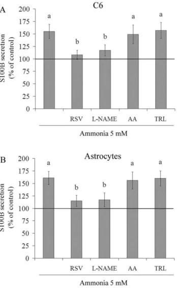
Figure 5. Effect of antioxidants on ammonia-induced S100B secretion in C6 astroglial cells and primary astrocyte cultures. C6 astroglial cells (A) and primary astrocytes (B) were pre-treated for 1 h with 100mM resveratrol (RSV), 500mM L-NAME, 100mM ascorbic acid (AA) or 50mM trolox (TRL). After pre-treatment, ammonia (5 mM) was added in the presence or absence of these antioxidants. The basal secretion level, assumed to be 100%, is indicated by the line. Data represent means 6 S.E.M of three experimental determinations performed in triplicate, analyzed statistically by two-way ANOVA followed by the Tukey’s test. (a) indicates significant differences from the control (P,0.05). (b) indicates significant differences from ammonia (P,0.05).
cetic acid. After centrifugation, the absorbance of the supernatant was measured at 530 nm and compared to a calibration curve of
c-glutamylhydroxamate treated with ferric chloride reagent. Results are expressed as percentages of the control value.
S100B Secretion Assay
S100B secretion was measured by an enzyme-linked immuno-sorbent assay, as previously described [46]. Briefly, 50mL of sample and 50mL of Tris buffer were incubated for 2 h on a microtiter plate previously coated with monoclonal anti-S100B (SH-B1). Next, the samples were incubated with polyclonal anti-S100B for 30 min, and then, peroxidase-conjugated anti-rabbit antibody was added for a further 30 min incubation period. A colorimetric reaction with o-phenylenediamine was observed at 492 nm. Results are expressed as percentages of the control value.
Figure 6. Effect of simultaneous resveratrol and L-NAME treatment and SNP on ammonia-induced S100B secretion in C6 astroglial cells. Cells were pre-treated with 100mM resveratrol (RSV) and 500mM L-NAME simultaneously, in the presence of 5 mM ammonia for 24 h (A). Cells were pre-treated for 1 h with 100mM resveratrol (RSV) or 500mM L-NAME, followed by incubation with 40mM SNP for 24 h (B). The basal secretion level, assumed to be 100%, is indicated by the line. Data represent means 6 S.E.M of three experimental determinations performed in triplicate, analyzed statisti-cally by two-way ANOVA followed by the Tukey’s test. (a) indicates significant differences from the control (P,0.05). (b) indicates significant differences from ammonia (P,0.05). (c) indicates significant differences from SNP (P,0.05).
Tumor Necrosis FactoraMeasurement
TNF-a assay was carried out in extracellular medium, using a rat TNF-aELISA from PeproTech (USA).
Interleukins Measurement
IL-1band IL-6 was carried out in cell culture supernatant, using the rat ELISA for IL-1band IL-6, respectively, from eBioscience (USA).
Nuclear Factor-kB Measurement
The levels of NF-kB p65 were measured using an ELISA commercial kit from Invitrogen (USA).
Protein Determination
Protein content was measured by Lowry’s method using bovine serum albumin as the standard [47].
Statistical Analysis
Data are presented as mean6 S.E.M. Each experiment was performed in triplicate from at least three independent cultures. The data were subjected to one/two-way analysis of variance (ANOVA) followed by the Tukeys test. Values of P,0.05 were considered significant. All analyses were performed using the Statistical Package for the Social Sciences (SPSS) software.
Results
The integrity and metabolic activity of C6 astroglial cells incubated with ammonia for 24 h, as evaluated by measuring PI incorporation, extracellular LDH content and MTT reduction (Table S1), did not significantly change relative to control conditions. The antioxidants resveratrol, L-NAME, ascorbic acid and trolox also did not produce significant changes in these cell viability parameters (Table S2), with the exception of 100mM trolox. Morphological studies showed that ammonia exposure induced astroglial swelling and cell body retraction and resveratrol totally prevented this effect (Figure S1).
The production of ROS was monitored using DCFH oxidation. The level of DCFH oxidation in C6 astroglial cells increased by about 40% and 55% following treatment for 24 h with 5 and 10 mM ammonia, respectively, indicating an increase in ROS production (Figure 1A). All of the antioxidants tested decreased the basal level of DCFH oxidation (Figure 1B). Resveratrol was the most active (35%), followed by L-NAME (25%), trolox (19%) and ascorbic acid (15%). Next, we evaluated the effect of antioxidants on ROS production in the presence of ammonia (Figure 1C). Resveratrol and L-NAME successfully protected the cells; the DCFH oxidation levels dropped from 14768.7% to 10367.6% and 11469%, respectively. The production of NO was indirectly measured by the formation of nitrite (Figure 2A). Both resveratrol and L-NAME decreased nitrite production, 22% and 16%, respectively, compared to the control conditions. Ammonia increased nitrite formation up to 25%. This effect was completely
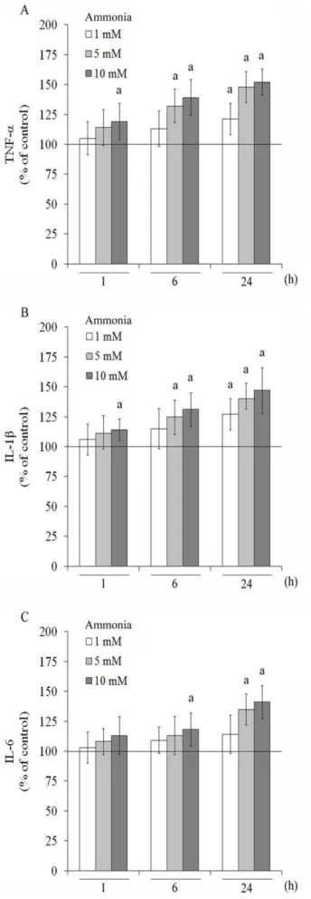
Figure 7. Effect of ammonia on cytokines release in C6 astroglial cells.Cells were incubated for 1, 6 and 24 h with ammonia (1, 5 and 10 mM). TNF-a(A), IL-1b(B) and IL-6 (C) levels were measured as described in the Materials and methods section. The basal cytokines levels, assumed to be 100%, is indicated by the line. Data represent means 6 S.E.M of three experimental determinations performed in triplicate, analyzed statistically by one-way ANOVA followed by the Tukey’s test. (a) indicates significant differences from the control (P,0.05).
doi:10.1371/journal.pone.0052164.g007
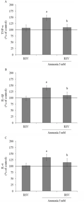
Figure 8. Effect of resveratrol on ammonia-induced cytokines in C6 astroglial cells.Cells were pre-treated for 1 h with 100mM resveratrol (RSV). After pre-treatment, ammonia (5 mM) was added in the presence or absence of resveratrol. The basal cytokines levels, assumed to be 100%, is indicated by the line. Data represent means6
S.E.M of three experimental determinations performed in triplicate, analyzed statistically by two-way ANOVA followed by the Tukey’s test. (a) indicates significant differences from the control (P,0.05). (b) indicates significant differences from ammonia (P,0.05).
prevented by resveratrol and L-NAME. SNP, a NO donator, was used as a positive control for NO production (Figure 2B). SNP (40mM –24 h) increased by about 35% the nitrite levels compared to basal cultures. Ammonia plus SNP also increased nitrite levels (45%). Resveratrol and L-NAME totally prevent this effect, restoring levels to control conditions.
Because GS is primarily responsible for clearing ammonia in the CNS, we measured the GS activity of C6 astroglial cells following exposure to ammonia (Figure 3A). GS activity decreased by about 19% compared to control conditions after exposure for 24 h to 5 mM ammonia. Because resveratrol and L-NAME effectively prevented the induction of increased ROS production by ammonia, we evaluated their effects on GS activity. Resveratrol per seincreased the activity of GS, by about 20% and completely prevented the ammonia-induced decrease in GS activity (8165.7% to 10267.9%), but L-NAME did not.
SNP was used indirectly to investigate whether the effect of resveratrol on GS activity was mediated by NO (Figure 3B). SNP decreased GS activity (16%) and also potentiated the decreased on GS activity induced by ammonia (27%). Resveratrol co-incubated with SNP and under ammonia exposure was able to prevent the decrease. As expected, L-NAME blocked the effect of SNP on GS activity. These data indicate that nitrosative stress and nitric oxide synthase (NOS) activity can mediate the effect of resveratrol on GS activity.
The secretion of S100B by C6 astroglial cells was not affected by incubation with 1, 5 or 10 mM of ammonia for 1 h (Figure 4A). After 6 h of treatment with 10 mM of ammonia, however, S100B secretion increased by about 20%. Following 24 h of exposure to ammonia, the level of extracellular S100B increased significantly, by about 55% and 48% with 5 mM and 10 mM of ammonia, respectively. Because changes in cell integrity were not observed, the increased levels of extracellular S100B most likely resulted from secretion. The intracellular S100B content, however, was not affected by ammonia (Figure 4B). This result may indicate glial activation because S100B was released without being over-expressed.
Under ammonia-induced oxidative stress, resveratrol decreased S100B secretion from 155614% to 10869%, relative to ammonia per se (Figure 5A). L-NAME produced a similar effect. Ascorbic acid and trolox maintained or did not significantly reduce the level of ammonia-induced S100B secretion. As C6 cells and primary astrocytes have shown contrasting profile of in vitro S100B secretion [48], here we also examined the effect of ammonia and antioxidants in primary astrocytes. Both stimuli produced similar effects on S100B secretion by primary astrocytes (Figure 5B), confirming in our study the similar responses between these cells. It is important to mention that antioxidantsper sedid not affect the levels of extracellular S100B in primary astrocytes (data not shown).
Simultaneous addition of resveratrol and L-NAME reduced S100B in C6 astroglial cells by about 55%, compared to ammonia alone (Figure 6A). SNP was used to examine whether alterations on S100B secretion was dependent of NO production (Figure 6B). SNP increased 51% the S100B release. Resveratrol and L-NAME restore the levels to near the basal values, indicating that the increase in S100B is related to NO metabolism.
ROS play a critical role in inflammatory response. Moreover, neuroinflammation has been described in a wide variety of neurological disorders, including HE. In this sense, we evaluated
Figure 9. Effect of resveratrol on NF-kB levels in C6 astroglial cells. Cells were pre-treated for 1 h with 100mM resveratrol (RSV) followed by incubation with 5 mM ammonia for 24 h. The basal NF-kB level, assumed to be 100%, is indicated by the line. Data represent means 6 S.E.M of three experimental determinations performed in triplicate, analyzed statistically by two-way ANOVA followed by the Tukey’s test. (a) indicates significant differences from the control (P,0.05). (b) indicates significant differences from ammonia (P,0.05). doi:10.1371/journal.pone.0052164.g009
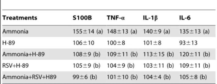
Table 1.PKA pathway modulates ammonia-induced cytokines release.
Treatments S100B TNF-a IL-1b IL-6
Ammonia 155614 (a) 148613 (a) 14069 (a) 135613 (a)
H-89 106610 10068 10168 93613
Ammonia+H-89 10869 (b) 109611 (b) 113615 (b) 120611 (b)
RSV+H-89 10569 (b) 10469 (b) 103611 (b) 109611 (b)
Ammonia+RSV+H89 9966 (b) 101610 (b) 10464 (b) 10568 (b)
C6 astroglial cells were pre-incubated with 10mM PKA inhibitor (H-89), followed by treatment with 100mM resveratrol (RSV) and 5 mM ammonia for 24 h. Data
are expressed as percentage of control values and represent means6S.E.M of three experimental determinations performed in triplicate, analyzed statistically by two-way ANOVA followed by the Tukey’s test. (a) indicates significant differences from the control (P,0.05). (b) indicates significant differences from ammonia (P,0.05).
doi:10.1371/journal.pone.0052164.t001
Table 2.Resveratrol protects ammonia-induced cytokines release through ERK signaling pathway.
Treatments S100B TNF-a IL-1b IL-6
Ammonia 153613 (a) 148613 (a) 14069 (a) 135613 (a)
PD 8969 (b) 103610 (b) 102611 (b) 93610 (b)
Ammonia+PD 11569 (a,b,c) 109610 (b) 108612 (b) 10969 (b)
RSV+PD 7265 (a,b,c) 88611 (b,c) 8769 (b,c) 8067 (a,b,c)
Ammonia+RSV+PD 8767 (a,b) 8569 (a,b,c) 81610 (a,b,c) 8369 (a,b,c)
C6 astroglial cells were incubated with 5mM MEK/ERK inhibitor (PD98059– referred in the table as PD), followed by treatment with 100mM resveratrol
(RSV) and 5 mM ammonia for 24 h. Data are expressed as percentage of control values and represent means6S.E.M of three experimental determinations performed in triplicate, analyzed statistically by two-way ANOVA followed by the Tukey’s test. (a) indicates significant differences from the control (P,0.05). (b) indicates significant differences from ammonia (P,0.05). (c) indicates significant differences from MEK/ERK inhibitor (P,0.05).
the classical proinflammatory cytokine, TNF-a(Figure 7A). After 1 h of exposure, ammonia 10 mM increased TNF-a release by about 19%. At 6 h, 5 and 10 mM ammonia increased
TNF-arelease by about 32% and 39%, compared to control conditions, respectively. Following 24 h of exposure to ammonia, all concentrations increased TNF-a levels. IL-1b, another proin-flammatory cytokine, was also measured (Figure 7B) and a significant increase was observed at 1 h of 10 mM ammonia exposure. An increase of 25% and 31% in IL-1b levels was observed with 5 and 10 mM of ammonia, respectively, after 6 h exposure. IL-1b increased from 1 mM ammonia and higher concentrations at 24 h of exposure. Ammonia also modulated positively the release of IL-6 (Figure 7C). At 6h, 10 mM ammonia increased 18% IL-6 levels, compared to control conditions. After 24 h, 5 and 10 mM of ammonia induced a significant increase (by about 35% and 41%, respectively) in the cytokine IL-6.
To determine whether resveratrol could modulate the ammo-nia-stimulated proinflammatory cytokines, we evaluated the levels of TNF-a, IL-1band IL-6 in C6 astroglial cells. Resveratrolper se did not affect the levels of these cytokines, but is able to completely prevent the augment induced by ammonia exposure, decreasing TNF-a levels from 148613% to 109613% (Figure 8A); IL-1b
levels from 14069% to 110613% (Figure 8B) and IL-6 from 135613% to 115616% (Figure 8C), values expressed as percentage of control.
NF-kB is a transcription factor that induces the expression of genes involved in many biological responses including inflamma-tion [49]. Ammonia increased the NF-kB levels by about 43% (Figure 9). Resveratrol alone did not significantly change the levels, but it was able to prevent the increase induced by ammonia exposure from 143616% to 105612%. As expected, H-89 and PD98059 inhibited the NF-kB translocation (data not shown).
The effect of ammonia on cytokines release can involve many pathways, one them involves cAMP. In fact, cAMP mediate ammonia toxicity [50,51] and we investigated whether PKA was involved in the increase of S100B secretion and proinflammatory cytokines provoked by ammonia in C6 astroglial cells (Table 1). Cells were pre-treated with a specific PKA inhibitor, H-89 (10mM), and then incubated with ammonia. The PKA inhibitor prevented the increase in S100B release after 24 h of incubation with ammonia. H-89 also prevented the increase in ammonia-stimulated proinflammatory cytokines. The co-incubation with resveratrol, ammonia and H-89 did not show results different from ammonia and H-89, indicating that resveratrol probably does not act directly by PKA.
ERK pathway has also been implicated in the regulation of glial inflammatory responses following an insult [52]. In this sense, we examined the S100B and proinflammatory cytokines release using a MEK/ERK inhibitor, PD98059 (Table 2). Under ammonia exposure, MEK/ERK inhibitor prevented the increase in all cytokines. Moreover, resveratrol potentiated the effect of PD98059
Figure 10. Proposed schematics of ammonia-induced cytokines and resveratrol protection in astroglial cells.Ammonia induces increase in intracellular ROS production and cytokines, probably through cAMP/PKA, ERK and NO. Resveratrol (RSV) decreases the levels of ROS and cytokines. The protective mechanism by which resveratrol prevents cytokines production is through inhibition of ERK signaling pathway and NF-kB and reduction of NO levels. Thus, RSV protects astroglial cells against ammonia-induced oxidative stress and/or cytokines release.
in astroglial cells, decreasing the release of S100B, TNF-a, IL-1b
and IL-6 lower that ammonia plus PD98059. These results indicated the involvement of ERK signaling in the protective mechanism of resveratrol against hyperammonemia.
Discussion
Oxidative stress has been implicated in ammonia neurotoxicity [10]. In the brainin vivoand in cultured cells, this condition results in diminished antioxidant defense activity [53,54]. In this study, we observed that treatment of C6 astroglial cells with ammonia increased the level of DCFH oxidation and nitrite production. Previously, Haghighat et al.[55] showed that ammonia interfered with ATP production, leading to reduced levels of NADH and increased NO levels, with subsequent nitration/inactivation of enzymes [56]. We found that resveratrol and L-NAME, two important antioxidants with many biological effects, prevented the ammonia-induced increase of ROS/RNS production. This effect may arise from improved antioxidant defense and/or from inhibition of NOS.
In the CNS, ammonia is primarily detoxified in astrocytes by the enzyme GS, which catalyses the ATP-dependent amidation of glutamate to form glutamine. Continuing the glutamate-glutamine cycle, the glutamine thus formed is exported to neurons, allowing the synthesis of glutamate [57,58]. GS is very sensitive to oxidative and nitrosative stress. Consistent with the results ofLeite et al.[16], we have demonstrated that ammonia toxicity decreases GS activity in astroglial cells. Moreover, the NO metabolism seems to be related to GS activity failure (Figure 3). Resveratrol acted as scavenger more efficiently than L-NAME. Previously, we have demonstrated that resveratrol per se increases values of glial function parameters such as glutamate uptake, GS activity and glutathione content under oxidative stress [27,36,38,39]. Recent reports showed that HO1 (heme oxygenase 1) and its transcription factor regulator, nuclear factor erythroid-2 related factor-2 (Nrf2), might control the neuroprotective effect of resveratrol [59,60]. Resveratrol may therefore be effective against ammonia toxicity either by its ROS/RNS-scavenging effect directly and/or by inhibiting iNOS, a protein downstream of HO1 (Quincozes-Santos, manuscript in revision) [61]. Additionally, the increase in the activity of GS could be related to increased production of glutathione, the main antioxidant of the CNS [22,62,63,64]. It is important to mention that Nrf2 also facilitates the GSH synthesis [65].
During this study, we also investigated the effect of ammonia on the release of S100B from astroglial cells. Elevated serum levels of S100B were observed in HE [66], and increased levels of S100B secretion were also observed in astrocytes exposed to ammonia [16]. We verified that S100B secretion was stimulated by ammonia in the C6 astrocyte cell line. This effect cannot be attributed to astroglial cell death because cell viability was not altered by ammonia. It is widely known that S100B secretion is affected by redox conditions and metabolic stress [38,48,67,68,69]. Increases in extracellular S100B concentrations may be related to brain damage/glial reactivity, and persistent high levels could be involved in neurodegenerative disorders [14]. We observed that increased S100B release was accompanied by increased ROS production and decreased GS activity, indicating a glial response. These effects may be associated with the impairment of several cellular functions.
Hyperammonemia in glial cells results in increased cellular levels of calcium and cAMP [3,9,51,70]. Both calcium and cAMP have been proposed to mediate S100B release [71,72]. Treatment with the PKA inhibitor H-89 completely prevented the
ammonia-induced increase of extracellular S100B levels. Moreover, ammo-nia induces ERK activation [13] and the effect of S100B release was totally abolished by MEK/ERK inhibitor, indicating that cAMP/PKA and ERK are involved in this effect.
When astroglial cells were incubated with antioxidants and ammonia, only resveratrol and L-NAME (an NOS-inhibitor) protected against ammonia toxicity. Data on resveratrol and L-NAME indicate that NO and/or its toxic derivative peroxynitrite are probably involved in these effects. Co-incubation with resveratrol and L-NAME produced a reduction in S100B secretion, probably indicating an interaction between the two compounds. We observed that SNP, a NO donator, stimulated the S100B secretion, and resveratrol and L-NAME were able to prevent this effect, indicating that both could act in the NO pathway. Then, the mechanism by which resveratrol modulates S100B secretion may therefore involve NO. It is important to point out that the mechanisms of S100B secretion and action of resveratrol are not well defined.
Ammonia also increased the main proinflammatory cytokines: TNF-aand IL-1b. These cytokines have a major role in initiating a cascade of activation of other cytokines and growth factor in inflammatory response [73]. TNF-a is synthesized mainly by microglia and astrocytes, and has several important functions in the CNS, including astrocytes activation and glutamatergic gliotransmission [30,73,74]. Recent studies show that proinflam-matory cytokines are increased in HE [75,76], may be used as a marker for encephalopathy grade. TNF-aand IL-1bacts since acute inflammatory responses and both induces IL-6 synthesis [73]. IL-6 is also produced in microglia, astrocytes and neurons, and it plays a pivotal role in a variety of CNS functions such as induction and modulation of astrocytes reactivation, pathological inflammatory and neuroprotection. Assuming S100B as a cytokine, it present the same profile of augment IL-6 and different of other cytokines their present a duality of actions: degenerative and reparative and/or anti and proinflammatory [77,78,79]. More-over, the negative effects of these cytokines may be mediated by TNF-aand IL-1b. Interestingly, as well as the effect on S100B was via PKA and ERK, the increases of other cytokines were also cAMP/PKA/ERK-dependent pathways.
The mechanism of NF-kB activation and subsequent trans-location into the nucleus involves a variety of signaling pathways and modifications, which are not completely understood [80]. A significant increase in NF-kB levels was observed after treatment of C6 cells with ammonia. NF-kB is a transcription factor involved in immune and inflammatory reactions [81], which was inhibited by resveratrol. This regulation could explain the decrease in cytokines release. Besides, PKA and ERK signaling pathway can activate NF-kB [41,49]. Resveratrol potentiated the effect of MEK/ERK inhibitor and this data may provide a mechanistic explanation for the protective effect of resveratrol against in-flammatory cytotoxicity induced by ammonia. NF-kB also induces the expression of iNOS [82]. Thus, we demonstrated that resveratrol decreased S100B-ammonia induced through ERK signaling, inhibition of NF-kB and NO metabolism.
and oxidation of RNA [85,86]. Our group recently demonstrated that resveratrol prevented astroglial cells against H2O2-induced genotoxicity [26,28]. Even though its mechanism of action remains a mystery, resveratrol may exert its effects by antiox-idant/scavenger activity, by modulated NO metabolism or by anti-inflammatory effect [32,34,87,88]. As well, clinical studies showed that N-acetylcysteine (NAC), an antioxidant precursor of glutathione, present protection averts neuroinflammation due to HE [75].
Our results suggest, for the first time, that resveratrol prevents ammonia toxicity in astroglial cells by attenuating oxidative and nitrosative stress, GS activity and cytokines release. Treatment of C6 astroglial cells with ammonia induced increases in S100B, TNF-a, IL-1band IL-6 after 24 h of exposure via a mechanism dependent upon PKA, ERK and NO. Resveratrol prevented ammonia-induced cytokines release through inhibition of ERK signaling pathway and NF-kB and reduction of NO levels. The main conclusions about cytokines in this study are depicted in Figure 10. Overall, these observations suggest that resveratrol may potentially be useful for protection against oxidative stress and inflammatory response in ammonia-induced gliopathy. However, further studies are necessary to investigate the neuroprotective effects of resveratrol against in vivo hyperammonemia in both animal models and clinical trials.
Supporting Information
Figure S1 C6 astroglial cells morphology. Cells were incubated for 24 h with 5 mM ammonia in the presence or absence of 100mM resveratrol (RSV). Under normal conditions (Basal), the cells present polygonal morphology as shown by phase contrast microscopy. Ammonia induced astrocyte swelling and
body retraction and RSV prevents this effect. Representative images of three experiments performed in triplicate.
(TIF)
Table S1 Effect of ammonia on membrane integrity and metabolic activity in C6 astroglial cells.C6 astroglial cells were incubated with ammonia (1, 5 and 10 mM) for 24 h. Membrane integrity and metabolic activity were measured as described in the Materials and methods section. Data are expressed as percentage of control values and represent means
6 S.E.M of three experimental determinations performed in triplicate, analyzed statistically by one-way ANOVA followed by the Tukey’s test.
(DOCX)
Table S2 Effect of antioxidants on membrane integrity and metabolic activity in astroglial cells.C6 astroglial cells were incubated for 24 h in the presence of antioxidants -resveratrol (RSV), L-NAME, ascorbic acid (AA) and trolox (TRL) - at the indicated concentrations. Membrane integrity and metabolic activity were measured as described in the Materials and methods section. Data are expressed as percentage of control values and represent means 6 S.E.M of three experimental determinations performed in triplicate, analyzed statistically by one-way ANOVA followed by the Tukey’s test. (a) indicates significant differences from the control (P,0.05). (DOCX)
Author Contributions
Conceived and designed the experiments: LDB AQS CAG CG. Performed the experiments: LDB AQS MCG MCL. Analyzed the data: LDB AQS MCG MCL DOS CAG CG. Contributed reagents/materials/analysis tools: AQS DOS CAG CG. Wrote the paper: LDB AQS CAG CG.
References
1. Albrecht J, Jones EA (1999) Hepatic encephalopathy: molecular mechanisms underlying the clinical syndrome. J Neurol Sci 170: 138–146.
2. Buzanska L, Zablocka B, Dybel A, Domanska-Janik K, Albrecht J (2000) Delayed induction of apoptosis by ammonia in C6 glioma cells. Neurochem Int 37: 287–297.
3. Felipo V, Butterworth RF (2002) Neurobiology of ammonia. Prog Neurobiol 67: 259–279.
4. Felipo V, Hermenegildo C, Montoliu C, Llansola M, Minana MD (1998) Neurotoxicity of ammonia and glutamate: molecular mechanisms and pre-vention. Neurotoxicology 19: 675–681.
5. Monfort P, Kosenko E, Erceg S, Canales JJ, Felipo V (2002) Molecular mechanism of acute ammonia toxicity: role of NMDA receptors. Neurochem Int 41: 95–102.
6. Monfort P, Munoz MD, ElAyadi A, Kosenko E, Felipo V (2002) Effects of hyperammonemia and liver failure on glutamatergic neurotransmission. Metab Brain Dis 17: 237–250.
7. Hillmann P, Kose M, Sohl K, Muller CE (2008) Ammonium-induced calcium mobilization in 1321N1 astrocytoma cells. Toxicol Appl Pharmacol 227: 36–47. 8. Norenberg MD (2003) Oxidative and nitrosative stress in ammonia
neurotox-icity. Hepatology 37: 245–248.
9. Zielinska M, Zablocka B, Dybel A, Albrecht J (2005) The role of protein kinase C and cyclic AMP in the ammonia-induced shift of the taurine uptake/efflux balance towards efflux in C6 cells. Neurochem Res 30: 349–354.
10. Jayakumar AR, Panickar KS, Murthy Ch R, Norenberg MD (2006) Oxidative stress and mitogen-activated protein kinase phosphorylation mediate ammonia-induced cell swelling and glutamate uptake inhibition in cultured astrocytes. J Neurosci 26: 4774–4784.
11. Jayakumar AR, Rao KV, Murthy Ch R, Norenberg MD (2006) Glutamine in the mechanism of ammonia-induced astrocyte swelling. Neurochem Int 48: 623– 628.
12. Haussinger D, Gorg B (2010) Interaction of oxidative stress, astrocyte swelling and cerebral ammonia toxicity. Curr Opin Clin Nutr Metab Care 13: 87–92. 13. Norenberg MD, Rama Rao KV, Jayakumar AR (2009) Signaling factors in the
mechanism of ammonia neurotoxicity. Metab Brain Dis 24: 103–117. 14. Rothermundt M, Peters M, Prehn JH, Arolt V (2003) S100B in brain damage
and neurodegeneration. Microsc Res Tech 60: 614–632.
15. Schroeter ML, Steiner J, Mueller K (2011) Glial pathology is modified by age in mood disorders–a systematic meta-analysis of serum S100B in vivo studies. J Affect Disord 134: 32–38.
16. Leite MC, Brolese G, de Almeida LM, Pinero CC, Gottfried C, et al. (2006) Ammonia-induced alteration in S100B secretion in astrocytes is not reverted by creatine addition. Brain Res Bull 70: 179–185.
17. He F, Sun YE (2007) Glial cells more than support cells? Int J Biochem Cell Biol 39: 661–665.
18. Markiewicz I, Lukomska B (2006) The role of astrocytes in the physiology and pathology of the central nervous system. Acta Neurobiol Exp (Wars) 66: 343– 358.
19. Barbeito LH, Pehar M, Cassina P, Vargas MR, Peluffo H, et al. (2004) A role for astrocytes in motor neuron loss in amyotrophic lateral sclerosis. Brain Res Brain Res Rev 47: 263–274.
20. Belanger M, Allaman I, Magistretti PJ (2011) Brain energy metabolism: focus on astrocyte-neuron metabolic cooperation. Cell Metab 14: 724–738.
21. McKenna MC (2007) The glutamate-glutamine cycle is not stoichiometric: fates of glutamate in brain. J Neurosci Res 85: 3347–3358.
22. Mates JM, Perez-Gomez C, Nunez de Castro I, Asenjo M, Marquez J (2002) Glutamine and its relationship with intracellular redox status, oxidative stress and cell proliferation/death. Int J Biochem Cell Biol 34: 439–458.
23. Lemberg A, Fernandez MA (2009) Hepatic encephalopathy, ammonia, glutamate, glutamine and oxidative stress. Ann Hepatol 8: 95–102.
24. Benda P, Lightbody J, Sato G, Levine L, Sweet W (1968) Differentiated rat glial cell strain in tissue culture. Science 161: 370–371.
25. Funchal C, Latini A, Jacques-Silva MC, Dos Santos AQ, Buzin L, et al. (2006) Morphological alterations and induction of oxidative stress in glial cells caused by the branched-chain alpha-keto acids accumulating in maple syrup urine disease. Neurochem Int 49: 640–650.
26. Quincozes-Santos A, Andreazza AC, Nardin P, Funchal C, Goncalves CA, et al. (2007) Resveratrol attenuates oxidative-induced DNA damage in C6 Glioma cells. Neurotoxicology 28: 886–891.
27. dos Santos AQ, Nardin P, Funchal C, de Almeida LM, Jacques-Silva MC, et al. (2006) Resveratrol increases glutamate uptake and glutamine synthetase activity in C6 glioma cells. Arch Biochem Biophys 453: 161–167.
28. Quincozes-Santos A, Andreazza AC, Goncalves CA, Gottfried C (2010) Actions of redox-active compound resveratrol under hydrogen peroxide insult in C6 astroglial cells. Toxicol In Vitro 24: 916–920.
30. Tanabe K, Kozawa O, Iida H (2011) Midazolam suppresses interleukin-1beta-induced interleukin-6 release from rat glial cells. J Neuroinflammation 8: 68. 31. Gutteridge JM, Halliwell B (2010) Antioxidants: Molecules, medicines, and
myths. Biochem Biophys Res Commun 393: 561–564.
32. Baur JA, Sinclair DA (2006) Therapeutic potential of resveratrol: the in vivo evidence. Nat Rev Drug Discov 5: 493–506.
33. Delmas D, Jannin B, Latruffe N (2005) Resveratrol: preventing properties against vascular alterations and ageing. Mol Nutr Food Res 49: 377–395. 34. Vang O, Ahmad N, Baile CA, Baur JA, Brown K, et al. (2011) What is new for
an old molecule? Systematic review and recommendations on the use of resveratrol. PLoS One 6: e19881.
35. de Almeida LM, Leite MC, Thomazi AP, Battu C, Nardin P, et al. (2008) Resveratrol protects against oxidative injury induced by H2O2 in acute hippocampal slice preparations from Wistar rats. Arch Biochem Biophys 480: 27–32.
36. de Almeida LM, Pineiro CC, Leite MC, Brolese G, Tramontina F, et al. (2007) Resveratrol increases glutamate uptake, glutathione content, and S100B secretion in cortical astrocyte cultures. Cell Mol Neurobiol 27: 661–668. 37. Vieira de Almeida LM, Pineiro CC, Leite MC, Brolese G, Leal RB, et al. (2008)
Protective effects of resveratrol on hydrogen peroxide induced toxicity in primary cortical astrocyte cultures. Neurochem Res 33: 8–15.
38. Quincozes-Santos A, Nardin P, de Souza DF, Gelain DP, Moreira JC, et al. (2009) The janus face of resveratrol in astroglial cells. Neurotox Res 16: 30–41. 39. Quincozes-Santos A, Gottfried C (2011) Resveratrol modulates astroglial
functions: neuroprotective hypothesis. Ann N Y Acad Sci 1215: 72–78. 40. Tiwari V, Chopra K (2011) Resveratrol prevents alcohol-induced cognitive
deficits and brain damage by blocking inflammatory signaling and cell death cascade in neonatal rat brain. J Neurochem 117: 678–690.
41. Lee EO, Park HJ, Kang JL, Kim HS, Chong YH (2010) Resveratrol reduces glutamate-mediated monocyte chemotactic protein-1 expression via inhibition of extracellular signal-regulated kinase 1/2 pathway in rat hippocampal slice cultures. J Neurochem 112: 1477–1487.
42. Fukui M, Choi HJ, Zhu BT (2010) Mechanism for the protective effect of resveratrol against oxidative stress-induced neuronal death. Free Radic Biol Med 49: 800–813.
43. Kwon KJ, Kim JN, Kim MK, Lee J, Ignarro LJ, et al. (2011) Melatonin synergistically increases resveratrol-induced heme oxygenase-1 expression through the inhibition of ubiquitin-dependent proteasome pathway: a possible role in neuroprotection. J Pineal Res 50: 110–123.
44. Leite MC, Galland F, de Souza DF, Guerra MC, Bobermin L, et al. (2009) Gap junction inhibitors modulate S100B secretion in astrocyte cultures and acute hippocampal slices. J Neurosci Res 87: 2439–2446.
45. Hu J, Castets F, Guevara JL, Van Eldik LJ (1996) S100 beta stimulates inducible nitric oxide synthase activity and mRNA levels in rat cortical astrocytes. J Biol Chem 271: 2543–2547.
46. Leite MC, Galland F, Brolese G, Guerra MC, Bortolotto JW, et al. (2008) A simple, sensitive and widely applicable ELISA for S100B: Methodological features of the measurement of this glial protein. J Neurosci Methods 169: 93– 99.
47. Lowry OH, Rosebrough NJ, Farr AL, Randall RJ (1951) Protein measurement with the Folin phenol reagent. J Biol Chem 193: 265–275.
48. Nardin P, Tramontina F, Leite MC, Tramontina AC, Quincozes-Santos A, et al. (2007) S100B content and secretion decrease in astrocytes cultured in high-glucose medium. Neurochem Int 50: 774–782.
49. King CC, Sastri M, Chang P, Pennypacker J, Taylor SS (2011) The rate of NF-kappaB nuclear translocation is regulated by PKA and A kinase interacting protein 1. PLoS One 6: e18713.
50. Liskowsky DR, Norenberg LO, Norenberg MD (1986) Effect of ammonia on cyclic AMP production in primary astrocyte cultures. Brain Res 386: 386–388. 51. Svoboda N, Zierler S, Kerschbaum HH (2007) cAMP mediates ammonia-induced programmed cell death in the microglial cell line BV-2. Eur J Neurosci 25: 2285–2295.
52. Fernandes A, Falcao AS, Silva RF, Brito MA, Brites D (2007) MAPKs are key players in mediating cytokine release and cell death induced by unconjugated bilirubin in cultured rat cortical astrocytes. Eur J Neurosci 25: 1058–1068. 53. Kosenko E, Kaminsky Y, Kaminsky A, Valencia M, Lee L, et al. (1997)
Superoxide production and antioxidant enzymes in ammonia intoxication in rats. Free Radic Res 27: 637–644.
54. Kosenko E, Felipo V, Montoliu C, Grisolia S, Kaminsky Y (1997) Effects of acute hyperammonemia in vivo on oxidative metabolism in nonsynaptic rat brain mitochondria. Metab Brain Dis 12: 69–82.
55. Haghighat N, McCandless DW, Geraminegad P (2000) Responses in primary astrocytes and C6-glioma cells to ammonium chloride and dibutyryl cyclic-AMP. Neurochem Res 25: 277–284.
56. Rose C, Felipo V (2005) Limited capacity for ammonia removal by brain in chronic liver failure: potential role of nitric oxide. Metab Brain Dis 20: 275–283. 57. Westergaard N, Sonnewald U, Schousboe A (1995) Metabolic trafficking between neurons and astrocytes: the glutamate/glutamine cycle revisited. Dev Neurosci 17: 203–211.
58. Pellerin L, Bonvento G, Chatton JY, Pierre K, Magistretti PJ (2002) Role of neuron-glia interaction in the regulation of brain glucose utilization. Diabetes Nutr Metab 15: 268–273; discussion 273.
59. Bastianetto S, Quirion R (2010) Heme oxygenase 1: another possible target to explain the neuroprotective action of resveratrol, a multifaceted nutrient-based molecule. Exp Neurol 225: 237–239.
60. Sakata Y, Zhuang H, Kwansa H, Koehler RC, Dore S (2010) Resveratrol protects against experimental stroke: putative neuroprotective role of heme oxygenase 1. Exp Neurol 224: 325–329.
61. Quincozes-Santos A, Bobermin L, Goncalves C, Gottfried C, Souza D (2011) Resveratrol modulates astroglial functions. Supplement to GLIA 59(S1): S80. 62. Hertz L, Zielke HR (2004) Astrocytic control of glutamatergic activity: astrocytes
as stars of the show. Trends Neurosci 27: 735–743.
63. Dringen R, Hirrlinger J (2003) Glutathione pathways in the brain. Biol Chem 384: 505–516.
64. Allaman I, Belanger M, Magistretti PJ (2011) Astrocyte-neuron metabolic relationships: for better and for worse. Trends Neurosci 34: 76–87.
65. Escartin C, Won SJ, Malgorn C, Auregan G, Berman AE, et al. (2011) Nuclear factor erythroid 2-related factor 2 facilitates neuronal glutathione synthesis by upregulating neuronal excitatory amino acid transporter 3 expression. J Neurosci 31: 7392–7401.
66. Wiltfang J, Nolte W, Otto M, Wildberg J, Bahn E, et al. (1999) Elevated serum levels of astroglial S100beta in patients with liver cirrhosis indicate early and subclinical portal-systemic encephalopathy. Metab Brain Dis 14: 239–251. 67. Donato R, Sorci G, Riuzzi F, Arcuri C, Bianchi R, et al. (2009) S100B’s double
life: intracellular regulator and extracellular signal. Biochim Biophys Acta 1793: 1008–1022.
68. Quincozes-Santos A, Rosa RB, Leipnitz G, de Souza DF, Seminotti B, et al. (2010) Induction of S100B secretion in C6 astroglial cells by the major metabolites accumulating in glutaric acidemia type I. Metab Brain Dis 25: 191– 198.
69. Funchal C, Tramontina F, Quincozes dos Santos A, Fraga de Souza D, Goncalves CA, et al. (2007) Effect of the branched-chain alpha-keto acids accumulating in maple syrup urine disease on S100B release from glial cells. J Neurol Sci 260: 87–94.
70. Rose C, Kresse W, Kettenmann H (2005) Acute insult of ammonia leads to calcium-dependent glutamate release from cultured astrocytes, an effect of pH. J Biol Chem 280: 20937–20944.
71. Davey GE, Murmann P, Heizmann CW (2001) Intracellular Ca2+and Zn2+ levels regulate the alternative cell density-dependent secretion of S100B in human glioblastoma cells. J Biol Chem 276: 30819–30826.
72. Goncalves D, Karl J, Leite M, Rotta L, Salbego C, et al. (2002) High glutamate decreases S100B secretion stimulated by serum deprivation in astrocytes. Neuroreport 13: 1533–1535.
73. Tanabe K, Matsushima-Nishiwaki R, Yamaguchi S, Iida H, Dohi S, et al. (2010) Mechanisms of tumor necrosis factor-alpha-induced interleukin-6 synthesis in glioma cells. J Neuroinflammation 7: 16.
74. Santello M, Bezzi P, Volterra A (2011) TNFalpha controls glutamatergic gliotransmission in the hippocampal dentate gyrus. Neuron 69: 988–1001. 75. Butterworth RF (2011) Neuroinflammation in acute liver failure: Mechanisms
and novel therapeutic targets. Neurochem Int 59: 830–836.
76. Butterworth RF (2011) Hepatic encephalopathy: a central neuroinflammatory disorder? Hepatology 53: 1372–1376.
77. Van Eldik LJ, Wainwright MS (2003) The Janus face of glial-derived S100B: beneficial and detrimental functions in the brain. Restor Neurol Neurosci 21: 97–108.
78. Guerra MC, Tortorelli LS, Galland F, Da Re C, Negri E, et al. (2011) Lipopolysaccharide modulates astrocytic S100B secretion: a study in cerebro-spinal fluid and astrocyte cultures from rats. J Neuroinflammation 8: 128. 79. Farina C, Aloisi F, Meinl E (2007) Astrocytes are active players in cerebral innate
immunity. Trends Immunol 28: 138–145.
80. Baltimore D (2011) NF-kappaB is 25. Nat Immunol 12: 683–685.
81. Gerlo S, Kooijman R, Beck IM, Kolmus K, Spooren A, et al. (2011) Cyclic AMP: a selective modulator of NF-kappaB action. Cell Mol Life Sci 68: 3823– 3841.
82. Wakabayashi N, Slocum SL, Skoko JJ, Shin S, Kensler TW (2010) When NRF2 Talks, Who’s Listening? Antioxid Redox Signal.
83. Shen HM, Pervaiz S (2006) TNF receptor superfamily-induced cell death: redox-dependent execution. FASEB J 20: 1589–1598.
84. Hamby ME, Gragnolati AR, Hewett SJ, Hewett JA (2008) TGF beta 1 and TNF alpha potentiate nitric oxide production in astrocyte cultures by recruiting distinct subpopulations of cells to express NOS-2. Neurochem Int 52: 962–971. 85. Gorg B, Qvartskhava N, Keitel V, Bidmon HJ, Selbach O, et al. (2008) Ammonia induces RNA oxidation in cultured astrocytes and brain in vivo. Hepatology 48: 567–579.
86. Schliess F, Gorg B, Haussinger D (2009) RNA oxidation and zinc in hepatic encephalopathy and hyperammonemia. Metab Brain Dis 24: 119–134. 87. Csiszar A (2011) Anti-inflammatory effects of resveratrol: possible role in
prevention of age-related cardiovascular disease. Ann N Y Acad Sci 1215: 117– 122.