from cortical astroglial cells
Gloria Queiroz
Faculty of Pharmacy
Glutamate-evoked release of ATP from
cortical astroglial cells
Dissertação de candidatura ao grau de Doutor
apresentada à Faculdade de Farmácia da
Universidade do Porto
Maria da Glória Correia da Silva Queiroz
(SFB 505) and by the European Commission (BMH4 CT96-0676).
The results presented in this thesis were published in:
Queiroz G, Gebicke-Haerter PJ, Schobert A, Starke K, von Kugelgen I (1997) Release of
ATP from cultured rat astrocytes elicited by glutamate receptor activation. Neuroscience 78, 1203-12088.
Queiroz G, Meyer DK, Meyer A, Starke K, von Kùgelgen I (1999) A study of the mechanism
of the release of ATP from rat cortical astroglial cells evoked by activation of glutamate
Summary
Resumo
Résumé
A b b r e v i a t i o n s
Glial Cells
The astrocyte
Classification
Astrocyte physiology
Communication between glial cells and neurones
Glial cells as targets of substances released by
Glial cells as modulators of neuronal activity
Control of neuronal microenvironment
Release of neuroactive mediators
Study of glial cells
Astrocytes in culture
Glutamate in the CNS
lonotropic glutamate receptors
AM PA receptors
Kainate receptors
NMDA receptors
Metabotropic glutamate receptors
Glutamate receptors on glial cells
Cellular c o m m u n i c a t i o n via ATP in CNS
Purinergic receptors
Role of ATP in the CNS
Cell cultures 32
Immunocytochemistry 33
ATP release 33
Lactate dehydrogenase release 35
Materials 36
Statistics 37
Results 38
ATP release: effect of glutamate receptor agonists 38
ATP release: fraction released 41
ATP release: possible contribution of neurones 43
ATP release: interaction with antagonists 46
Lactate dehydrogenase release 46
ATP release: effect of ionomycin 49
ATP release: omission of extracellular calcium 49
ATP release: effect of blockers of voltage-dependent Ca2 + channels 53
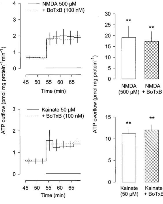
ATP release: effect of a-latrotoxin and botulinum toxin B 53
ATP release: interaction with atractyloside and DIDS 57
ATP release: interaction with glibenclamide and DPC 57
ATP release: interaction with lithium 62
Discussion 63
The role of Ca2+ in ATP release 67
On the mechanism of ATP release 68
Neurone-like exocytosis? 69
Membrane passage by means of channels or transporters? 70
The effect of lithium 72
Conclusions 7 5
References 7 6
Summary
Excitatory amino acids are known to release adenyl compounds in the brain. Both the
possibility that astroglial cells may release ATP in response to excitatory amino acids and the
mechanism of that release were studied in astrocyte cultures derived from the brain
hemispheres of newborn rats. ATP was measured with the luciferine-luciferase assay.
In primary cultures of astrocytes, glutamate receptor agonists did not increase the
release of lactate dehydrogenase but elicited the release of ATP. There was a basal efflux of
ATP, which was increased up to 19-fold by glutamate (100-1000 uM), N-methyl-D-aspartate
(NMDA; 20-500 uM), a-amino-3-hydroxy-5-methylisoxazole-4-propionate (AMPA; 30-100
uM), kainate (20 uM) and (1S,3R)-1-amino-cyclopentane-1,3-dicarboxylic acid (frans-ACPD;
100-1000 uM). The NMDA receptor-selective antagonist 2-amino-5-phosphonopentanoate
(AP5, 100 uM) blocked the effect of NMDA but not the effects of AMPA, kainate and
glutamate. The AMPA receptor-selective antagonist
2,3-dihydroxy-6-nitro-7-sulfamoyl-benzo(f)quinoxaline (NBQX; 30 uM) blocked the effect of AMPA and also of glutamate and
NMDA but not the effect of kainate. The kainate receptor-selective antagonist
y-D-glutamyl-amino-methanesulfonate (GAMS; 30 uM) blocked the effect of kainate but not of glutamate.
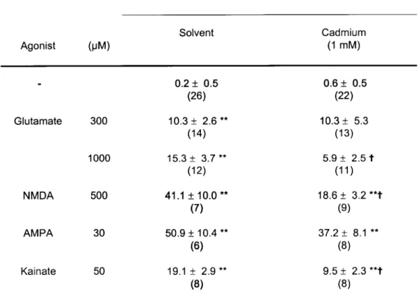
The calcium ionophore ionomycin (5 uM) elicited release of ATP but only in the
presence of external calcium. The release of ATP elicited by NMDA and kainate was
abolished (or greatly reduced) by withdrawal of extracellular calcium as well as by cadmium
(1 mM) and nicardipine (10 uM). In contrast, the release of ATP elicited by AMPA was not
by withdrawal of external calcium. a-Latrotoxin failed to elicit ATP release. Some putative
blockers of ATP transporters or channels were tested on the ATP release elicited by
glutamate receptor agonists. Only the two blockers of the cystic fibrosis transmembrane
conductance regulator (CFTR), glibenclamide (100 uM) and diphenylamine-2-carboxylate
(500 uM) changed the release of ATP. Both reduced the effect of AMPA without changing
the effects of NMDA and kainate (only glibenclamide tested). Furthermore, lithium (1 mM)
abolished the release of ATP evoked by glutamate and AMPA but only reduced the release
elicited by NMDA and kainate.
The present study shows that cultured astroglial cells respond to activation of NMDA,
AMPA, and kainate receptors with release of ATP. Two different mechanisms seem to be
involved in the release of ATP from astroglial cells upon activation of ionotropic glutamate
receptors: one involved in the NMDA- and kainate-induced release of ATP, the other in
AMPA-induced release. The NMDA- and kainate-induced release of ATP requires an influx
of calcium, is not due to neurone-like exocytosis, is not mediated by CFTR or a mechanism
regulated by CFTR, and is reduced but not abolished by lithium. The AMPA-induced release
does not require extracellular calcium may be mediated by CFTR or a mechanism regulated
by CFTR, and is abolished by lithium.
By showing that astrocytes may release a neuroactive substance, namely ATP, with
known modulatory and trophic actions in the central nervous system, this study supports the
view that glial cells are intimately involved in the active control of neuronal activity and should
Resumo
Vários estudos demonstraram que os aminoácidos excitatórios induzem a libertação
de purinas (nomeadamente adenosina) no cérebro. Utilizando como modelo culturas
primárias de astrócitos obtidas de cérebro de rato recém-nascido, estudou-se a
possibilidade das células gliais, em particular os astrócitos, poderem libertar ATP no cérebro
em resposta é estimulação com aminoácidos excitatórios bem como o mecanismo de
libertação envolvido. O ATP libertado foi quantificado pelo método da luciferina-luciferase.
Em culturas primárias de astrócitos, nenhum dos agonistas dos receptores do
glutamato testados aumentou a libertação da enzima lactato desidrogenase, embora o
efluxo basal de ATP tenha sido aumentado (até cerca de 19 vezes) pelo glutamato
(100-1000 uM), N-metil-D-aspartato (NMDA; 20-500 uM), ácido
a-amino-3-hidroxi-5-metilisoxazol-4-propiónico (AMPA; 30-100 uM), kainato (20 uM) e pelo ácido
(1S,3R)-1-amino-ciclopentano-1,3-dicarboxilico (trans-ACPD; 100-1000 uM). O antagonista selectivo dos receptores do NMDA, o ácido 2-amino-5-fosfono pentanóico (AP5, 100 uM), bloqueou o
efeito do NMDA mas não modificou os efeitos do AMPA, kainato e glutamato. O antagonista
selectivo dos receptores do AMPA, 2,3-dihidroxi-6-nitro-7-sulfamoil-benzo(f)quinoxalina
(NBQX; 30 uM) antagonizou o efeito do AMPA e também os efeitos do glutamato e NMDA,
mas não modificou a libertação de ATP induzida pelo kainato. O antagonista selectivo dos
receptores do kainato, y-D-glutamil-amino-metanosulfonato (GAMS; 30 uM), bloqueou o
A ionomicina (5 |jM), um ionóforo do cálcio, induziu a libertação de ATP mas apenas
na presença de cálcio no meio extracelular. A libertação de ATP induzida pelo NMDA e pelo
kainato foi abolida ou significativamente reduzida com a omissão de cálcio do meio
extracelular, na presença de cádmio (1 mM) ou na presença de nicardipina (10 uM). Pelo
contrário, a libertação de ATP induzida pelo AMPA não sofreu qualquer alteração pelas
intervenções referidas e a libertação de ATP induzida pelo trans-ACPD também não sofreu alteração quando o cálcio extracelular foi omitido do meio. A a-latrotoxina não teve qualquer
efeito sobre a libertação de ATP. Alguns compostos com potencialidade de bloquear
transportadores ou canais permeáveis ao ATP foram testados sobre a libertação de ATP
induzida pelos agonistas dos receptores do glutamato. Dos compostos testados apenas dois
bloqueadores do CFTR (cystic fibrosis transmembrane conductance regulator), a
glibenclamida (100 uM) e o ácido difenilamino-2-carboxílico (500 uM), modificaram a
libertação de ATP. Ambos os compostos reduziram o efeito do AMPA, mas não modificaram
o efeito do NMDA e kainato (apenas a glibenclamida foi testada). O lítio (1 mM) aboliu a
libertação de ATP induzida pelo glutamato e AMPA e reduziu significativamente a libertação
de ATP induzida pelo NMDA e kainato.
Os resultados obtidos demonstram que a activação dos receptores do NMDA, AMPA,
e kainato induz a libertação de ATP de astrócitos em cultura. Dois mecanismos distintos
parecem estar envolvidos na libertação de ATP: um envolvido na libertação de ATP induzida
pelo NMDA e kainato, e um outro mecanismo envolvido na libertação de ATP induzida pelo
AMPA. A libertação de ATP induzida pelo NMDA e kainato requer o influxo de cálcio, não
envolve um processo de exocitose semelhante ao que ocorre nos neurónios, não é mediada
pelo CFTR ou por um mecanismo regulado pelo CFTR e é reduzida pelo lítio. A libertação
de ATP induzida pelo AMPA não é dependente do cálcio extracelular, podendo ser mediada
pelo CFTR ou por um mecanismo regulado pelo CFTR e é abolida pelo lítio.
A demonstração de que os astrócitos libertam um composto neuroactivo, o ATP, com
conhecidas acções moduladoras e tróficas no sistema nervoso central, vem corroborar com
a tese de que as células gliais estão intimamente envolvidas no controlo da actividade
Résumé
Il est resté démontré, en divers études, que les aminoacides excitateurs induisent la
libération de dérivés de purines (nommément l'adénosine) dans le cerbère. En utilisant
comme modèle des cultures primaires d'astrocytes obtenues à partir de cerbère de rat
nouveau-né, on a cherché à déterminer la possibilité des cellules gliales, en particulier des
astrocytes, pouvoir libérer ATP dans le cerbère et son mécanisme de libération, en réponse
à l'estimulation avec des aminoacides excitateurs. L'ATP libéré a été quantifié par la
méthode de luciférine-luciférase.
Aucun des agonistes des récepteurs du glutamate testés, a augmenté la libération de
l'enzyme lacticodéshydrogénase, mais l'eflux basai de ATP a été augmenté (jusqu'à 19 fois,
moins au plus) par le glutamate (100-1000 uM), N-méthyle-D-aspartate (NMDA; 20-500 uM),
a-amino-3-hydroxy-5-méthylisoxasole-4-propionate (AMPA; 30-100 uM), kainate (20 uM) et
par l'acide (1S,3R)-1-amino-ciclopentane-1,3-dicarboxilique (trans-ACPD; 100-1000 uM).
L'antagoniste sélectif des récepteurs du NMDA, l'acide 2-amino-5-fosfonopentanoïque
(AP5;100 uM), a bloqué l'effet du NMDA, mais n'a eu aucune influence sur les effets du
AMPA, kainate et glutamate. L'antagoniste sélectif des récepteurs du AMPA,
2,3-dihydroxy-6-nitro-7-sulfamoyl-benzo(f)quinoxaline (NBQX; 30 uM) a bloqué l'effet du AMPA bien que
les effets du glutamate et NMDA, mais il n'a eu aucun effet sur la libération de ATP induite
par le kainate. L'antagoniste sélectif des récepteurs du kainate, acide
y-D-glutamyl-amino-méthanesulfonique (GAMS; 30 uM), a bloqué l'effet du kainate, mais il n'a pas modifié
L'ionomycine (5 uM), un ionophore du calcium, a induit la libération de ATP
seulement en présence de calcium au milieu extracellulaire. La libération de ATP induite par
le NMDA et par le kainate a été anéantie ou significativement réduite dans l'absence de
calcium extracellulaire, en présence de cadmium (1 mM) ou en présence de nicardipine (10
uM). Au contraire, la libération de ATP induite par le AMPA n'a subi aucune altération par les
interventions déjà référées et la libération de ATP induite par le trans-ACPD n'a pas subi aussi d'altération quand le calcium extracellulaire a été omis du milieu. La a-latrotoxine n'a
eu aucun effet sur la libération de ATP. Parmi les divers composés testés avec potentiel
d'empêché transporteurs ou canaux perméables à le ATP, seulement deux empêcheurs du
CFTR (cystic fibrosis transmembrane conductance regulator), la glibenclamide (100 uM) et
l'acide diphenylamine-2-carboxílique (500 uM) ont modifié la libération de ATP. Tous les
deux composés ont réduit l'effet du AMPA, mais n'ont pas modifié l'effet du NMDA et du
kainate (la glibenclamide, c'est la seule qui a été testée). Le lithium (1 mM) a anéanti la
libération de ATP induite par le glutamate et AMPA et a réduit la libération de ATP induite
par le NMDA et kainate.
Les résultats obtenus démontrent que l'activation des récepteurs du NMDA, AMPA
and kainate induit la libération de ATP de astrocytes en culture. Deux mécanismes différents
semblent être engagés dans la libération de ATP: l'un engagé dans la libération de ATP
induite par NMDA et kainate et, un autre mécanisme engagé dans la libération de ATP
induite par le AMPA. La libération de ATP induite par NMDA et kainate a besoin d'un influx
de calcium; n'engage pas un procès d'exocytose semblable à ce qui pourvoit dans les
neurones, n'est pas moyennée par le CFTR ou par un mécanisme régulé par le CFTR et est
réduite par le lítio. La libération de ATP induite par le AMPA n'a pas besoin du calcium
extracellulaire pouvant être moyennée par le CFTR ou par un mécanisme régulé par le
CFTR et elle est anéantie par le lítio.
La situation qui démontre que les astrocytes libèrent un composé neuroactif, le ATP,
avec des actions modulatrices et trophiques dans le système nerveux central, vient confirmer
la thèse qui défend que les cellules gliales son intimement engagés dans le contrôle de
Abbreviations
ADP adenosine-5'-diphosphate
AMP adenosine-5'-monophosphate
AM PA a-amino-3-hydroxy-5-methylisoxazole-4-propionate
AP5 (±)-2-amino-5-phosphonopentanoicacid
ATP adenosine-5'-triphosphate
cAMP 3',5'-cyclic adenosine phosphate
CFTR cystic fibrosis transmembrane conductance regulator
cGMP 3',5'-cyclic guanosine phosphate
CNS central nervous system
DIDS 4,4'-diisothiocyanato-stilbene-2,2'-disulfonate
DPC diphenylamine-2-carboxylate
GABA y-aminobutyric acid
GAMS y-D-glutamyl-amino-methanesulfonicacid
GFAP glial fibrillary acidic protein
HEPES N-[2-hydroxyethyl]piperazine-N'-[2-ethanosulfonicacid]
INT iodonitrotetrazolium
IP3 inositol trisphosphate
LDH lactate dehydrogenase
LPS lipopolysaccharide
MAP2a,b microtubule associated protein-2a,b
NBQX 2,3-dihydroxy-6-nitro-7-sulfamoyl-benzo(f)-quinoxaline
NMDA N-methyl-D-aspartate
NO nitric oxide
frans-ACPD (1 S,3R)-1 -aminocyclopentane-1,3-dicarboxylic acid
Glial Cells
The number of neurones in the central nervous system (CNS) is enormous but they
are exceeded in number 5-10 fold by cells collectively called neuroglia or glial cells. These
cells were described for the first time in 1846 by Rudolf Virchow, but only in the beginning of
this century the major glial populations: astrocytes, oligodendrocytes, and microglial cells
were identified.
Glial cells were defined for a long time as neurone supporting cells, representing the
CNS version of connective tissue cells. Over the last 30 years, the information obtained from
the study of glial cell physiology has completely changed this view. Nowadays, glial cells are
considered more than merely mechanical supportive cells. They are known to have an active
role in the CNS, assisting nerve cells in the performance of their integrative and
communicative functions. They are able to respond to extracellular messengers and to
release neuroactive mediators, the same mediators neurones use to communicate. The
present work demonstrates that astrocytes, like neurones, are the target of neurotransmitters
The astrocyte
Classification
Astrocytes are a heterogeneous population of cells and, based on anatomic and
morphologic criteria, it is possible to distinguish two major populations: the fibrous and the
protoplasmic astrocytes. Fibrous astrocytes are found mainly in the white matter of the CNS.
They have long thin processes that branch infrequently and contain abundant intermediate
filaments. Protoplasmic astrocytes are found mainly in the grey matter of the CNS. They
have a stellate form with multiple highly branched processes, which contain fewer
intermediate filaments than fibrous astrocytes, have abundant cytoplasm and a nucleus that
is larger than the nucleus of any the other type of glial cells.
Another approach to identify and classify astrocytes has emerged from in vitro studies, namely astrocytes in culture (also called astroglia). It is based on astroglia
morphology and on antigenic phenotypes, using immunocytochemical methods.
Glial filaments are known to contain glial fibrillary acidic glycoprotein (GFAP) and the
detection of GFAP is used to identify these cells. Astroglia are, thus, GFAP positive. Based
on the morphology of GFAP positive cells and on the use of antibodies directed against
astroglia antigens it was possible to identify two subtypes of astroglial cells, named type 1
and type 2 astroglia (Raff et al., 1983a). The monoclonal antibody A2B5, which binds to
polysialogangliosides, was the first immunological cell marker used to distinguish between
these two subtypes of astroglia. Type 1 astroglia are polygonal, GFAP positive and A2B5
negative cells. Type 2 astroglia are process bearing, GFAP positive, and A2B5 positive cells.
Type 1 and type 2 astroglia were originally proposed to be the cell culture subtypes
equivalent to fibrous and protoplasmic astrocytes, respectively (Miller and Raff, 1984). In
and fibrous to classify astrocytes in vivo and type 1 and type 2 to classify astrocytes in culture (Miller et al., 1989).
Astrocyte physiology
Astrocytes have an important role in homeostasis in the CNS. They regulate the
composition of extracellular fluid and synaptic transmission; they remove the excess of K+
ions that accumulate in the extracellular space following neuronal activity and take up and
inactivate neurotransmitters.
Astrocytes also play an important role in neuronal development and neuronal
plasticity (Rakic, 1981; Muller and Best, 1989). In the developing nervous system, they
provide scaffoldings for the inward migration of young neurones to their final position and, in
the adult, they form an elaborate framework in which neurones are deployed.
The intimate relationship between astrocyte processes and blood vessels (astrocyte
end-feet are in contact with capillaries and arterioles) suggests that they may play important
roles in neuronal energy supply and reserve (astrocytes are the main glycogen storage cells
in the CNS). They also participate in the formation of the blood-brain barrier and in the
regulation of blood flow during regional increases of neuronal activity (Janzer and Raff, 1987;
Paulson and Newman, 1987).
Astrocytes and astroglia are extensively coupled through gap junctions resulting in
the formation of a cellular syncytium (Bennett et al., 1991). Communication between
astrocytes through gap junctions is though to underlie their ability to redistribute the excess
of extracellular K+ ions that occurs following neuronal activity. It has been suggested that gap
and distribution of glucose (Tabernero et al., 1996); regulation of cell volume and proliferation
(Kimelberg and Kettenmann, 1990); and intracellular Ca2+ mediated communication between
astrocytes (Cornell-Bell et al., 1990) and between astrocytes and neurones (Nedergaard,
1994).
Like neurones, astrocytes and astroglia express voltage-dependent ion-channels
(Barres et al., 1990a), namely voltage-dependent K+, Na+ and Ca2+ channels (MacVicar,
1984; Barres et al., 1990a). In general, the density of K+ channels in glial cells and in
neurones is similar, while the densities of Na+ and Ca2+ channels in astroglia are
considerably lower than in neurones. Furthermore, the properties of astrocyte ion channels
seem to be different from those of neurones; Na+ channels in type 1 astroglia open at more
negative potentials and more slowly than neuronal Na+ channels in neurones (Barres et al.,
1989).
Glial cells, in spite of the presence of Na+ channels, are not able to generate action
potentials; thus, the role of these channels in glia is still a matter of debate. It has been
proposed that glial cells synthesise Na+ channels and donate them to adjacent neurones,
reducing the neuronal biosynthetic demands. Na+-channels may also act as sensors to
detect electric activity in neighbouring neurones or be coupled to Na+/K+-ATPase,
generating the Na+ gradient needed for the uptake of K+ from the extracellular space
(Sontheimeretal., 1996).
Astroglia responds to a variety of neurotransmitters (Bevan, 1990). Several studies
have demonstrated that glial cells express one or several types of glutamate- and
GABA-receptor gated ion channels (Bormann and Kettenmann, 1988). In addition to these
ionotropic receptors, astrocytes in culture also express many metabotropic receptors coupled
to adenylate cyclase, guanylate cyclase, phospholipases A2 and C and nitric oxide synthase.
Glial metabotropic receptors modulate the activity of ion channels and enzymes and,
messengers diacylglycerol, inositol trisphosphate (IP3), Ca2+, cAMP and cGMP (Murphy and
Pearce, 1987; Teichberg, 1991).
Communication between glial cells and neurones
Glial cells as targets of substances released by neurones
Substances released from neurones including K+, neurotransmitters, and metabolites
(such as C02) may act on glial cells and influence glial metabolism. For instance, the K+
released during neuronal activity causes a rapid astrocyte depolarisation and generates K+
spatial-buffering currents within astroglia that lead to extracellular K+ redistribution (Newman,
1986). An increase in extracellular K+ also causes glial metabolic changes by stimulation of
glycogenosis (Reichenbach et al., 1993).
Astrocytes are also targets for neurotransmitters. For example, a rise in intracellular
Ca2+ can be detected in glial cells after glutamate release from synaptic nerve terminals
(Porter and McCarthy, 1996). This effect can be due to activation of astrocyte glutamate
receptors or may be secondary to glutamate-receptor induced release of a mediator, namely
NO. This possibility is supported by the observation that glutamate induces synthesis of NO
in neurones by activation of NO synthase (Garthwaite et al., 1989a,b). Since NO is
membrane soluble, it can diffuse into neurones and glia and activate their guanylate cyclase.
An increase in cGMP may decrease astrocyte coupling through gap-junctions or modulate
Glial cells as modulators of neuronal activity
Control of neuronal microenvironment
Glial cells can modulate neuronal activity and synaptic transmission by controlling the
concentration of ions and neurotransmitters in the extracellular fluid. For example, they take
up the excess of K+ that accumulates during neuronal activity. A decrease in the efficacy of
K+ removal causes K+ accumulation and a consequent increase on neuronal excitability,
altering spontaneous and impulse-mediated neurotransmitter release.
Additionally astrocytes may modulate neuronal excitability through interference with
the extracellular Ca2 + concentration and pH. Astrocytes take up Ca2+ and changes in the
efficacy of this system may have important consequences for the Ca2+-dependent
transmitter release (Barres, 1991). Through the operation of a Na+-HC03" transport system
present in glia but not in neurones astrocytes regulate the extracellular pH with consequent
changes on neuronal excitability (Ransom and Orkand, 1996).
Astrocytes located close to synapses regulate neurotransmission by participating in
neurotransmitter inactivation, providing synaptic insulation, and preventing neurotransmitter
diffusion to the nearby synapses. One of the best examples of glial regulation of synaptic
transmission is glutamatergic transmission. Glial cells take up glutamate using high affinity
carriers (Rothstein et al., 1994). Glutamate is subsequently transformed to glutamine, by
glutamine synthetase; an enzyme synthesised by astrocytes but not by neurones (Derouiche
and Frotscher, 1991). Glial glutamine serves to maintain levels of extracellular glutamine,
which in turn can be used to replenish neuronal glutamate pools. Changes in the glial
glutamate-uptake system, namely a decrease in glutamate uptake, would increase baseline
glutamate concentration in the synaptic cleft causing either an enhancement of glutamatergic
Release of neuroactive mediators
Glial cells may also synthesise and release neuroactive mediators such as glutamate
(Parpura et al., 1994) and this observation supports the existence of a glutamatergic
bi-directional communication between neurones and astrocytes. Glutamate release may occur
either by Ca2+-dependent (Jeftinija et al., 1996) or Ca2+-independent mechanisms
(Szatkowski et al., 1990; Rutledge and Kimelberg, 1996). Glutamate released from glial cells
may be the mediator of a feedforward mechanism that regulates neuronal activity and
synaptic strength. For example, glutamate released from glial cells activates neuronal
glutamate receptors and evokes glutamate release from neuronal cells (Parpura et al., 1994;
Bezzietal., 1998).
Astroglia also synthesises and releases glycine (co-agonist of glutamate receptors),
quinolinic and homocysteic acids (agonists of glutamate receptors) and kynurenic acid
(antagonist of glutamate receptors) which may modulate responses to glutamate (see
Barres, 1991 for review). Glial cells also synthesise and release arachidonic acid (Stella et
al., 1994). Arachidonic acid causes a sustained inhibition of the electrogenic glutamate
uptake-system (Barbour et al, 1989) and may potentiate glutamate transmission (Williams et
al., 1989).
Study of glial cells
the abundance of astrocytes in the CNS, relatively few studies have addressed the role of
these cells on normal brain functions.
In the early eighties some new techniques became available to be used in the study
of glial functions. Among these new techniques are the methods to obtain purified cell
cultures, the availability of specific cell markers to identify cell-types, the use of imaging
techniques to measure intracellular Ca2 + changes and the patch-clamp technique to study
ionic currents. Although some of these new techniques can be applied on intact systems
such as brain slices, the study of astrocytes in this type of preparations still raises some
problems. Slice preparations do not usually provide exposed membrane surfaces for
patch-clamp measurements, or accessibility for the antibodies used in cell identification. Moreover,
glial cells are connected through gap junctions, and the electrical properties of the syncytium
they form raises difficulties in the control of membrane potential and the measurement of
ionic currents. Thus, most of the knowledge of glial physiology came from in vitro studies, namely on cell cultures and acutely dissociated cells. Acute dissociation of cells has a
serious disadvantage; it often shears off the astrocyte fine processes that may contain
certain channel types or receptors. In culture, these processes may regrow, but it is not
certain whether astroglial cells in culture have the same properties as astrocytes in vivo.
Astrocytes in culture
The study of astroglia has been simplified since it became possible to prepare
cultures of glial cells from immature brain tissue with a higher efficacy (McCarthy and de
Vellis, 1980). Another important step was the development of immunological markers to
Primary cultures of astroglial cells, especially of cortical astrocytes (referred as
cortical type 1 astrocytes), are used as standard preparations to study glial functions. The
reason for their wide application is the fact that they are easily prepared. Since contamination
with other cell types can be minimised by the use of appropriated conditions, almost pure
cultures can be obtained. The few oligodendrocytes or their precursors, which appear on the
top of the astroglial layer, can be removed by vigorous shaking (McCarthy and de Vellis,
1980). Contaminating microglial cells can either be removed by replating, or severely
reduced in their proliferation, by adding lipopolyssacharide (LPS; Gebicke-Haerter et al.,
1989).
The wide use of astroglia cell cultures allowed important advances in the knowledge
of glial physiology, namely:
Receptor expression - More than 25 different types of receptors are known to be expressed by type 1 astrocytes, coupled to transducing systems that control ion channels to
gene expression (Arenander et al., 1989a,b).
Lineage studies and cell differentiation - For example, it was shown that the stem cell 0-2A progenitor cell has the potential to became a oligodendrocyte or a type 2 astrocyte
depending on the culture medium (Raff et al., 1983b).
Neuronal-glial interactions - Based on results obtained in cell cultures, it is now accepted that interactions between neurones and glial cells occur in brain, particularly during
development, that may be critical to normal brain function. The nature of such interactions
was, for a long time, ignored or unknown due to the impossibility of their study in situ. Studies carried out with co-cultures of astrocytes and neurones confirmed the existence of
neuronal-glia interactions, namely the ability of astroneuronal-glia to support neurones in culture (Banker 1980;
Unsicker et al., 1987). Astroglial cells grown in the presence of neurones also show marked
astrocytes and neurones (Lerea and McCarthy, 1990), and also of the release of paracrine
factors.
Although being a very convenient method to study the glial cells properties, the
extrapolation of data obtained in vitro to in vivo has important limitations that must be considered.
First, astroglial cultures are generally prepared from immature brain tissue and the
cells obtained do not mature to the same degree that occurs in vivo. It is possible to prepare cultures from mature brains, but the cells obtained most likely represent stem (immature)
cells that still might be present in adult tissue. Since there are no specific markers of
astrocytic maturation, it is prudent to consider cultured astroglia as immature cells with
properties similar to the properties that astroblasts have in vivo.
A second limitation involves the existence of sub-populations of astroglia. Because
there are no biochemical criteria that allow distinction different populations of astroglia cells,
and because they have a homogeneous morphology, cultured astrocytes are thought to
represent a single population of cells. However, receptor expression by different
sub-populations of glial cells can vary considerably, depending on factors such as the culture
conditions, developmental stage, or duration of cell maintenance in culture. Even within the
same culture, different cells can express different sets of receptors (see Steinhãuser and
Gallo, 1996). These observations reflect a high flexibility of glial cells with respect to receptor
expression and suggest that, in pharmacological terms, astroglia in culture is a
heterogeneous population of cells.
A third limitation is the lack of a clear relationship between the subtypes of astrocytes
It is also uncertain whether the protoplasmic and fibrous phenotypes reflect intrinsic or
environmental differences of astrocytes (see Barres, 1991).
Another limitation of these single cell-type cultures is the possibility that certain
properties of astroglial cells depend on interactions with specific neuronal populations. For
example, type 1 astrocytes in culture do not have processes while, in situ, they show many processes. This difference may be due to the lack of some "neuronal influence" since the
presence of neurones induces the formation of processes by astroglia (Hatten, 1985).
The influence of neurones is also detectable in the membrane properties of astroglia.
Neurones influence the expression of ion channels (Barres et al., 1990b; Corvalan et al.,
1990), neurotransmitter receptors (Maderspach and Fajszi, 1983) and neurotransmitter
inactivation systems by astroglia (Westergaard et al., 1991).
In conclusion, the development of astroglial cultures allowed the characterisation of
these cells in terms of receptor expression, secretion of neurotrophic factors, ion fluxes,
enzyme induction, protein synthesis, and phosphorylation, uptake of neurotransmitters,
metabolic processes and lipid metabolism. However, the possibility of complex interactions
between neurones and glial cells suggests caution in the extrapolation of data obtained from
Glutamate in the CNS
The amino acid glutamate is a ubiquitous transmitter in the CNS and the main
neurotransmitter involved on the fast excitatory transmission. Aspartate and homocysteate
may also mimic glutamate effects.
Glutamate does not cross the blood-brain barrier; it is synthesised within the brain.
The pathways for glutamate synthesis include transamination, reduction of 2-oxoglutarate by
glutamate dehydrogenase and deamination of glutamine by glutaminase (Nicholls, 1994).
In the neurones, glutamate is stored in synaptic vesicles and is mainly released by
Ca2+-dependent exocytosis (McMahon and Nicholls, 1991). Release of glutamate can also
occur by Ca2+-independent mechanisms: by reversal of the glutamate transporter (Attwell et
al., 1993) or by a swelling-induced mechanism (Kimelberg et al., 1990). Once released
glutamate is removed from the synaptic cleft by Na+-coupled transporters present on glia
cells and on neurones. Glial cells convert glutamate to glutamine by the enzyme glutamine
synthase. Glutamine then diffuses to the cerebrospinal fluid where it is present at high
concentrations (0.5 mM), and enters the neurone where it can be transformed to glutamate
after hydrolysis by the mitochondrial glutaminase.
Two distinct classes of receptors mediate the effects of glutamate: the ionotropic
permeable to Na+, K+ and (some of them) to Ca2+. The metabotropic receptors are coupled
to G-proteins and mediate changes on intracellular second messenger levels.
lonotropic glutamate receptors
lonotropic glutamate receptors have been classified into three subclasses named
according to their selective agonists, AM PA
(a-amino-3-hydroxy-5-methyl-4-isoxazole-propionate), kainate, and NMDA (N-methyl-D-aspartate).
AMPA receptors - AMPA receptors were initially named quisqualate receptors. However, they were renamed AMPA since quisqualate was found to act also on
metabotropic glutamate receptors and AMPA was a more selective agonist.
AMPA receptors are either homomeric or heteromeric oligomers, composed of
multiple subunits. Up to now four different subunits have been cloned: GluR1, GluR2, GluR3,
and GluR4 (Keinànen et al., 1990).
Native AMPA receptors show remarkable differences in functional properties, as
consequence of differences on subunit composition. The GluR2 subunit determines the
channel conductance and Ca2+ permeability. For example, homomeric receptors made of
GluR1, GluR3 or GluR4 subunits show high Ca2+ permeability. In contrast, homomeric
receptors made of GluR2 subunits, as well as heteromeric receptors having the GluR2
subunit, show low permeability to Ca2+ (Burnashev et al., 1992; Jonas et al., 1994). AMPA,
glutamate and kainate are agonists of AMPA receptors and, with the exception of kainate,
these agonists evoke rapid and desensitising responses (Trussell et al., 1988; Tang et al.,
AMPA receptors have three separate binding sites at which agonists or antagonists
can act: the agonist binding site, the desensitisation, and the intra-ion channel binding sites.
The agonist binding site is also the binding site for the competitive antagonist
2,3-dihydroxy-6-nitro-7-sulphamoyl-benzo(f)quinoxaline (NBQX; Sheardown et al., 1990) and
6-(1H-imidazol-1-yl)-7-nitro-2,3(1H,4H)-quinoxalinedione (YM90K; Ohmori et al., 1994).
Some drugs such as cyclothiazide, aniracetam or diazoxide prevent the AMPA and
glutamate induced receptor-desensitisation (Yamada and Rothman, 1992; Patneau et al.,
1993). The joro spider toxin (JSTX) and analogues bind to a site located within the channel
and block ion flows (Blaschke et al., 1993; lino et al., 1996). Some recently developed
2,3-benzodiazepines are also highly selective non-competitive antagonists at AMPA receptors
and seem to interact with the desensitising site (Donevan and Rogawski, 1993; Palmer and
Lodge, 1993). Among them are 1-(4-aminophenyl)-4-methyl-7, 8-methylenedioxy-5H-2,
3-benzodiazepine (GYKI 52466) and
1-(4-aminophenyl)-4-methyl-7,8-methylenedioxy-5H-(3N-methylcarbamate)-2,3-benzodiaze-pine (GYKI 53655).
AMPA receptors mediate fast excitatory transmission in most of the synapses in the
CNS. Several studies also suggest that Ca2+ entry through Ca2+-permeable AMPA
receptors plays a role in modulation of long-term synaptic function and participate in the
regulation of synaptic plasticity in the hippocampus (Gu et al., 1996; Jia et al., 1996).
Kainate receptors - Although kainate is a potent agonist at AMPA receptors, it also
activates a distinct class of ionotropic glutamate receptors, called kainate-preferring
receptors or simply kainate receptors (Egebjerg et al., 1991).
Kainate receptors are made up of the GluR5, GluR6, GluR7, KA1, and KA2 subunits
(Chittajallu et al., 1999). Kainate-evoked responses have been observed upon activation of
homomeric receptors made up of GluR5 or GluR6 subunits (Hollmann and Heinemann,
not form functional kainate receptors (Herb et al., 1992). Homomeric GluR5 or GluR6
channels are highly permeable to Ca2+ and are desensitised after a long exposition to
kainate (Herb et al., 1992).
Pharmacological characterisation of kainate receptors has been difficult to establish
because of the lack of selectivity of kainate and also because the desensitising
kainate-receptor mediated response is easily masked by the non-desensitising AMPA-kainate-receptor
mediated responses to kainate.
Some recent pharmacological tools may contribute to the pharmacological
characterisation of kainate receptors, namely concanavalin A, a lectin that selectively
prevents desensitisation of kainate receptor-mediated responses without affecting AMPA
receptor-mediated responses (Partin et al., 1993);
5-nitro-6,7,8,9-tetrahydrobenzo(g)indole-2,3-dione-3-oxine (NS-102), an antagonist that selectively and reversibly blocks kainate
responses with almost no effect on AMPA receptors (Johansen et al., 1993; Lerma et al.,
1993); and 2S,4R-4-methylglutamate (SYM2081), a glutamate analogue that selectively
desensitises kainate receptor-mediated currents (Zhou et al., 1997).
Kainate receptors have a widespread distribution throughout the CNS. Acting on
presynaptic kainate receptors, kainate seems to exert modulatory actions that may vary
according to the synapse; suppression vs. potentiation of synaptic transmission in CA1 and CA3 regions of the hippocampus, respectively (Malva et al., 1995, 1996; Chittajallu et al.,
1996). It also has a potent convulsive action when applied in vivo probably due to the presynaptic action on mossy fibre terminals, leading to a massive glutamate release
(Debonnel et al., 1989; Gaiarsa et al., 1994).
NMDA receptors - NMDA receptors are highly permeable to Ca2+ (Mayer and
Westbrook, 1987), with slow gating kinetics (Lester et al., 1990), and are gated by Mg2+ in a
Molecular cloning studies have shown that the NMDA receptor is composed of two
types of subunits, NMDAR1, and NMDAR2, the latter consisting of four homologous
isoforms, NMDAR2A, NMDAR2B, NMDAR2C, and NMDAR2D (Hollmann and Heinemann,
1994). NMDAR1 is a fundamental subunit to form a functional NMDA receptor (Moriyoshi et
al., 1991). In contrast, NMDAR2 receptor subunits will not form functional, homologous
channels but rather functional heteromeric channels composed of NMDAR1 and one or more
NMDAR2 subunits (Laurie and Seeburg, 1994).
NMDA receptors have several recognition sites, where agonists, modulators and
non-competitive antagonists bind: the agonist site, the glycine site, the channel, and modulatory
sites (such as the redox modulatory site, proton sensitive site, Z n2 + site, and the polyamine
site; Sucher et al., 1996). Glutamate, ibotenate, quisqualate, and homocysteate are all potent
NMDA receptor agonists and their effect is due to the binding at the glutamate/NMDA
recognition site of the NMDA receptor. The NMDA-receptor competitive antagonists
D-(-)-2-amino-5-phosphovalerate (AP5) and 3-[(±)-2-carboxypiperazin-4-yl]-propyl-1-phosphonate
(CGS-19755) also act on the glutamate/NMDA recognition site.
NMDA receptors are implicated in phenomena of synaptic plasticity, such as
long-term potentiation and long-long-term depression in the hippocampus and neocortex (Kaczmarek
et al., 1997). Under certain pathophysiological conditions such hypoglycaemia, ischemic
insults, head trauma, or epileptic seizures, glutamate levels increase in the brain and can
cause overactivation of NMDA receptors. Overactivation of NMDA receptors may lead to a
variety of neurological disorders (Javitt and Zukin, 1991), or can cause activation of a
cascade of cellular events that lead to neuronal cell death (Choi, 1988; Choi and Rothman,
Metabotropic glutamate receptors
Glutamate activates also G-protein coupled receptors, the metabotropic glutamate
receptors. The gene family of metabotropic glutamate receptors comprises eight members
identified as mGluR1-mGluR8. Although structurally related (some exhibiting more than 40 %
homology), mGluR subtypes are highly heterogeneous in what concerns their agonist
selectivity, signal transduction mechanisms and distribution in the CNS (Pin and Duvoisim,
1995).
Glutamate, quisqualate, and ibotenate activate metabotropic glutamate receptors.
Furthermore, some glutamate analogues, such as 1-aminocyclopentane-1,3-dicarboxylic
acid (ACPD) and s-2-amino-4-phosphonobutyrate (L-AP4), are specific for mGluRs, while
their potencies differ for each mGluR subtype. The eight mGluR subtypes have been
classified into three subgroups based on the extent of homology of their amino acid
sequences, agonist selectivity and associated signal transduction mechanisms (Nakanishi,
1992).
Group I receptors include mGluRi and mGluRs. They are coupled to a Gq/11
-phospholipase C system and stimulate IP3 formation and intracellular Ca2+ mobilisation
(Pickering et al., 1993). Group II receptors include mGlu2 and mGlu3. They are negatively
coupled to adenylyl cyclase and inhibit the formation of cAMP (Thomsen et al., 1992; Kemp
et al., 1994). The response to mGlu stimulation is pertussis toxin-sensitive, thus suggesting
the involvement of a Gi protein. The group III receptors include mGlu4 6 7 a n d 8. Like the
mGluR of group II, they are also coupled to Gi but show a different pattern of agonist and
antagonist selectivity (Prézeau et al., 1994; Bedingfield et al., 1995). Some mGluRs seem to
be coupled to phospholipase D but the nature of the (novel?) receptor involved has not been
elucidated (Holler at al., 1993).
The mGluR-mediated modulation seems to involve both direct excitatory postsynaptic
effects (Charpak et al., 1990; Crépel at al., 1994) and inhibitory presynaptic effects at both
excitatory and inhibitory synapses (see Nakanishi, 1994 for review).
Glutamate receptors on glial cells
Glial cells express ionotropic and metabotropic glutamate receptors (see Steinhãuser
and Gallo, 1996). The existence of functional NMDA receptors is still questionable, but there
is some indirect evidence suggesting that astrocytes express some of the NMDA receptor
subunits (Aoki at al., 1994; Luque and Richards, 1995). NMDA-induced currents have been
observed in Bergmann glia (Mùller et al., 1993) and in hippocampal and cortical astrocytes
(Steinhãuser et al., 1994; Porter and McCarthy, 1995). However, in these studies it was not
possible to exclude an indirect effect of NMDA, via activation of neuronal NMDA-receptors
with subsequent glutamate release. The presence of functional NMDA channels in glia has
been more clearly demonstrated in a recent study on retinal Mùller cells (Puro et al., 1996).
Glutamate may cause glial cell depolarisation (Bowman and Kimelberg, 1984),
change of second messengers levels (Pin and Duvoisin, 1995; Porter and McCarthy, 1995),
and release of autocrine and paracrine messengers that mediate glial-glial and glial-neuronal
communication (Bezzi et al., 1998).
Glutamate also seems to cause long-term changes in glial-cell function or
development, through modulation of gene expression (Sanchez and Ortega, 1994; Condorelli
et al., 1993; Mack et al., 1994). For instance, glial glutamate receptors, activated by
glutamate released during neuronal activity, may regulate glial proliferation and differentiation
by modulating the expression of distinct sets of voltage-gated ionic channels (Gallo and
Cellular communication via ATP in the CNS
Drury and Szent-Gyórgyi (1929) published the first report about potent actions of
adenyl purines. Three decades later it was described that ATP was released from sensory
neurones supplying the rabbit ear artery when submitted to antidromic stimulation (Holton,
1959). This observation was the base to propose ATP as a neurotransmitter. The role of ATP
as neurotransmitter in the peripheral nervous system was supported in the coming years
(Burnstock, 1972, 1990a). In the peripheral nervous system ATP is co-released with
acetylcholine or noradrenaline, being a neurotransmitter involved in fast synaptic
transmission (Burnstock, 1990b; von Kugelgen and Starke, 1991a). Furthermore, ATP also
has excitatory effects on dorsal horn neurones (Evans et al., 1992; Gu and MacDermott,
1997) and in the CNS (see Inoue et al., 1996).
ATP can be rapidly metabolised in the synaptic cleft giving rise to adenosine which is
a potent neuromodulator in both the peripheral and CNS (Ribeiro, 1995).
In addition to the short-term transmitter or modulator effects, purines may also
participate in long-term effects (so called trophic effects). Purines seem to play important
roles in embryonic development, growth, and cell proliferation and apoptosis. Together with
growth factors, purines activate glial cells and neurones, and participate in modulation of
Purinergic receptors
The responses to extracellular purines are mainly due to ATP or one of its metabolic
products, ADP, AMP, or adenosine. Receptors for ATP and other purines have been named
purinergic receptors. These receptors are subdivided into two major classes: Pi
purinoceptors (also called adenosine receptors) and P2 purinoceptors (also called receptors
for nucleotides; Bumstock, 1978).
Pi-purinoceptors have been subdivided in A-i, A2A- A2B and A3 subtypes (Fredholm et
al., 1994). All are G-protein coupled receptors. P2-purinoceptors receptors are further
subdivided in two main classes: the ionotropic receptors (P2X) and metabotropic receptors
(P2Y)- P2X-purinoceptors are ligand-gated ion channels, mediating Na+ and Ca2+ influx
(Bean, 1992). P2Y receptors are G protein-coupled receptors often coupled to activation of
phospholipase C and IP3 formation, to cAMP generation and arachidonic acid mobilisation
(see Windscheif, 1996 for review).
Recently, it has been demonstrated that some of the P2-purinoceptors can also be
activated by pyrimidine nucleotides and that there are P2-purinoceptors activated by UTP (a
pyrimidine nucleotide) but not by adenine nucleotides (Lazarowski and Harden, 1994). To
overcome the nomenclature problems of having P2 purinoceptors which are not activated by
adenine nucleotides, P2-purinoceptors are now called P2-receptors and include all receptors
responsive to purine and/or pyrimidine nucleotides or dinucleotides (see Fredholm et al.,
RoleofATPintheCNS
Contrasting with the strong evidence that ATP acts as an excitatory neurotransmitter
at synapses in the peripheral nervous system very little is known about ATP effects in the
CNS. In the hippocampus, ATP is released particularly during high frequency stimulation
(Wieraszko and Ehrlich, 1994). In some CNS synapses, ATP acts as a fast excitatory
neurotransmitter. In slices from medial habenula (Edwards et al., 1992; Spérlagh et al., 1995)
and locus coeruleus (Nies et al., 1996), and in cultured neurones from the hippocampus
(Inoue et al., 1992; 1995) ATP induces fast synaptic currents with pharmacological and
electrophysiological properties consistent with the activation of receptors of the P2X subtype.
ATP also regulates neurotransmitter release, an effect mediated by P2Y receptors. In
cortical brain slices, activation of P2Y receptors causes an inhibition of noradrenaline (von
Kùgelgen et al., 1994a) and serotonin (von Kùgelgen et al., 1997) and facilitation of
glutamate release (Motin and Bennett, 1995). A P2Y-receptor mediated inhibition of
noradrenaline release was also described in hippocampal slices (Koch et al., 1997).
ATP can induce a different type of responses not mediated by P2-receptors.
Extracellular ATP may be involved on phosphorylation of membrane proteins, an effect
catalysed by ecto-protein kinases. This type of phosphorylation that seems to occur in a
variety of cellular systems can cause, for example, stimulation of noradrenaline uptake into
synaptosomes (Hardwick et al., 1989) or inhibition of nerve growth factor-induced neurite
extension (Pawlowska et al., 1993).
ATP also acts on glial cells, stimulating Ca2+ influx, IP3 formation, and mobilisation of
intracellular Ca2+ (Kastritis et al., 1992; Pearce and Langley, 1994). Furthermore, a
P2y-receptor mediated arachidonic acid mobilisation and stimulation of prostaglandin synthesis in
Adenine nucleotides and nucleosides can cause long-term (trophic) effects both on
neurones and glial cells, by influencing cell proliferation, growth and cytotoxicity (Abbracchio
et al., 1996). ATP released during tissue injury is known to exert P2-receptor mediated
morphogenic and mitogenic effects on astrocytes (Abbrachio et al., 1994; Neary et al., 1994).
The fact neurones and glial cells both express P2-receptors and respond to ATP suggests
that it may be a messenger involved in communication between astrocytes and neurones.
ATP sources and release mechanisms
ATP is present in millimolar concentrations (-3-5 mM) in the cytosol of all cell types.
However, extracellular levels of the nucleotide are normally extremely low for two reasons.
First, membrane permeability to ATP or MgATP (the predominant cytosolic form) is minimal.
Second, ubiquitous ecto-ATPases and ectophosphatases rapidly and efficiently hydrolyse
extracellular ATP (Ziganshin et al., 1994; Zimmermann, 1994). Thus, significant increases of
extracellular ATP levels occur only transiently and in response to specific physiological
and/or pathological stimuli.
In the CNS, release of ATP from synaptosomal preparations can be evoked by high
KCI or veratridine (Potter and White, 1980). In slices of medial habenula (Sperlágh et al.,
1995) and Schaffer collateral-comissural afférents in hippocampal slices (Wiercaszko et al.,
1989) release of ATP may be elicited by electrical stimulation.
Release of adenine nucleotides and/or nucleosides evoked by glutamate is also a
frequent observation in the CNS. Jhamandas and Dumbrille (1980) have shown that
glutamate and aspartate can evoke release of [3H]-adenosine and [3H]-adenine nucleotides
pH]-purines. In cortex and hippocampus slices, activation of any of the three ionotropic glutamate
receptors evokes release of adenosine (Hoehn and White, 1990a; Pedata et al., 1991; Craig
and White, 1992).
Furthermore, it has been suggested that the type of adenyl compound released may
be dependent on the type of ionotropic glutamate receptor involved. For example, in brain
cortex slices, activation of NMDA receptors primarily releases a nucleotide, whereas
activation of AMPA and kainate receptors releases adenosine (Craig and White, 1993).
ATP is released from neurones by exocytosis of secretory granules or vesicles that
store it. This mechanisms is also involved in the release of ATP from certain types of
secretory cells such as platelets, adrenal chromaffin cells, mast cells and basophils (see
Dubyak and El-Moatassim, 1993). Furthermore, ATP release is a common observation due
to membrane leakage of damaged cells after an ischemic insult and cell necrosis.
ATP can also be released from sites that apparently lack vesicular storage organelles
and in the absence of irreversible cytolysis. For example, smooth muscle cells and
endothelial cells release ATP upon activation of specific membrane receptors by a
mechanism that is neither due to cell damage nor exocytosis (Yang et al., 1994). Recently, it
has been shown that two members of the ABC (ATbinding cassette) protein family, the
P-glycoprotein and the cystic fibrosis transmembrane conductance regulator (CFTR), are
ATP-conductive channels (Abraham et al., 1993; Reisin et al., 1994; Schwiebert at al., 1995). This
suggests that ATP release may be through a carrier or channel. Whether an intrinsic plasma
membrane channel or carrier is a generalised mechanism involved in ATP release,
particularly from non-neuronal cells, remains an open question.
The data available strongly support the view that in the brain, glutamate evokes
release of adenyl compounds; however, several points deserve some clarification. First,
which purine or purines is (are) released in first place upon activation of glutamate receptors.
and the possibility that activation of glutamate receptors releases primarily endogenous ATP
was never clearly demonstrated.
The second question raised concerns the source of these purines. Since ATP is a
central transmitter, neurones are a likely source. Astroglial cells are also another possibility;
they constitute most of the brain's mass, they express glutamate receptors and there is
evidence that electrical stimulation evokes the release of [3H]-adenyl compounds from glial
cells (Caciagli et al., 1988). However, the possibility of a glutamate receptor evoked release
of endogenous ATP from astroglial cells and the question whether there is a preferential
glutamate receptor involved on ATP release, has not been investigated.
Aims
Experimental Procedures
Cell cultures
Primary astroglial cultures were prepared from cerebral hemispheres of newborn
Wistar rats as described by Keller et al. (1985). The brains were placed in ice-cold
phosphate-buffered saline (PBS) containing 0.2% glucose and hemispheres were freed from
meninges and blood vessels. After washing twice with ice-cold PBS, the hemispheres were
cut into small pieces in "culture medium", i.e. Dulbecco's Modified Eagle Medium containing
1 g/l N-acetyl-alanyl-L-glutamine and supplemented with 10% foetal calf serum, 50 U/ml
penicillin, 50 ug/ml streptomycin and 10 ng/ml LPS (Gebicke-Haerter et al., 1989). Tissue
from 2 to 4 hemispheres was then dissociated by trituration in 10 ml culture medium. The cell
suspension was passed through a 50-um pore nylon mesh and centrifuged at 200g for 5 min.
The pellet was resuspended in medium and the suspension was centrifuged again at 200g
for 5 min. Resuspension and centrifugation were repeated and the final pellet suspended in 1
ml medium. The number of cells (with 3.4 urn diameter) was determined and adjusted to
approximately 200,000 cells/ml. One hundred microliter of this cell suspension was seeded
on 5-mm diameter Thermanox coverslips (Nunc, Wiesbaden, Germany) placed in the wells
of 96-well microtiter plates. The cultures were incubated at 37° C in a humidified atmosphere
of 95% air and 5% C02.
twice a week unless stated otherwise. Confluent 13- to 16-day-old cultures were used for
immunostaining and for release experiments. In some cases, cultures were incubated with
botulinum toxin B for 16 h prior to the release experiment.
Immunocytochemistry
Immunocytochemistry was performed using antibodies against the glial fibrillary acidic
protein (GFAP) and against the microtubule associated protein-2a,b (MAP-2a,b). The
cultures were fixed in phosphate buffer (100 mM NaH2P04, 50 mM NaCI, pH adjusted to 7.3)
containing 4% formaldehyde and 4% sucrose. Non-specific binding was prevented by
incubation with normal goat serum. The cells were then incubated with the primary antibody
directed against the protein of interest.
GFAP was labelled with a rabbit polyclonnal anti-GFAP antiserum (1:400) at 4° C
overnight. For detection of the immune complex a secondary antibody (anti-rabbit, 1:50)
labelled with fluorescein isothiocyanate (FITC; Biozol, Eching, Germany) was used.
For immunostaining of MAP-2a,b a mouse monoclonal antibody (Sigma, Munchen,
Germany) was used at a concentration of 5 ug per ml. The resulting immune complex was
detected with a biotinylated secondary antibody. The colour reaction was enhanced with the
Vectastain ABC kit and 3,3'-diaminobenzidine as a substrate (Sigma).
ATP release
Two coverslips were transferred to each of twelve 0.3-ml superfusion chambers and
showed that the basal efflux of ATP was lower, whereas the glutamate receptor
agonist-evoked overflow of ATP was similar at 25° C as compared to 37° C. Unless stated otherwise,
the buffer contained (mM) 110 NaCI, 5.4 KCI, 0.8 MgS04, 1.8 CaCI2, 1.0 NaH2P04, 25
NaHC03, 10 glucose, 1.0 sodium pyruvate and was gassed with a mixture of 95% 02 and
5% C02. The pH was adjusted to 7.4 with NaOH (1 M). After 45 minutes of superfusion,
3-min fractions of superfusate were collected for 24 3-minutes. Glutamate receptor agonists,
ionomycin and a-latrotoxin were added 54 minutes after onset of superfusion for the
remaining 15 min of the experiment. For Ca2+-free experiments, cells were superfused with
Ca2+-free buffer (the buffer had the same composition as described, except that CaCI2 was
replaced by NaCI) for 30 minutes before and during stimulation with the agonists or
ionomycin. In experiments performed in the presence of cadmium, the buffer had the
following composition (mM): 135 NaCI, 5.4 KCI, 0.8 MgS04, 1.8 CaCI2, 10 HEPES, 10
glucose, 1.0 sodium pyruvate, pH adjusted to 7.4 with NaOH (1 M). Glutamate receptor
antagonists, cadmium, nicardipine, 4,4'-diisothio-cyanato-stilbene-2, 2'-disulfonate (DIDS),
atractyloside, glibenclamide, diphenylamine-2-carboxylate (DPC) and lithium were present
from the beginning of superfusion.
In some experiments, cells were lysed at the end of superfusion by incubation for 20
min in 300 pi of ice-cold water. After taking a 90 pi sample for protein determination (see
below), the suspension was centrifuged at 4000g for 3 min, and a 100 pi aliquot was taken
from the supernatant.
ATP was measured in the superfusat.es, and in some experiments in the supernatant
of lysed cells, with the luciferin-luciferase technique using the ATP bioluminescence HS
assay kit (Sigma). The general chemical reactions of the ATP bioluminescence HS assay kit
ATP +Luciferin
Firefly Luciferase
-z >
Mg2+
Adenyl-luciferin +PPi
(1)
Adenyl-luciferin +O2 ■ > Oxyluciferin+AMP +C02 +Light (2)
ATP is consumed and light isemittedwhenfirefly luciferase catalysestheoxidationof D-luciferin. Reaction (1) is reversible and the equilibrium lies far to the right. Reaction (2) is essentially irreversible. When ATP is the limiting reagent, the light emitted is proportional to
the ATP present. Drugs present throughout superfusion were also included in blank and calibration curve media. The protein content of the cells was determined at the end of experiments. For protein determination, cells were dissolved in NaOH 0.1 N and protein
contentwasdetermined accordingto Lowry etal.(1951), using bovine albuminasstandard.
The outflow of ATP was expressed as pmol per mg protein per minute, and cellular
ATP as nmol per mg protein. The overflow of ATP induced by glutamate receptor agonists and ionomycin was calculated by subtraction of the basal outflow from the total outflow of ATPmeasured inthe 15minafter addition ofstimulant; thebasal outflow wastakento bethe
average outflow in the 9 min before stimulant addition. Experiments with solvent instead of stimulant were evaluated in the same manner. None of the drugs present throughout superfusion changedthe basal efflux ofATP.
Lactate dehydrogenase release
Celldamage wasassessedquantitatively bymeasuring lactate dehydrogenase (LDH)
incubated under gentle shaking in 100 pi of "superfusion buffer" at 37° C in an atmosphere
of 95% air and 5% CO2 for two consecutive periods of 15 min each, in the first 15-min period
without, in the second with glutamate receptor agonists. After 15 min, the medium was
removed for determination of LDH activity and a second 100 pi sample of either superfusion
buffer or buffer containing glutamate receptor agonists was added. The cells were incubated
for another 15 min, and a second sample was withdrawn. The cells where then disrupted by
addition of 10 pi of Triton X-100 at 9% (v/v), the mixture was centrifuged at 4000g for 10 min,
and a final 100 pi aliquot was taken from the supernatant. LDH released in each period and
after cell lysis was measured in aliquots of 50 pi with the CytoTox 96 Non-Radioactive
Cytotoxicity Assay (Promega, Madison, Wl, USA). The general chemical reactions of
CytoTox 96 Assay are as follows:
LDH
NAD+ + Lactate > pyruvate + NAD H
Diaphorase
NADH + INT > NAD+ + formazan (red)
The light absorbance at 490 nm is proportional to the amount of LDH released. Results were
expressed as percentage of the total LDH released after cell lysis.
Materials
The following materials were used: (1S,3R)-1-amino-cyclopentane-1,3-dicarboxylic
(±)-oc-amino-3-hydroxy-5-methylisoxazole-4-propionic acid hydrobromide (AMPA),
2,3-dihydroxy-6-nitro-7-sulfamoyl-benzo(f)-quinoxaline (NBQX), glibenclamide, y-D-glutamyl-amino-methanesulfonic
acid (GAMS) (Research Biochemicals, Kóln, Germany); (±)-2-amino-5-phosphonopentanoic
acid (AP5), atractyloside sodium salt, botulinum toxin B,
4,4'-diisothiocyanato-stilbene-2,2'-disulfonic acid dissodium salt (DIDS), ionomycin calcium salt, kainic acid, L-glutamic acid
monosodium salt, lithium chloride, nicardipine hydrochloride and N-methyl-D-aspartic acid
(NMDA; Sigma, Deisenhofen, Germany); cadmium chloride monohydrate (Merck, Darmstadt,
Germany); diphenyl-amine-2-carboxylate (DPC) (Fluka, Neu-Ulm, Germany); Dulbecco's
Modified Eagle Medium (Seromed, Biochrom, Berlin, Germany); foetal calf serum (Serva,
Heidelberg, Germany); lipopolysaccharide from Salmonella typhimurium (LPS; Sebak, Aidenbach, Germany); penicillin G and streptomycin (Gibco, Eggenstein, Germany).
Glutamate receptor agonists, AP5, GAMS, cadmium, atractyloside and lithium were
dissolved in the superfusion medium; nicardipine in dimethyl sulfoxide (final concentration 7
uM); ionomycin, DIDS and glibenclamide and NBQX in dimethyl sulfoxide (final concentration
14 uM); DPC in ethanol (final concentration 0.4%); a-latrotoxin in water.
Statistics
Results are given as arithmetic means ± S.E.M. from n observations. Differences
between means were tested for significance by the Mann-Whitney test. A P-value less than
0.05 was taken to be statistically significant. For multiple comparisons with the same control,
Results
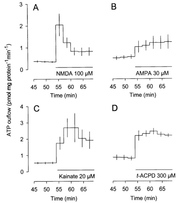
ATP release: effect of glutamate receptor agonists
The basal outflow of ATP in the 9 min before stimulation by glutamate receptor
agonists averaged 0.60 ± 0.02 pmol per mg protein per min (n = 564).
Glutamate and the three receptor type-selective agonists NMDA, AMPA and kainate
all caused significant and, where several concentrations were tested,
concentration-dependent increases in the outflow of ATP (Fig. 1). Trans-ACPD, a selective agonist at
metabotropic glutamate receptors and devoid of activity at ionotropic glutamate receptors
(Palmer et al. 1989; Manzoni et al., 1990), also caused a concentration-dependent release of
ATP from astrocytes (Fig. 1). The order of potency was AMPA > NMDA > trans-ACPD >
glutamate. AMPA had the maximal effect.
Glutamate itself elicited a transient overflow of ATP at the lowest concentration (100
uM; Fig. 2A) but a long-lasting overflow at higher concentrations (Fig. 2B and C). The
120
-î
100
80
-Q. O)
E
õ
3
60
I
>
O CL
40
-20
-0
JKainate
AMPA
trans-ACPD
Glutamate
■ ■ i l i i i i r1 1 1 1—i—i—i i i|
10
100
1000
uM
Figure
1.
ATP
release
evoked
by
glutamate
receptor
agonists
from
superfused
primary
c
^E
'c
'd)2
E
õ
E
Q . O ( ^ -I—» O Q.5
1.5
1.0
0.5
0.0
Glutamate 100 u M
i i i i i
45 50 55 60 65
c
E
B
T
c 3
-Q)
2
Q.
1
2
"
O
E
S 1 -
I
4 1
1 +
o
4—
O ,-»
n ° "
LL
I-<
Glutamate 300 |JM
LL
I-< i
45
I50
55 60 65
Time (min)
H 1—+
Glutamate 1000 uM
— i 1 1