The Combined Deficiency of
Immunoproteasome Subunits Affects Both
the Magnitude and Quality of Pathogen- and
Genetic Vaccination-Induced CD8
+
T Cell
Responses to the Human Protozoan Parasite
Trypanosoma cruzi
Jonatan Ersching1¤, José R. Vasconcelos1,2, Camila P. Ferreira1, Braulia C. Caetano3, Alexandre V. Machado4, Oscar Bruna–Romero5, Monique A. Baron6, Ludmila R. P. Ferreira6,7, Edécio Cunha-Neto6, Kenneth L. Rock3, Ricardo T. Gazzinelli3,4,8☯*,
Maurício M. Rodrigues1☯†
1Centro de Terapia Celular e Molecular and Departamento de Microbiologia, Imunologia e Parasitologia, Universidade Federal de São Paulo - Escola Paulista de Medicina, São Paulo, São Paulo, Brazil,
2Departamento de Biociências, Universidade Federal de São Paulo, Santos, São Paulo, Brazil,
3Departments of Medicine and Pathology, University of Massachusetts Medical School, Worcester, Massachusetts, United States of America,4Centro de Pesquisas René Rachou, FIOCRUZ, Belo Horizonte, Minas Gerais, Brazil,5Departamento de Microbiologia, Imunologia e Parasitologia, Universidade Federal de Santa Catarina, Florianópolis, Santa Catarina, Brazil,6Instituto do Coração (InCor), Faculdade de Medicina
- Universidade de São Paulo, São Paulo, São Paulo, Brazil,7Universidade Santo Amaro, São Paulo, São
Paulo, Brazil,8Departamento de Bioquímica e Imunologia, Instituto de Ciências Biológicas, Universidade
Federal de Minas Gerais, Belo Horizonte, Minas Gerais, Brazil
†Deceased.
☯These authors contributed equally to this work.
¤ Current address: Whitehead Institute for Biomedical Research, Cambridge, Massachusetts, United States
of America
Abstract
Theβ1i,β2iandβ5iimmunoproteasome subunits have an important role in defining the rep-ertoire of MHC class I-restricted epitopes. However, the impact of combined deficiency of the three immunoproteasome subunits in the development of protective immunity to intra-cellular pathogens has not been investigated. Here, we demonstrate that immunoprotea-somes play a key role in host resistance and genetic vaccination-induced protection against the human pathogenTrypanosoma cruzi(the causative agent of Chagas disease), immunity to which is dependent on CD8+T cells and IFN-γ(the classical immunoproteasome
inducer). We observed that infection withT.cruzitriggers the transcription of immunoprotea-some genes, both in mice and humans. Importantly, genetically vaccinated orT.cruzi -infectedβ1i,β2iandβ5itriple knockout (TKO) mice presented significantly lower frequen-cies and numbers of splenic CD8+effector T cells (CD8+CD44highCD62Llow) specific for the previously characterized immunodominant (VNHRFTLV) H-2Kb-restrictedT.cruziepitope. a11111
OPEN ACCESS
Citation:Ersching J, Vasconcelos JR, Ferreira CP, Caetano BC, Machado AV, Bruna–Romero O, et al. (2016) The Combined Deficiency of
Immunoproteasome Subunits Affects Both the Magnitude and Quality of Pathogen- and Genetic Vaccination-Induced CD8+T Cell Responses to the
Human Protozoan ParasiteTrypanosoma cruzi. PLoS Pathog 12(4): e1005593. doi:10.1371/journal. ppat.1005593
Editor:Ingrid Müller, Imperial College London, UNITED KINGDOM
Received:August 28, 2015
Accepted:April 1, 2016
Published:April 29, 2016
Copyright:© 2016 Ersching et al. This is an open access article distributed under the terms of the
Creative Commons Attribution License, which permits unrestricted use, distribution, and reproduction in any medium, provided the original author and source are credited.
Data Availability Statement:All relevant data are within the paper and its Supporting Information files.
Not only the quantity, but also the quality of parasite-specific CD8+T cell responses was
altered in TKO mice. Hence, the frequency of double-positive (IFN-γ+/TNF+) or
single-posi-tive (IFN-γ+) cells specific for the H-2Kb-restricted immunodominant as well as subdominant
T.cruziepitopes were higher in WT mice, whereas TNF single-positive cells prevailed among CD8+T cells from TKO mice. Contrasting with their WT counterparts, TKO animals
were also lethally susceptible toT.cruzichallenge, even after an otherwise protective vacci-nation with DNA and adenoviral vectors. We conclude that the immunoproteasome subunits are key determinants in host resistance toT.cruziinfection by influencing both the magni-tude and quality of CD8+T cell responses.
Author Summary
CD8+t lymphocytes are cells of the immune system that mediate control of intracellular infections by viruses, prokaryote as well as eukaryote pathogens. To confer protection, these lymphocytes need to be elicited by pathogen peptides that are presented in associa-tion with MHC class I molecules. The degradaassocia-tion of self and microbial proteins by cata-lytic domains of the cytosolic proteasomeβ1,β2 andβ5 subunits is intimately linked to the generation of MHC class I-restricted epitopes, which in turn are important determinants of the kinetics, specificity and efficiency of CD8+T cell-mediated immunity. Importantly, inflammatory stimuli trigger the expression of the inducible alternativeβ1i,β2i andβ5i subunits that form the immunoproteasomes. The qualitative and quantitative importance of immunoproteasomes in generating CD8+T cell epitopes has recently been demon-strated in mice that are simultaneously devoid of theβ1i,β2i andβ5i subunits. In this study, we explored the role of immunoproteasomes in host resistance toTrypanosoma cruzi, a protozoan parasite that causes Chagas disease. We found thatβ1i,β2i andβ5i tri-ply deficient mice have an impaired response of CD8+T cells and are highly susceptible to primary infection withT.cruzi. We also demonstrated that host resistance induced by a genetic vaccine able to protect normal mice fromT.cruzichallenge fails to do so in the immunoproteasome-deficient mice. Our study provides strong evidences thatβ1i,β2i and
β5i immunoproteasome subunits are important determinants of both the magnitude and quality of CD8+T cell responses as well as immune-mediated host resistance to a human pathogen.
Introduction
CD8+T cells are important mediators of pathogen control during intracellular infections. Suffi-cient induction of these cells leads to pathogen elimination [1–8], whereas weak or exacerbated CD8+T cell stimulation may lead to pathology [9–17]. Therefore, the proper induction of CD8+T cells must be tightly regulated and may be co-opted in the development of new vac-cines against intracellular pathogens [18–22].
Critical in the process of CD8+T cell induction is the kinetics and efficiency of the provision of MHC class I-restricted epitopes recognized by these lymphocytes, which is linked to the deg-radation of mature proteins and defective ribosomal products in the cytosol by barrel-shaped structures denoted proteasomes, as recently reviewed [23,24]. The catalytic activity of protea-somes is attributed to 3 subunits (β1 (Psmb6),β2 (Psmb7), andβ5 (Psmb5)) located in each of the two innerβrings of the 20S core. In addition, proteasomes featuring the alternative (INCTV-CNPq). JE is a recipient of fellowship from
FAPESP. JRV received funding from FAPESP 2012/ 22514-3. CPF received funding from FAPESP 2015/ 08814-2. OBR, RTG and MMR are recipients of fellowships from CNPq. The funders had no role in study design, data collection and analysis, decision to publish, or preparation of the manuscript.
catalytic subunitsβ1i (LMP2 or Psmb9),β2i (MECL1 or Psmb10) andβ5i (LMP7 or Psmb8) are named immunoproteasomes, which are constitutively expressed in some hematopoietic cells and may be induced by inflammatory stimuli such as IFN-γ, IFN-βor TNF in other cell types (reviewed in [25]).
The immunoproteasomes enhance the quantity and diversity of MHC class I-restricted pep-tides generated and their consequent impact on the magnitude and breath of protective responses of CD8+T cells against intracellular pathogens has long been studied by several groups. However, theβ1i,β2i andβ5i proteins have a redundant role, and only partial pheno-type is observed in mice lacking a single functional gene encoding one of the immunoprotea-some subunits. Only recently a mouse concomitantly defective of all three immunoproteaimmunoprotea-some genes (TKO mouse) was made available, allowing the observation that immunoproteasomes are more relevant for the repertoire of MHC class I-presented peptides than thought before, so much so that WT splenocytes are rejected by TKO mice [26]. However, the participation of immunoproteasomes in the control of infections by most pathogens studied so far was only incremental.
Here, we evaluated the role of immunoproteasomes in the CD8+T cell-mediated immunity, resistance to infection, and protective immunity conferred by genetic vaccination against Try-panosoma cruzi, a human protozoan parasite and causative agent of Chagas disease. Because control of infection byT.cruziis critically dependent on CD8+T cells and IFN-γ(reviewed in [27]), we reasoned that the immunoproteasome could be specially relevant for protection in this model. Confirming our hypothesis, we found that CD8+T cell immune responses toT.
cruziepitopes were remarkably weaker in TKO mice infected withT.cruzior immunized with an adenoviral vaccine vector expressing an immunodominant parasite antigen. Not only the quantity, but also the quality of parasite-specific CD8+T cell responses was altered in TKO mice, as indicated by the higher frequency of double-positive (IFN-γ+/TNF+) or single-positive (IFN-γ+) cells specific for immunodominant as well as subdominantT.cruziepitopes in WT versus TKO mice. Finally, another highly relevant finding was that bothnaïveand vaccinated TKO mice were extremely susceptible to experimental infection, with most animals succumb-ing to an otherwise non-lethal challenge. These observations establish that immunoprotea-somes play a critical role in the generation of immunogenic peptides and the development of protectiveT.cruzi-specific CD8+T lymphocytes.
Results
Reduced antigen presentation of MHC class I-restricted
T
.
cruzi
epitopes
by immunoproteasome-deficient dendritic cells
in vitro
Because dendritic cells constitutively express immunoproteasomes, we examined whetherin vitro-generated bone marrow-derived dendritic cells (BMDC) from TKO mice differed from WT BMDC in antigen presentation capacity upon exposure toT.cruzitrypomastigotes or par-ticles of the adenoviral vaccine vector expressing the immunodominantT.cruziantigen ASP-2 (AdASP-2) [28]. Upon stimulation with parasites or adenovirus, we observed that WT and TKO BMDCs upregulated the costimulatory marker CD86 equally wellin vitro, whereas the expression of H-2Kbmolecules by TKO BMDC was lower than by their WT counterparts (Fig 1a and 1b). In addition, IL-12 p70 concentrations in the culture supernatants were similar between WT BMDC and TKO BMDC (Fig 1c).
Consistent with the result showing lower MHC class I expression, TKO BMDC exposed toT.
cruzior AdASP-2 stimulated significantly fewer IFN-γ-producing CD8+T cells than WT BMDC did, but no difference was observed when BMDC were incubated with synthetic VNHRFTLV peptide corresponding to the immunodominant H-2Kb-restricted epitope from ASP-2, or the ANYKFTLV and ANYDFTLV peptides that correspond to the respective sub-dominant epitopes, thus indicating that the reduction in antigen presentation capacity from TKO BMDC is due to the impaired processing of MHC class I-restricted epitopes (p<0.001,Fig
1d). Conversely, we observed that TKO and WT BMDC exposed toT.cruzior AdASP-2 were equally able to present MHC class II-restricted epitopesinvitro, as measured by their capacity to stimulate similar numbers of IFN-γ-producing CD4+T cells (Fig 1e). WhenT.cruzi- or AdASP-2-exposed BMDC were co-cultured with CD4+or CD8+T cells isolated fromnaïve
mice, no IFN-γsecretion was detected.
These results thus suggested the contribution of immunoproteasomes for the processing of MHC class I-restrictedT.cruziepitopes delivered by the parasite itself or by an adenoviral vac-cine vector.
Fig 1. Reducedin vitropresentation of MHC class I epitopes fromT.cruziby immunoproteasome-deficient BMDC.In vitro-generated WT (red) or TKO (blue) BMDC were incubated for 24 h with
trypomastigotes ofT.cruziY strain (m.o.i. = 3), the adenoviral vector AdASP-2 (m.o.i. = 50), or left untreated. (a) Staining of H-2Kband (b) CD86 gated in CD11c+BMDC. (c) IL-12 concentration in culture supernatants obtained after 24 h of cultivation as measured by ELISA. (d) Purified CD8+or (e) CD4+T cells were obtained from the spleens of WT mice infected withT.cruzi15 days earlier. The purified T cells were admixed with WT or TKO BMDC and incubated overnight. Where indicated, BMDC were loaded with the peptides VNHRFTLV, ANYKFTLV, or ANYDFLTV, corresponding to H-2Kb-restricted epitopes fromT.cruzi. The frequency of
IFN-γ-producing cells was detected by ELISPOT. SFC: spot-forming cells. Results are shown as individual values and as the mean±SEM for each group (n = 3). One of two independent experiments is presented. Asterisks indicate that the values observed in TKO mice were significantly lower than those in WT mice
(****P<0.0001).
T
.
cruzi
induces the transcription of immunoproteasome genes in mice
and humans
To further investigate the participation of immunoproteasomes in the response toT.cruzi
infectionin vivo, we performed semi-quantitative real-time PCR with primers specific toβ1,
β2,β5 and the respective surrogateβ1i,β2i, andβ5i genes using cDNA obtained from heart samples ofnaïveandT.cruzi-infected mice 12 dpi. Additionally, the corresponding human mRNAs were quantified in heart samples from healthy and chronic chagasic patients with car-diomyopathy. These experiments demonstrated thatin vivoinfection withT.cruziinduces the transcription of immunoproteasome genes in both mice and humans (Fig 2).
Impaired immunity of specific CD8
+T cells and higher tissue parasitism
upon
T
.
cruzi
infection of immunoproteasome-deficient mice
Following experimental infection, we evaluated the expression of MHC molecules by splenic antigen-presenting cells (CD11c+I-Ab+CD3-CD19-) fromnaïveanimals and from mice chal-lenged withT.cruzitwenty days earlier. As expected,T.cruziinfection induced the upregula-tion of MHC class I in both WT and TKO cells (p<0.001 in both cases), suggesting the
participation of the conventional immunoproteasome catalytic subunits in the processing of H-2Kb-restricted epitopes. Nevertheless, the expression of H-2Kbmolecules on the surface of TKO cells fromnaïveor infected mice was significantly lower than on their counterpart WT cells (p<0.01,Fig 3a), indicating the contribution of immunoproteasomes to the processing of
T.cruziepitopes. In contrast, the expression of I-Abmolecules on the surface of TKO antigen-presenting cells fromnaïveor infected mice was similar to their WT counterparts (Fig 3b). Accordingly,naïveand infected TKO mice presented lower numbers of total CD8+T cells in the spleen in comparison to WT animals, whereas no difference in total CD4+T cell numbers was observed between WT and TKO mice (Fig 3c and 3d). When results were expressed in cell frequencies, these differences remained exclusive for the CD8+T cell compartment (S2 Fig).
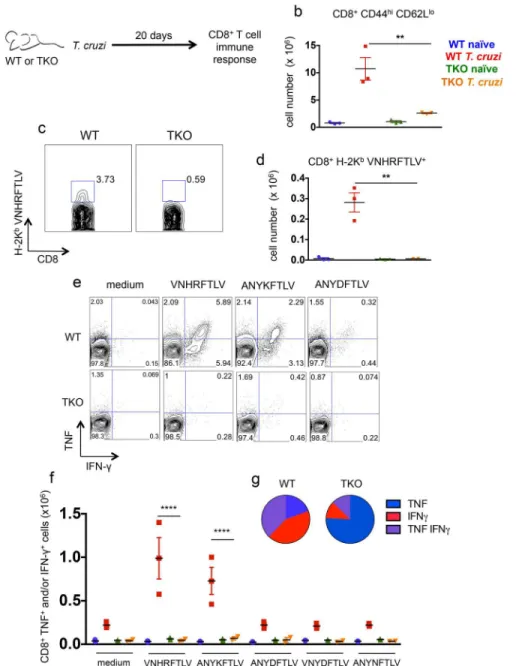
The immune response mediated by CD8+T cells was evaluated in detail in WT and TKO mice 20 days after infection withT.cruzi. The total numbers of splenic CD8effectorcells (CD8 +-CD44highCD62Llow) in infected TKO mice were significantly lower than the corresponding numbers in infected WT animals (p<0.01,Fig 4b). Using pentamer staining, we also measured
the numbers of CD8+T cells specific for the previously characterized immunodominant, H-2Kb-restricted,T.cruziepitope VNHRFTLV [29,30]. Again, these numbers were considerably lower in TKO mice than those observed in infected WT mice (p<0.01,Fig 4c and 4d). We
fur-ther evaluated the function of CD8+T cells through their pattern of IFN-γ/TNF production as assessed byex vivorestimulation of splenocytes with the peptides VNHRFTLV, ANYKFTLV, and ANYDFTLV (corresponding toT.cruziH-2Kb-restricted epitopes) followed by intracellu-lar staining. In presence of the two former peptides, higher numbers of CD8+T cells from infected WT mice produced IFN-γand/or TNF in comparison to the infected TKO animals, whereasnaïvemice did not respond regardless of their background (p<0.0001,Fig 4e and 4f).
Fig 2.T.cruziinduces the transcription of immunoproteasome genes in mice and humans.Relative quantification (RQ) of the indicated mRNA levels was measured by real-time PCR in myocardial tissue samples from (a) WT mice infected withT.cruzi(Y strain) 12 dpi or from (b) chronic chagasic patients with cardiomyopathy as compared to samples obtained fromnaïvemice or healthy subjects, respectively. Results are shown as individual values and as the mean±SEM for each group. For mouse groups, n = 5, pooled from
two independent experiments, and for human samples, n = 14. Asterisks indicate that the values observed for immunoproteasome genes were significantly higher than those for conventional proteasome genes (*P<0.05
***P<0.001****P<0.0001).
doi:10.1371/journal.ppat.1005593.g002
Fig 3. Mice lacking immunoproteasomes have diminished expression of MHC class I molecules and reduced numbers of CD8+T cells.WT and TKO mice were infecteds.c. with 104T.cruziparasites (Y strain) or left uninfected. Twenty days later, spleen cells were collected and stained for surface markers. (a) gMFI (geometric mean of fluorescence intensity) of H-2Kband (b) I-Abstainings are shown after gating in CD11c+ I-Ab+CD3-CD19-cells. Total numbers of CD8+and CD4+T cells in the spleen of these animals are shown in (c) and (d), respectively. Results are shown as individual values and as the mean±SEM for each group (n = 3). One representative of at least two independent experiments is shown. Asterisks indicate that the values observed for TKO mice were significantly different than those for WT mice (*P<0.05**P<0.01
***P<0.001).
Fig 4. Immunoproteasome-deficient mice present impaired immunity of specific CD8+T cells upon infection withT.cruzi.(a) Experiment design: WT and TKO mice were infecteds.c. with 104T.cruzi parasites (Y strain) or left uninfected. Twenty days later, the response of CD8+T cells was assessed in the spleen. (b) Total numbers of CD8+CD44highCD62Llowcells. (c) Representative samples and (d) total numbers of specific CD8+T cells stained with H-2Kb-VNHRFTLV pentamers. (e) Representative samples and (f) numbers of CD8+splenic cells positively stained with anti-TNF and/or anti-IFN-γafterex vivo restimulation with the indicated peptides corresponding to known or hypotheticalT.cruziMHC class I-restricted epitopes. (g) Combination of cytokines stained in responder CD8+T cells from spleens ofT.cruzi -infected mice restimulatedex vivowith VNHRFTLV peptide. Results are shown as individual values and as the mean±SEM for each group (n = 3). One representative of two independent experiments is shown. Asterisks indicate that the values observed for TKO mice were significantly lower than those for WT mice (**P<0.01****P<0.0001).
TKO animals. Among the cells that produced any cytokine, the frequencies of double-positive (IFN-γ+/TNF+) or single-positive (IFN-γ+) cells after restimulation with VNHRFTLV peptide were higher in infected WT mice compared with cells from infected TKO mice (p<0.01,Fig
4g), whereas TNF single-positive cells prevailed among CD8+T cells from TKO mice (Fig 4g). We also compared the frequencies of splenic CD4effector(CD4+CD44highCD62Llow) cells in mice infected 20 days earlier, and we also observed that upon infection, CD4+T cells express-ing IFN-γand/or TNF can be detected withoutex vivorestimulation, as previously reported [31]. No difference was observed in the effector phenotype or function of CD4+T cells between WT and TKO mice infected withT.cruzi(S4 Fig).
Based on the experiments above, we concluded that following infection withT.cruzi, the generation of specific CD8+T cells was severely impaired and profile of cytokine production altered in TKO mice.
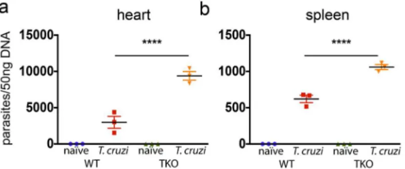
To test whether the impaired immunity of CD8+T cells in TKO mice correlates with reduced resistance to infection withT.cruzi, we further estimated the amount of parasite DNA in infected WT and TKO mice. As shown inFig 5, in the heart and spleen, the quantity ofT.
cruziDNA was significantly higher in TKO mice as compared with WT animals (p<0.0001).
Failure of genetic vaccination in mice devoid of immunoproteasomes
Given that CD8+T cells are critical for immunity againstT.cruziinfection, we developed an immunization regimen that successfully vaccinates highly susceptible mice against systemic lethal infection [32–34]. For that purpose, we used recombinant plasmid DNA for priming and human replication-defective recombinant adenovirus type 5 for boost, both vectors expressing ASP-2 ofT.cruzi. This vaccination protocol elicited a long-lived protective immune response mediated by CD8+effector and effector memory T cells [35,36].
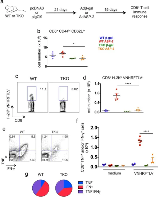
When we compared the response of CD8+T cells, we found significantly lower numbers of CD8effectorcells in ASP-2-vaccinated TKO mice than in ASP-2-vaccinated WT animals (p<0.05,Fig 6b). As in the case ofT.cruzi-infected mice, the numbers of cells specific for the
VNHRFTLV epitope among the splenic CD8+T cells of ASP-2-vaccinated TKO mice were sig-nificantly lower than those observed in ASP-2-vaccinated WT mice (p<0.0001,Fig 6c and 6d).
In addition, the numbers of specific CD8+T cells producing IFN-γand/or TNF uponex vivo
restimulation with the peptide VNHRFTLV were also lower in the population of splenic CD8+ T cells from ASP-2-vaccinated TKO mice compared with cells from ASP-2-vaccinated WT mice (p<0.0001,Fig 6e and 6f). The quality of the immune response of the CD8+T cells from
ASP-2-vaccinated WT mice in comparison to TKO animals was not as different as observed in
T.cruzi-infected mice, and IFN-γsingle-positive or IFN-γ/TNF double-positive cells predomi-nated in both WT and TKO animals (Fig 6g). When expressed in cell frequencies, or when the cytokine response was assessed by ELISPOT, similar differences between WT and TKO mice were found (S5 Fig). Moreover, the response of CD4+T cells to genetic vaccination withAsp-2
was similar between WT and TKO animals, as measured byin vivoincorporation of BrdU in CD44highcells or intracelllular staining of IFN-γafterex-vivorestimulation with AdASP-2-infected cells (S6 Fig). Overall, we concluded that following genetic immunization or infec-tion withT.cruzi, TKO mice were severely impaired in the generation of CD8+T cell-mediated immune responses to the VNHRFTLV epitope.
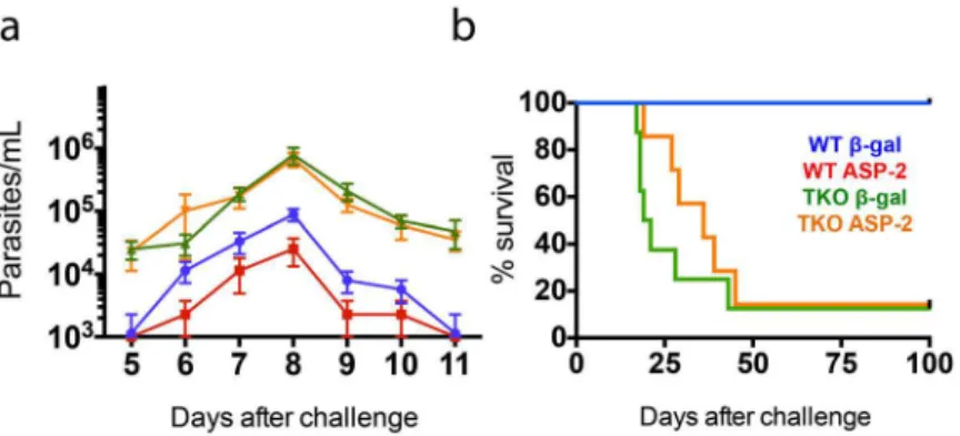
challenge, TKO mice vaccinated with theβgal unrelated control or with ASP-2 presented levels of parasitemia that were about one order of magnitude higher than those in WT mice vacci-nated with theβgal control (p<0.01,Fig 7a). Notably, the parasitemia detected in TKO mice
previously immunized with ASP-2 was indistinguishable from that observed among TKO mice that had received the unrelatedβgal-expressing vector. In contrast, and as previously described [32], the parasitemia observed after challenge of ASP-2-vaccinated WT mice was significantly lower than that measured amongβgal-vaccinated control animals (p<0.01,Fig 7a). Not only
did TKO mice have higher parasitemia, but most of them also succumbed to an otherwise non-lethal infection. A total of 87.5% of TKO mice vaccinated with theβgal control succumbed before 45 days after infection. ASP-2-vaccinated TKO mice survived slightly longer, but still, 85.7% of the mice died before day 50 after challenge (Fig 7b). The increase in survival was sta-tistically significant in the groups of WT mice compared with TKO mice (p<0.001). These
results support the association between the decrease in the frequency of specific splenic CD8+ T cells and the limited parasite control in TKO mice.
Because different strains ofT.cruzimay present distinct patterns of infectivity in mice, we also tested whether TKO mice were more susceptible to infection with parasites of the CL strain. Similar to the case of mice infected with parasites of the Y strain, we observed that TKO mice were highly susceptible to infection and unable to control their parasitemia, succumbing before 25 days following infection with an otherwise non-lethal challenge (S7 Fig).
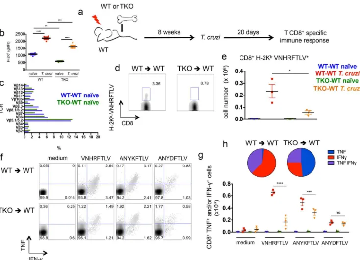
An altered CD8+T cell repertoire has been previously described in TKO mice in compari-son to WT animals [26]. This was explained by a different repertoire of immunogenic peptides presented by thymic epithelial cells from TKO mice. To test whether a difference in the T cell repertoire accounted for most of the defective immune response observed in TKO mice, we generated bone marrow chimeras. Irradiated WT mice were reconstituted with bone marrow from either WT (WT-WT) or TKO (TKO-WT) animals and after 8 weeks these mice were infected withT.cruzi. After 20 days of infection, the response of CD8+T cells was assessed (Fig 8a). In this set up, bone marrow-derived antigen presenting cells lack immunoproteasomes in TKO-WT chimeras, as indicated by the lower expression of H-2Kbmolecules on CD11c+ splenic cells from these animals in comparison to WT-WT mice (Fig 8b). Conversely, epithelial thymic stromal cells are WT in both WT-WT and TKO-WT chimeras, thus allowing similar selection of CD8+T cells. Similar CD8+T cell repertoire between WT-WT and TKO-WT chi-meras was inferred by the indistinguishable staining of CD8+T cells with TCR Vβantibodies panel (Fig 8c). UponT.cruziinfection, the chimeric TKO-WT animals presented lower Fig 5. Mice devoid of immunoproteasomes present higher tissue parasitism upon infection withT.
cruzi.WT and TKO mice were infecteds.c. with 104T.cruziparasites (Y strain) or left uninfected. Twenty
days later, their (a) hearts and (b) spleens were collected, and the number of parasites/50 ng genomic DNA was measured by real-time PCR. Results are shown as individual values and as the mean±SEM for each group (n = 3). One representative of two independent experiments is shown. Asterisks indicate that the values observed for TKO mice were significantly higher than those for WT mice (****P<0.0001).
Fig 6. Immunoproteasome-deficient mice present impaired immunity of specific CD8+T cells upon genetic vaccination againstT.cruzi.(a) Experiment design: WT and TKO mice were primed with empty plasmid DNA (pcDNA3) or a vector expressing ASP-2 (pIgCl9) and boosted after 21 days with adenovirus 5 expressing beta-galactosidase (Adβ-gal) or ASP-2 (AdASP-2), respectively. Fifteen days later, the response of CD8+T cells was assessed in the spleen. (b) Total numbers of CD8+CD44highCD62Llowcells. (c) Representative samples and (d) total numbers of specific CD8+T cells stained with H-2Kb-VNHRFTLV pentamers. (e) Representative samples and (f) numbers of CD8+splenic cells positively stained for TNF and/ or IFN-γafterex vivorestimulation with the peptide VNHRFTLV corresponding to the immunodominantT.
cruziMHC class I-restricted epitope from ASP-2. (g) Combination of cytokines stained in responder CD8+T cells from spleens of ASP-2-vaccinated mice restimulatedex vivowith VNHRFTLV peptide. Results are shown as individual values and as the mean±SEM for each group (n = 4). One representative of two
independent experiments is shown. Asterisks indicate that the values observed for TKO mice were significantly lower than those for WT mice (*P<0.05****P<0.0001).
numbers of VNHRFTLV-specific CD8+T cells stained with pentamers (p<0.05,Fig 8d and 8e)
and lower numbers of cytokine-producing CD8+T cells specific to VNHRFTLV and
ANYKFTLV epitopes (p<0.001,Fig 8f and 8g), although the response to the subdominant
epi-tope ANYDFTLV was comparable between WT-WT and TKO-WT chimeras (Fig 8f and 8g). Once again, the frequency of double-positive (IFN-γ+/TNF+) or single-positive (IFN-γ+) CD8+ T cells after restimulation with VNHRFTLV peptide was higher in infected WT-WT chimeric mice compared with cells from infected TKO-WT mice (p<0.01,Fig 8h).
Moreover, WT-WT and TKO-WT chimeras were vaccinated with AdASP-2 and the specific CD8+T cell response was evaluated after 20 days (Fig 9a). Again, splenic CD11c+cells from TKO-WT mice presented lower expression of H-2Kb(Fig 9b), whereas the staining of CD8+T cells with TCR Vβantibodies panel was similar between TKO-WT and WT-WT animals (Fig 9c). Despite homogenous repertoire of CD8+T cells, the number and cytokine effector function of VNHRFTLV-specific CD8+T cells was significantly reduced in AdASP-2-vaccinated TKO-WT mice in comparison to AdASP-2-vaccinated WT-WT mice (Fig 9d–9h).
Because even WT-WT chimeric mice became highly susceptible toT.cruzichallenge, suc-cumbing to infection from 22 days after challenge, comparisons between WT-WT and TKO-WT animals in terms of resistance to infection and vaccine-induced protection were not possible. Nonetheless, these experiments sustain the notion that the impaired immunity of CD8+T cells inT.cruzi-infected or genetically vaccinated animals lacking all immunoprotea-some genes could not be solely attributed to an altered T cell repertoire.
Discussion
The recognition by CD8+T lymphocytes of antigens displayed on the context of MHC class I molecules is a fundamental requirement for the control of tumors and intracellular infections by viruses, fungi, bacteria and parasites. Proteasomes are of paramount importance in the Fig 7. Failure of genetic vaccination-induced protection againstT.cruziin mice lacking
immunoproteasomes.WT and TKO mice (n = 7–8) were primed with empty plasmid DNA (pcDNA3) or a plasmid vector expressing ASP-2 (pIgCl9) and boosted after 21 days with adenovirus 5 expressing beta-galactosidase (Adβ-gal) or ASP-2 (AdASP-2), respectively. Fifteen days later, the mice were challengeds.c. with 104T.cruziparasites (Y strain). (a) Parasitemia and (b) Kaplan-Meier survival curves for each mouse group are presented. Parasitemia is expressed as the mean±SEM. The values of peak parasitemia (day 8) were log transformed and compared by one-way ANOVA followed by Tukey’s HSD test, which showed that i) ASP-2-immunized WT mice displayed levels of parasitemia significantly lower than those of the other mouse groups (p<0.01 in all cases), ii) the parasitemia values of TKO mice (vaccinated or control) were higher than those of WT mice (p<0.01 in all cases), and iii) no difference between vaccinated and control mice was
detected among TKO animals. Comparison of the survival curves using the log-rank (Mantel-Cox) test indicated that i) vaccinated and control WT mice survived significantly longer (p<0.001) than TKO mice did, and ii) although vaccinated TKO mice survived slightly longer than control TKO mice did, no statistical significance was reached. Data was pooled from three independent experiments (n = 7–8).
processing of antigens in the MHC class I pathway. However, the specific contribution of alter-native proteasome catalytic subunitsβ1i,β2i andβ5i (as opposed to the canonical active sites) to the generation of MHC class I-restricted epitope repertoire was thought to be, at most, incre-mental until the recent development of a mouse model simultaneously devoid ofβ1i,β2i and
β5i subunits (TKO mice) [26]. In that study, major differences in the quantity and quality of epitopes recognized by CD8+T cells was reported, suggesting that models where only one or two subunits are targeted might underestimate the relevance of immunoproteasomes in anti-gen processing and presentation. Although it is reasonable to assume that immunoprotea-some’s role in epitope abundance and specificity may lead to altered resistance to a pathogen, supportive data to this assumption remain relatively scarce. Here we explored the TKO mouse model to further investigate the role of immunoproteasomes in the development of protective Fig 8. Impaired immunity of specific CD8+T cells upon infection withT.cruziof WT mice reconstituted with immunoproteasome-deficient bone marrow.(a) Experiment design: WT mice were irradiated and reconstituted with WT (WT-WT) or TKO (TKO-WT) bone marrow. After 8 weeks, the chimeric animals were infecteds.c. with 104T.cruziparasites or left uninfected. Twenty days later, the response of CD8+T cells was assessed in the spleen. (b) gMFI (geometric mean of fluorescence intensity) of H-2Kbstaining of CD11c+splenic cells from WT-WT and TKO-WT chimeras. (c) Staining of TCR Vβchains gated in CD8+ T cells fromnaïveWT-WT and TKO-WT chimeras. (d) Representative samples and (e) total numbers of specific CD8+T cells stained with H-2Kb-VNHRFTLV pentamers. (f) Representative samples and (g) total numbers of CD8+splenic cells positively stained with anti-TNF and/or anti-IFN-γafter
ex vivorestimulation with the indicated peptides corresponding to known or hypotheticalT.cruziMHC class I-restricted epitopes. (h) Combination of cytokines stained in responder CD8+T cells from spleens ofT.cruzi-infected mice restimulatedex vivowith VNHRFTLV peptide. Results are shown as individual values and as the mean±SEM for each group (n = 3). One representative of two independent experiments is shown. Asterisks indicate that the values observed for TKO mice were significantly lower than those for WT mice (*P<0.05**P<0.01***P<0.001****P<0.0001).
immunity to a pathogen.Trypanosoma cruzi, a neglected human parasite, was of didactical use as a model, since protective immunity against it is highly dependent on CD8+T cell function and IFN-γ(the major inducer of immunoproteasomes). The TKO mice exhibited a drastically reduced response of CD8+T cells specific for immunodominant and subdominant epitopes afterT.cruziinfection, whereas the response of CD4+T cells was unaltered. Surprisingly, natu-ral resistance to infection in the absence of immunoproteasomes was seriously compromised and even more unexpectedly the protection induced by genetic vaccination was completely abolished, indicating that the immunoproteasome catalytic subunits, rather than the conven-tional proteolytical sites, are essential for the processing ofT.cruziepitopes related to Fig 9. Impaired immunity of specific CD8+T cells upon genetic vaccination of WT mice reconstituted with immunoproteasome-deficient bone marrow.(a) Experiment design: WT mice were irradiated and reconstituted with WT (WT-WT) or TKO (TKO-WT) bone marrow. After 8 weeks, chimeric mice were vaccinated with adenovirus 5 expressing beta-galactosidase (Adβ-gal) or ASP-2 (AdASP-2). Twenty days later, the response of CD8+T cells was assessed in the spleen. (b) gMFI (geometric mean of fluorescence intensity) of H-2Kbstaining of CD11c+splenic cells from WT-WT and TKO-WT chimeras. (c) Staining of TCR Vβchains gated in CD8+ T cells from WT-WT and TKO-WT chimeras genetically immunized with Adβ-gal. (d) Representative samples and (e) total numbers of specific CD8+T cells stained with H-2Kb-VNHRFTLV pentamers. (f) Representative samples and (g) total numbers of CD8+splenic cells positively stained with anti-TNF and/or anti-IFN-γafterex vivorestimulation with the peptide VNHRFTLV corresponding to the immunodominant MHC class I-restricted epitope from ASP-2. (h) Combination of cytokines stained in responder CD8+T cells from spleens of AdASP-2-vaccinated mice
restimulatedex vivowith VNHRFTLV peptide. Results are shown as individual values and as the mean±SEM for each group (n = 3). One representative of
two independent experiments is shown. Asterisks indicate that the values observed for TKO mice were significantly lower than those for WT mice (**P<0.01
***P<0.001****P<0.0001).
protective immunity generated during infection or vaccination. Our observations from chime-ric mice are consistent with the hypothesis that the defect in TKO mice is the inability of anti-gen presenting cells to process protective epitopes to CD8+T cells, rather than the variation of naïve CD8+T cell repertoire. Also, we were unable to detect any shift in the immunodomi-nance pattern of TKO mice relatively to WT counterparts.
We have previously reported that the induction of CD8+T cell responses toT.cruziis remarkably delayed and suboptimal, with most cells expressing high levels of the proapoptotic marker CD95 [16]. Conversely, genetic vaccination with adenoviral vectors induces a rapid, expanded, polyfunctional and highly viable CD8+T cell response that differentiates into a long-lasting memory pool, as reported forT.cruzias well as other pathogens [33–36]. Here, we report that the function ofT.cruzi-specific CD8+T cell responses was altered in the absence of immunoproteasomes, as indicated by the higher frequency of TNF single-positive cells in TKO animals as opposed to a prevailing compartment of IFN-γsingle-positive or IFN-γand TNF double-positive cells in immunoproteasome-sufficient mice. This difference, however, was more pronounced during infection than upon genetic vaccination withAsp-2. It is acknowl-edged that IFN-γis of major importance for control ofT.cruziinfection and its induction is tightly regulated, whereas TNF, expressed throughout the course of infection, is of secondary importance to parasite control [13,27,37]. Our results may suggest that in our model the threshold of antigen presentation required to induce TNF production by CD8+T cells might be lower than the levels of stimulation necessary to induce IFN-γ. Consistently, the data pre-sented here reinforce the notion that AdASP-2 is remarkably more potent thanT.cruziat inducing CD8+T cell responses, since even in the absence of immunoproteasomes the viral vector was able to stimulate the production of IFN-γ-secreting cells. Therefore, even in the absence of immunoproteasomes, the antigen pool generated by the adenoviral vaccine could be cleaved by conventional proteasomes without severely compromising the induction of polyfun-cional CD8+T cells (yet lower number of cells are induced), in contrast to the observations from experimental infection. In fact, we have previously observed that CD8+T cells specific to subdominant epitopes fromT.cruzican be induced by adenoviral vaccination, but not by pro-tozoan infection, in line with the idea that the provision of epitopes is enhanced during genetic vaccination [33].
Presumably, the lower expression of MHC class I in TKO mice is inextricably linked to a defect in generating peptides that bind to H-2 molecules. We found that upon infection withT.
cruzior adenovirus expressing ASP-2, dendritic cells from TKO background were highly impaired at presenting antigen to WT CD8+T lymphocytes explanted from either infected or vaccinated mice. These results contrasted with the fact that WT and TKO BMDC loaded with VNHRFTLV, ANYKFTLV, or ANYDFTLV peptides were equally able to stimulateT.cruzi -specific CD8+T cellsin vitro. These results suggest that the defects in antigen presentation capacity of TKO cells are due to the compromised processing of epitopes. These findings are consistent with a previous study indicating that in absence of immunoproteasomes the presen-tation of protein antigens that need to be cleaved is compromised, whereas no defect is observed when the processed epitope is directly expressed by a minigene [26]. Nonetheless, dendritic cells lacking immunoproteasomes still upregulate MHC class I molecules and present epitopes recognized by CD8+ T cells upon infection withT.cruzior genetic vaccination with
Asp-2, an effect most likely attributed to the processing of antigen by the canonical catalytic subunits of the proteasome.
Independently of their specificities or hierarchies, the CD8+T cells activated in TKO mice were always reduced in comparison to WT counterparts.
An altered CD8+T cell repertoire has been previously described in TKO mice in compari-son to WT animals [26]. Hence, distinct naïve T cell repertoires could also explain the discrep-ancies in CD8+T cell response between TKO and WT mice [26,41]. It is assumed that this difference in the repertoire of CD8+T cells from WT and TKO mice is due to MHC I-mediated presentation of a different repertoire of peptides by epithelial cells from thymus during T cell development. To address this issue we performed experiments using chimeras, where recipient WT mice received bone marrows from either WT or TKO mice. The results obtained from these experiments suggest that it is unlikely that a difference in the T cell repertoire accounted for most of the limited immune response observed in TKO mice. In the same direction, the transfer into TKO mice of WT CD8+T cells or of the P14 transgenic CD8+T cell clone specific to GP33 epitope was unable to restore the capacity of TKO animals to optimally respond to LCMV infection [26].
Not only did TKO mice have diminished CD8+T cell immune responses to parasite-derived epitopes, but they were also no longer resistant to infection withT.cruzi. It is reasonable to hypothesize that the susceptibility to infection and the weak CD8+T cell-mediated immunity are linked because in the mouse models that we used (naïveand vaccinated), CD8+T cells have been described as being critically involved in the control of parasitemia and in survival [30,32]. Nevertheless, in addition to the low numbers of specific CD8+T cells, other factor(s) may also account for the extreme susceptibility that we observed. For instance, a second likely source of reduced resistance may stem from the effector phase of the immune response. DuringT.cruzi
infection, large amounts of IFN-γare produced and can be detected in the serum [42]. As immunoproteasomes become the dominant form of proteasomes in IFN-γ-activated cells, their absence may drastically reduce the antigen presentation capacity of target cells infected withT.
cruzi. This aspect may further contribute to the impaired efficacy of the few CD8+T cells gen-erated during the priming phase.
It is worth mentioning that the susceptibility of TKO mice occurred in parallel to undetect-able changes in presentation of MHC II-restricted epitopes to CD4+T cells and in the genera-tion of CD4effectorcells during infection. These findings are also in agreement with previous observations that CD4+T cell-mediated immune responses were similar in TKO and WT mice in different infection models [26].
The susceptibility of TKO mice may also be linked to a larger inflammatory reaction and to more severe myocardial tissue damage. Recently, immunoproteasomes have been described as protecting the heart from excessive inflammatory tissue damage due to acute coxsackievirus B3 (CVB3)-induced myocarditis [43]. BecauseT.cruzimay infect myocardial cells, whether a sim-ilar event is associated with susceptibility to infection should be further investigated. This topic may not be as important in the model of infection used in most of our studies, considering that infection with parasites of the Y strain targets the spleen as well as the heart tissues [44]. How-ever, otherT.cruzistrains, such as the CL or the Colombian strain, cause severe acute heart muscle injury and deserve further investigation [45].
In other words, these protozoan infection models were instrumental at evidencing a specific role of immunoproteasomes for the generation of critical epitopes required for protective immunity.
By employing adenoviral vectors, our group developed genetic vaccination strategies against
T.cruzi. The immunization regimens confer protection to wild-type mice of different strains, including the C57BL/6, BALB/c and A/Sn [16,28]. From these, the A/Sn mice are the most sus-ceptible and succumb to infection in less than 30 days, even when challenged with as low as 150 parasites ofT.cruziY strain. Conversely, A/Sn mice surviveT.cruziinfection if immunized with AdASP-2 even on the same day of challenge [16]. At least in our animal facility, the C57BL/6 mouse lineage is more resistant toT.cruziinfection and survives challenge with high doses (10,000) of blood forms ofT.cruzi. Therefore, the C57BL/6 mouse serves as a model to study different aspects of the CD8+T cell response related to resistance. Here, we observed that mice devoid of immunoproteasomes in a C57BL/6 background are highly susceptible toT.
cruzichallenge. Unexpectedly, vaccination of these mice with AdASP-2 does not have any effect in protecting the mice upon challenge, whereas in normal C57BL/6 mice the parasitemia is reduced in about one order of magnitude. These results thus indicate that the efficacy of our vaccination regimen is extremely dependent on the expression ofβ1i,β2i andβ5i subunits of the immunoproteasome, rather than the conventional proteolytical sites. In accordance with this finding, a recent clinical trial aimed at testing the efficacy of the RTS,S vaccine against malaria identified the upregulation of immunoproteasome genes among protected individuals after challenge [47]. Altogether, these results may point to the induction of immunoprotea-some genes as pivotal targets to be considered in the design of successful vaccination strategies aimed at inducing CD8+T cells.
In conclusion, we report that immunoproteasomes, rather than canonical proteasomes, have a potentially underestimated role in inducing protective immunity both in primary infec-tion as well as genetic vaccinainfec-tion against a human pathogen.
Materials and Methods
Ethics statement
This study was carried out in strict accordance with the recommendations in the Guide for the Care and Use of Laboratory Animals of the Brazilian National Council of Animal Experimen-tation (http://www.cobea.org.br/). The protocol was approved by the Committee on the Ethics of Animal Experiments of the Institutional Animal Care and Use Committee at the Federal University of Sao Paulo (Id # CEP 0426/09). The protocol using human samples was approved by the Institutional Review Board of the University of São Paulo School of Medicine (Protocol number 739/2005) and written informed consent was obtained from the patients. In the case of samples from heart donors, written informed consent was obtained from their families.
Patients
Myocardial left ventricular free wall heart samples were obtained from end-stage heart failure chronic chagasic patients with cardiomyopathy (7 females, 7 males, 15–61 years old). Control adult heart tissue from the left ventricular-free wall was obtained from nonfailing donor hearts not used for cardiac transplantation due to size mismatch with available recipients (males, 17–
46 years old). Hearts were explanted at the time of heart transplantation at the Heart Institute
Mice and parasites
Five- to 8-week-old female C57BL/6 mice were purchased from CEDEME (Federal University of São Paulo). TKO mice were generated as described by Kincaidet al. [26] and were bred in our own animal facility. Bloodstream trypomastigotes of the Y or CL strain ofT.cruziwere obtained from mice infected 7 or 15 days earlier. The concentration of parasites was estimated and adjusted to 105parasites/mL. Each mouse was inoculated with 104trypomastigotes diluted in 0.1 mL PBS administered subcutaneously (s.c.) at the base of the tail. Parasitemia was assessed on the days indicated in each figure, which involved counting the number of parasites per 5μL blood.
Genetic vaccination
The plasmid pIgSP Cl.9 and AdASP-2 were generated, grown and purified as described previ-ously [28,32]. Control mice were immunized with pcDNA3 and human replication-deficient adenovirus type 5 expressingβgal (Adβ-gal). The mice were inoculated intramuscularly (i.m.) with 50μg plasmid DNA into eachtibialis anteriorismuscle. A total of 21 days later, these mice received 50μL of a viral suspension containing 2 X 10⁸plaque-forming units (pfu) of adenovi-rus via the same locations. Immunological assays were performed on the days indicated in each figure.
Peptides
Synthetic peptides VNHRFTLV, ANYKFTLV, and ANYDFTLV were purchased from Gen-Script (Piscataway, NJ). The peptide purity was higher than 90%. Peptide identities were con-firmed using a Q-Tof Micro equipped with an electrospray ionization source (Micromass, UK). The pentamer H-2Kb-VNHRFTLV was purchased from ProImmune Inc. (Oxford, UK).
BMDC generation
Progenitor cells were flushed from femurs and culturedin vitroin RPMI 1640 supplemented with 10 mM HEPES, 0.2% sodium bicarbonate, 59 mg/L penicillin, 133 mg/L streptomycin, 10% HyClone fetal bovine serum, 2 mM L-glutamine, 1 mM sodium pyruvate, 55μM 2-mer-captoethanol and 20 ng/mL GM-CSF (R&D Systems) at a concentration of 2 X 10⁵cells/mL. After 4 days in culture, half of the medium volume was replaced with fresh medium. At day 6, the resulting BMDCs were exposed to tissue-culture trypomastigotes ofT.cruziat a ratio of 3 parasites/cell or to AdASP-2 at a ratio of 50 pfu/cell for an additional 24 h. After 7 days in cul-ture (and 24 h ofT.cruzior AdASP-2 exposure), the BMDCs were employed inin vitroantigen presentation assays.
Immunological assays
The presence of IL-12p70 in the supernatant of the BMDCs was assessed after 7 days in culture (and 24 h ofT.cruzior AdASP-2 exposure) using an ELISA (BD).
To perform thein vitroantigen presentation assays, spleen cells from naïve animals or mice infected withT.cruzi15 days earlier were harvested, and the CD8+T cell population was iso-lated through negative selection with a CD8+T Cell Isolation Kit using MACS beads (Miltenyi) according to the manufacturer’s specification, followed by staining with anti-CD8 PerCP (53–
unexposed at a ratio of 1 BMDC to 5 CD8 or CD4 cells for 24 h, and the number of IFN-γ -secreting cells was determined by ELISPOT, as described elsewhere [31].
For flow cytometry analyses, we used mouse splenocytes treated with ACK buffer (NH4Cl, 0.15 M; KHCO3, 10 mM; Na2-EDTA 0.1 mM; pH = 7.4) for lysing the erythrocytes. Single-cell suspensions were washed in PBS, stained for 10 min at RT with biotinylated MHC I multimer H-2Kb-VNHRFTLV, and stained for 20 min at 4°C with streptavidin-APC and CD8 FITC (53–6.7). For the analyses of other cell-surface markers, single-cell suspensions from spleens of mice were stained with CD11c APCCy7 (HL3), CD86 APC (GL1), CD11b PerCP (M1/70), CD19 PE (ID3), CD3 PE (17A2), H-2KbFITC (AF6-88.5), IAb biotinylated (25-9-17), strepta-vidin PECy7, CD4 PeCy7 (RM4-5), CD8 Pacific Blue (53–6.7), CD62L APC (MEL-14), and CD44 PE (IM7). Staining of TCR Vβchains was performed using TCR VβScreening Panel Kit (BD Pharmingen). Isotype control antibodies were PE-labeled hamster IgG1k and rat IgG1k, and FITC-labeled IgG2ak. All antibodies and streptavidins were purchased from BD Pharmin-gen. At least 500,000 cells were acquired on a BD FACS Canto II flow cytometer and analyzed with FlowJo 8.7 (Tree Star, Ashland, OR).
For the intracellular staining (ICS) of cytokines (IFN-γand TNF), splenocytes collected from C57BL/6 or TKO mice were treated with ACK buffer. ICS was performed afterin vitroculture of splenocytes in the presence or absence of the peptides indicated in each figure. Cells were washed 3 times in plain RPMI and re-suspended in cell culture medium consisting of RPMI 1640 medium supplemented with 10 mM Hepes, 0.2% sodium bicarbonate, 59 mg/L of penicil-lin, 133 mg/L of streptomycin, 10% Hyclone fetal bovine serum, 2 mM L-glutamine, 1 mM sodium pyruvate, 55μM 2-mercaptoethanol. The viability of the cells was evaluated using 0.2% trypan blue exclusion dye to discriminate between live and dead cells. Cell concentration was adjusted to 5 X 10⁶cells/mL in cell culture medium containing anti-CD28 (2μg/mL), BDGolgi-Plug (1μL/mL) and monensin (5μg/mL). In half of the cultures, a final concentration of 10μM of the VNHRFTLV peptide was added. The cells were cultivated in V-bottom 96-well plates (Corning) in a final volume of 200μL in duplicate, at 37°C in a humid environment containing 5% CO2. After 8h incubation, cells were stained for surface markers with CD4 FITC (GK1.5) and CD8 PerCP (53–6.7) on ice for 20 min. To detect IFN-γand TNF by intracellular staining, cells were then washed twice in buffer containing PBS, 0.5% BSA, and 2 mM EDTA, fixed and permeabilized with BD perm/wash buffer. After being washed twice with BD perm/wash buffer, cells were stained for intracellular markers using APC-labeled anti-IFN-γ(XMG1.2) and PE-labeled anti-TNF (MP6-XT22) for 20 minutes on ice. Finally, cells were washed twice with BD perm/wash buffer and fixed in 1% PBS-paraformaldehyde. At least 800,000 cells were acquired on a BD FACS Canto II flow cytometer and then analyzed with FlowJo.
Real-time PCR
Total RNA was isolated from mice and human myocardial tissues with Trizol (Invitrogen), fol-lowed by purification with Quick RNA Miniprep columns and DNAse I treatment (Zymo Research). cDNA was synthesized with High Capacity cDNA Reverse Transcription Kit (Applied Biosystems). To confirm the absence of genomic DNA control samples were used without reverse transcriptase. RT-PCR was performed using Power SYBR PCR Mastermix (Thermo Scientific) and StepOne Plus thermo cycler (Applied Biosystems). mRNA levels were normalized to HPRT (mouse) and beta-actin (human). Primer sequences are reported else-where for murine (procedure B in [48]) and human [49] samples. Relative quantification was calculated overnaïve(mouse) or healthy (human) controls usingΔ ΔCT method [50]. The pro-tocol for quantification ofT.cruziDNA in heart and spleen samples was performed as
Bone marrow chimeras
Eight-week-old female C57BL/6 mice were irradiated at 900 Rads. Each irradiated animal received 10 × 106bone marrow cellsi.v. isolated from C57BL/6 or TKO female mice. Before transfer, bone marrow cell suspensions were depleted of T cells with CD8a (Ly-2) MicroBeads and CD4 (L3T4) MicroBeads (Miltenyi Biotec). Mice were given medicated water containing sulfamethoxazole (200 mg/mL) and trimethroprim (40 mg/mL). Experiments were performed 8 weeks after bone marrow transfer.
Statistical analysis
Groups were compared using One Way ANOVA followed by Tukey’s HSD test (http://faculty. vassar.edu/lowry/VassarStats.html). Parasitemia values were log transformed before compari-son. The Log-rank (Mantel-Cox) test was used to compare mouse survival rates after challenge withT.cruzi(http://bioinf.wehi.edu.au/software/russell/logrank/). The differences were con-sidered significant when the P value was<0.05.
Supporting Information
S1 Fig.T.cruziMHC class I epitopes are processed through the cytosolic pathway.(a) TAP-1-deficient mice had their phenotype confirmed by the absence of CD8+T cells in the spleen. (b) WT and TAP-1-deficient BMDC were incubated with LPS,T.cruzior AdASP-2 and the expression of MHC and co-stimulatory molecules on CD11c+cells was assessed by flow cytom-etry. These BMDC were co-cultured with (c) CD8+or (d) CD4+T cells isolated from the spleen ofT.cruzi-infected mice and the ability to present antigen was assessed through ELISPOT to detect spot forming cells (SFC) secreting IFN-γ. (e) WT BMDC were incubated withT.cruzior AdASP-2 in presence or absence of the proteasome inhibitor epoxomicin (EPO) 1μM and the expression of MHC and co-stimulatory molecules on CD11c+cells was assessed by flow cytom-etry. (f) These BMDC were co-cultured with CD8+T cells isolated from the spleen ofT.cruzi -infected mice and the ability to present antigen was assessed through ELISPOT to detect spot forming cells (SFC) secreting IFN-γ.
(TIF)
S2 Fig. Reduced frequencies of CD8+T cells in TKO animals.WT and TKO mice were infecteds.c. with 104T.cruziparasites or left uninfected. Frequencies of (a) CD8+and (b) CD4+T cells in the spleen of these animals are shown. Results are expressed as individual val-ues and the mean ± SEM for each group. Asterisks indicate that the valval-ues observed for TKO mice were significantly lower than those for WT mice (P<0.05P<0.001).
(TIF)
S3 Fig. Impaired immunity of CD8+T cells in TKO animals infected withT.cruzi.WT and TKO mice were infecteds.c. with 104T.cruziparasites or left uninfected. Twenty days later, the response of CD8+T cells was assessed in the spleen. (a) Frequencies of CD8+CD44high
CD62Llowcells. (b) Frequencies of specific CD8+T cells stained with H-2Kb-VNHRFTLV pen-tamers. (c) Frequencies of CD8+splenic cells positively stained with TNF and/or anti-IFN-γafterex vivorestimulation with the indicated peptides corresponding to known or hypo-theticalT.cruziMHC class I-restricted epitopes. (d) Numbers of spot forming cells (SFC) secreting IFN-γand (e) representative samples from ELISPOT of spleen cells upon restimula-tion with the indicated peptides. Results are shown as individual values and as the mean ± SEM for each group. Asterisks indicate that the values observed for TKO mice were significantly lower than those for WT mice (P<0.05P<0.01P<0.001P<0.0001).
S4 Fig. Unaltered immunity mediated by CD4+T cells in TKO animals infected withT.
cruzi.WT and TKO mice were infecteds.c. with 104T.cruziparasites or left uninfected. Twenty days later, their spleens were collected and the frequencies of (a) CD4+CD44high CD62Llowcells and (b) CD4+T cells producing IFN-γand/or TNF were estimated by intracel-lular staining. The results are expressed as individual values and as the mean ± SEM for each group.
(TIF)
S5 Fig. Impaired immunity of CD8+T cells in TKO animals genetically vaccinated against
T.cruzi.WT and TKO mice were primed with empty plasmid DNA (pcDNA3) or a plasmid vector expressing ASP-2 (pIgCl9) and boosted after 21 days with adenovirus 5 expressing beta-galactosidase (Adβ-gal) or ASP-2 (AdASP-2), respectively. Fifteen days later, the response of CD8+T cells was assessed in the spleen. (a) Frequencies of specific CD8+T cells stained with H-2Kb-VNHRFTLV pentamers. (b) Frequencies of CD8+splenic cells positively stained with anti-TNF and/or anti-IFN-γafterex vivorestimulation VNHRFTLV peptide. (c) Numbers of spot forming cells (SFC) secreting IFN-γdetected by ELISPOT of spleen cells upon restimula-tion with the peptide VNHRFTLV. Results are shown as individual values and as the
mean ± SEM for each group. Asterisks indicate that the values observed for TKO mice were significantly lower than those for WT mice (P<0.0001).
(TIF)
S6 Fig. Unaltered response of CD4+T cells in TKO animals genetically immunized with Asp-2.WT and TKO mice were immunized with plasmid DNA encodingAsp-2and boosted after 21 days with the viral vector AdASP-2. Following immunization, mice were given 2 mg BrdU i.p. every other day. Fifteen days after boost, their spleens were collected and the frequen-cies of CD8+CD44highBrdU+and CD4+CD44highBrdU+cells were determined by flow cytom-etry. These results are expressed as individual values and as the mean ± SEM for each group (n = 3). Asterisks indicate that the values observed for TKO mice were significantly lower than those for WT mice (P<0.05). Alternatively, splenocytes from WT and TKO immunized mice
were re-stimulatedex vivowith AdASP-2-infected BMDC followed by IFN-γstaining in CD4+ and CD8+cells.
(TIF)
S7 Fig. Susceptibility of TKO animals to challenge with CL strain ofT.cruzi.WT and TKO mice were challenged intraperitoneally with 100 bloodstream parasites. (a) Parasitemia at dif-ferent time points after infection. (b) Mortality rates after challenge. The parasitemia values were compared by one-way ANOVA, which showed that WT mice displayed levels of parasite-mia that were significantly lower than those of TKO animals (p<0.01 in all cases). Comparison
of the survival curves using the log-rank (Mantel-Cox) test indicated that WT mice survived significantly longer (p<0.01) than TKO mice did.
(TIF)
Acknowledgments
This work is a tribute to the memory of Professor Maurício Martins Rodrigues.
Author Contributions
References
1. Morrot A, Zavala F. Effector and memory CD8+ T cells as seen in immunity to malaria. Immunological reviews. 2004; 201:291–303. PMID:15361248
2. Goldszmid RS, Sher A. Processing and presentation of antigens derived from intracellular protozoan parasites. Current opinion in immunology. 2010; 22(1):118–23. doi:10.1016/j.coi.2010.01.017PMID: 20153156
3. Jordan KA, Hunter CA. Regulation of CD8+ T cell responses to infection with parasitic protozoa. Experi-mental parasitology. 2010; 126(3):318–25. doi:10.1016/j.exppara.2010.05.008PMID:20493842
4. Padilla AM, Bustamante JM, Tarleton RL. CD8+ T cells in Trypanosoma cruzi infection. Current opinion in immunology. 2009; 21(4):385–90. doi:10.1016/j.coi.2009.07.006PMID:19646853
5. Bertholet S, Goldszmid R, Morrot A, Debrabant A, Afrin F, Collazo-Custodio C, et al. Leishmania anti-gens are presented to CD8+ T cells by a transporter associated with antigen processing-independent pathway in vitro and in vivo. Journal of immunology. 2006; 177(6):3525–33.
6. Rosenberg CS, Martin DL, Tarleton RL. CD8+ T cells specific for immunodominant trans-sialidase epi-topes contribute to control of Trypanosoma cruzi infection but are not required for resistance. Journal of immunology. 2010; 185(1):560–8.
7. Harris TH, Banigan EJ, Christian DA, Konradt C, Tait Wojno ED, Norose K, et al. Generalized Levy walks and the role of chemokines in migration of effector CD8+ T cells. Nature. 2012; 486(7404):545–
8. doi:10.1038/nature11098PMID:22722867
8. Van Braeckel-Budimir N, Harty JT. CD8 T-cell-mediated protection against liver-stage malaria: lessons from a mouse model. Frontiers in microbiology. 2014; 5:272. doi:10.3389/fmicb.2014.00272PMID: 24936199
9. Claser C, Malleret B, Gun SY, Wong AY, Chang ZW, Teo P, et al. CD8+ T cells and IFN-gamma medi-ate the time-dependent accumulation of infected red blood cells in deep organs during experimental cerebral malaria. PloS one. 2011; 6(4):e18720. doi:10.1371/journal.pone.0018720PMID:21494565
10. Howland SW, Poh CM, Gun SY, Claser C, Malleret B, Shastri N, et al. Brain microvessel cross-presen-tation is a hallmark of experimental cerebral malaria. EMBO molecular medicine. 2013; 5(7):916–31. doi:10.1002/emmm.201202273PMID:23681698
11. Pai S, Qin J, Cavanagh L, Mitchell A, El-Assaad F, Jain R, et al. Real-time imaging reveals the dynam-ics of leukocyte behaviour during experimental cerebral malaria pathogenesis. PLoS pathogens. 2014; 10(7):e1004236. doi:10.1371/journal.ppat.1004236PMID:25033406
12. Novais FO, Carvalho LP, Graff JW, Beiting DP, Ruthel G, Roos DS, et al. Cytotoxic T cells mediate pathology and metastasis in cutaneous leishmaniasis. PLoS pathogens. 2013; 9(7):e1003504. doi:10. 1371/journal.ppat.1003504PMID:23874205
13. Silverio JC, Pereira IR, Cipitelli Mda C, Vinagre NF, Rodrigues MM, Gazzinelli RT, et al. CD8+ T-cells expressing interferon gamma or perforin play antagonistic roles in heart injury in experimental Trypano-soma cruzi-elicited cardiomyopathy. PLoS pathogens. 2012; 8(4):e1002645. doi:10.1371/journal.ppat. 1002645PMID:22532799
14. Joshi T, Rodriguez S, Perovic V, Cockburn IA, Stager S. B7-H1 blockade increases survival of dysfunc-tional CD8(+) T cells and confers protection against Leishmania donovani infections. PLoS pathogens. 2009; 5(5):e1000431. doi:10.1371/journal.ppat.1000431PMID:19436710
15. Bhadra R, Khan IA. Redefining chronic toxoplasmosis—a T cell exhaustion perspective. PLoS patho-gens. 2012; 8(10):e1002903. doi:10.1371/journal.ppat.1002903PMID:23071434
16. Vasconcelos JR, Bruna-Romero O, Araujo AF, Dominguez MR, Ersching J, de Alencar BC, et al. Path-ogen-induced proapoptotic phenotype and high CD95 (Fas) expression accompany a suboptimal CD8 + T-cell response: reversal by adenoviral vaccine. PLoS pathogens. 2012; 8(5):e1002699. doi:10. 1371/journal.ppat.1002699PMID:22615561
17. Horne-Debets JM, Faleiro R, Karunarathne DS, Liu XQ, Lineburg KE, Poh CM, et al. PD-1 dependent exhaustion of CD8+ T cells drives chronic malaria. Cell reports. 2013; 5(5):1204–13. doi:10.1016/j. celrep.2013.11.002PMID:24316071
18. Hoft DF, Eickhoff CS, Giddings OK, Vasconcelos JR, Rodrigues MM. Trans-sialidase recombinant pro-tein mixed with CpG motif-containing oligodeoxynucleotide induces protective mucosal and systemic trypanosoma cruzi immunity involving CD8+ CTL and B cell-mediated cross-priming. Journal of immu-nology. 2007; 179(10):6889–900.
19. Hill AV, Reyes-Sandoval A, O'Hara G, Ewer K, Lawrie A, Goodman A, et al. Prime-boost vectored malaria vaccines: progress and prospects. Human vaccines. 2010; 6(1):78–83. PMID:20061802
prime-boost vaccination. Frontiers in immunology. 2012; 3:358. doi:10.3389/fimmu.2012.00358PMID: 23264773
21. Ewer KJ, O'Hara GA, Duncan CJ, Collins KA, Sheehy SH, Reyes-Sandoval A, et al. Protective CD8+ T-cell immunity to human malaria induced by chimpanzee adenovirus-MVA immunisation. Nature com-munications. 2013; 4:2836. doi:10.1038/ncomms3836PMID:24284865
22. Morrot A, Rodrigues MM. Tissue signatures influence the activation of intrahepatic CD8(+) T cells against malaria sporozoites. Frontiers in microbiology. 2014; 5:440. doi:10.3389/fmicb.2014.00440 PMID:25202304
23. Rock KL, Farfan-Arribas DJ, Colbert JD, Goldberg AL. Re-examining class-I presentation and the DRiP hypothesis. Trends in immunology. 2014; 35(4):144–52. doi:10.1016/j.it.2014.01.002PMID:24566257
24. McCarthy MK, Weinberg JB. The immunoproteasome and viral infection: a complex regulator of inflam-mation. Frontiers in microbiology. 2015; 6:21. doi:10.3389/fmicb.2015.00021PMID:25688236
25. Kruger E, Kloetzel PM. Immunoproteasomes at the interface of innate and adaptive immune
responses: two faces of one enzyme. Current opinion in immunology. 2012; 24(1):77–83. doi:10.1016/ j.coi.2012.01.005PMID:22296715
26. Kincaid EZ, Che JW, York I, Escobar H, Reyes-Vargas E, Delgado JC, et al. Mice completely lacking immunoproteasomes show major changes in antigen presentation. Nature immunology. 2012; 13 (2):129–35.
27. Junqueira C, Caetano B, Bartholomeu DC, Melo MB, Ropert C, Rodrigues MM, et al. The endless race between Trypanosoma cruzi and host immunity: lessons for and beyond Chagas disease. Expert reviews in molecular medicine. 2010; 12:e29. doi:10.1017/S1462399410001560PMID:20840799
28. Machado AV, Cardoso JE, Claser C, Rodrigues MM, Gazzinelli RT, Bruna-Romero O. Long-term pro-tective immunity induced against Trypanosoma cruzi infection after vaccination with recombinant ade-noviruses encoding amastigote surface protein-2 and trans-sialidase. Human gene therapy. 2006; 17 (9):898–908. PMID:16972758
29. Tzelepis F, de Alencar BC, Penido ML, Gazzinelli RT, Persechini PM, Rodrigues MM. Distinct kinetics of effector CD8+ cytotoxic T cells after infection with Trypanosoma cruzi in naive or vaccinated mice. Infection and immunity. 2006; 74(4):2477–81. PMID:16552083
30. Tzelepis F, de Alencar BC, Penido ML, Claser C, Machado AV, Bruna-Romero O, et al. Infection with Trypanosoma cruzi restricts the repertoire of parasite-specific CD8+ T cells leading to immunodomi-nance. Journal of immunology. 2008; 180(3):1737–48.
31. Oliveira AC, de Alencar BC, Tzelepis F, Klezewsky W, da Silva RN, Neves FS, et al. Impaired innate immunity in Tlr4(-/-) mice but preserved CD8+ T cell responses against Trypanosoma cruzi in Tlr4-, Tlr2-, Tlr9- or Myd88-deficient mice. PLoS pathogens. 2010; 6(4):e1000870. doi:10.1371/journal.ppat. 1000870PMID:20442858
32. de Alencar BC, Persechini PM, Haolla FA, de Oliveira G, Silverio JC, Lannes-Vieira J, et al. Perforin and gamma interferon expression are required for CD4+ and CD8+ T-cell-dependent protective immu-nity against a human parasite, Trypanosoma cruzi, elicited by heterologous plasmid DNA prime-recom-binant adenovirus 5 boost vaccination. Infection and immunity. 2009; 77(10):4383–95. doi:10.1128/IAI. 01459-08PMID:19651871
33. Dominguez MR, Silveira EL, de Vasconcelos JR, de Alencar BC, Machado AV, Bruna-Romero O, et al. Subdominant/cryptic CD8 T cell epitopes contribute to resistance against experimental infection with a human protozoan parasite. PloS one. 2011; 6(7):e22011. doi:10.1371/journal.pone.0022011PMID: 21779365
34. Dominguez MR, Ersching J, Lemos R, Machado AV, Bruna-Romero O, Rodrigues MM, et al. Re-circu-lation of lymphocytes mediated by sphingosine-1-phosphate receptor-1 contributes to resistance against experimental infection with the protozoan parasite Trypanosoma cruzi. Vaccine. 2012; 30 (18):2882–91. doi:10.1016/j.vaccine.2012.02.037PMID:22381075
35. Rigato PO, de Alencar BC, de Vasconcelos JR, Dominguez MR, Araujo AF, Machado AV, et al. Heter-ologous plasmid DNA prime-recombinant human adenovirus 5 boost vaccination generates a stable pool of protective long-lived CD8(+) T effector memory cells specific for a human parasite, Trypano-soma cruzi. Infection and immunity. 2011; 79(5):2120–30. doi:10.1128/IAI.01190-10PMID:21357719
36. Reyes-Sandoval A, Berthoud T, Alder N, Siani L, Gilbert SC, Nicosia A, et al. Prime-boost immunization with adenoviral and modified vaccinia virus Ankara vectors enhances the durability and polyfunctional-ity of protective malaria CD8+ T-cell responses. Infection and immunpolyfunctional-ity. 2010; 78(1):145–53. doi:10. 1128/IAI.00740-09PMID:19858306
38. Chapiro J, Claverol S, Piette F, Ma W, Stroobant V, Guillaume B, et al. Destructive cleavage of anti-genic peptides either by the immunoproteasome or by the standard proteasome results in differential antigen presentation. Journal of immunology. 2006; 176(2):1053–61.
39. Chen W, Norbury CC, Cho Y, Yewdell JW, Bennink JR. Immunoproteasomes shape immunodomi-nance hierarchies of antiviral CD8(+) T cells at the levels of T cell repertoire and presentation of viral antigens. The Journal of experimental medicine. 2001; 193(11):1319–26. PMID:11390439
40. Pang KC, Sanders MT, Monaco JJ, Doherty PC, Turner SJ, Chen W. Immunoproteasome subunit defi-ciencies impact differentially on two immunodominant influenza virus-specific CD8+ T cell responses. Journal of immunology. 2006; 177(11):7680–8.
41. Osterloh P, Linkemann K, Tenzer S, Rammensee HG, Radsak MP, Busch DH, et al. Proteasomes shape the repertoire of T cells participating in antigen-specific immune responses. Proceedings of the National Academy of Sciences of the United States of America. 2006; 103(13):5042–7. PMID: 16549793
42. Caetano BC, Carmo BB, Melo MB, Cerny A, dos Santos SL, Bartholomeu DC, et al. Requirement of UNC93B1 reveals a critical role for TLR7 in host resistance to primary infection with Trypanosoma cruzi. Journal of immunology. 2011; 187(4):1903–11.
43. Opitz E, Koch A, Klingel K, Schmidt F, Prokop S, Rahnefeld A, et al. Impairment of immunoproteasome function by beta5i/LMP7 subunit deficiency results in severe enterovirus myocarditis. PLoS pathogens. 2011; 7(9):e1002233. doi:10.1371/journal.ppat.1002233PMID:21909276
44. Melo RC, Brener Z. Tissue tropism of different Trypanosoma cruzi strains. The Journal of parasitology. 1978; 64(3):475–82. PMID:96243
45. Michailowsky V, Silva NM, Rocha CD, Vieira LQ, Lannes-Vieira J, Gazzinelli RT. Pivotal role of interleu-kin-12 and interferon-gamma axis in controlling tissue parasitism and inflammation in the heart and cen-tral nervous system during Trypanosoma cruzi infection. The American journal of pathology. 2001; 159 (5):1723–33. PMID:11696433
46. Tu L, Moriya C, Imai T, Ishida H, Tetsutani K, Duan X, et al. Critical role for the immunoproteasome sub-unit LMP7 in the resistance of mice to Toxoplasma gondii infection. European journal of immunology. 2009; 39(12):3385–94. doi:10.1002/eji.200839117PMID:19830724
47. Vahey MT, Wang Z, Kester KE, Cummings J, Heppner DG Jr., Nau ME, et al. Expression of genes associated with immunoproteasome processing of major histocompatibility complex peptides is indica-tive of protection with adjuvanted RTS,S malaria vaccine. The Journal of infectious diseases. 2010; 201(4):580–9. doi:10.1086/650310PMID:20078211
48. Freudenburg W, Gautam M, Chakraborty P, James J, Richards J, Salvatori AS, et al. Reduction in ATP levels triggers immunoproteasome activation by the 11S (PA28) regulator during early antiviral response mediated by IFNbeta in mouse pancreatic beta-cells. PloS one. 2013; 8(2):e52408. doi:10. 1371/journal.pone.0052408PMID:23383295
49. Morawietz L, Martinez-Gamboa L, Scheffler S, Hausdorf G, Dankof A, Kuckelkorn U, et al. Expression of proteasomal immunosubunit beta1i is dysregulated in inflammatory infiltrates of minor salivary glands in Sjogren's syndrome. The Journal of rheumatology. 2009; 36(12):2694–703. doi:10.3899/ jrheum.081098PMID:19833746
50. Livak KJ, Schmittgen TD. Analysis of relative gene expression data using real-time quantitative PCR and the 2(-Delta Delta C(T)) Method. Methods. 2001; 25(4):402–8. PMID:11846609