Research Article
Immunoregulatory Effects of Bone Marrow-Derived
Mesenchymal Stem Cells in the Nasal Polyp Microenvironment
Rogério Pezato,
1,2Danilo Cândido de Almeida,
3Thiago Freire Bezerra,
4Fernando de Sá Silva,
5Claudina Perez-Novo,
1Luís Carlos Gregório,
2Richard Louis Voegels,
4Niels Olsen Câmara,
5and Claus Bachert
11Upper Airway Research Laboratory (URL), Department of Otorhinolaryngology, Ghent University Hospital,
Ghent University, 9000 Ghent, Belgium
2Department of Otolaryngology-Head and Neck Surgery, Federal University of S˜ao Paulo, Rua dos Otonis,
700 Piso Superior, Vila Clementino, 04025-002 S˜ao Paulo, SP, Brazil
3Nephrology Division, Department of Medicine, Federal University of S˜ao Paulo, 04025-002 S˜ao Paulo, SP, Brazil 4Department of Otorhinolaryngology and Ophthalmology, University of S˜ao Paulo, 05508-070 S˜ao Paulo, SP, Brazil 5Department of Immunology, Institute of Biomedical Sciences IV, University of S˜ao Paulo, 05508-070 S˜ao Paulo, SP, Brazil
Correspondence should be addressed to Rog´erio Pezato; [email protected]
Received 21 August 2013; Revised 20 December 2013; Accepted 26 December 2013; Published 13 February 2014
Academic Editor: Bruno L. Diaz
Copyright © 2014 Rog´erio Pezato et al. his is an open access article distributed under the Creative Commons Attribution License, which permits unrestricted use, distribution, and reproduction in any medium, provided the original work is properly cited.
Nasal polyposis is a severe, chronic inlammatory condition of the paranasal sinuses and is frequently associated with asthma and aspirin sensitivity. Mesenchymal stem cells exhibit a potent immunosuppressive efect in several inlammatory conditions, and their role in nasal polyposis remains little explored. Hence, we investigated whether bone marrow-derived mesenchymal stem cells could modulate cell phenotype in the nasal polyp milieu. Ater coculture with mesenchymal stem cells, the frequency of these inlammatory cells was found to decrease. Furthermore, mesenchymal stem cells promoted strong inhibition of CD4+ and CD8+ T cell proliferation, increased the frequency of CD4+CD25+Foxp3 T cells, and changed the global cytokine proile from an inlammatory to an anti-inlammatory response. We believe that mesenchymal stem cells may be a very useful adjunct for investigation of the inlammatory process in nasal polyposis, contributing to better understanding of the inlammatory course of this condition.
1. Introduction
Nasal polyposis (NP) is a severe, chronic inlammatory condition of the paranasal sinuses, with a prevalence ranging
from 1% to 4% in the general population [1]. It is frequently
associated with asthma and aspirin sensitivity [1]. NP in
combination with aspirin-induced asthma (AIA) represents
the most severe form of airway inlammation [2].
NP is characterized by overgrowth of nasal mucosa caused by the inlux of a variety of inlammatory cells. he inlammatory process characteristic of NP is deined mainly by T-cell activation and arrest of regulatory T-cell function, with a decrease in Foxp3 expression and concomitant
upreg-ulation of T-bet and GATA-3 levels [3]. he predominance
of T-efector cells in polyp tissue is closely associated with
patient ethnicity. In white European patients a h2-driven response is predominant, whereas in Chinese patients, a
h1/h17-driven response has been demonstrated [4].
How-ever, little is known about the inlammatory milieu of nasal polyposis, and understanding of this process can play an important role in deining the course of the disease.
he most important features of NP concern its unique remodeling process, which is characterized by low
produc-tion of transforming growth factor-� (TGF-�) associated
with a lack of collagen as compared with healthy subjects
[5,6]. In NP, matrix metalloprotease-7 (MMP-7) and matrix
metalloprotease-9 (MMP-9) levels are increased and tis-sue inhibitor of metalloproteinases 1 (TIMP-1) levels are, inversely, decreased as compared with normal nasal mucosa
[7]. his imbalance can be partially explained by the lack of
TGF-�1 in NP and its inhibitory efect on MMP-9 activity
via TIMP-1 release [8]. TGF-�1-activated PAI-1 (plasminogen
activator inhibitor-1) is also found decreased in NP, leading to an increase in the levels of plasminogen activator and MMP
levels when compared with controls [6,9].
Due to the similarities between upper and lower airway mucosa, the last one has been used as a model to understand
NP [10]. It is demonstrated an epithelium disruption in nasal
tissue less extensive than in the lungs [11], so to repair the
injured epithelium less TGF-�is produced in nasal mucosa
when compared to bronchial mucosa [12]. Supporting these
indings it is reported that the basement membrane in nasal mucosa has limited pseudothickening with signiicant less elastase positive cells comparatively to bronchial mucosa
[11].
Furthermore, histological examination of biopsy speci-mens shows a sot tissue with clear lack of extracellular matrix
[13], major edema, albumin-illed pseudocysts, and
alpha-2-macroglobulin [14].
Multipotent stromal cells or mesenchymal stem cells (MSC) are adult, adherent, nonhematopoietic stem cells with the ability to diferentiate into several mesenchymal cell lines (chondrocytes, adipocytes, and osteocytes) beyond to pro-mote a prominent immunomodulatory efects on inlamed environment. MSCs retain low immunogenicity and exert
immunosuppressive efects in allogeneic transplantation [15].
hese cells exhibit reduced expression of both major histo-compatibility complex (MHCs) and costimulatory molecules (CD80, CD86, and CD40) and have emerged as a very useful tool for therapeutic use, including in regenerative medicine
and tissue bioengineering [16,17].
MSCs have been used in experiments involving a very broad range of diseases, including repair of acutely injured tissue, chronic diseases, grat rejection, and autoimmune
conditions [18]. Such widespread use of these cells is based on
their distinct natural properties, namely, stromal cell difer-entiation, soluble factor secretion stimulating hematopoiesis,
ECM maintenance, and immunoregulatory efects [19]. he
immunomodulatory role of MSCs has been demonstrated in manyin vitroandin vivostudies and consists essentially of downmodulation of the inlammatory process, inhibiting T-cell, B T-cell, NK T-cell, and APC cell proliferation via a paracrine
secretory mechanism [20–22].
Most soluble factors produced by MSCs are associated with their immunoregulatory properties, including
TGF-�1, prostaglandin-E2 (PGE-2), IDO, IL-10, IL-6, MMP, and
TIMP [17], and could interact with severely inlamed tissues
(such as the NP environment) to restore a balanced T cell-mediated response. Hence, we hypothesized that MSCs could be able to modulate allergic inlammation in the context of nasal polyposis.
Using MSCs as an immunosuppressive tool, we irst characterized the iniltrating polyp-derived cells and found that, ater coculture with MSCs, the frequency and activation of inlammatory cells had changed. We also observed that MSC promoted modulation of the cytokine proile, inhibition of T-cell proliferation, and expansion of CD4+CD25+Foxp3
cells in anin vitroassay.
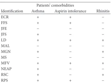
Table 1
Patients’ comorbidities
Identiication Asthma Aspirin intolerance Rhinitis
ECR + + −
FFS + + −
JFE − − −
JFS + − −
LD + + +
MAL − − −
MGN + − +
MS − − −
MFV + + +
NEAP + − −
RSC + − +
RPS + + −
2. Material and Methods
2.1. Patients and Clinical Diagnosis. Nasal polyp tissue sam-ples of 12 patients with known NP were obtained during functional endoscopic sinus surgery (FESS) performed at the Department of Otorhinolaryngology, University of S˜ao Paulo, Brazil. he study was approved by the local Research Ethics Committee and written informed consent was obtained from each patient before sample collection. he diagnosis of NP was based on medical history, clinical examination, nasal endoscopy, and computed tomography (CT) of the paranasal sinuses according to the European Position Paper on the Primary Care Diagnosis and Management of Rhinosinusitis
and Nasal Polyps 2012 [23].
All subjects underwent a skin prick test for common inhalant allergens. he diagnosis of asthma was obtained from the Department of Pulmonology at the University of S˜ao Paulo. Information on aspirin intolerance was collected from
patient histories (Table 1).
2.2. MSC Isolation and Preparation of Nasal Polyp Single-Cell Suspension. Human mesenchymal stem cells (MSCs) were isolated by rinsing of the cells remaining in bone marrow collection tubes, kindly provided by the Children’s Hospital of S˜ao Paulo and GRAACC (Support Group for Children and Adolescents with Cancer), with ethical approval and the informed consent of the donors (Protocol no. 45/09). he tubes were washed with PBS, cells were isolated by the Ficoll-Hypaque protocol (Sigma, USA), and culture was performed as previously described by Lennon and Caplan (2006) and
Pittenger et al. (1999) [24, 25]. he cells were cultured
in basal medium containing DMEM-low glucose medium (Gibco, USA) supplemented with 10% HyClone fetal bovine serum (hermo, USA), antibiotic (100 U/mL penicillin and
100�g/mL streptomycin, Gibco), and 100 mM of nonessential
amino acids (Gibco). he MSCs were subcultured for 10 passages and then used for coculture experiments with polyp-derived cells. All polyp-polyp-derived cells were isolated from polyp tissues by mechanical separation (with surgical scissors and forceps) followed by digestion in collagenase IV (1 mg/mL,
in complete medium (10% serum), iltered in a 70�m cell strainer (BD Biosciences, USA), suspended, and seeded onto six well plates containing R10 medium: RPMI-1640 culture medium (Gibco), supplemented with 10% FBS (Gibco) and
antibiotic (100 U/mL penicillin and 100�g/mL streptomycin;
Gibco). During coculture assays, both MSCs and
polyp-derived cells were incubated at 37∘C in a 5% CO2atmosphere
for 72 hours.
2.3. Phenotype and Diferentiation Capacity of MSCs. To characterize MSC phenotype, the cells were harvested by treatment with trypsin (Gibco), washed, and suspended in
phosphate-bufered saline (PBS), and approximately 1×10⁵
cells were incubated with conjugated monoclonal antibodies (1 : 100) against CD73, CD90, CD105, CD54, CD45, HLA-DR, PDL-1, PDL-2, and CTLA-4 (conjugated with PE, FITC, or PerCP luorochrome; BD Biosciences). he FACSCanto II low cytometer (BD, Biosciences) was used for acquisition, and analysis was performed using the FlowJo sotware, ver-sion 7.2.4 (Tree Star, USA). To assess the ability to diferentiate into several mesenchymal cell lines, the cells were subjected to adipogenic, chondrogenic, and osteogenic diferentiation
protocols, as described by Pittenger et al., 1999 [25]. he stem
cells were cultivated at 95% conluence and multipotential diferentiation was induced with speciic agents using the Mesenchymal Stem Cell Adipogenesis, Osteogenesis, and Chondrogenesis Kit, according to manufacturer instructions (Chemicon, USA). he medium was replaced every 4 days for 21 days. Adipocyte-like cells were stained with Oil Red O to assess neutral lipid accumulation in fat vacuoles. Chondrocyte-like cells were stained using Safranin O, and osteogenesis specimen cells were stained with Alizarin Red for intracellular calcium compounds.
2.4. Cellular Phenotyping of Nasal Polyp-Derived Tissue. To determine the immune cell proile of whole polyp cells, fresh and cultured (for 72 hours) cells were harvested (both adherent and nonadherent cells) without enzymatic dissoci-ation and labeled with a set of various speciic luorescent antibodies, such as CD4-Pacifc Blue, CD8-PECy7, CD11c-PE, CD14-PerCPCy5.5, CD19-Viollet, CD25-APC, CD40-FITC, CD69-FITC, and NK1.1-PE (all from BD Biosciences). hese
sets of antibodies were used for both fresh and in vitro
coculture analysis. To investigate the CD4+CD25+Foxp3+ T lymphocyte proile, intracellular staining for FoxP3 expres-sion was performed ater 72 hours in culture with or without the presence of MSCs. he cells were ixed and permeabilized using the Fix/Perm Bufer Set Kit (BD Biosciences). Staining was performed using FoxP3 (PE), CD4 (APC-Cy7), and CD25 (APC) antibodies at 1 : 100 dilution (BD Biosciences). Analysis was determined by low cytometry (FACSCanto II cytometer, BD Biosciences) within FSC and SSC gate in CD4+ T cells. All acquisitions were performed using a FACSCanto II low cytometer (BD Biosciences), and analysis was again done using the FlowJo 7.2.4 sotware (Tree Star). During analysis, gates for mononuclear cells were performed in FSC and SSC parameters ater doublet exclusion. Results are presented as cell frequency.
2.5. T-Cell Proliferation Assay. To determine the ability of MSCs to modulate the polyp microenvironment, fresh whole
polyp cells were labeled with 5�M carboxyluorescein
suc-cinimidyl ester (CFSE, BD Bioscience) and seeded in culture with or without MSCs for 5 days. MSCs from nonrelated
donors were cocultured with 1 ×10⁵ whole polyp cells in
a 1 : 3 ratio in R10 medium supplemented with 100 mM of vitamin complex (hermo), antibiotic (Pen/Strep, 100 U/mL, Gibco), 100 mM L-Glutamine Mixture (Gibco), 100 mM MEM nonessential amino acids (Gibco), and 1 mM HyClone sodium pyruvate (hermo). Ater 5 days, all polyp cells were stained with anti-CD4 and anti-CD8 antibodies (1 : 100 ratio, BD Biosciences), and CFSE dilution was read in FITC channel by low cytometry analysis (FACSCanto II cytometer, BD Biosciences). his analysis was performed within the FSC and SSC gate using histograms for the T-cell type of interest
(CD4+ or CD8+). Phytohemagglutinin A (1�g/mL) (PHA,
Invitrogen, USA) was used as a polyclonal-positive stimulus.
2.6. Cytokine Proile of Nasal Polyp-Derived Cells. Isolated polyp culture and coculture (MSC and polyps) supernatants (from 6 out of 12 NP samples) were further harvested ater 72 hours for cytokine quantiication, using the Cytometric Bead Array (CBA) h1/h2/h17 Cytokine Kit, according to manufacturer recommendations (BD, Becton Dickinson Biosciences). Acquisition was performed in the FACSCanto II cytometer (BD Biosciences) and the samples analyzed using FCAP Array sotware v3.0 (Sot Flow Inc., HUN).
2.7. Statistical Analysis. Results were assessed for normal distribution by the Kolmogorov-Smirnov test. Categorical variables were expressed as percentages (%), and continuous
variables (data) were presented as means ±standard
devi-ations. he Mann-Whitney nonparametric test was used to assess between-group diferences. For analysis among three or more groups, the Kruskal-Wallis nonparametric test for one-way analysis of variance was used. For all analyses,
a P value ≤ 0.05 was considered statistically signiicant.
All statistical testing and plotting were performed using GraphPad Prism 5 (GraphPad sotware, Inc., USA).
3. Results
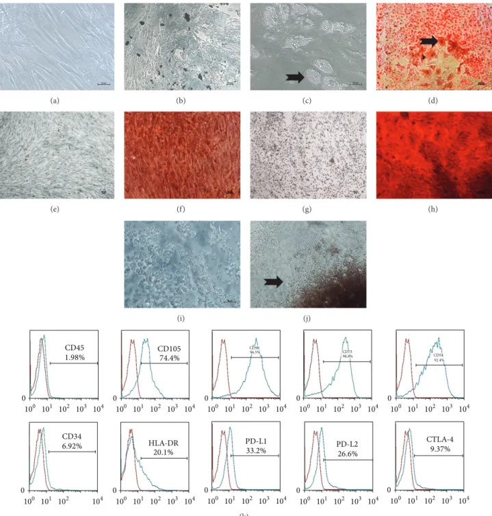
3.1. Characterization of Mesenchymal Stem Cells and Polyp Iniltrating Cells. he bone marrow-derived mesenchymal stem cells used in this study exhibited most important characteristics of MSCs, such as a plastic-adherent growth
pattern (Figure 1), ibroblast-like morphology (Figure 1(a)),
expression of speciic surface antigens (CD105, CD54, CD90, and CD73), and immunoregulatory molecules (CTLA-4, PD-L1, and PD-L2), and no expression of the immunogenic and hematopoietic surface markers HLA-DR and CD45,
respectively (Figure 1(k)). Additionally, MSCs demonstrated
the ability to diferentiate into adipocytes (Figures1(c)-1(d)),
chondrocytes (Figure 1(f)), and osteocytes (Figure 1(h)) in
vitro. he polyp-derived cells spread from the whole polyp in culture and exhibited spheroid-like morphology (Figures
(a) (b) (c) (d)
(e) (f) (g) (h)
(i) (j)
CD45
1.98% CD74.1054%
CD90
96.5% CD73
90.8% CD54
92.4%
CD34
6.92% HLA-DR
20.1%
PD-L1
33.2% PD-L26.6%2
CTLA-4 9.37%
0
100 101 102 103 104
0
100 101 102 103 104
0
100 101 102 103 104
0
100 101 102 103 104
0
100 101 102 103 104
0
100 101 102 103 104
0
100 101 102 103 104
0
100 101 102 103 104
0
100 101 102 103 104
0
100 101 102 104
(k)
Figure 1: Characterization of bone marrow-derived mesenchymal stem cells and polyp-derived cells. (a) MSCs exhibiting ibroblastic-like morphologyin vitro; (b) MSC culture in adipogenic control medium showing absence of lipid vesicles; (c) MSCs containing lipid vesicles (black arrow) in adipogenic diferentiation medium without Oil Red stain; (d) MSC culture in adipogenic diferentiation medium stained with Oil Red (black arrow); (e) MSC culture in chondrogenic control medium; (f) MSC culture in chondrogenic diferentiation medium stained with Safranin O; (g) MSC culture in osteocyte control medium; (h) MSC culture in osteogenic diferentiation medium stained with Alizarin Red; ((i)-(j)) representative view of polyp-derived cells spreading and growing in culture (black arrow); and (k) immunophenotypic signature of MSCs in culture, demonstrating absence of hematopoietic (CD45 and CD34) and immunogenic (HLA-DR) markers and expression of CD105, CD90, CD73, and CD54, as well as immunoregulatory receptors such as PDL-1 and -2 and CTLA-4.
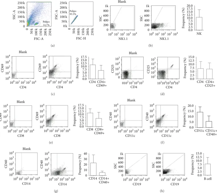
he immunophenotype of whole NP-derived cells sho-wed the presence of distinct immune cells ater single-cell suspension. NK cells, T cells, dendritic cells, monocytes,
and B cells were present (Figures2(a)–2(h)). he frequency
of each subtype of immune cells was as follows: NK
cells, 10.60 ± 6.28%; CD4+,11.26 ± 4.70%; CD4+CD69+,
6.71 ± 4.31%; CD4+CD25+, 6.77 ± 4.35%; CD8+, 11.19 ±
250k 200k 150k 100k 50k 0 0 25 0 k 200 k 15 0 k 100 k 50 k 0 25 0 k 200 k 15 0 k 100 k 50 k 250k 200k 150k 100k 50k 0k SSC-A FSC-A FSC-A FSC-H Polipo Polipo 14.7% 93.5% (a) 1k 800 600 400 200 0 1k 800 600 400 200 0 SSC SSC NK1.1 NK1.1
100101102103104 100101102103104 NK
20.0
F
req
uenc
y (%) 15.012.5 10.0 7.5 5.0 2.5 0.0 17.5 Blank (b)
Q4 Q4
CD4 CD4 CD 69 CD 69 100 101 102 103 104
100101102103104
100
101
102
103
104
100101102103104
Blank F req uenc y (%) 17.5 15.0 12.5 10.0 7.5 5.0 2.5 0.0 CD4CD4+ CD69+ (c) CD4 CD4 CD 25 CD 25 102 103 104 105 102 103 104 105 101
102103104105
101
102103104105
0 0 Blank F req uenc y (%) 15.0 12.5 10.0 7.5 5.0 2.5 0.0 CD4 CD4+ CD25+ (d)
Q8 Q8
CD8 CD8 CD 69 CD 69 100 101 102 103 104
100101102103104
100
101
102
103
104
100101102103104
Blank F req uenc y (%) 17.5 15.0 12.5 10.0 7.5 5.0 2.5 0.0 CD8 CD8+ CD69+ (e)
Q8 Q8
100
101
102
103
104
100101102103104
100
101
102
103
104
100101102103104
Blank
F
req
uenc
y (%) 15.0
10.0 5.0 0.0 20.0 CD 40 CD 40 CD11c CD11c CD11c CD11c+ CD40+ (f) 100 101 102 103 104
100101102103104
100
101
102
103
104
100101102103104
Blank F req uenc y (%) CD 40 CD 40 CD14 CD14 CD14 40 30 20 10 0
Q4 Q4
CD14+ CD40+ (g) 1k 800 600 400 200 SSC SSC
100101102103104
F req uenc y (%) 15.0 12.5 10.0 7.5 5.0 2.5 0.0 0 1k 800 600 400 200
100101102103104
0
B cell
CD19 CD19
(h)
Figure 2: Phenotypic aspects of polyp-derived cells. (a) Gate strategies for selection of the mononuclear fraction of polyp-derived cells; (b) NK cells (NK1.1+); (c) CD4 and CD4+CD69+ T cells; (d) CD4+CD25+ T cells; (e) CD8 and CD8+CD69+ T cells; (f) CD11c+ and CD11c+CD40+ dendritic cells; (g) CD14+ and CD14+CD40+ monocytes and (h) B cells (CD19+). Several types of activated and nonactivated immune cells were observed within the polyp parenchyma.
CD11c+CD40+, 5.52 ± 5.14%; CD14+, 22.88 ± 15.06%;
CD14+CD40+, 5.06 ± 2.92%; and CD19+, 7.88 ± 4.36%
(Figures2(a)–2(h)). In our samples, the ratio of activated cells
to nonactivated cells within the polyp ranged from 60% to 20%, depending on cell type. For CD4+ and CD8+ T cells, this index was approximately 60%, for dendritic cells 35%, and for
monocytes 20% (Figures2(a)–2(h)).
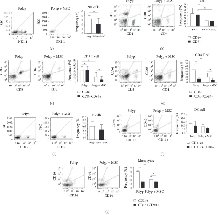
3.2. Efect of MSCs on Nasal Polyp-Derived Cells. To evaluate the role of MSCs in the modulation of the NP microen-vironment, we carried out a coculture assay for 72 hours and assessed the frequency of polyp-derived iniltrating cells. We found a signiicant decrease in the frequency of most inlammatory cells. NK cells (NK1.1+), T helper cells (CD4+),
and cytotoxic T cells (CD8+) showed a signiicant decrease in frequency when cocultured with MSC; however, no changes in the activated T cell compartment (CD4+CD69+ and
CD8+CD69+) were observed (Figures 3(a)–3(d)). he
fre-quency of B cells, monocytes, and dendritic cells also tended
to decrease ater coculture with MSCs.De novo, the fraction
of activated cells included in the monocyte and dendritic cell subpopulations was not altered in comparison with cultures
performed in the absence of MSCs (Figures3(e)–3(g)).
SSC SSC
NK1.1 NK1.1
0 0
102103104105
Polyp Polyp+MSC
Polyp Polyp+MSC
NK cells F req uenc y (%) 50 40 30 20 10 0 250k 200k 150k 100k 50k 0 0
102103104 105
250k 200k 150k 100k 50k ∗ (a) 101 102 103 104 105
101102103104105 101
102
103 104 105
101102103104105
CD4 CD4 CD 8 CD 8 CD4+ CD8+ T cell
Polyp Polyp+MSC
Polyp Polyp+MSC
F req uenc y (%) 80 70 60 50 40 30 20 10 0 ∗ ∗ (b) 101 102 103 104 105 101 102 103 104 105
101102103104105 101102103104105
Polyp Polyp+MSC
Polyp Polyp+MSC
F req uenc y (%) 80 70 60 50 40 30 20 10 0 ∗ ∗ ∗ CD8 CD8 CD 69 CD 69 CD8+ CD8+CD69+
CD8T cell
(c) 101 102 103 104 105 101 102 103 104 105
101102103104105 101102103104105
Polyp Polyp+MSC
Polyp Polyp+MSC
F req uenc y (%) 80 70 60 50 40 30 20 10 0 ∗ ∗ ∗ CD4 CD4 CD 69 CD 69 CD4+ CD4+CD69+
CD4T cell
(d)
SSC SSC
0 0
102 103104105
Polyp Polyp+MSC
Polyp Polyp+MSC
F req uenc y (%) 250k 200k 150k 100k 50k 0 0
102 103104 105 250k 200k 150k 100k 50k B cells 20.0 17.5 15.0 12.5 10.0 7.5 5.0 2.5 0.0 CD19 CD19 (e)
Polyp Polyp+MSC
Polyp Polyp+MSC
F
req
uenc
y (%)
0 102103104105 0102103104105 0 102 103 104 105 0 102 103 104 105 20.0 15.0 10.0 5.0 0.0 CD 40 CD 40 CD11c CD11c DC cell CD11c+ CD11c+CD40+ 25.0 (f)
Polyp Polyp+MSC
Polyp Polyp+MSC
F
req
uenc
y (%)
0 102103104105 0
102 103 104
105
0 102103104 105 0 102 103 104 105 CD 40 CD 40 CD14 CD14 Monocytes CD14+ CD14+CD40+ 50 40 30 20 10 0 ∗ ∗ ∗ (g)
Figure 3: Bone marrow-derived mesenchymal stem cells modulating the polyp microenvironmentin vitro. (a) NK cell (NK1.1+) frequency alone and in coculture with MSCs; (b) CD4+ and CD8+ frequencies alone and in coculture with MSCs; (c) CD8+ and CD69+ frequencies alone and in coculture with MSCs; (d) CD4+ and CD69+ frequencies alone and in coculture with MSCs; (e) B cell (CD19+) frequency alone and in coculture with MSCs; (f) CD11c+ and CD11c+CD40+ dendritic cell frequencies alone and in coculture with MSCs and CD14+ and CD14+CD40+ monocyte frequencies alone and in coculture with MSCs. he presence of MSCs may immunomodulate the phenotype of polyp-derived cells toward an immunosuppressive proile.
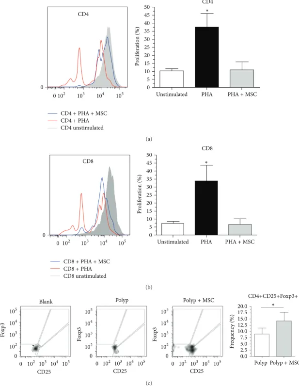
we sought to investigate whether MSCs could promote the functional ability to arrest NP-derived T-cell proliferation
in vitro. Surprisingly, we observed signiicant inhibition of expansion/proliferation of both T helper cells (CD4+) and cytotoxic T cells (CD8+) derived from NP stimulated with PHA in compared with cells stimulated but cultured in the
absence of MSCs (Figures4(a)and4(b)). he
immunosup-pressive efect of MSCs on NP-derived T cells was evident,
considering that the index of proliferation of CD4+ and CD8+ T cells cocultured with MSCs did not difer from that of control unstimulated T cells.
CD4+PHA+MSC
CD4+PHA
CD4unstimulated
105
104
103
102
0 0
CD4
PHA+MSC
PHA Unstimulated
CD4 ∗ 50
45 40 35 30 25 20 15 10 5 0
P
ro
lif
era
tio
n (%)
(a)
CD8+PHA+MSC
CD8+PHA
CD8unstimulated
105
104
103
102
0 0
CD8
PHA+MSC
PHA Unstimulated
CD8
∗ 50
45 40 35 30 25 20 15 10 5 0
P
ro
lif
era
tio
n (%)
(b)
Polyp Polyp+MSC
Polyp Polyp+MSC
F
req
uenc
y (%)
0 102 103 104 105
0
102
103
104
105
0 102 103 104 105
0
102
103
104
105
0 102 103 104 105
0
102
103
104
105 20.017.5
15.0 12.5 10.0 7.5 5.0 2.5 0.0 Blank
Fox
p
3
Fox
p
3
Fox
p
3
CD25 CD25 CD25
∗ CD4+CD25+Foxp3+
(c)
Figure 4: Bone marrow-derived mesenchymal stem cells exhibit functional immunosuppressive action on polyp-derived cellsin vitro. (a, b) Proliferative index of CD4+ and CD8+ cells, respectively, stimulated with phytohemagglutinin (PHA), in the presence or absence of MSCs; (c) frequency of CD4+CD25+Fpxp3+ cells in the presence or absence of MSCs. Functionally, MSCs exhibited immunosuppressive action on polyp-derived cells, as represented by inhibition of proliferation of CD4+ and CD8+ cells with a concomitant increase in CD4+CD25+Fpxp3+ frequency.
number of CD4+CD25+Foxp3+ T cells was observed in the presence of MSCs as compared with cultures of T cells alone (Figure 4(c)), suggesting that these cells can play an essential role in the development of the chronic inlammatory process in NP.
15000
12500
10000
7500
5000
2500
0
Polyp Polyp+MSC Polyp Polyp+MSC
IL-6
∗ ∗
IL-10 20.0
17.5 15.0 12.5 10.0 7.5 5.0 2.5 0.0
(pg/mL) (pg/mL)
(a)
Polyp Polyp+MSC Polyp Polyp+MSC Polyp Polyp+MSC
∗ ∗ ∗
IL-2 4.0
3.5 3.0 2.5 2.0 1.5 1.0 0.5 0.0
TNF-� 10
9 8 7 6 5 4 3 2 1 0
IFN-𝛾 50
45 40 35 30 25 20 15 10 5 0
(pg/mL) (pg/mL) (pg/mL)
(b)
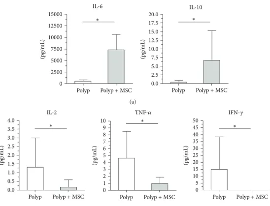
Figure 5:In vitrocytokine proile of polyp-derived cells cultured in the presence or absence of bone marrow-derived mesenchymal stem cells. A decrease in inlammatory cytokines (IL-2, TNF-�, and IFN-�) and enhancement of anti-inlammatory cytokine (IL-10 and IL-6) levels was observed when MSCs were cocultured with polyp-derived cells.
prominent shit from an inlammatory to an anti-inla-mmatory cytokine proile. In coculture, MSCs promoted an increase in anti-inlammatory molecules such as IL-10, with a concomitant decrease in inlammatory cytokines such as
IL-2, TNF-�, and IFN-�(Figure 5).
4. Discussion
Nasal polyposis is characterized by the most severe upper-airway inlammatory process observed in clinical practice. his process is crucial to understand the mechanisms that underlie development of polyposis. It is known that MSCs have immunomodulatory properties, as demonstrated in some organs such as the brain, kidney, heart, bone, and lung
[26–30]. MSC-mediated immunomodulation can occur via
cell-to-cell contact or by release of soluble factors, which are associated with many regulatory efects of these cells in a tissue inlammation context. he best documented
MSC-secreted cytokines are TGF-�1, PGE-2, IDO, IL-10, IL-6,
MMP-2,9, TIMP-2,3, nitric oxide (NO), chemokine ligand 2 and 5 (CCL2,5), human leukocyte antigen 5 (HLA-G5), heme oxygenase-1 (HO-1), hepatocyte growth factor (HGF),
and leukemia inhibitory factor (LIF) [17]. On the basis
of their properties, MSCs have been explored in a wide range of experiments and have been used for therapeutic purposes more extensively than any other subtype of stem cell. hese cells also retain further important features, such as low immunogenicity, and promote inhibition of
prolifera-tion/activation in allogeneic lymphocytes [18].
hus, in the present study, we assessed the impact of MSCs on modulation of the inlamed NP microenvironment. Firstly, we demonstrated that several types of inlammatory cells (NK cells, B cells, T cells, monocytes, and dendritic cells) are found in this milieu. Ater characterization of MSC phenotype and diferentiation, these cells were cultured with polyp-derived cells, and a direct immunomodulatory efect on the inlammatory NP cell compartment was observed. here was a signiicant decrease in the frequency of CD4+, CD8+, CD14+, and NK cells, as well as a signiicant increase in CD4+CD25+Foxp3+ T cells, when MSCs were cocultured with NP-derived cells. A decrease in regulatory T cells has been described as a feature of the NP disease course. his is an important inding, and the increase in CD4+CD25+Foxp3 cells induced by presence of MSCs in the present study may help in our understanding of the progression of NP. Func-tionally, we observed that MSCs inhibited both CD4+ and CD8+ polyp-derived T cell proliferation and eiciently chan-ged the local inlammatory pattern, promoting a shit from an inlammatory to an anti-inlammatory cytokine proile.
he stem cells actions are widely dependent on the disease and time of therapy. Contact-dependent immunosuppression is one mechanism of MSC action and can be associated with the expression of immunoregulatory molecules expressed on the surface of MSCs or by delivering microvesicles carrying
bioactive molecules [31]. CTLA-4, PDL-1, and PDL-2 were
has been reported extensively in the stem cell literature [32,
33].
MSC is also capable of exerting inluence on inlamed
process via paracrine action [34] even far from the injured
tis-sue. he soluble factors released by MSCs are another mecha-nism that may also have inluenced modulation of the polyp-derived cell microenvironment. Many studies have
demon-strated the efect of TGF-�secreted by MSCs on suppression
of peripheral blood mononuclear cell (PBMC) proliferation
[35]. TGF-� is also capable of increasing the frequency of
regulatory T cells, especially when associated with PGE2 [36–
38]. In a model of asthmatic mice, TGF-�secreted by MSCs
showed a beneicial efect by decreasing levels of IL-4, IL-5, IL-13, and immunoglobulin in bronchoalveolar lavage luid
[38].
Although the role of TGF-�on remodeling process is still
controversial in the literature, there is a strong evidence in NP
that the lack of TGF-�is involved in decreasing extracellular
matrix formation [5, 6, 39] highlighting the importance
of this molecule on the development of inlammation in NP.
In addition, PGE2 is another molecule which is found
in low levels in NP tissue [40] and MSCs produce PGE2.
PGE2 exerts an immunosuppressive action on T cells and,
consequently, promotes a decrease in IFN-�and TNF-�levels
[41,42]. Our study corroborates these indings,
demonstra-ting decreased IFN-�and TNF-�expression in NP cells
cocu-ltured with MSCs in comparison with cultures of NP-derived cells alone; maybe PGE2 is contributing to decrease these interleukins.
In mice, macrophages from septic lungs produced higher levels of IL-10 when treated with MSCs than untreated macrophages. he authors suggested that the EP2 and EP4 receptors (prostaglandin receptors) were responsible for this
increase in IL-10 production [43]. In this context, IL-10 is
considered to be the main immunosuppressive interleukin, and we found higher levels of IL-10 in NP cells treated with MSCs than in untreated cells.
Consistently with what is found in a variety of other
dise-ases [44, 45], the presence of MSCs in NP cell cultures
increased the expression of IL-6 (an interleukin mainly prod-uced by h1 cells). Paradoxically, although IL-6 is associated
with increased production of IL-2 [46], we found decreased
levels of this inlammatory interleukin in NP cells treated with MSCs. One plausible explanation is the fact that MSCs
can secrete TGF-�and IL-6 and that diferentiation of Treg/
h17 depends on the proportion of these two cytokines [47].
In the present study, we did not detect IL-17 production in NP cell cultures, with or without the presence of MSCs. his may suggest that the interaction of distinct soluble factors with diferent cell types could alter the immune context of NP.
Furthermore, indoleamine 2,3-dioxygenase (IDO), a rate-limiting enzyme that catalyzes the degradation of trypto-phan, is found at increased levels during chronic inlammat-ory diseases induced by inlammatinlammat-ory mediators such as IFNs
and IL-6 [48]. Elevated IDO expression has been observed in
nasal polypoid tissue as compared with healthy nasal mucosa
[49]. IDO is an immunoregulatory enzyme and belongs to
the MSC arsenal. he role of MSCs in induction of tolera-nce in renal allograt recipients was not conirmed in IDO knockout mice, showing the crucial importance of IDO to the immunosuppressive efect of MSCs via regulatory T cells
[50].
In addition, we observed an elevated presence of
IFN-� and IL-6 in cultures of NP-derived cells. Ater coculture
with MSCs, IL-6 levels increased whereas IFN-�declined. It
is important to note that a decrease in IFN-�could indicate a
decrease in h1 cells activation.
Diferent types of T cells are implicated in the pathogen-esis and progression of NP, but, in populations of European descent, h2-driven disease is still a hallmark of the condi-tion. In the Asian population NP is a h17/h1-driven disease with increase of neutrophils. We characterize our patients as descendants of European and none was of Asian origin. In our sample the inlammatory cells proile was similar to the one found in European population (increase of h2 cells and eosinophils).
Some studies have demonstrated that MSCs are able to promote conversion of the h1 phenotype into a h2 response
[51]. In this sense, MSCs could contribute to NP, considering
the microenvironment already saturated with h2-dependent interleukins in this setting. In the present study, we did not detect IL-4 which is considered an important interleukin that induces diferentiation from h0 to h2 immune response in NP-derived cell cultures, regardless of the presence of MSCs. his would suggest that in our coculture experiments with NP-derived cells, MSCs could not intensify a speciic h2 response.
Additionally, in the presence of MSCs, h1 cytokines proile was altered, h2 cytokines were not detected, and IL-10 was increased. hese results indirectly suggest that the T-cell proile may have been directed to a regulatory pattern, considering the prominent increase of CD4+CD25+Foxp3+ T cell frequency in our MSC cocultures.
he NP treatment is based on two main pillars: oral and topical steroids and surgery, with the recurrence of NP ater surgery being usual. he understanding of MSC mechanism in decreasing the inlammatory process in NP could be helpful to reduce the intake of steroids and the surgery indications.
In conclusion, we demonstrated that MSCs can be a useful tool for the investigation of the inlammatory
microenviron-ment of NP. hese results were obtained entirely in vitro,
and any conclusions about the actual efects of MSCs in NP in vivo remain to be explored. However, our indings clearly demonstrate an immunoregulatory efect of MSCs on immune cells (especially T cells) derived from nasal tissue afected by polyposis. Finally, we hope that further studies will be performed in the search for an understanding of the mechanism of MSC activity in the context of NP inlammation.
Conflict of Interests
Acknowledgments
We would like to thank the collaborators, donors and students for all support in this work. his work also was supported by Fundac¸˜ao de Amparo `a Pesquisa do Estado de S˜ao Paulo (FAPESP) and linked to the inancial protocol Fapesp number 2012/02270-2 and 2010/12295-7. Rog´erio Pezato and Danilo Cˆandido de Almeida are contributed equally to the paper.
References
[1] D. Hastan, W. J. Fokkens, C. Bachert et al., “Chronic rhinos-inusitis in Europe—an underestimated disease. A GA2LEN study,”Allergy, vol. 66, no. 9, pp. 1216–1223, 2011.
[2] R. Pezato, M. ´Swierczy´nska-Krępa, E. Ni˙zankowska-Mogilni-cka, L. Derycke, C. Bachert, and C. A. P´erez-Novo, “Role of imbalance of eicosanoid pathways and staphylococcal super-antigens in chronic rhinosinusitis,”Allergy, vol. 67, no. 11, pp. 1347–1356, 2012.
[3] N. van Bruaene, C. A. P´erez-Novo, T. M. Basinski et al., “T-cell regulation in chronic paranasal sinus disease,”Journal of Allergy and Clinical Immunology, vol. 121, no. 6, pp. 1435–1441, 2008. [4] N. Zhang, T. van Zele, C. Perez-Novo et al., “Diferent types of
T-efector cells orchestrate mucosal inlammation in chronic sinus disease,”Journal of Allergy and Clinical Immunology, vol. 122, no. 5, pp. 961–968, 2008.
[5] N. van Bruaene, L. Derycke, C. A. Perez-Novo et al., “TGF-�signaling and collagen deposition in chronic rhinosinusitis,”
Journal of Allergy and Clinical Immunology, vol. 124, no. 2, pp. 253–259, 2009.
[6] L. Balsalobre, R. Pezato, C. Perez-Novo et al., “Epithelium and stroma from nasal polyp mucosa exhibits inverse expression of TGF-�1 as compared with healthy nasal mucosa,”Journal of Otolaryngology—Head & Neck Surgery, vol. 42, no. 1, article 29, 2013.
[7] J. B. Watelet, C. Bachert, C. Claeys, and P. van Cauwenberge, “Matrix metalloproteinases MMP-7, MMP-9 and their tissue inhibitor TIMP-1: expression in chronic sinusitis vs nasal polyposis,”Allergy, vol. 59, no. 1, pp. 54–60, 2004.
[8] Y.-M. Lee, S.-S. Kim, H.-A. Kim et al., “Eosinophil inlammation of nasal polyp tissue: relationships with matrix metallopro-teinases, tissue inhibitor of metalloproteinase-1, and transform-ing growth factor-�1,”Journal of Korean Medical Science, vol. 18, no. 1, pp. 97–102, 2003.
[9] T. Sejima, G. Holtappels, and C. Bachert, “he expression of ibrinolytic components in chronic paranasal sinus disease,”
American Journal of Rhinology and Allergy, vol. 25, no. 1, pp. 1–6, 2011.
[10] R. Pezato and R. L. Voegels, “Why do we not ind polyps in the lungs? Bronchial mucosa as a model in the treatment of polyposis,”Medical Hypotheses, vol. 78, no. 4, pp. 468–470, 2012. [11] J. Bousquet, W. Jacquot, A. M. Vignola, C. Bachert, and P. van Cauwenberge, “Allergic rhinitis: a disease remodeling the upper airways?”Journal of Allergy and Clinical Immunology, vol. 113, no. 1, pp. 43–49, 2004.
[12] S. T. Holgate, J. Holloway, S. Wilson, F. Bucchieri, S. Puddi-combe, and D. E. Davies, “Epithelial-mesenchymal communi-cation in the pathogenesis of chronic asthma,”Proceedings of the American horacic Society, vol. 1, no. 2, pp. 93–98, 2004. [13] L.-F. Wang, C.-Y. Chien, F.-Y. Chiang, C.-Y. Chai, and C.-F.
Tai, “Corelationship between matrix metalloproteinase 2 and
9 expression and severity of chronic rhinosinusitis with nasal polyposis,”American Journal of Rhinology and Allergy, vol. 26, no. 1, pp. e1–e4, 2012.
[14] X. Li, J. Meng, X. Qiao et al., “Expression of TGF, matrix metalloproteinases, and tissue inhibitors in Chinese chronic rhinosinusitis,”Journal of Allergy and Clinical Immunology, vol. 125, no. 5, pp. 1061–1068, 2010.
[15] K. le Blanc, L. Tammik, B. Sundberg, S. E. Haynesworth, and O. Ringd´en, “Mesenchymal stem cells inhibit and stimulate mixed lymphocyte cultures and mitogenic responses independently of the major histocompatibility complex,”Scandinavian Journal of Immunology, vol. 57, no. 1, pp. 11–20, 2003.
[16] S. M. Devine, C. Cobbs, M. Jennings, A. Bartholomew, and R. Hofman, “Mesenchymal stem cells distribute to a wide range of tissues following systemic infusion into nonhuman primates,”
Blood, vol. 101, no. 8, pp. 2999–3001, 2003.
[17] ˆE. J. Bassi, D. C. de Almeida, P. M. M. Moraes-Vieira, and N. O. S. Cˆamara, “Exploring the role of soluble factors associated with immune regulatory properties of mesenchymal stem cells,”
Stem Cell Reviews and Reports, vol. 8, no. 2, pp. 329–342, 2012. [18] B. Parekkadan and J. M. Milwid, “Mesenchymal stem cells as
therapeutics,”Annual Review of Biomedical Engineering, vol. 12, pp. 87–117, 2010.
[19] F. S. Silva, P. N. Almeida, J. V. Rettore et al., “Toward personal-ized cell therapies by using stem cells: seven relevant topics for safety and success in stem cell therapy,”Journal of Biomedicine and Biotechnology, vol. 2012, Article ID 758102, 12 pages, 2012. [20] K. le Blanc and O. Ringd´en, “Immunomodulation by
mes-enchymal stem cells and clinical experience,”Journal of Internal Medicine, vol. 262, no. 5, pp. 509–525, 2007.
[21] A. J. Nauta and W. E. Fibbe, “Immunomodulatory properties of mesenchymal stromal cells,”Blood, vol. 110, no. 10, pp. 3499– 3506, 2007.
[22] M. Di Nicola, C. Carlo-Stella, M. Magni et al., “Human bone marrow stromal cells suppress T-lymphocyte proliferation induced by cellular or nonspeciic mitogenic stimuli,”Blood, vol. 99, no. 10, pp. 3838–3843, 2002.
[23] W. J. Fokkens, V. Lund, J. Mullol et al., “European position paper on rhinosinusitis and nasal polyps 2012,”Rhinology, vol. 50, supplement 23, pp. 1–298, 2012.
[24] D. P. Lennon and A. I. Caplan, “Isolation of human marrow-derived mesenchymal stem cells,” Experimental Hematology, vol. 34, no. 11, pp. 1604–1605, 2006.
[25] M. F. Pittenger, A. M. Mackay, S. C. Beck et al., “Multilineage potential of adult human mesenchymal stem cells,”Science, vol. 284, no. 5411, pp. 143–147, 1999.
[26] L. T. Galindo, T. R. Filippo, P. Semedo et al., “Mesenchymal stem cell therapy modulates the inlammatory response in experimental traumatic brain injury,”Neurology Research Inter-national, vol. 2011, Article ID 564089, 9 pages, 2011.
[27] P. Semedo, C. Donizetti-Oliveira, M. Burgos-Silva et al., “Bone marrow mononuclear cells attenuate ibrosis development ater severe acute kidney injury,”Laboratory Investigation, vol. 90, no. 5, pp. 685–695, 2010.
[28] S.-L. Chen, W.-W. Fang, F. Ye et al., “Efect on let ventricular function of intracoronary transplantation of autologous bone marrow mesenchymal stem cell in patients with acute myocar-dial infarction,”American Journal of Cardiology, vol. 94, no. 1, pp. 92–95, 2004.
stimulate growth in children with osteogenesis imperfecta: implications for cell therapy of bone,” Proceedings of the National Academy of Sciences of the United States of America, vol. 99, no. 13, pp. 8932–8937, 2002.
[30] M. Rojas, J. Xu, C. R. Woods et al., “Bone marrow-derived mesenchymal stem cells in repair of the injured lung,”American Journal of Respiratory Cell and Molecular Biology, vol. 33, no. 2, pp. 145–152, 2005.
[31] A. Fierabracci, A. del Fattore, R. Luciano, M. Muraca, A. Teti, and M. Muraca, “Recent advances in mesenchymal stem cell immunomodulation. he role of microvisicles,”Cell Transplan-tation, 2013.
[32] ˆE. J. Bassi, P. M. Moraes-Vieira, C. S. Moreira-S´a et al., “Immune regulatory properties of allogeneic adipose-derived mesenchy-mal stem cells in the treatment of experimental autoimmune diabetes,”Diabetes, vol. 61, no. 10, pp. 2534–2545, 2012. [33] F. Dai, D. Shi, W. He et al., “hCTLA4-gene modiied human
bone marrow-derived mesenchymal stem cells as allogeneic seed cells in bone tissue engineering,”Tissue Engineering, vol. 12, no. 9, pp. 2583–2590, 2006.
[34] J. Huang, J. Guo, F. Beigi et al., “HASF is a stem cell paracrine factor that activates PKC epsilon mediated cytoprotection,”
Journal of Molecular and Cellular Cardiology, vol. 66, pp. 157– 164, 2014.
[35] S. Tomic, J. Djokic, S. Vasilijic et al., “Immunomodulatory pro-perties of mesenchymal stem cells derived from dental pulp and dental follicle are susceptible to activation by toll-like receptor agonists,”Stem Cells and Development, vol. 20, no. 4, pp. 695– 708, 2011.
[36] K. English, J. M. Ryan, L. Tobin, M. J. Murphy, F. P. Barry, and B. P. Mahon, “Cell contact, prostaglandin E2and transforming growth factor beta 1 play non-redundant roles in human mes-enchymal stem cell induction of CD4+CD25Highforkhead box P3+regulatory T cells,”Clinical and Experimental Immunology, vol. 156, no. 1, pp. 149–160, 2009.
[37] S. A. Patel, J. R. Meyer, S. J. Greco, K. E. Corcoran, M. Bryan, and P. Rameshwar, “Mesenchymal stem cells protect breast cancer cells through regulatory T cells: role of mesenchymal stem cell-derived TGF-�,”Journal of Immunology, vol. 184, no. 10, pp. 5885–5894, 2010.
[38] K. Nemeth, A. Keane-Myers, J. M. Brown et al., “Bone marrow stromal cells use TGF-� to suppress allergic responses in a mouse model of ragweed-induced asthma,”Proceedings of the National Academy of Sciences of the United States of America, vol. 107, no. 12, pp. 5652–5657, 2010.
[39] Y. C. Yang, N. Zhang, K. van Crombruggen, G. H. Hu, S. L. Hong, and C. Bachert, “Transforming growth factor-beta1 in inlammatory airway disease: a key for understanding inlam-mation and remodeling,”Allergy, vol. 67, no. 10, pp. 1193–1202, 2012.
[40] C. A. P´erez-Novo, C. Claeys, P. van Cauwenberge, and C. Bachert, “Expression of eicosanoid receptors subtypes and eosi-nophilic inlammation: implication on chronic rhinosinusitis,”
Respiratory Research, vol. 7, article 75, 2006.
[41] K. English, F. P. Barry, C. P. Field-Corbett, and B. P. Mahon, “IFN-�and TNF-�diferentially regulate immunomodulation by murine mesenchymal stem cells,”Immunology Letters, vol. 110, no. 2, pp. 91–100, 2007.
[42] K. Chen, D. Wang, W. T. Du et al., “Human umbilical cord mesenchymal stem cells hUC-MSCs exert immunosuppressive activities through a PGE2-dependent mechanism,” Clinical Immunology, vol. 135, no. 3, pp. 448–458, 2010.
[43] K. N´emeth, A. Leelahavanichkul, P. S. T. Yuen et al., “Bone mar-row stromal cells attenuate sepsis via prostaglandin E 2-dependent reprogramming of host macrophages to increase their interleukin-10 production,”Nature Medicine, vol. 15, no. 1, pp. 42–49, 2009.
[44] F. Djouad, L.-M. Charbonnier, C. Boui et al., “Mesenchymal stem cells inhibit the diferentiation of dendritic cells through an interleukin-6-dependent mechanism,”Stem Cells, vol. 25, no. 8, pp. 2025–2032, 2007.
[45] G. Xu, Y. Zhang, L. Zhang, G. Ren, and Y. Shi, “he role of IL-6 in inhibition of lymphocyte apoptosis by mesenchymal stem cells,”Biochemical and Biophysical Research Communications, vol. 361, no. 3, pp. 745–750, 2007.
[46] T. Kishimoto, “Interleukin-6: discovery of a pleiotropic cytokine,”Arthritis Research and herapy, vol. 8, supplement 2, p. S2, 2006.
[47] X.-J. Liu, J.-F. Zhang, B. Sun et al., “Reciprocal efect of mesenchymal stem cell on experimental autoimmune encepha-lomyelitis is mediated by transforming growth factor-� and interleukin-6,”Clinical and Experimental Immunology, vol. 158, no. 1, pp. 37–44, 2009.
[48] L. Huang, B. Baban, B. A. Johnson III, and A. L. Mellor, “Den-dritic cells, indoleamine 2,3 dioxygenase and acquired immune privilege,”International Reviews of Immunology, vol. 29, no. 2, pp. 133–155, 2010.
[49] T. Honkanen, A. Luukkainen, M. Lehtonen et al., “Indoleamine 2,3-dioxygenase expression is associated with chronic rhinosi-nusitis with nasal polyps and antrochoanal polyps,”Rhinology, vol. 49, no. 3, pp. 356–363, 2011.
[50] W. Ge, J. Jiang, J. Arp, W. Liu, B. Garcia, and H. Wang, “Regula-tory T-cell generation and kidney allograt tolerance induced by mesenchymal stem cells associated with indoleamine 2,3-dioxygenase expression,”Transplantation, vol. 90, no. 12, pp. 1312–1320, 2010.
Submit your manuscripts at
http://www.hindawi.com
Stem Cells
International
Hindawi Publishing Corporation
http://www.hindawi.com Volume 2014
Hindawi Publishing Corporation
http://www.hindawi.com Volume 2014
INFLAMMATION
Hindawi Publishing Corporation
http://www.hindawi.com Volume 2014
Behavioural
Neurology
Endocrinology
International Journal ofHindawi Publishing Corporation
http://www.hindawi.com Volume 2014
Hindawi Publishing Corporation
http://www.hindawi.com Volume 2014
Disease Markers
Hindawi Publishing Corporation
http://www.hindawi.com Volume 2014
BioMed
Research International
Oncology
Journal ofHindawi Publishing Corporation
http://www.hindawi.com Volume 2014
Hindawi Publishing Corporation
http://www.hindawi.com Volume 2014 Oxidative Medicine and Cellular Longevity Hindawi Publishing Corporation
http://www.hindawi.com Volume 2014
PPAR Research
The Scientiic
World Journal
Hindawi Publishing Corporation
http://www.hindawi.com Volume 2014
Immunology Research
Hindawi Publishing Corporation
http://www.hindawi.com Volume 2014 Journal of
Obesity
Journal ofHindawi Publishing Corporation
http://www.hindawi.com Volume 2014
Hindawi Publishing Corporation
http://www.hindawi.com Volume 2014
Computational and Mathematical Methods in Medicine
Ophthalmology
Journal ofHindawi Publishing Corporation
http://www.hindawi.com Volume 2014
Diabetes Research
Journal of Hindawi Publishing Corporationhttp://www.hindawi.com Volume 2014
Hindawi Publishing Corporation
http://www.hindawi.com Volume 2014
Research and Treatment
AIDS
Hindawi Publishing Corporation
http://www.hindawi.com Volume 2014
Gastroenterology Research and Practice
Hindawi Publishing Corporation
http://www.hindawi.com Volume 2014
Parkinson’s
Disease
Evidence-Based Complementary and Alternative Medicine
Volume 2014